Article

Canine and Human Red Blood Cells: Biochemical Mechanisms for the Control of Heat Dissipation

Department of Chemical, Biological, Pharmaceutical and Environmental Science, University of Messina, 98166 Messina, Italy
*
Authors to whom correspondence should be addressed.
Stresses 2024, 4(4), 787-799; https://doi.org/10.3390/stresses4040052
Submission received: 27 September 2024 / Revised: 30 October 2024 / Accepted: 18 November 2024 / Published: 22 November 2024
(This article belongs to the Collection Feature Papers in Human and Animal Stresses)

Abstract

:
Dogs, unlike humans, are equipped with a reduced number of sweat glands, which makes it difficult for them to dissipate heat, especially in conditions of intense activity that lead to a significant increase in body temperature. The study aims to investigate the metabolic differences between canine and human red blood cells (RBCs) and the hemoglobin (Hb) functionality focusing on their roles in heat dissipation. In detail, we evaluated the Band 3 protein (AE1) kinetic flux by observing that in canine red blood cells the anion exchange rate is higher than in humans (Rate Constant: 0.0438 min−1 and 0.012 min−1, respectively). Furthermore, we investigated the rate of ATP production and release to evaluate the possible variation of nucleotide concentration in the two species, observing a lower intracellular ATP concentration (101.80 μM and 297.90 μM) but a higher ATP release (3 μM and 2.65 μM) in canine RBCs compared to humans respectively. Subsequently, we evaluated the involvement of canine hemoglobin in heat dispersion; in detail, the ΔH= −5.15 Kcal/mol recorded in dog hemolysate at pH 7.5 shows an exothermic Hb-O2 bond that may be useful for further dispersing heat from the lungs. The peculiar oxygen-binding properties of dog Hb may also promote oxygen release in hyperventilation characterized by alkaline pH.

1. Introduction

Red blood cells (RBCs) represent one of the three types of mammalian cells that are biologically functional even if they lack a nucleus or organelles [1]. Erythrocytes participate in different physiological activities, such as the transport of oxygen (from the lungs to the tissues) and carbon dioxide (from the tissues to the lungs), act as a buffer system, preserving the systemic acid-base balance; they contribute to the maintenance of cardiovascular homeostasis, thanks to their additional functions, such as nitric oxide (NO) metabolism and control of blood rheology. In addition, they also perform an erythrocrine function as they could release signal molecules, including NO, metabolites, and adenosine triphosphate (ATP) [2]. The absence of the nucleus limits the metabolic activities of RBCs; in fact, the Krebs cycle and oxidative phosphorylation are missing, so the energy production is regulated by the glycolytic pathway; the typical metabolic pathways of erythrocytes are anaerobic glycolysis which allows the production of energy, during the state of low oxygenation (LOS), and the pentose phosphate pathway (PPP) which leads to the production of nicotinamide adenine dinucleotide reduced phosphate (NADPH) important for the defense against oxidative stress in the high oxygenated state (HOS) [3,4]. The transition between the two metabolic pathways is related to the conformational states of hemoglobin (T deoxygenated, and R oxygenated) and to the consequent interaction of the protein with the cytoplasmic domain of band 3 protein (CDB3). In HOS, the R conformational state of hemoglobin (Hb) loses its affinity for CDB3 allowing the binding and the consequent inhibition of some glycolysis enzymes (GE), thus reducing the speed of the metabolic pathway. Therefore, in HOS, the slowing down of the glycolysis shifts the glucose metabolism mainly towards the PPP pathway, resulting in the production of NADPH and optimization of cellular antioxidant defenses. On the other hand, in LOS, the high affinity of Hb in the T conformational state for CDB3 favors the detachment and consequent activation of glycolytic enzymes, activating the pathway and promoting the production of ATP [5,6]. In addition, oxygen-linked conformational transition of Hb (T→R) relates to a reduction of anion exchange transport across the band 3 protein (B3 or anion exchanger 1 AE1) of about 50% in human LOS RBCs. This is due to the higher affinity of T-Hb towards CDB3 with respect to R-Hb, the increase of T-Hb-CDB3 binding causes an increase of structural hindrance on going from HOS to LOS [7].
Considering the different characteristics of canine and human red blood cells, and the different amino acid sequence of Hb, we hypothesized that the bond between O2 and the protein might be involved in heat dissipation in dogs, promoting the restoration of normal physiological conditions. The aim of this study was to investigate the RBCs metabolism of dog comparing to human and the role played by Hb in the heat acclimatization, since heat transfer and blood flow are closely related, red blood cells may also have an impact on the body’s heat regulation. Xie et al., have shown that the heat transfer efficiency of blood is mainly due to the presence of RBCs and is higher than that of water [8]. However, at the same time, Hb has a quaternary structure that can generate and store energy and release it appropriately under the mechanical thrust of oxygen, at the time of the T-R respiratory transition [9]. After all, the key role Hb plays in oxygen delivery, heat dissipation, and thermoregulation has already been demonstrated [10,11,12,13,14,15,16,17]. In addition, dogs, unlike humans and other mammals, despite possessing two types of sweat glands, produce only a very small amount of sweat that does not seem very effective in cooling the body. These mammals rely on evaporation rather than sweating, dogs possess well-developed nasal turbinates that allow heat exchange between small arteries and veins in a countercurrent heat exchange system [18,19]. Panting increases airflow through the wet surface of the tongue and lungs, causing evaporation and heat transfer. Exhaled air at a temperature of 33 °C, being saturated with aqueous vapor, is an efficient way to disperse heat. When the temperature is too high and panting is not enough to help thermoregulation, there is an increase in salivation and blood flow to the tongue and exposed areas of the skin such as the ears and feet [20,21]. In this way, peripheral vasodilation and increased cardiac output increase skin circulation, allowing greater heat loss and promoting thermoregulation [22,23]. Only a few studies have examined the binding properties of O2 in relation to the thermodynamic characteristics of Hb in dogs. Electrophoretic models of canine blood have been shown to contain one or two types of Hb with non-significant differences between male and female [24,25].

2. Results

In order to evaluate the potential metabolic differences of canine and human RBCs, we focused on evaluating the activity of the two major erythrocyte proteins, Hb and Band 3, cytosolic and membrane proteins, respectively. First, RBCs were lysed to characterize Hb. In detail, the electrophoretic analysis of canine hemolysate (dog Hb) shows two main components named HbA1 and HbA2, each accounting for approximately 90% and 10% of the total pigment. Given the small size of the HbA2 component (less than 10%), the study of oxygen-binding behaviours was conducted on total hemolysate.

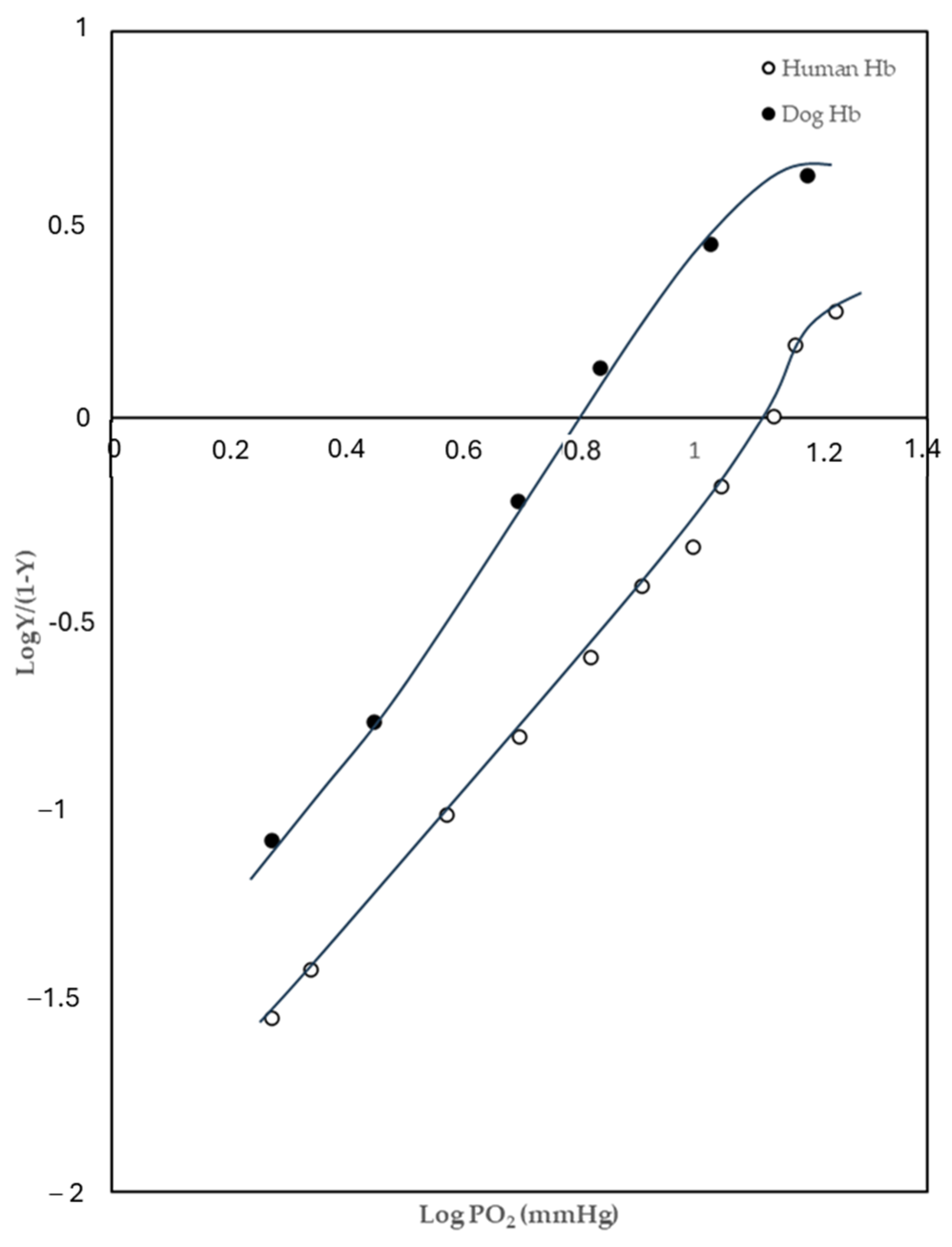
2.1. Oxygen-Binding Behaviours

Considering the known functional interaction between AE1 and Hb, the oxygen-hemoglobin binding properties of dog hemolysate have been evaluated comparing them with humans. In Figure 1, the Hill plot of oxygen binding by dog and human hemolysate shows a higher oxygen affinity of dog Hb in respect to humans.
Further experiments were performed in a pH range from 5.50 to 8.50 at a temperature of 20 °C and 37 °C under full stripped conditions, i.e., in the absence of both organic phosphates and chloride and in the presence of 2,3-BPG and lactic acid. The results are displayed in Figure 2 and Figure 3.
In details, the O2 binding properties of canine Hb are expressed as logP50, defined as the oxygen pressure required to bind 50% of the hemes. The lower the logP50, the higher the protein’s affinity for O2; because of logP50 is inversely proportional to the affinity constant of the Hb for O2.
The analysis of the curves at different temperature indicates an effectiveness of Bohr effect for pH ranges shifted towards alkaline pHs at 37 °C (see Figure 2). While, at 20 °C the curve gets narrower and the affinity for the oxygen is modulated by more acidic pHs values (see Figure 3). Both heterotrophic modulators influence the oxygen affinity of Hb at 20 and 37 °C increasing the amount of oxygen unloaded to the tissues for all pH values tested. Overall, the shape of Bohr curves is not altered by modulators except for the curve in the presence of 2.3-BPG at 37 °C. In this case, the curve shift suggests a higher impact of alkaline pH on the modulation of oxygen release from Hb. In consideration of the greater blood values of 2,3-BPG [26], the effects of this phosphate and of lactate on the oxygen binding properties of dog Hb have been investigated in more detail, at pH 7.0 and 6.5 as reported in Figure 4. Section A shows the effect on log P50 induced by increasing concentrations of 2,3-BPG at 7.0 pH and 20 °C, the modulating effect of 2,3-BPG on Hb oxygen affinity is evident. In fact, the addition of 2,3-BPG leads to an increase in logP50 corresponding to the oxygen release from Hb. In Section B, at constant concentration of 2,3-BPG (3 mM), pH 6.5 and 37 °C, the increase in lactate concentration (from 0 to 10 mM) covers up the modulating effect of 2,3-BPG. Taken together these findings suggest competitive interaction between lactate ions and 2,3-BPG anions for the same cavity on the Hb β-chains. Our data, in accordance with Böning et al., show that lactate at all concentrations tested slightly increased O2 affinity [27,28].
Figure 5 shows the oxygen binding properties of canine hemolysate in comparison with those of human haemoglobin (HbA) at 37 °C, as a function of pH in the absence and in the presence of 2,3-BPG. Results display a Bohr effect similar in amplitude for both Hbs but the oxygen-modulating effect due to the 2,3-BPG-Hb interaction appears to be lower in dog than in human. Bohr effect is highly sensitive to structural changes and these differences may be due to the amino acid residues located in a specific “Bohr group positions” in human and dog Hb. In detail, in human Hb there are fourteen amino acids of the two eliches that make positive contributions to the Bohr effect: His50α, His72α, His89α, His97β, His116β, His117β, His146β [29], and the N-terminal group of Val1α [30]. In dog Hb, three of fourteen positions on the two eliches are occupied by non-ionizable amino acid residues, in particular the substitution of His50α with Pro50α drives the protein to a structural change that could be responsible for the observable changes between Hbs [31].

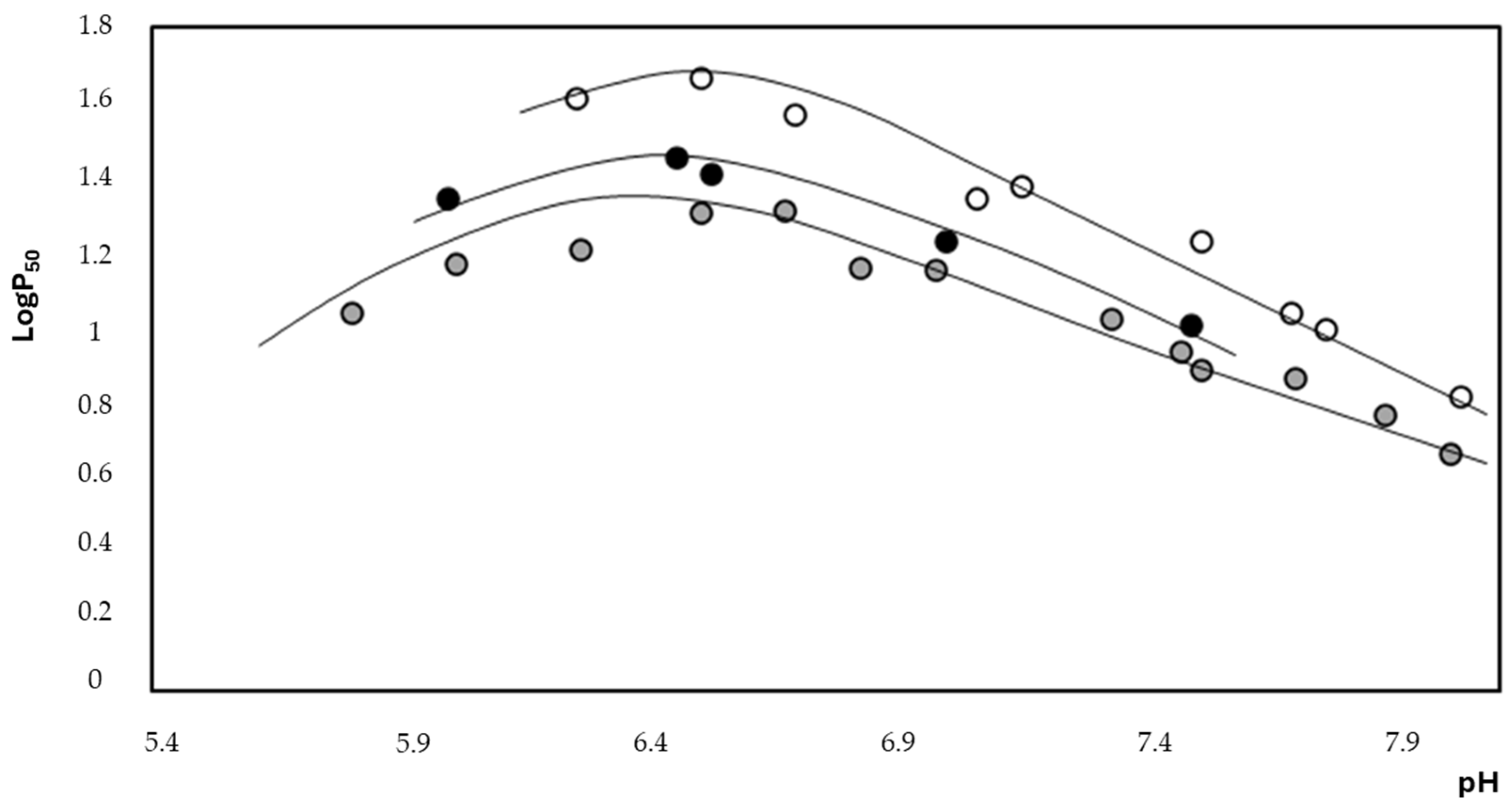
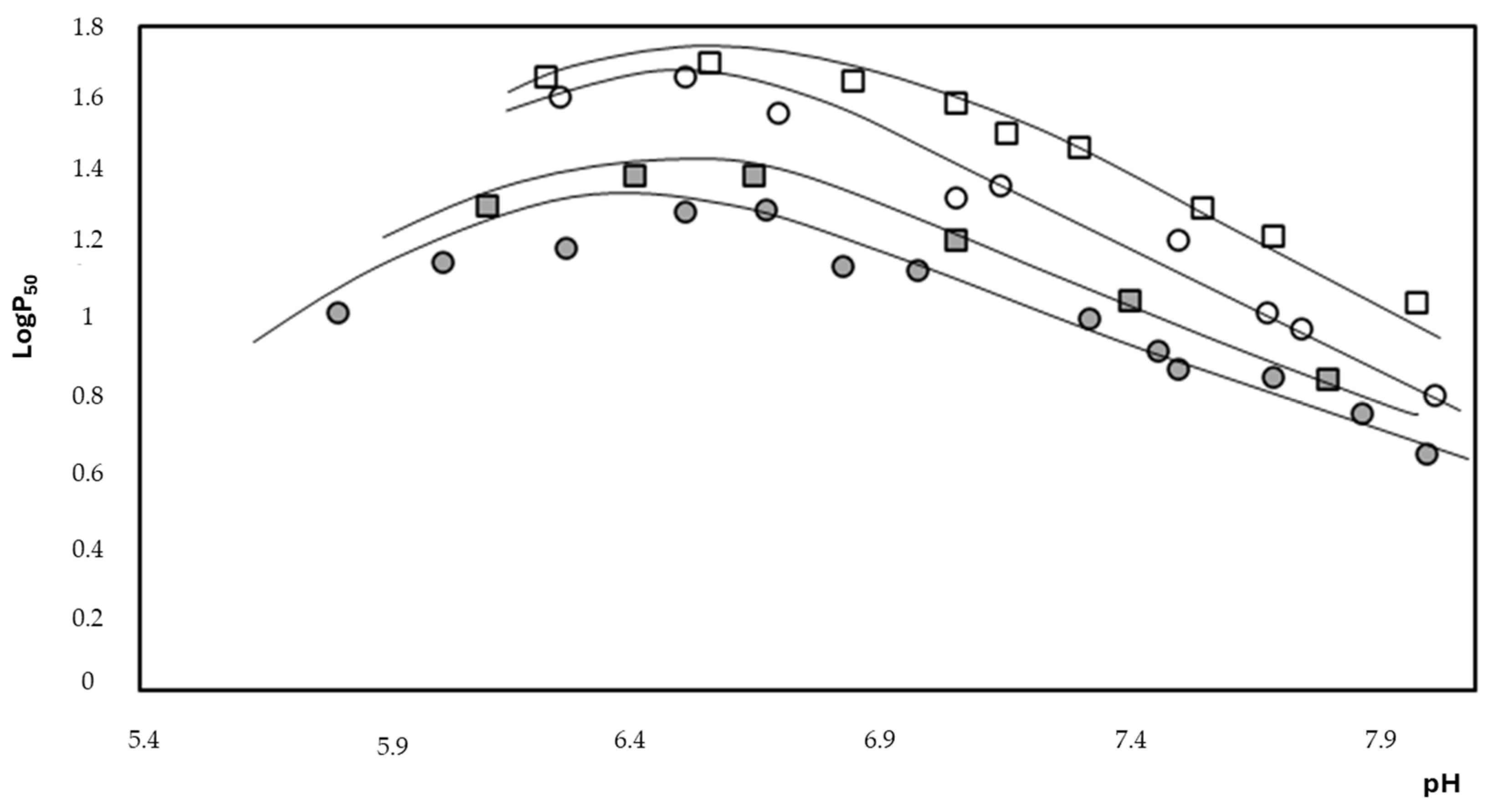
2.2. Thermodynamic Evaluation

However, the most intriguing characteristics of dog Hb concerns its response to changes in temperature, the value of the enthalpy involved in the T-R conformational variation of Hb obtained from the Van’t Hoff equation (see Figure 6). In this regard, it is important to remember that the oxygenation of Hb, being generally exothermic (ΔH negative), is favoured by a decrease in temperature (such as in the lungs at pH) and occurs with heat release.
ΔH value in stripped conditions, i.e., in the absence of organic phosphates, varies from −15.31 Kcal/mol (at pH 7.50) to −5.55 Kcal/mol (at pH 6.50). Addition of 2,3-BPG resulted in a less exothermic oxygenation (i.e., a less negative overall ΔH) which is shifted from −5.17 Kcal/mol (at pH 7.50) to −2.49 Kcal/mol (at pH 6.50), respectively (see Table 1).

2.3. Anion Exchange Evaluation

The anion exchanger 1 (AE1) kinetic of dog RBCs was evaluated performing a spectrophotometric analysis and comparing the S O 4 2 transport values with that of human RBCs. The use of S O 4 2 exchange to monitor AE1 activity allows the use of simple experimental techniques because transport times are much slower and easily measurable. The results shown in Figure 7 indicate that the rate of sulphate influx into canine RBCs (rate constant 0.0438 min−1) is remarkably higher than that of human RBCs (rate constant 0.012 min−1).

2.4. Intra- and Extracellular ATP Measurements

It is known that in human RBCs, AE1 functionality is modulated by Hb and that Hb conformational states are also related to the production and release of ATP by the cells [32,33], then intracellular and extracellular ATP levels were assessed. The results in Figure 8 show significantly lower levels of ATP in canine RBCs (101.80 μM) than in human (297.90 μM) (A); the same trend is visible in section B where there is a slight but not significant increase in ATP released by the dog’s RBCs (3 μM) compared to the human (2.65 μM).

3. Discussion

Although direct evidence on the CDB3-Hb interaction cannot be established in dog RBCs, all the data obtained here can be rationalized based on this binding. Dog RBCs show an increased anion flux, compared to human erythrocytes, which may be linked with the higher oxygen affinity of its Hb (see Figure 1). In this context since anion transport by AE1 is oxygen-dependent the stabilization of Hb in the R conformational state could favor the ion flux through the membrane [7]. Moreover, the increased AE1 functionality may have an indirect antioxidant effect because the rapid removal of carbon dioxide prevents the generation of free radicals such as nitrogen dioxide. Moreover, the increase in canine anion exchanger could be related to hyperventilation. In this context, the animal that pants to thermoregulation uses an efficient anion exchanger perfectly suited to its physiological needs. In addition, R-Hb lower binding affinity for CDB3 favors the GE-CDB3 binding. This condition would lead to a potential reduction in the glycolytic rate with a lower production of ATP. The results in Figure 8, section A showing significantly lower levels of ATP support this assumption. These considerations are further supported by the lower rates of glycolysis discovered in canine erythrocytes than human RBCs and by Miseta A, et al., that measured a reduced intracellular ATP in canine RBCs compared to human [34,35]. Moreover, the high thermal capacity of the blood makes it, a very efficient system for transferring heat from the deep parts of the body to the superficial areas, where heat is dispersed by perspiration and irradiation [8]. It should be noted that humans and dogs have different body temperatures, and in general humans have a lower body temperature than dogs. In several studies, by Nemeth N. et al., it has been demonstrated that RBCs are affected by temperature variations, going through deformations and morphological changes [36]. Furthermore, studies have shown that the sensitivity of RBCs is related to the organism examined, and that canine RBCs are less affected by temperature-induced stress, compared to human RBCs [37]. The involvement of erythrocytes in heat dissipation is also improved by the action carried out by canine Hb. The results of our study on the thermodynamics of Hb binding to O2 have highlighted a peculiar role played by Hb in canine thermal regulation. Dogs achieve cooling through a particular method of hyperventilation which consists of very fast shallow breathing that increases evaporation through the air. Panting by the animal brings airflow to wet surfaces that promotes evaporation. In detail, the exothermic value of the enthalpy of oxygenation (ΔH = −5.15 Kcal/mol) measured at pH 7.50 in the presence of 2,3-BPG indicates an exothermic binding of hemoglobin oxygen according to the reaction: Hb + 4O2 → Hb(O2)4. The dog can benefit from this exothermic reaction that allows it to lose heat from the lungs. It is therefore evident that the pulmonary tract represents an important means of heat dispersion for the dog as its skin surface equipped with sweat glands represents less than 2% of the body surface and has practically no weight in thermoregulation. In addition, in the case of excessive body heat, the dog begins to pant, i.e., to increase the amount of air inhaled and exhaled, this causes hyperoxygenation and heat dispersion. In fact, under normal conditions, the dog’s inhalation and exhalation occur only through the nose, when the caloric load to be disposed of is more substantial, the dog intermittently presents a thermal polypnea which consists of an increase in respiratory rate. Under normal conditions, the respiratory rate can be 30–40 breaths per minute, but under conditions of thermal polypnea, the rate suddenly jumps to 300–400 breaths per minute, and a one-way flow of air inhaled through the nose and exhaled through the mouth is established. The tongue, being a very vascularized organ, is a good means of heat dispersion because its wet surface helps to humidify the exhaled air. During hyperthermia, the blood flow of the tongue increases 4–5 times, its size increases, salivary secretion and muscle activity are more active with the increase in lactate [23]. These conditions favor the lowering of pH with a shift of ΔH of oxygenation towards less exothermic values (−2.49 Kcal/mol) which could constitute a further energy advantage for thermoregulation. In this district, the release of O2 by Hb would absorb a certain amount of heat that is subtracted from the total heat that must be dispersed to keep body temperature constant. The animal that pants to thermoregulate can also use an efficient anion exchanger perfectly adapted to the physiological needs. The increased AE1 functionality allows for rapid exchange of bicarbonate and chloride across the plasma membrane of RBC. This favors the hydration of bicarbonate and the fast CO2 diffusion out of RBCs to be expired by the lung. However, the animal that pants to thermoregulate also encounters another drawback, hyperventilation causes the loss of excess carbon dioxide from the lungs which leads to a severe state of alkalosis. The dog can fight this apparently dangerous condition, thanks to the peculiar oxygen-binding properties of Hb. Therefore, if a normal human Hb, during a state of alkalosis, tends to bind oxygen more tenaciously, hindering its release, in the same conditions the dog Hb shows a decreased affinity for oxygen (evidenced by the shift of the Bohr curve) which allows an effective release to the tissues even in the presence of high pH. Shifting the Bohr curve would compensate for the more difficult release of oxygen in an alkaline medium and increasing the synthesis of 2,3-BPG would help promote the release of oxygen from Hb. On the other hand, the release of oxygen from Hb is also linked to the carbamylation of the amino groups of the protein, which would dangerously contribute to further alkalinization of the blood. This condition could be partly balanced by the Hb-CDB3 interaction caused by oxygen release from Hb. The consequent detachment and activation of GE increases the glycolytic rate with the production of lactic acid which helps to compensate for the pH. Conversely, in the lungs increasing oxygen pressure results in a decreased affinity of Hb for CO2 (Haldane effect) and the oxygenation reaction release hydrogen ions to produce and facilitate the release of carbon dioxide in the lungs.

4. Materials and Methods

4.1. Reagents and Compounds

All reagents were purchased from Sigma Aldrich (St. Louis, MO, USA). Canine blood was collected during the routine clinical examination of dogs. RBCs were collected, in tubes with ethylenediaminetetraacetic acid (EDTA), used as anticoagulants and used fresh for the experiments. Concentrated stock solutions of 2,3-biphosphoglyceric acid (2,3-BPG) were prepared by dissolving the sodium salt of 2,3-BPG (Sigma) in Hepes buffer.
The animals enrolled in the study have not been subjected to stressful conditions experimentally. The blood sampling was performed during the routine clinical examination of dogs; therefore, it does not need any separate ethical approval according to Italian legislation. The protocol of animal husbandry and experimentation were reviewed and approved in accordance with the standards recommended by the Guide for the Care and Use of Laboratory Animals and Directive 2010/63/EU for animal experiments. Agreeing to the Guide for the Care and Use of Laboratory Animals and Directive 2010/63/EU for animal experiment, blood sampling procedures have been carried out according to the Section III of the cited Directive (Examples of different types of procedure assigned to each of the severity categories based on factors related to the type of the procedure) point (b). The blood sampling was performed by trained veterinarian, while the owners were present. Moreover, each animal was enrolled in the study for free choice of the owners who have issued their informed consent.

4.2. Preparation of Erythrocytes

Briefly, freshly blood samples, from human (10 volunteers, aged between 30 and 45 years), and canine (10 dogs aged between 2 and 5 years), were collected in EDTA or Heparin like anticoagulants and washed with iso-osmotic NaCl (0.9%) solution by centrifugation at 3000 rpm for three times at 4 °C. Then, RBCs were lysed by adding two volumes of cold hypotonic buffer (Tris 5mM, KCl 5 mM) and the stroma were removed by centrifugation at 12,000 rpm for 30 min.

4.3. Haemolysis Percentage and Methaemoglobin Calculation

After the washes, we determined the percentage of hemolysis and the meta-Hb by spectrophotometric analysis, with Beckman spectrophotometer DU 640 (Harbor Boulevard, Fullerton, CA, USA). For hemolysis, the measurement was made at 576 nm, making the ratio of the Hb released from the cells to the total Hb contained in the cells after total hemolysis with ultrapure water. The percentage of hemolysis was calculated using the following formula:
H   ( % ) = A / B × 100 %
where H (%) represents the percentage of hemolysis achieved, A is the level of hemoglobin released from the samples, and B represents the maximum hemoglobin released following total hemolysis with ultrapure water. Methaemoglobin was determined according to the method of W.G. Zijlstra et al. [25]. Under all the experimental conditions, the meta-Hb values of canine Hb, and percentage of haemolysis were always under 3%.

4.4. Kinetic Measurements

AE1 is one of the fastest transporters; it exchanges about 105 Cl- per second per molecule, this speed does not allow to correctly evaluate the kinetics of anion exchange [38]. The use of S O 4 2 exchange to monitor AE1 activity allows the use of simple experimental techniques because transport times are much slower and easily measurable [39]. After washing, the canine and human red blood cells were re-suspended in an incubation buffer (35 mM Na2SO4, 90 mM NaCl, 25 mM HEPES buffer, and 1.5 mM MgCl2), pH 7.4, at 25 °C. At different time intervals (5, 15, 30, 60, 90, and 120 min), 10 μM of 4-acetamido-40-isothiocyanostilbene-2,20-disulfonic acid (SITS) was added to stop the reaction. Subsequently, the red blood cells were washed three times at 4 °C, and treated with perchloric acid and H2O; then, the lysates were centrifuged for 10 min at 4000 rpm, at 4 °C, with a J2-HS Centrifuge, Beckman, and removed the supernatant. A solution of glycerol and distilled water (1:1, v/v), 4 M NaCl, 1 M HCl, and 1.23 M BaCl2·2H2O was added to remove sulfate ions from the supernatant. The absorbance of suspension was then measured within the range of 350–425 nm with Beckman spectrophotometer DU 640 (Harbor Boulevard, Fullerton, CA, USA). The sulfate concentration was determined through a calibrated standard curve, established by measuring the absorbance of the suspensions containing known quantities of sulphate [7,40]. Experimental data on the change in sulfate concentration over time were analyzed through the following equation:
c   ( t ) = c     ( 1 e k t )
-
c(t) is the concentration of sulfate at time t;
-
c∞ is the concentration of intracellular sulfate at equilibrium;
-
k is the rate constant of the sulfate inflow.

4.5. Measurement of ATP

ATP levels were measured via the luciferin-luciferase method, as reported by Tellone et al. The level of light emitted is directly proportional to the concentration of ATP detected within the samples. Briefly, RBCs were diluted with buffer (35 mM Na2SO4, 90 mM NaCl, 25 mM HEPES, 1.5 mM MgCl2) and incubated for 1 h with Mastoparan 7 (Mas 7). To further arrest ATP production, the samples were deproteinized with trichloroacetic acid (TCA, 15%) and subsequently, after centrifuging at 3000 rpm at 4 °C for 10 min, a solution of D-luciferin and Firefly Lantern Extract (FLE 250) was added to the samples (pellet) in a ratio of 1:1. The emitted light was recorded using a Bio Orbit Luminometer 1251 (Bio-Orbit Oy, Turku, Finland). Supernatant was used to determine extracellular ATP following the same procedure [40].

4.6. Purification of Haemoglobin

The presence of different haemoglobin fractions was checked by cellulose acetate electrophoresis. Organic phosphates and of chloride ions were removed by passing the hemolysate through a Sephadex G-25 column, equilibrated with 0.01 M Tris-HC1 buffer pH 8.0 containing 0.1 M NaCl and afterwards through a column of mixed-bed ion-exchange resin Bio-Rad 501X8. (Hercules, CA, USA)

4.7. Measurement of Oxygen Dissociation Curves (ODC)

Hb concentration (heme basis) was prepared using the Hb extinction coefficients at a protein concentration of 3–5 mg/mL [41]. Oxygen equilibrium isotherms were determined by the tonometric method by Giardina and Amiconi 1981 in the absence and in the presence of anionic cofactors such as lactate (0.1 mM) and/or 2,3-BPG (3 mM) [42]. Tonometer is an ampoule of 70 mL with an attached 1 cm path length cuvette. Spectrophotometric measurements were carried out with a Beckman DU 70 spectrophotometer. The parameters P50 (the partial pressure of the ligand at which 50% of heme molecules are oxygenated) and n50 (Hill cooperativity coefficient) were calculated by fitting the Hill’s sigmoidal equation:
l o g   ( Y / 1 y ) = n l o g   p O 2 + K
where Y is the fractional saturation of Hb with oxygen and K the apparent binding constant. Experiments were done at different pH values to estimate the Bohr effect and at different temperature to estimate the enthalpy of oxygenation. Determination of the overall oxygenation enthalpy corrected for the solubilization heat of oxygen was calculated from the integrated Van’t Hoff equation:
H = 4.574 ·   [ ( T l × T 2 )   /   ( T I T 2 ) ] × l o g P 50 / 1000   kcal / m o l 1
in which, T was the absolute temperature in Kelvin, logP50 indicated the partial pressure of the ligand at which 50% of hemes is oxygenated and ΔH expressed the heat liberated upon oxygenation. Van’t Hoff plots were linear within the experimental error, over the entire temperature range (from 20 °C to 37 °C) explored.

5. Conclusions

In conclusion, our results show that canine Hb has a higher affinity for oxygen, compared with human Hb. The stabilization in R conformational state of Hb would cause the increased rate of anion flux, potentially changes metabolic parameters in canine cells, compared with humans. In addition, the peculiar functional properties of canine Hb allow to positively contribute to heat dissipation in the animal under hyperthermia conditions, promoting the restoration of normal physiological conditions.

Author Contributions

Conceptualization, A.R. and E.T.; methodology, A.R., G.T.P., S.P. and E.T.; software, S.P.; validation, A.R., E.T., S.P. and G.T.P.; formal analysis, A.R., G.T.P. and E.T.; investigation, A.R., G.T.P., S.P., S.F., D.B. and G.L.; data curation, A.R. and E.T.; writing—original draft preparation, A.R., S.P. and G.T.P.; writing—review and editing, A.R. and G.T.P.; visualization, E.T.; supervision, E.T.; project administration, E.T. and D.B.; funding acquisition, E.T. and D.B. All authors have read and agreed to the published version of the manuscript.

Funding

This research received no external funding.

Institutional Review Board Statement

The human study was approved by a Local Ethics Committee (prot. 71-23 del 5 April 2023) in accordance with the Helsinki Declaration.

Informed Consent Statement

Informed consent was obtained from all subjects involved in the study.

Data Availability Statement

The data that support the findings of this study are available from the corresponding author upon reasonable request.

Conflicts of Interest

The authors declare no conflict of interest.

Abbreviations

red blood cells (RBCs); nitric oxide (NO); adenosine triphosphate (ATP); state of low oxygenation (LOS); pentose phosphate pathway (PPP); oxygenated state (HOS); Nicotinamide adenine dinucleotide phosphate (NADP); cytoplasmic domain of band 3 protein (CDB3); band 3 (AE1); hemoglobin (Hb); ethylenediaminetetraacetic acid (EDTA); 2,3-biphosphoglyceric acid (2,3-BPG); 4-acetamido-40-isothiocyanostilbene-2,20-disulfonic acid (SITS); Mastoparan 7 (Mas 7); trichloroacetic acid (TCA); Measurement of oxygen dissociation curves (ODC); the oxygen partial pressure at half saturation (P50); Hill cooperativity coefficient (n50); human haemoglobin (HbA); methemoglobin (MetHb); N-(2-hydroxyethyl)-piperazine-N1-2 ethanesulfonic acid (HEPES);

References

  1. Yeo, J.H.; Lam, Y.W.; Fraser, S.T. Cellular dynamics of mammalian red blood cell production in the erythroblastic island niche. Biophys. Rev. 2019, 11, 873–894. [Google Scholar] [CrossRef]
  2. Cortese-Krott, M.M.; Kelm, M. Endothelial nitric oxide synthase in red blood cells: Key to a new erythrocrine function? Redox Biol. 2014, 2, 251–258. [Google Scholar] [CrossRef] [PubMed]
  3. Castagnola, M.; Messana, I.; Sanna, M.T.; Giardina, B. Oxygen-linked modulation of erythrocyte metabolism: State of the art. Blood Transfus. 2010, 8, s53–s58. [Google Scholar] [PubMed]
  4. Messana, I.; Misiti, F.; el-Sherbini, S.; Giardina, B.; Castagnola, M. Quantitative determination of the main glucose metabolic f luxes in human erythrocytes by 13C- and 1H-MR spectroscopy. J. Biochem. Biophys. Methods 1999, 39, 63–84. [Google Scholar] [CrossRef]
  5. Campanella, M.E.; Chu, H.; Low, P.S. Assembly and regulation of a glycolytic enzyme complex on the human erythrocyte membrane. Proc. Natl. Acad. Sci. USA 2005, 102, 2402–2407. [Google Scholar] [CrossRef] [PubMed]
  6. De Rosa, M.C.; Carelli Alinovi, C.; Galtieri, A.; Russo, A.; Giardina, B. Allosteric properties of hemoglobin and the plasma membrane of the erythrocyte: New insights in gas transport and metabolic modulation. IUBMB Life 2008, 60, 87–93. [Google Scholar] [CrossRef] [PubMed]
  7. Galtieri, A.; Tellone, E.; Romano, L.; Misiti, F.; Bellocco, E.; Ficarra, S.; Russo, A.; Di Rosa, D.; Castagnola, M.; Giardina, B.; et al. Band-3 protein function in human erythrocytes: Effect of oxygenation-deoxygenation. Biochim. Biophys. Acta 2002, 1564, 214–218. [Google Scholar] [CrossRef]
  8. Xie, H.; Zhang, Y. The effect of red blood cells on blood heat transfer. Int. J. Heat. Mass. Transf. 2017, 113, 840–849. [Google Scholar] [CrossRef]
  9. Farsaci, F.; Tellone, E.; Galtieri, A.; Ficarra, S. A new model for thermodynamic characterization of hemoglobin. Fluids 2019, 4, 135. [Google Scholar] [CrossRef]
  10. Tellone, E.; Clementi, M.E.; Russo, A.M.; Ficarra, S.; Lania, A.; Lupi, A.; Giardina, B.; Galtieri, A. Oxygen Transport and Diving Behaviour: The Haemoglobin from Dolphin Tursiops truncatus. In Hemoglobin Function in Vertebrates; Springer: Milan, Italy, 2000; pp. 77–82. [Google Scholar]
  11. Giardina, B.; Scatena, R.; Clementi, M.E.; Cerroni, L.; Nuutinen, M.; Brix, O.; Sletten, S.N.; Castagnola, M.; Condò, S.G. Physiological relevance of the overall delta H of oxygen binding to fetal human hemoglobin. J. Mol. Biol. 1993, 229, 512–516. [Google Scholar] [CrossRef]
  12. Giardina, B.; Condò, S.G.; Petruzzelli, R.; Bardgard, A.; Brix, O. Thermodynamics of oxygen binding to arctic hemoglobins. The case of reindeer. Biophys. Chem. 1990, 37, 281–286. [Google Scholar] [CrossRef] [PubMed]
  13. Giardina, B.; Corda, M.; Pellegrini, M.G.; Sanna, M.T.; Brix, O.; Clementi, M.E.; Condo, S.G. Flight and heat dissipation in birds. A possible molecular mechanism. FEBS Lett. 1990, 270, 173–176. [Google Scholar] [CrossRef] [PubMed]
  14. Clementi, M.E.; Cataldi, E.; Capo, C.; Petruzzelli, R.; Tellone, E.; Giardina, B. Purification and characterization of the hemoglobin components of Adriatic sturgeon (Acipenser naccarii) blood. J. Appl. Ichthyol. 1999, 15, 78–80. [Google Scholar] [CrossRef]
  15. De Rosa, M.C.; Sanna, M.T.; Messana, I.; Castagnola, M.; Galtieri, A.; Tellone, E.; Scatena, R.; Botta, B.; Botta, M.; Giardina, B. Glycated human hemoglobin (HbA(1c)): Functional characteristics and molecular modeling studies. Biophys. Chem. 1998, 72, 323–335. [Google Scholar] [CrossRef] [PubMed]
  16. Tellone, E.; Russo, A.; Giardina, B.; Galtieri, A.; Ficarra, S. Metabolic effects of endogenous and exogenous heterotropic hemoglobin modulators on anion transport: The case of pig erythrocytes. OALIB 2015, 2, 1–11. [Google Scholar] [CrossRef]
  17. Atyabi, N.; Rahbarghazi, R.; Araghi, A.; Neqouiejahromi, O.A. Haemoglobin typing and its variations in Iranian domestic dogs. Comp. Clin. Pathol. 2012, 21, 1515–1519. [Google Scholar] [CrossRef]
  18. Schmidt-Nielsen, K.; Bretz, W.L.; Taylor, C.R. Panting in dogs: Unidirectional air flow over evaporative surfaces. Science 1970, 169, 1102–1104. [Google Scholar] [CrossRef]
  19. Goldberg, M.B.; Langman, V.A.; Taylor, R.C. Panting in dogs: Paths of air flow in response to heat and exercise. Respir. Physiol. 1981, 43, 327–338. [Google Scholar] [CrossRef]
  20. Pleschka, K.; Krönert, H. Thermoregulatory adjustment of lingual blood flow in the conscious dog at high ambient temperature. Isr. J. Med. Sci. 1976, 12, 1077–1078. [Google Scholar]
  21. Hales, J.R. Effects of exposure to hot environments on the regional distribution of blood flow and on cardiorespiratory function in sheep. Pflug. Arch. 1973, 344, 133–148. [Google Scholar] [CrossRef]
  22. Potter, A.W.; Berglund, L.G.; O'Brien, C. A canine thermal model for simulating temperature responses of military working dogs. J Therm. Biol. 2020, 91, 102651. [Google Scholar] [CrossRef] [PubMed]
  23. Krönert, H.; Pleschka, K. Lingual blood flow and its hypothalamic control in the dog during panting. Pflug. Arch. 1976, 367, 25–31. [Google Scholar] [CrossRef] [PubMed]
  24. Kitchen, H.; Brett, I. Embryonic and fetal hemoglobin in animals. Ann. N. Y. Acad. Sci. 1974, 241, 653–671. [Google Scholar] [CrossRef] [PubMed]
  25. Zijlstra, W.G.; Buursma, A.; Meeuwsen-van der Roest, W.P. Absorption spectra of human fetal and adult oxyhemoglobin, de-oxyhemoglobin, carboxyhemoglobin, and methemoglobin. Clin. Chem. 1991, 37, 1633–1638. [Google Scholar] [CrossRef]
  26. Rodriguez-Franco, F.; Tesouro-Diez, M.A.; Rodriguez-Sanchez, M. Concentrations of 2,3-diphosphoglycerate (2,3-DPG) in canine blood (healthy dogs, dogs with cardiopulmonary insufficiency and dogs with renal insufficiency). Zentralbl Vet. B 1994, 41, 9–16. [Google Scholar] [CrossRef]
  27. Böning, D.; Schünemann, H.J.; Maassen, N.; Busse, M.W. Reduction of oxylabile CO2 in human blood by lactate. J. Appl. Physiol. 1993, 74, 710–714. [Google Scholar] [CrossRef]
  28. Nielsen, M.S.; Weber, R.E. Antagonistic interaction between oxygenation-linked lactate and CO2 binding to human hemoglobin. Comp. Biochem. Physiol. A Mol. Integr. Physiol. 2007, 146, 429–434. [Google Scholar] [CrossRef]
  29. Lukin, J.A.; Ho, C. The structure–function relationship of hemoglobin in solution at atomic resolution. Chem. Rev. 2004, 104, 1219–1230. [Google Scholar] [CrossRef]
  30. Garner, M.H.; Bogardt, R.A.; Gurd, F.R.N. Determination of the pK values for the α-amino groups of human hemoglobin. J. Biol. Chem. 1975, 250, 4398–4404. [Google Scholar] [CrossRef]
  31. Okonjo, K.O. Bohr effect of hemoglobins: Accounting for differences in magnitude. J. Theor. Biol. 2015, 380, 436–443. [Google Scholar] [CrossRef]
  32. Jagger, J.E.; Bateman, R.M.; Ellsworth, M.L.; Ellis, C.G. Role of erythrocyte in the regulation of local O2 release mediated by hemoglobin oxygenation. Am. J. Physiol. Heart Circ. Physiol. 2001, 280, 2833–2839. [Google Scholar] [CrossRef] [PubMed]
  33. McMahon, T.J.; Darrow, C.C.; Hoehn, B.A.; Zhu, H. Generation and export of red blood cell ATP in health and disease. Anterior Physiol. 2021, 12, 754638. [Google Scholar] [CrossRef] [PubMed]
  34. Burr, M.J. The relationship between pH and aerobic glycolysis in human and canine erythrocytes. Comp. Biochem. Physiol. Part B Comp. Biochem. 1972, 41, 687–694. [Google Scholar] [CrossRef] [PubMed]
  35. Miseta, A.; Bogner, P.; Berényi, E.; Kellermayer, M.; Galambos, C.; Wheatley, D.N.; Cameron, I.L. Relationship between cellular ATP, potassium, sodium and magnesium concentrations in mammalian and avian erythrocytes. Biochim. Biophys. Acta 1993, 1175, 133–139. [Google Scholar] [CrossRef]
  36. Nemeth, N.; Baskurt, O.K.; Meiselman, H.J.; Kiss, F.; Uyuklu, M.; Hever, T.; Sajtos, E.; Kenyeres, P.; Toth, K.; Furka, I.; et al. Storage of laboratory animal blood samples causes hemorheological alterations: Inter-species differences andthe effects of duration and temperature. Korea-Aust. Rheol. J. 2009, 21, 127–133. [Google Scholar]
  37. Matrai, A.A.; Varga, G.; Tanczos, B.; Barath, B.; Varga, A.; Horvath, L.; Bereczky, Z.; Deak, A.; Nemeth, N. In vitro effects of temperature on red blood cell deformability and membrane stability in human and various vertebrate species. Clin. Hemorheol. Microcirc. 2021, 78, 291–300. [Google Scholar] [CrossRef]
  38. Passow, H. Molecular aspects of band 3 protein-mediated anion transport across the red blood cell membrane. Rev. Physiol. Biochem. Pharmacol. 1986, 103, 61–203. [Google Scholar]
  39. Jennings, M.L. Proton fluxes associated with erythrocyte membrane anion exchange. J. Membr. Biol. 1976, 28, 187–205. [Google Scholar] [CrossRef]
  40. Russo, A.; Patanè, G.T.; Putaggio, S.; Lombardo, G.E.; Ficarra, S.; Barreca, D.; Giunta, E.; Tellone, E.; Laganà, G. Mechanisms Underlying the Effects of Chloroquine on Red Blood Cells Metabolism. Int. J. Mol. Sci. 2024, 25, 6424. [Google Scholar] [CrossRef]
  41. Antonini, E.; Brunori, M. Hemoglobin and Myoglobin in Their Reactions with Ligands; North-Holland Pub. Co.: Amsterdam, The Netherlands, 1971. [Google Scholar]
  42. Giardina, B.; Amiconi, G. Measurement of binding of gaseous and nongaseous ligands to hemoglobins by conventional spectrophotometric procedures. Methods Enzymol. 1981, 76, 417–427. [Google Scholar]
Figure 1. Hill plot for the binding of oxygen to canine hemoglobin (closed symbols) and human hemoglobin (open symbols). Conditions: HEPES buffer 0.1 M plus 0.1 M NaCl and 3 mM 2,3-BPG at pH 7.4 and 37 °C.
Stresses 04 00052 g001
Figure 2. Shows the Bohr effect (affinity of Hb for oxygen expressed as logP50 as a function of pH) of the dog hemolysate in the absence (grey circles) and presence of 0.1 mM lactate (black circles) and in the presence of 3 mM 2,3-BPG (white circles). Conditions: Mes or Hepes 0.1 M at 37 °C. Data were expressed as means of five experiments.
Stresses 04 00052 g002
Figure 3. Shows the Bohr effect (oxygen affinity of Hb expressed as logP50 as a function of pH) of the dog hemolysate in the absence (grey circles) and the presence of 0.1 mM lactate (black circles) and in the presence of 3 mM 2,3-BPG (white circles). Conditions: Mes or Hepes 0.1 M at 20 °C. Data were expressed as means of 5 experiments.
Stresses 04 00052 g003
Figure 4. Variation of oxygen affinity as function of 2,3-BPG for dog Hb 7.0 pH and 20 °C (A) and variation of oxygen affinity as function of lactate concentrations in the presence of 3 mM 2,3-BPG for dog Hb at 6.5 pH and 37 °C (B). Data were expressed as means of 5 experiments.
Stresses 04 00052 g004
Figure 5. Variation of the oxygen affinity as a function of pH for dog Hb (circles) and human Hb (squares) in the absence (grey symbols) and in the presence of three mM 2,3-BPG (white symbols) at 37 °C. Data were expressed as means of five experiments.
Stresses 04 00052 g005
Figure 6. Overall ΔH values (expressed as Kcal/mol of O2, and corrected for the heat contribution of oxygen in solution) calculated by the integrated Van‘t Hoff equation. pH 6.5+2,3-BPG 3 mM (orange circles), pH 7.5+2,3- BPG 3 mM (black circles), pH 6.5 (white circles), pH 7.5 (grey circles). Data were expressed as means of five experiments.
Stresses 04 00052 g006
Figure 7. Sulphate anion flux in canine and human red blood cells in high oxygenation state at 25 °C. Results are from four independent experiments ± standard deviation.
Stresses 04 00052 g007
Figure 8. Intracellular (A) and extracellular (B) ATP concentration in canine and human red blood cells at 25 °C. Results are from four independent experiments ± standard deviation.
Stresses 04 00052 g008
Table 1. ΔH values (expressed as Kcal/mol of O2 and corrected for the thermal contribution of oxygen in solution) for dog Hb in the presence and absence of 2,3-BPG (3 mM). Conditions: Hepes 0.1 M, NaCI 0.1 M. Data were expressed as means of 5 experiments.
SamplepHΔH (Kcal/mol)
Hb6.5−5.55
7.5−15.31
Hb + 2,3-BPG (3 mM)6.5−2.49
7.5−5.17
Disclaimer/Publisher’s Note: The statements, opinions and data contained in all publications are solely those of the individual author(s) and contributor(s) and not of MDPI and/or the editor(s). MDPI and/or the editor(s) disclaim responsibility for any injury to people or property resulting from any ideas, methods, instructions or products referred to in the content.

