Article

Jaw Clenching Alters Neuromuscular Coordination in Dynamic Postural Tasks: A Pilot Study on Single-Leg Sit-to-Stand Movements

1
Department of Special Care Dentistry, Osaka Dental University Hospital, Osaka 540-0008, Japan
2
Department of Physical Therapy, Faculty of Health Care, Takasaki University of Health and Welfare, Takasaki 370-0033, Japan
*
Author to whom correspondence should be addressed.
Biomechanics 2025, 5(4), 89; https://doi.org/10.3390/biomechanics5040089
Submission received: 25 September 2025 / Revised: 27 October 2025 / Accepted: 30 October 2025 / Published: 4 November 2025
(This article belongs to the Section Neuromechanics)

Abstract

Background/Objective: Postural stability and motor coordination require precise regulation of agonist and antagonist muscle activities. Jaw clenching modulates neuromuscular control during static and reactive postural tasks. However, its effects on dynamic voluntary movement remain unclear. This pilot study aimed to investigate the effects of jaw clenching on muscle activity and kinematics during repetitive single-leg sit-to-stand task performance. Methods: Eleven healthy adults (age: 21.2 ± 0.4 years; 6 males and 5 females; height: 167.9 ± 9.6 cm; body weight: 59.7 ± 8.1 kg) performed repetitive single-leg sit-to-stand tasks for 30 s under jaw-clenching and control conditions. Electromyography (EMG) signals from eight muscles and kinematic data from 16 inertial measurement unit sensors were analyzed, focusing on the seat-off phase. Results: Jaw clenching resulted in a significantly lower success rate than the control condition (success rate: 0.96 ± 0.13 vs. 0.78 ± 0.29, p = 0.047). Under the jaw clenching condition, failed trials exhibited higher medial gastrocnemius and masseter EMG activity (p < 0.001), lower erector spinae longus EMG activity (p < 0.001), and altered kinematics, including increased trunk yaw and roll angles (p < 0.001). Jaw clenching increased the coactivation of the gastrocnemius and tibialis anterior muscles (p < 0.001), disrupting the reciprocal muscle patterns critical for task performance. Conclusions: These findings suggest that jaw clenching may reduce task performance by altering neuromuscular coordination during dynamic postural tasks.

1. Introduction

Postural stability and mobility are essential for daily activities and require adjustments in body position and orientation with respect to the environment. The central nervous system coordinates these adjustments by regulating agonist and antagonist muscle activities based on inputs from multiple sensory systems, including the visual, vestibular, and somatosensory systems. Muscle activation between agonist and antagonist pairs occurs in either a reciprocal or non-reciprocal manner depending on task demands and sensory feedback. Reciprocal muscle activity allows smooth joint displacement and offers advantages, such as quick, precise, and flexible movement adjustments [1,2]. However, non-reciprocal muscle activity or coactivation involves the simultaneous contraction of agonist and antagonist muscles, increasing joint rigidity. Non-reciprocal muscle activity provides benefits in weight-bearing and static postural control [3,4]. Increased muscle coactivation has been reported in individuals with neurological [5,6] and musculoskeletal disorders [7] and in aging populations [8]. Modulation of reciprocal and non-reciprocal muscle activity is essential for performing postural and motor tasks. Stomatognathic motor activity modulates lower limb muscle activity during postural balance and voluntary movements. Jaw clenching results in reduced body sway during static standing tasks [9] and altered coactivation patterns between agonist and antagonist muscles in the lower limbs [10]. Conversely, jaw clenching increases postural sway in 9–12-year-old children [11]. We previously reported that jaw clenching increased anticipatory activities of the trunk and lower limb muscles prior to external postural perturbations, whereas postural stability was not altered [12]. The effects of jaw clenching on postural and motor tasks have been widely investigated in tasks in which participants are required to maintain the center of mass (COM) position during quiet standing [13,14,15,16,17] or in response to expected [12,18] or unexpected [19] postural perturbations. However, the effect of jaw clenching on dynamic voluntary movements that require dynamic COM displacement remains unclear.
The single-leg sit-to-stand task requires dynamic anteroposterior and vertical displacement of the COM position while maintaining body orientation relative to the environment to sustain balance. Widely used in sports performance evaluation and assessments of mobility decline and postural balance, this task is a performance metric and training exercise for lower limbs. It requires strength, balance, and coordination; hence, it is a valuable tool for evaluating lower limb function and postural stability [20,21,22]. A critical event of the movement is the seat-off phase, in which a smooth transition from forward trunk motion to lifting the entire body upward determines the success of the task. Effective reciprocal activation of the trunk and lower limb muscles during the seat-off phase is essential for successful task execution [23,24]. Investigating the effects of jaw clenching on the seat-off phase of single-leg sit-to-stand tasks will elucidate the interaction between stomatognathic motor activity and activation of the trunk and lower limb muscles as well as its effect on motor performance.
This study aimed to examine the effects of jaw clenching on muscle activity and kinematics during repetitive single-leg sit-to-stand movements. We hypothesized that jaw clenching affects muscle activity and movement coordination, potentially impairing task performance by altering reciprocal muscle activity.

2. Materials and Methods

2.1. Participants

Eleven healthy young adults (six men and five women; age: 21.2 ± 0.4 years; height: 167.9 ± 9.6 cm; body weight: 59.7 ± 8.1 kg; Table 1) participated in this study after providing written informed consent. The required sample size was estimated a priori based on the effect size reported in a previous study comparing postural balance with and without clenching (effect size = 0.934) [25]. Using a Wilcoxon signed-rank test with a two-sided significance level of 0.05 and statistical power of 0.80, the minimum sample size was calculated to be 10 participants. Therefore, we considered 11 subjects to be sufficient to detect the expected difference in balance performance between the conditions. This study was approved by the Ethics Committee of Osaka Dental University (Approval Number: 111088) and conducted in accordance with the Declaration of Helsinki. The participants had no history of musculoskeletal or neurological disorders that could affect their task performance. A dentist assessed the participants’ oral health, confirming the presence of a Class I incisor relationship, absence of periodontal disease, and no mobile teeth (Miller Classification [26] II or III: >1 mm horizontal or vertical mobility). A temporomandibular disorder screening questionnaire ruled out temporomandibular dysfunction and associated symptoms [27]. The participants were university students recruited through flyers posted on campus. Individuals who met the inclusion criteria and did not meet the exclusion criteria were enrolled in the study.

2.2. Experimental Settings

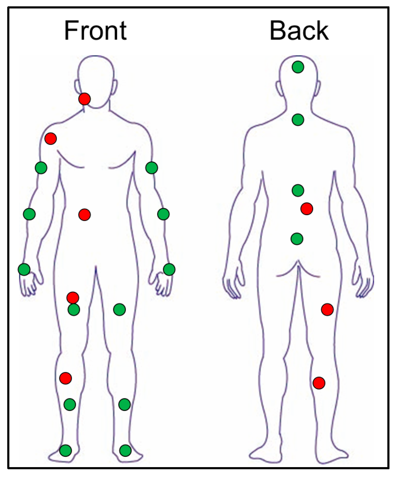
The participants sat on a height-adjustable platform with their right foot on a force platform (FDM-S; Zebris Medical GmbH, Isny, Germany; Figure 1A). They performed single-leg sit-to-stand movements for 30 s using their dominant legs. The electromyographic (EMG) activity of eight muscles was recorded at 2000 Hz (Ultium, Noraxon, Scottsdale, AZ, USA). After cleaning the skin, disposable surface electrodes (G207, Nihon Kohden, Japan) were placed on the belly of the following muscles: masseter, anterior deltoid, rectus abdominis, erector spinae longus, rectus femoris, biceps femoris, tibialis anterior, and medial gastrocnemius (Figure 2). The electrodes were placed according to the SENIAM guidelines [28]. Kinematic data were collected using 16 inertial measurement unit (IMU) sensors (Figure 2; myoMOTION, Noraxon, Scottsdale, AZ, USA) at a sampling frequency of 100 Hz. The sensors were securely attached to anatomical landmarks on the trunk and limbs using elastic straps and medical-grade double-sided adhesive tape to minimize movement relative to the skin. Segment orientations and joint kinematics were computed in MR3 (Noraxon, Scottsdale, AZ, USA) software based on sensor fusion of tri-axial accelerometer, gyroscope, and magnetometer signals. The force platform, EMG, and IMU signals were synchronized using analog synchronization signals.

2.3. Experimental Procedures

Prior to the experiment, maximal voluntary contractions (MVCs) were measured to determine the 100% MVC for EMG normalization. The participants initially sat on a bench with a standardized height (set to the height of the lateral femoral epicondyle above the floor), with their right foot on the force platform and their left foot lifted off the floor (Figure 1A). The knee and hip joints were flexed at 90°. The participants were instructed to fold their arms across their chests and perform single-leg sit-to-stand movements for 30 s. On the command “go,” the participants were instructed to stand fully upright and sit back down firmly as many times as possible within 30 s (Figure 1B). A trial was considered successful if the participant used only the tested leg and fully extended their knees while standing. A trial was considered to have failed if the participant placed the opposite foot on the ground or did not fully extend the knee of the stance leg after the seat-off phase (Figure 1C). The number of successful and failed trials in each 30-s session was recorded.
The movements were performed under two conditions: clenching and no clenching. In the clenching condition, the participants maintained jaw clenching of at least 5% of MVC, as monitored using masseter EMG. Auditory feedback from the EMG measurement device ensured a consistent clenching intensity of >5% of MVC. In the control condition, participants relaxed their jaws and maintained their masseter in a physiological rest position. The order of the experimental conditions was randomized using a random number generator (RAND function in Microsoft Excel). A 5-min rest period between the conditions minimized the fatigue.

2.4. Data Processing

Data were analyzed using a custom MATLAB program (MATLAB 2017b; MathWorks, Natick, MA, USA). The following angular metrics were extracted from the IMU signals: trunk pitch, yaw, roll angles, hip flexion, abduction, internal rotation angles, knee flexion angle, ankle dorsiflexion angle, and hip angular velocity.
This study primarily focused on the timing of the seat-off phase during the single-leg sit-to-stand movements. Movements were identified using hip and knee joint angles measured using the IMUs, and vertical force measured using the force platform. Movement onset was defined as the time when the hip flexion angular velocity exceeded 0 for >100 ms, corresponding to the beginning of forward trunk motion. The seat-off phase was defined as the time at which the hip flexion angle reached its maximum value after movement onset. The duration of each movement was time-normalized from 0% (movement onset) to 100% (set-off) using the interp1 function in MATLAB.
The EMG signals were down-sampled to 100 Hz using a resampling function in MATLAB. The downsampled EMG signals were rectified and filtered using a second-order Butterworth bandpass filter (50–500 Hz). A moving average filter with a 20-ms window was applied to the EMG signals. For each participant, the EMG activity for each channel was normalized to the peak values recorded across all conditions, including the MVC trials. The coactivation index, which represents the proportion of antagonist muscle activity relative to the total agonist and antagonist muscle activity, was calculated for three muscle pairs: the rectus abdominis-erector spinae (RA-ES), rectus femoris-biceps femoris (RF-BF), and medial gastrocnemius-tibialis anterior (MG-TA). EMG and kinematic data were extracted during the seat-off phase for between-condition comparisons.

2.5. Statistical Analysis

To compare EMG and kinematic outcomes between the control and clenching conditions, all trials (successful and failed) were analyzed at the trial level, and no averaging was performed per participant. A linear mixed-effects model (LMM) was fitted with Condition (control/clenching) as a fixed effect and Subject as a random intercept to account for within-subject correlations arising from repeated measurements. Subsequently, to examine performance-related differences within the clenching condition, a separate LMM was performed with Performance outcome (success/failure) as a fixed effect and Subject as a random intercept. This analysis aimed to clarify EMG and kinematic factors associated with task success under clenching conditions. Cohen’s d was calculated to quantify effect sizes, and statistical significance was set at p < 0.05. All statistical analyses were performed using R software (version 4.5.0; R Foundation for Statistical Computing, Vienna, Austria). The primary R scripts used for mixed-effects modeling are provided as Supplementary Materials.
Because the primary comparisons relied on a within-subject design with two levels (control vs. clenching), statistical power was approximated using a paired-sample framework. With n = 11, a two-sided α = 0.05, and 80% power, the study was sufficiently powered to detect large standardized mean differences. Assuming a plausible within-subject correlation (ρ) of 0.3–0.7, the minimum detectable effect size ranged from Cohen’s d = 0.65–1.00 (≈0.85 at ρ = 0.5). Thus, the current sample size was appropriate for identifying large effects, whereas moderate effects may have been underpowered.

3. Results

3.1. Performance Outcomes

A total of 214 trials from 11 participants were analyzed at the trial level, and within-subject dependencies due to repeated trials were modeled using Subject as a random effect in the linear mixed-effects model. The performance outcomes of single-leg sit-to-stand movements are summarized in Table 2. The success rate was significantly lower in the clenching condition (0.78 ± 0.29) than in the control condition (0.96 ± 0.13; p = 0.047, d = 0.69). Similarly, the number of successful trials was significantly lower under the clenching condition (8.18 ± 4.87) than under the control condition (9.45 ± 3.80; p = 0.031, d = 0.23). In contrast, the total number of attempts did not differ significantly between the conditions (9.91 ± 4.32 vs. 9.82 ± 3.52; p = 0.878, d = 0.02).

3.2. Comparisons of EMG and Kinematic Outcomes Between Control and Clenching Conditions

A comparison of the electromyographic (EMG) activity during the seat-off phase between the control and clenching conditions in the single-leg sit-to-stand task is summarized in Table 3. Masseter activity was significantly higher in the clenching condition (10.8 ± 7.0% MVC) than in the control condition (3.3 ± 5.8%MVC; p < 0.001, d = 1.323). In contrast, no significant differences were observed in the anterior deltoid (p = 0.845, d = 0.028), rectus abdominis (p = 0.961, d = 0.007), erector spinae (p = 0.375, d = 0.125), rectus femoris (p = 0.246, d = 0.163), biceps femoris (p = 0.839, d = 0.029), tibialis anterior (p = 0.303, d = 0.146), or medial gastrocnemius (p = 0.126, d = 0.217). Regarding the coactivation indices, no significant differences were found between the control and clenching conditions in the ES–RA (p = 0.448, d = 0.107), RF–BF (p = 0.169, d = 0.194), or MG–TA (p = 0.535, d = 0.088).
Table 4 presents a comparison of the kinematic variables during the seat-off phase between the control and clenching conditions in the single-leg sit-to-stand task. Significant increases were observed in trunk roll (6.3 ± 5.1° vs. 1.9 ± 5.6°; p < 0.001, d = 1.020), hip flexion (105.8 ± 15.9° vs. 101.7 ± 20.4°; p < 0.001, d = 0.698), hip abduction (−21.0 ± 10.8° vs. −20.6 ± 10.2°; p < 0.001, d = 0.549), hip internal rotation (17.5 ± 16.5° vs. 13.0 ± 15.4°; p < 0.001, d = 0.631), and knee flexion (113.1 ± 15.1° vs. 109.7 ± 14.1°; p < 0.001, d = 0.816). No significant differences were found in trunk pitch (p = 0.892, d = 0.076), yaw (p = 0.969, d = 0.006), ankle dorsiflexion (p = 0.145, d = 0.211), or hip angular velocity (p = 0.068, d = 0.258).

3.3. Comparisons of EMG and Kinematic Outcomes Between Successful and Gailed Trials Within the Clenching Condition

A comparison of the EMG activity at the seat-off phase between successful and failed trials during the single-leg sit-to-stand task with jaw clenching is summarized in Table 5. Masseter activity was significantly higher in failed trials (15.5 ± 13.4%MVC) than in successful trials (6.8 ± 9.7%MVC; p < 0.001, d = 0.831). Similarly, anterior deltoid (p = 0.022, d = 0.559) and medial gastrocnemius (p < 0.001, d = 1.200) activities were significantly greater in failed trials. In contrast, erector spinae activity was significantly lower in failed trials (27.9 ± 17.0%MVC) than in successful trials (44.9 ± 17.0%MVC; p < 0.001, d = 1.007). No significant differences were observed in the rectus abdominis (p = 0.768, d = 0.100), rectus femoris (p = 0.639, d = 0.147), biceps femoris (p = 0.260, d = 0.305), or tibialis anterior (p = 0.170, d = 0.387) between successful and failed trials. Regarding the coactivation indices, the ES–RA index was significantly lower in failed trials (p < 0.001, d = 1.042), whereas the MG–TA index was significantly higher (p < 0.001, d = 1.284). No significant difference was observed in the RF–BF coactivation index (p = 0.149; d = 0.420).
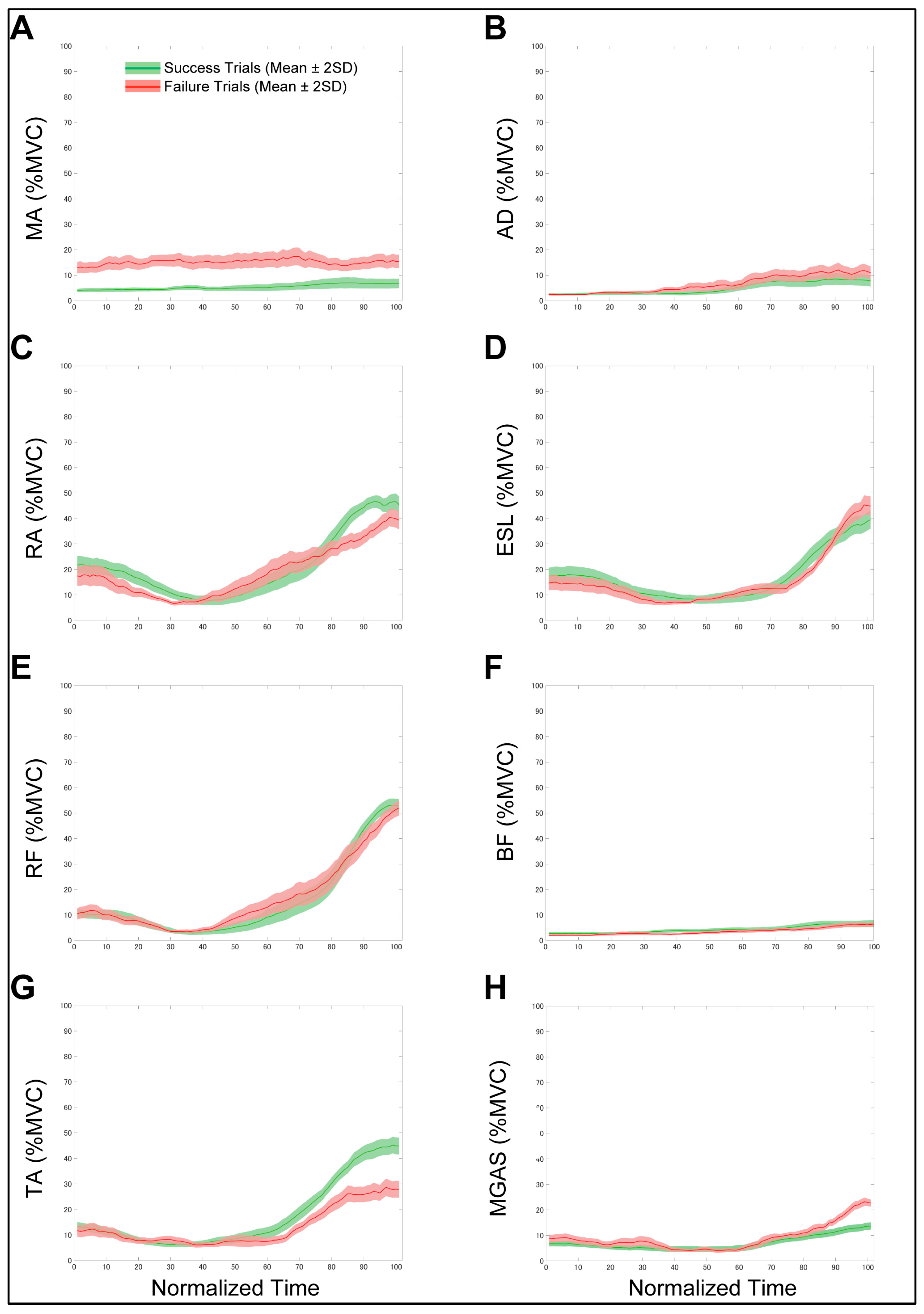
The means and 95% confidence intervals of time-normalized EMG activity for successful (n = 90) and failed (n = 19) trials are presented in Figure 3A–H. Table 6 presents a comparison of kinematic variables during the seat-off phase between successful and failed trials in the clenching condition, where the trunk yaw angle was significantly greater in failed trials (5.70 ± 9.19°) than in successful trials (−0.39 ± 6.72°; p = 0.001, d = 0.713). Similarly, the trunk roll angle was significantly larger in failed trials (8.83 ± 5.69°) than in successful trials (1.45 ± 6.60°; p < 0.001, d = 0.958). Significant increases were also observed in hip internal rotation (24.96 ± 16.90° vs. 15.80 ± 16.74°; p = 0.033, d = 0.518), knee flexion (122.11 ± 17.79° vs. 111.21 ± 13.78°; p = 0.004, d = 0.637), and ankle dorsiflexion (21.88 ± 10.74° vs. 17.53 ± 6.56°; p = 0.022, d = 0.556). In contrast, no significant differences were observed in trunk pitch (p = 0.236, d = 0.361), hip flexion (p = 0.120, d = 0.479), hip abduction (p = 0.549, d = 0.164), or hip angular velocity (p = 0.407, d = 0.206).

4. Discussion

This study investigated the effects of jaw clenching on muscle activity and kinematics during a repetitive single-leg sit-to-stand task. When comparing the control and clenching conditions, jaw clenching significantly reduced the success rate and the number of successful trials, although the total number of attempts did not differ between the conditions. Regarding neuromuscular and kinematic changes, masseter activity significantly increased under clenching conditions, confirming voluntary activation of the jaw muscles during the task. In contrast, no consistent changes were observed in most limb and trunk muscles. However, at the kinematic level, the clenching condition exhibited greater trunk roll and hip flexion angles as well as increased hip internal rotation and knee flexion angles. To further clarify the changes associated with jaw clenching, we compared successful and failed trials under the clenching condition. In this within-condition analysis, failed trials exhibited higher EMG activity in the masseter and medial gastrocnemius muscles, greater coactivation of the ankle muscles, lower erector spinae activity, and larger trunk roll, trunk yaw, hip internal rotation, knee flexion, and ankle dorsiflexion angles than the successful trials. Collectively, these results suggest that jaw clenching induces increased global muscle activation, which may alter trunk–lower limb coordination and lead to less successful performance. These findings suggest that jaw clenching affects neuromuscular control during dynamic voluntary movements, thereby reducing performance.
The increased medial gastrocnemius activation and reduced success rate observed during jaw clenching can be explained by spinal-level modulation driven by masseter spindle input. Masseter muscle spindles exhibit unique mechanosensory properties that precisely regulate isometric contraction and jaw stability [29,30]. Activation of these spindles during clenching facilitates motor neuron excitability in remote muscle groups through trigeminal–spinal pathways, a phenomenon known as the remote effect. Strong evidence for this mechanism is provided by H-reflex studies showing increased excitability of the soleus muscle during jaw clenching [31,32] and attenuation of reciprocal Ia inhibition between antagonist ankle muscles [33]. These neural changes promote non-reciprocal facilitation of both the agonist and antagonist muscles. This mechanism aligns with our findings that failure trials exhibited significantly increased gastrocnemius–tibialis anterior coactivation during the seat-off phase (Table 5). During this phase, successful sit-to-stand execution requires coordinated tibialis anterior activity to counteract the external dorsiflexion moment and stabilize the shank relative to gravity [34]. Excessive gastrocnemius facilitation may therefore disrupt optimal reciprocal control, resulting in inefficient moment generation and failure to achieve forward–upward progression.
Jaw clenching effects are highly task-specific. In dynamic or externally perturbed tasks, clenching facilitates anticipatory and reactive postural adjustments (APAs and RPAs) by increasing muscle activation amplitudes and advancing the onset timing [12,18]. Clenching has also been shown to improve dynamic steady-state balance with lasting effects, even after the novelty of the task diminishes, potentially reflecting neuromuscular optimization [15,16,17]. Moreover, increased joint stiffness resulting from non-reciprocal coactivation may help stabilize the stance under challenging static or perturbed conditions [13,14]. However, these same facilitatory mechanisms may become maladaptive when a task demands smooth, precise reciprocal coordination, such as during voluntary sit-to-stand. Our results showed that jaw clenching did not consistently elevate muscle activity across all trials (Table 3) yet selectively increased the likelihood of coordination failure (Table 2). This suggests that performance impairment is not due to cognitive or attentional interference. Indeed, previous research indicates minimal dual-task competition arising merely from the act of clenching [16]. Instead, trial-to-trial differences in the degree of masseter-driven spinal modulation may determine whether a heightened neuromuscular state improves or disrupts balance control. Taken together, these findings highlight that jaw clenching modifies neuromuscular coordination primarily by altering spinal inhibitory–excitatory balance rather than through central attentional competition. Facilitation of muscle activity through the trigeminal–spinal pathway can be advantageous for stability-dominant tasks but detrimental for coordination-dominant movements requiring finely tuned reciprocal control. Thus, the functional consequences of jaw clenching depend not only on postural task dynamics (stability vs. mobility demands), but also on whether the required balance control is voluntary or reactive. Further investigation is needed to determine the conditions under which jaw clenching contributes to functional improvement compared to neuromechanical interference.
These findings have several important clinical implications. In the context of rehabilitation and training, clinicians and trainers should be aware that oral habits such as jaw clenching may affect movement coordination and performance, particularly during dynamic voluntary movement. This highlights the need to assess and address excessive jaw clenching during dynamic voluntary tasks such as single-leg sit-to-stand tasks. Specific interventions (such as relaxation techniques, targeted oral motor training, and monitoring devices) can be used to minimize the negative effects of jaw clenching on motor performance [35,36,37]. Furthermore, jaw clenching plays a complex role in oral function and whole-body movements. Jaw clenching can enhance muscle activation and balance, as seen in vertical jump performance with custom mouthpieces [38]. Similarly, oral interventions, such as complete denture adjustments, improve gait performance [39]. However, our findings suggest that deliberate clenching above 5% may alter neuromuscular coordination and impair postural stability during dynamic tasks in healthy individuals. These results emphasize the importance of tailoring interventions to optimize the interaction between oral habits and whole-body coordination to ensure that jaw clenching enhances performance without compromising motor control during specific tasks. Furthermore, rehabilitation and training programs should carefully integrate strategies to balance these effects and maximize the functional and performance outcomes.
This study had several limitations. First, the small sample size of 11 participants may limit the generalizability of the findings to a larger population. Although our sample size was sufficiently large to detect the immediate effects of jaw clenching on sit-to-stand task performance, a larger cohort would provide more robust data and allow subgroup analyses based on factors such as age, sex, and habitual oral behaviors. Nevertheless, the effect sizes identified in this study provide valuable reference data for determining appropriate sample sizes and designing future studies to investigate the influence of jaw clenching on motor performance. Second, the analysis included a limited number of EMG channels and focused only on eight muscles. Although this approach allowed for a detailed investigation of the important muscles involved in the task, it may have overlooked the contributions of other muscles such as the contralateral limb muscles. Incorporating additional EMG channels in future studies could provide a more comprehensive understanding of muscle activation patterns. Third, although we investigated the immediate effects of jaw clenching on specific motor tasks, the long-term effects of habitual jaw clenching on motor performance remain unclear. It is unknown whether prolonged or chronic jaw clenching leads to adaptations in neuromuscular control, which may further affect performance on dynamic or static tasks. Finally, the relationship between jaw clenching and habitual oral behaviors such as bruxism has not been explored. These behaviors may have modulated the effects observed in this study and should be considered in future research to better understand the broader implications of jaw clenching in clinical and nonclinical populations. Addressing these limitations could significantly enhance the clinical relevance and applicability of our findings.

5. Conclusions

This study demonstrated that jaw clenching negatively affected performance in the single-leg sit-to-stand task. Specifically, task failure was associated with stronger jaw clenching, which in turn was related to increased EMG activity in the medial gastrocnemius, reduced activity in the erector spinae, and altered kinematics, including changes in the trunk and lower limb joint angles. These findings highlight the complex and task-specific neuromuscular effects of jaw clenching, suggesting that its facilitatory and inhibitory effects vary depending on the task demands and the specific muscle groups involved.

Supplementary Materials

The following supporting information can be downloaded at: https://www.mdpi.com/article/10.3390/biomechanics5040089/s1, S1. statistical analysis program.

Author Contributions

Conceptualization, Y.T. (Yuto Tanaka) and Y.T. (Yosuke Tomita); methodology, Y.T. (Yuto Tanaka) and Y.T. (Yosuke Tomita); software, Y.T. (Yosuke Tomita); validation, Y.T. (Yuto Tanaka) and Y.T. (Yosuke Tomita); formal analysis, Y.T. (Yosuke Tomita); investigation, Y.T. (Yuto Tanaka) and Y.T. (Yosuke Tomita); resources, Y.T. (Yuto Tanaka) and Y.T. (Yosuke Tomita); data curation, Y.T. (Yosuke Tomita); writing—original draft preparation, Y.T. (Yuto Tanaka) and Y.T. (Yosuke Tomita); writing—review and editing, Y.T. (Yuto Tanaka), Y.O. and Y.T. (Yosuke Tomita); visualization, Y.T. (Yuto Tanaka) and Y.T. (Yosuke Tomita); project administration, Y.T. (Yuto Tanaka); funding acquisition, Y.T. (Yuto Tanaka). All authors have read and agreed to the published version of the manuscript.

Funding

This work was supported by JSPS KAKENHI (Grant No. 25K14472).

Institutional Review Board Statement

The study was conducted in accordance with the Declaration of Helsinki and approved by the Ethics Committee of Osaka Dental University (approval number 111265-0).

Informed Consent Statement

Informed consent was obtained from all the participants involved in the study.

Data Availability Statement

The data that support the findings of this study are available from the corresponding author, Y.T., upon reasonable request.

Acknowledgments

We thank all participants who volunteered for this study.

Conflicts of Interest

The authors declare no conflicts of interest.

Abbreviations

The following abbreviations are used in this manuscript:
EMGelectromyography
COMcenter of mass
IMUinertial measurement unit
MVCmaximal voluntary contraction
IQRinterquartile range
MAmasseter
ADanterior deltoid
RArectus abdominis
ESLerector spinae longus
RFrectus femoris
BFbiceps femoris
TAtibialis anterior
MGASmedial gastrocnemius
APAsanticipatory postural adjustments
RPAsreactive postural adjustments

References

  1. Carroll, T.J.; Baldwin, E.R.; Collins, D.F.; Zehr, E.P. Corticospinal excitability is lower during rhythmic arm movement than during tonic contraction. J. Neurophysiol. 2006, 95, 914–921. [Google Scholar] [CrossRef] [PubMed]
  2. Floeter, M.K.; Danielian, L.E.; Kim, Y.K. Effects of motor skill learning on reciprocal inhibition. Restor. Neurol. Neurosci. 2013, 31, 53–62. [Google Scholar] [CrossRef] [PubMed]
  3. Pienciak-Siewert, A.; Horan, D.P.; Ahmed, A.A. Role of muscle coactivation in adaptation of standing posture during arm reaching. J. Neurophysiol. 2020, 123, 529–547. [Google Scholar] [CrossRef] [PubMed]
  4. Martino, G.; Beck, O.N.; Ting, L.H. Voluntary muscle coactivation in quiet standing elicits reciprocal rather than coactive agonist-antagonist control of reactive balance. J. Neurophysiol. 2023, 129, 1378–1388. [Google Scholar] [CrossRef]
  5. Mari, S.; Serrao, M.; Casali, C.; Conte, C.; Martino, G.; Ranavolo, A.; Coppola, G.; Draicchio, F.; Padua, L.; Sandrini, G.; et al. Lower limb antagonist muscle co-activation and its relationship with gait parameters in cerebellar ataxia. Cerebellum 2014, 13, 226–236. [Google Scholar] [CrossRef]
  6. Rinaldi, M.; Ranavolo, A.; Conforto, S.; Martino, G.; Draicchio, F.; Conte, C.; Varrecchia, T.; Bini, F.; Casali, C.; Pierelli, F.; et al. Increased lower limb muscle coactivation reduces gait performance and increases metabolic cost in patients with hereditary spastic paraparesis. Clin. Biomech. 2017, 48, 63–72. [Google Scholar] [CrossRef]
  7. Boudreau, S.A.; Falla, D. Chronic neck pain alters muscle activation patterns to sudden movements. Exp. Brain Res. 2014, 232, 2011–2020. [Google Scholar] [CrossRef]
  8. Nagai, K.; Yamada, M.; Uemura, K.; Yamada, Y.; Ichihashi, N.; Tsuboyama, T. Differences in muscle coactivation during postural control between healthy older and young adults. Arch. Gerontol. Geriatr. 2011, 53, 338–343. [Google Scholar] [CrossRef]
  9. Hellmann, D.; Giannakopoulos, N.N.; Blaser, R.; Eberhard, L.; Schindler, H.J. The effect of various jaw motor tasks on body sway. J. Oral. Rehabil. 2011, 38, 729–736. [Google Scholar] [CrossRef]
  10. Hellmann, D.; Stein, T.; Potthast, W.; Rammelsberg, P.; Schindler, H.J.; Ringhof, S. The effect of force-controlled biting on human posture control. Hum. Mov. Sci. 2015, 43, 125–137. [Google Scholar] [CrossRef]
  11. Tanaka, Y.; Yoshino, N.; Sako, K.; Ono, Y.; Maeda, Y. Occlusal support and postural stability in children: An observational study. Pediatr. Int. 2021, 63, 210–216. [Google Scholar] [CrossRef] [PubMed]
  12. Tomita, Y.; Tanaka, Y.; Sako, K.; Ono, Y.; Tanaka, M. Effect of jaw clenching on postural adjustments to a predictable external perturbation. J. Electromyogr. Kinesiol. 2021, 57, 102512. [Google Scholar] [CrossRef] [PubMed]
  13. Alghadir, A.H.; Zafar, H.; Iqbal, Z.A. Effect of three different jaw positions on postural stability during standing. Funct. Neurol. 2015, 30, 53–57. [Google Scholar]
  14. Ringhof, S.; Leibold, T.; Hellmann, D.; Stein, T. Postural stability and the influence of concurrent muscle activation—Beneficial effects of jaw and fist clenching. Gait Posture 2015, 42, 598–600. [Google Scholar] [CrossRef]
  15. Fadillioglu, C.; Kanus, L.; Möhler, F.; Ringhof, S.; Schindler, H.J.; Stein, T.; Hellmann, D. Influence of controlled masticatory muscle activity on dynamic reactive balance. J. Oral. Rehabil. 2022, 49, 327–336. [Google Scholar] [CrossRef]
  16. Fadillioglu, C.; Kanus, L.; Möhler, F.; Ringhof, S.; Hellmann, D.; Stein, T. Effects of jaw clenching on dynamic reactive balance task performance after 1-week of jaw clenching training. Front. Neurol. 2013, 14, 1140712. [Google Scholar] [CrossRef]
  17. Fadillioglu, C.; Kanus, L.; Möhler, F.; Ringhof, S.; Schmitter, M.; Hellmann, D.; Stein, T. Persisting effects of jaw clenching on dynamic steady-state balance. PLoS ONE 2024, 19, e0299050. [Google Scholar] [CrossRef]
  18. Sako, K.; Tanaka, Y.; Tomita, Y.; Yoshida, T.; Ono, Y.; Kashiwagi, K. Effect of jaw clenching on head acceleration during a predictable load impact. J. Oral. Rehabil. 2021, 48, 1327–1336. [Google Scholar] [CrossRef]
  19. Ringhof, S.; Stein, T.; Hellmann, D.; Schindler, H.J.; Potthast, W. Effect of jaw clenching on balance recovery: Dynamic stability and lower extremity joint kinematics after forward loss of balance. Front. Psychol. 2016, 7, 291. [Google Scholar] [CrossRef]
  20. Rainville, J.; Jouve, C.; Finno, M.; Limke, J. Comparison of four tests of quadriceps strength in L3 or L4 radiculopathies. Spine 2003, 28, 2466–2471. [Google Scholar] [CrossRef]
  21. Suri, P.; Rainville, J.; Katz, J.N.; Jouve, C.; Hartigan, C.; Limke, J.; Pena, E.; Li, L.; Swaim, B.; Hunter, D.J. The accuracy of the physical examination for the diagnosis of midlumbar and low lumbar nerve root impingement. Spine 2011, 36, 63–73. [Google Scholar] [CrossRef] [PubMed]
  22. Matsumoto, H.; Hagino, H.; Wada, T.; Kobayashi, E. Locomotive syndrome presents a risk for falls and fractures in the elderly Japanese population. Osteoporos. Sarcopenia 2016, 2, 156–163. [Google Scholar] [CrossRef] [PubMed]
  23. Silva, A.; Sousa, A.S.; Pinheiro, R.; Ferraz, J.; Tavares, J.M.; Santos, R.; Sousa, F. Activation timing of soleus and tibialis anterior muscles during sit-to-stand and stand-to-sit in post-stroke vs. healthy subjects. Somatosens. Mot. Res. 2013, 30, 48–55. [Google Scholar] [CrossRef] [PubMed]
  24. Jeon, W.; Dong, X.N.; Dalby, A.; Goh, C.H. The influence of smoothness and speed of stand-to-sit movement on joint kinematics, kinetics, and muscle activation patterns. Front. Hum. Neurosci. 2024, 18, 1399179. [Google Scholar] [CrossRef]
  25. Takahashi, M.; Bando, Y.; Fukui, T.; Sugita, M. Light Clenching Differentially Affects Balance Ability According to Occlusal Contact Stability. Appl. Sci. 2024, 14, 10314. [Google Scholar] [CrossRef]
  26. Lang, N.P.; Bartold, P.M. Periodontal health. J. Periodontol. 2018, 89, S9–S16. [Google Scholar] [CrossRef]
  27. Gonzalez, Y.M.; Schiffman, E.; Gordon, S.M.; Seago, B.; Truelove, E.L.; Slade, G.; Ohrbach, R. Development of a brief and effective temporomandibular disorder pain screening questionnaire: Reliability and validity. J. Am. Dent. Assoc. 2011, 142, 1183–1191. [Google Scholar] [CrossRef]
  28. Hermens, H.J.; Freriks, B.; Disselhorst-Klug, C.; Rau, G. Development of recommendations for SEMG sensors and sensor placement procedures. J. Electromyogr. Kinesiol. 2000, 10, 361–374. [Google Scholar] [CrossRef]
  29. Tsukiboshi, T.; Sato, H.; Tanaka, Y.; Saito, M.; Toyoda, H.; Morimoto, T.; Türker, K.S.; Maeda, Y.; Kang, Y. Illusion caused by vibration of muscle spindles reveals an involvement of muscle spindle inputs in regulating isometric contraction of masseter muscles. J. Neurophysiol. 2012, 108, 2524–2533. [Google Scholar] [CrossRef]
  30. Tanaka, Y.; Sato, H.; Toyoda, H.; Saito, M.; Maeda, Y.; Kang, Y. The mechanism for regulating the isometric contraction of masseter muscles is involved in determining the vertical dimension of occlusion. J. Neurophysiol. 2023, 129, 211–219. [Google Scholar] [CrossRef]
  31. Miyahara, T.; Hagiya, N.; Ohyama, T.; Nakamura, Y. Modulation of human soleus H reflex in association with voluntary clenching of the teeth. J. Neurophysiol. 1996, 76, 2033–2041. [Google Scholar] [CrossRef]
  32. Waldhelm, A.; Gubler, C.; Sullivan, K.; Witte, C.; Buchheister, D.; Bartz-Broussard, J. Inter-rater and test-retest reliability of two new single leg sit-to-stand tests. Int. J. Sports Phys. Ther. 2020, 15, 388–394. [Google Scholar] [CrossRef] [PubMed]
  33. Takada, Y.; Miyahara, T.; Tanaka, T.; Ohyama, T.; Nakamura, Y. Modulation of H reflex of pretibial muscles and reciprocal Ia inhibition of soleus muscle during voluntary teeth clenching in humans. J. Neurophysiol. 2000, 83, 2063–2070. [Google Scholar] [CrossRef] [PubMed]
  34. Goulart, F.R.; Valls-Solé, J. Patterned electromyographic activity in the sit-to-stand movement. Clin. Neurophysiol. 1999, 110, 1634–1640. [Google Scholar] [CrossRef] [PubMed]
  35. Bergmann, A.; Edelhoff, D.; Schubert, O.; Erdelt, K.J.; Pho Duc, J.M. Effect of treatment with a full-occlusion biofeedback splint on sleep bruxism and TMD pain: A randomized controlled clinical trial. Clin. Oral. Investig. 2020, 24, 4005–4018. [Google Scholar] [CrossRef]
  36. Yamaguchi, T.; Mikami, S.; Maeda, M.; Saito, T.; Nakajima, T.; Yachida, W.; Gotouda, A. Portable and wearable electromyographic devices for the assessment of sleep bruxism and awake bruxism: A literature review. Cranio 2023, 41, 69–77. [Google Scholar] [CrossRef]
  37. Colonna, A.; Lobbezoo, F.; Bracci, A.; Ferrari, M.; Val, M.; Manfredini, D. Long-term study on the fluctuation of self-reported awake bruxism in a cohort of healthy young adults. J. Oral. Rehabil. 2025, 52, 37–42. [Google Scholar] [CrossRef]
  38. Miró, A.; Buscà, B.; Arboix-Alió, J.; Huertas, P.; Aguilera-Castells, J. Acute effects of jaw clenching while wearing a customized bite-aligning mouthguard on muscle activity and force production during maximal upper body isometric strength. J. Exerc. Sci. Fit. 2023, 21, 157–164. [Google Scholar] [CrossRef]
  39. Okubo, M.; Fujinami, Y.; Minakuchi, S. Effect of complete dentures on body balance during standing and walking in elderly people. J. Prosthodont. Res. 2010, 54, 42–47. [Google Scholar] [CrossRef]
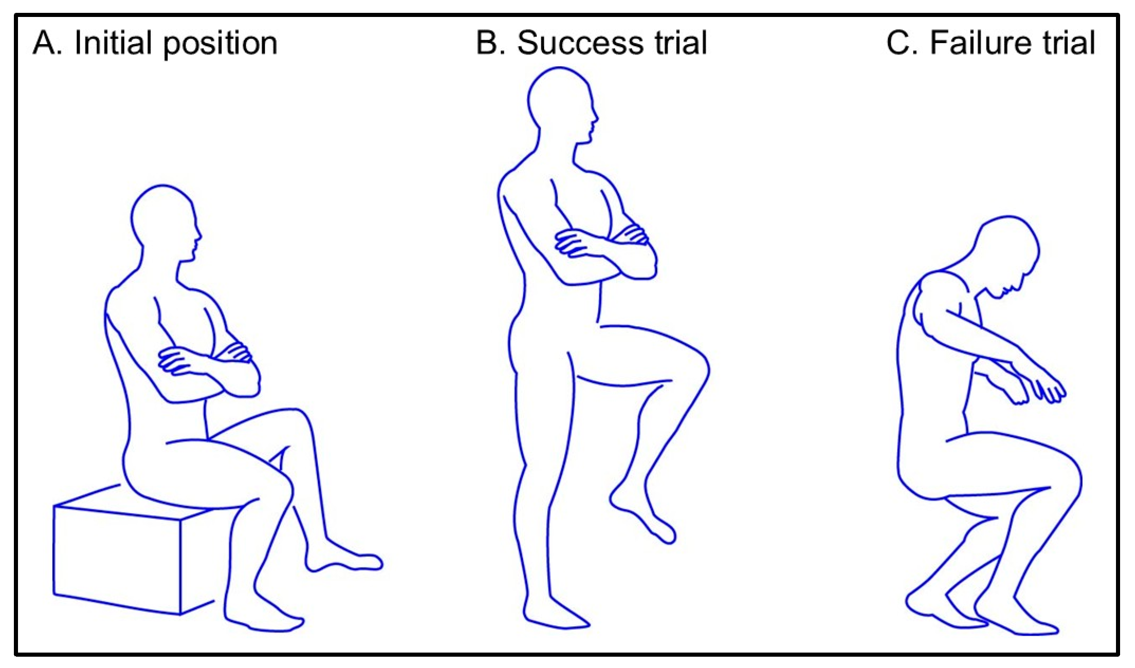
Figure 1. Experimental setup and criteria for the single-leg sit-to-stand movement. (A) Initial position: The participants began the single-leg sit-to-stand task seated on a height-adjustable platform with their right foot placed on a force platform and their left foot elevated off the ground. The knee and hip joints were flexed at 90°, and the participants were instructed to fold their arms across their chests. (B) Task execution: Upon the command “go,” participants performed the single-leg sit-to-stand movement by standing fully upright and returning to a seated position repeatedly for 30 s. Successful trials required participants to use only their tested leg and achieve full knee extension during the standing phase of the trial. (C) Failed trial: A trial was deemed unsuccessful if the participant placed the opposite foot on the ground or failed to fully extend the knee of the stance leg after the seat-off phase.
Biomechanics 05 00089 g001
Figure 2. Placement of EMG and IMU sensors. The electromyography (EMG) and inertial measurement unit (IMU) sensors are indicated by red and green, respectively. The EMG sensors were positioned on the right side of the body following the SENIAM recommendations [28]. A total of 16 IMU sensors were attached to the body at the following locations: the head, upper and lower trunk, bilateral upper arms, forearms, hands, thighs, shanks, and feet.
Biomechanics 05 00089 g002
Figure 3. Comparison of time-normalized EMG activity between successful and failed trials. The mean (solid lines) and 95% confidence intervals (shaded areas) of time-normalized EMG activity are shown for successful (n = 90; green) and failed trials (n = 19; red). Data are presented for the following eight muscles: (A) masseter (MA), (B) anterior deltoid (AD), (C) rectus abdominis (RA), (D) erector spinae longus (ESL), (E) rectus femoris (RF), (F) biceps femoris (BF), (G) tibialis anterior (TA), and (H) medial gastrocnemius (MGAS). In failed trials, the masseter and medial gastrocnemius muscles exhibited significantly higher EMG activity, whereas the erector spinae muscle showed lower EMG activity than in the successful trials. In failed trials, the masseter and medial gastrocnemius muscles demonstrated significantly higher EMG activity, whereas the erector spinae muscle exhibited lower EMG activity than in the successful trials.
Biomechanics 05 00089 g003
Table 1. Demographics, median ± interquartile range.
Age, years21.2 ± 0.4
Sex, male/female6:5
Height, mm167.9 ± 9.6
Body weight, kg59.7 ± 8.1
Dominant leg, right/left11:0
Table 2. Comparison of performance during the single-leg sit-to-stand task, mean ± standard deviation.
ControlClenchingESp-Value
Total attempts9.82 ± 3.529.91 ± 4.320.0210.878
Success count9.45 ± 3.808.18 ± 4.870.2280.031 *
Success rate0.96 ± 0.130.78 ± 0.290.6940.047 *
*: p < 0.05; ES: effect size estimated with Cohen’s d.
Table 3. Comparison of EMG metrics between control and clenching conditions during the single-leg sit-to-stand task with jaw clenching, mean ± standard deviation.
ControlClenchingESp-Value
EMG activity, %MVC
  Masseter3.3 ± 5.810.8 ± 7.01.323<0.001 *
  Anterior deltoid9.3 ± 14.28.3 ± 11.60.0280.845
  Rectus abdominis7.2 ± 7.86.7 ± 7.30.0070.961
  Erector spinae39.5 ± 16.941.9 ± 18.10.1250.375
  Rectus femoris57.9 ± 15.153.3 ± 13.70.1630.246
  Biceps femoris40.3 ± 18.940.5 ± 18.70.0290.839
  Tibialis anterior51.5 ± 18.244.3 ± 17.30.1460.303
  Medial gastrocnemius15.3 ± 8.016.6 ± 8.40.2170.126
Coactivation index
  ES-RA0.84 ± 0.150.85 ± 0.140.1070.448
  RF-BF0.39 ± 0.110.41 ± 0.120.1940.169
  MG-TA0.24 ± 0.130.26 ± 0.140.0880.535
*: p < 0.05; ES: effect size estimated with Cohen’s d; EMG: electromyography; MVC: maximum voluntary contraction; ES-RA: Coactivation index between the erector spinae and rectus abdominis, where a higher value indicates greater erector spinae activity relative to the rectus abdominis; RF-BF: Coactivation index between the rectus femoris and biceps femoris, where a higher value indicates greater rectus femoris activity relative to the biceps femoris; MG-TA: Coactivation index between the medial gastrocnemius and tibialis anterior, where a higher value indicates greater medial gastrocnemius activity relative to the tibialis anterior.
Table 4. Comparison of kinematics between control and clenching conditions during the single-leg sit-to-stand task with jaw clenching, mean ± standard deviation.
ControlClenchingESp-Value
Joint angle, degree
  Trunk pitch15.4 ± 17.215.6 ± 17.80.0760.892
  Trunk yaw2.3 ± 9.53.2 ± 7.60.0060.969
  Trunk roll1.9 ± 5.66.3 ± 5.11.020<0.001 *
  Hip flexion101.7 ± 20.4105.8 ± 15.90.698<0.001 *
  Hip abduction−20.6 ± 10.2−21.0 ± 10.80.549<0.001 *
  Hip internal rotation13.0 ± 15.417.5 ± 16.50.631<0.001 *
  Knee flexion109.7 ± 14.1113.1 ± 15.10.816<0.001 *
  Ankle dorsiflexion16.9 ± 7.817.8 ± 7.90.2110.145
  Hip angular velocity3.6 ± 8.56.0 ± 11.70.2580.068
*: p < 0.05; ES: effect size estimated with Cohen’s d.
Table 5. Comparison of EMG metrics between success and failure trials during the single-leg sit-to-stand task with jaw clenching, mean ± standard deviation.
Success
(n = 90)
Failure
(n = 19)
ESp-Value
EMG activity, %MVC
  Masseter6.8 ± 9.7115.5 ± 13.40.831<0.001 *
  Anterior deltoid7.7 ± 11.311.0 ± 12.90.5590.022 *
  Rectus abdominis6.8 ± 4.76.3 ± 4.70.1000.768
  Erector spinae44.9 ± 17.027.9 ± 17.01.007<0.001 *
  Rectus femoris53.7 ± 13.552.0 ± 15.20.1470.639
  Biceps femoris39.6 ± 18.544.9 ± 19.40.3050.260
  Tibialis anterior45.4 ± 17.039.34 ± 18.30.3870.170
  Medial gastrocnemius13.7 ± 7.322.6 ± 7.31.200<0.001 *
Coactivation index
  ES-RA0.88 ± 0.11±0.74 ± 0.221.042<0.001 *
  RF-BF0.41 ± 0.110.45 ± 0.150.4200.149
  MG-TA0.23 ± 0.110.39 ± 0.191.284<0.001 *
*: p < 0.05; ES: effect size estimated with Cohen’s d; EMG: electromyography; MVC: maximum voluntary contraction; ES-RA: Coactivation index between the erector spinae and rectus abdominis, where a higher value indicates greater erector spinae activity relative to the rectus abdominis; RF-BF: Coactivation index between the rectus femoris and biceps femoris, where a higher value indicates greater rectus femoris activity relative to the biceps femoris; MG-TA: Coactivation index between the medial gastrocnemius and tibialis anterior, where a higher value indicates greater medial gastrocnemius activity relative to the tibialis anterior.
Table 6. Comparison of kinematics between success and failure trials during the single-leg sit-to-stand task with jaw clenching, median ± interquartile range.
Success
(n = 90)
Failure
(n = 19)
95% Confidence Interval
(Lower Limit, Upper Limit)
ESp-Value
Joint angle, degree
  Trunk pitch18.15 ± 15.5912.76 ± 26.66−3.584, 14.3710.3610.236
  Trunk yaw−0.39 ± 6.725.70 ± 9.192.421, 8.9690.7130.001 *
  Trunk roll1.45 ± 6.608.83 ± 5.69−10.611, −4.1450.958<0.001 *
  Hip flexion104.68 ± 14.71110.93 ± 20.38−1.662, 14.1580.4790.120
  Hip abduction−20.69 ± 11.38−22.33 ± 7.110.763, 17.5470.1640.549
  Hip internal rotation15.80 ± 16.7424.96 ± 16.900.763, 17.5470.5180.033 *
  Knee flexion111.21 ± 13.78122.11 ± 17.793.629, 18.1780.6370.004 *
  Ankle dorsiflexion17.53 ± 6.5621.88 ± 10.740.629, 8.0650.5560.022 *
  Hip angular velocity5.86 ± 11.838.38 ± 12.74−3.477, 8.5180.2060.407
*: p < 0.05; ES: effect size estimated with Cohen’s d.
Disclaimer/Publisher’s Note: The statements, opinions and data contained in all publications are solely those of the individual author(s) and contributor(s) and not of MDPI and/or the editor(s). MDPI and/or the editor(s) disclaim responsibility for any injury to people or property resulting from any ideas, methods, instructions or products referred to in the content.