Article

Impact of Walking Path Length on Gait Parameters During the 2-Minute Walk Test in Healthy Young Adults

Department of Human Movement and Health Sciences, University of Rome ‘Foro Italico’, Piazzale Lauro De Bosis 6, 00135, Rome, Italy
*
Author to whom correspondence should be addressed.
Biomechanics 2025, 5(4), 82; https://doi.org/10.3390/biomechanics5040082
Submission received: 21 July 2025 / Revised: 10 September 2025 / Accepted: 30 September 2025 / Published: 10 October 2025

Abstract

Background/Objectives: The 2-minute walk test (2MWT) is a time-based gait assessment commonly employed for populations with limited walking ability for greater tolerability compared to the longer 6-minute test. The recommended distance to perform the tests is a 30 m straight path, a space requirement that is not always available in non-laboratory contexts. Shorter paths are therefore often adopted, but associated changes in gait patterns are not clear. The aim of the study is therefore to investigate how different walking path lengths affect gait patterns during the 2MWT. Methods: Twenty healthy young adults performed three walking trials on a straight hallway of 5 m, 15 m, and 30 m lengths. Spatiotemporal gait parameters were measured using three inertial measurement units on both distal tibiae and at pelvis level. Results: The 5 m path showed the greatest deviations, specifically in walking distance, walking speed, stride duration, stance time, swing time, single support time, and cadence, if compared to longer distances (p < 0.05). The 15 m path showed differences only in walking distance and walking speed (p < 0.05), if compared to the 30 m path. Conclusions: Shorter path lengths, particularly the 5 m, significantly impact gait patterns and should be considered when interpreting 2MWT results in clinical settings. The 30 m path is recommended as the gold standard, with 15 m as a viable alternative for assessing temporal parameters. Nevertheless, the extent to which each feature would be over/underestimated when walking in limited spaces is also addressed.

1. Introduction

Human walking is a complex motor function, integrating the motor and sensory systems [1,2]. To evaluate walking performance and estimate health status across populations, time-based tests like the 12-Minute Walk Test, and distance-based tests like the 10-Meter Walk Test, are commonly used. Shorter versions of time-based tests, such as the 6-Minute Walk Test (6MWT) and 2-Minute Walk Test (2MWT), are highly correlated with longer versions [3]. The 6MWT is popular due to its simplicity and similarity to daily living activities. However, for individuals with walking difficulties—such as amputees or the frail elderly—the 2MWT is preferred for its better tolerability and safety [4,5,6,7,8].
In time-based tests assessing functional capacity, total walking distance and walking speed are the most commonly measured parameters. More recently, wearable devices, like inertial measurement units (IMUs,) have been used for instrumenting different tests allowing spatiotemporal gait parameters, upper body accelerations, and gait variability to be captured, either in clinical or real-life settings [7,9,10,11,12]. Guidelines for the 6MWT recommend a straight path of at least 30 m to minimize turning points and maximize the distance walked [2].
However, such space may not always be available in clinical settings, thus motivating studies aiming to explore the feasibility of adopting shorter paths, such as 5 m, 10 m, 15 m, and 20 m [13,14]. Barnett et al. [15] compared the execution of the 6MWT across different path lengths and configurations (5 m, 10 m, 15 m, 30 m, rectangular, and continuous figure-of-eight), finding differences in walking speed, distance, and variability in stride length, stride width, and stride time, especially in shorter paths like 5 m.
Since the 6MWT can be time-consuming and fatiguing, especially for patients with walking impairments, the 2MWT has been adopted as a shorter and tolerable alternative [16]. Despite the recommendation of a 30 m straight path for the 2MWT [17], shorter or more discontinuous paths are often used in clinical environments. A recent study [18] compared 2MWT performance across six clinical settings with varying path lengths, obstacles, and surfaces. Results showed significant differences in total walking distance compared to normative reference values of research laboratory studies, indicating the impact of environmental factors. However, the study did not explore spatiotemporal gait parameters, leaving a gap in understanding how short path lengths influence gait patterns.
Thus, to the author’s knowledge, there is no clear indication in the literature as to the extent to which different gait parameters change because of shorter path lengths. An enhanced understanding of how much gait patterns are under-/overestimated when using shorter paths could be informative for guiding the choice of optimal settings for administering gait test protocols and for better interpreting the results when these settings are unavailable. Hence, this study aims to determine whether and how manipulations in walking path length would affect 2MWT gait patterns in healthy adult individuals.

2. Materials and Methods

Twenty healthy young adults (10 males and 10 females; mean age: 27.3 ± 2.9 years; body mass index (BMI): 22.8 ± 2.4 kg/m2) were recruited (Power Analysis, G*Power 3: effect size f = 0.4, power (1-β error = 0.95, α = 0.05)) for this cross-sectional study. Exclusion criteria were psychological, neurological, musculoskeletal disorders, or any condition affecting gait, and injuries occurred in the last six months.
Participants performed three 2MWT trials, one for each straight path of 5 m, 15 m, and 30 m in a randomized order. A rest period of at least 3 min was provided between trials, and participants were asked whether they felt ready to continue before starting the next trial. Participants were instructed to walk at their normal, self-selected pace, wearing comfortable shoes. Turning points (180°) were marked at the ends of each path (Figure 1b), and verbal cues were used to signal the start and stop of each trial. Three synchronized IMUs (Movit® by Captiks Srl, Rome, Italy, 100 Hz, ±1.7 g range) were placed on both lateral distal tibiae and at L5 level (Figure 1a). Sensors were secured using Velcro straps provided by Captiks Srl and positioned according to the manufacturer’s guidelines.
Raw accelerometer and gyroscope data were processed with the manufacturer’s validated algorithms using Motion Analyzer software (Captiks srl) which automatically detects gait events and compute spatio-temporal parameters. The validity and reliability of these algorithms for spatio-temporal gait analysis have been previously reported [19,20]. The following gait parameters were extracted: walking distance (WD) (m), walking speed (WS) (m/s), cadence (steps/min), stride duration (s), double support time (s), single support time (s), stance and swing phases (s).
Statistical analyses were conducted in SPSS Version 29 (IBM Corporation US) using repeated measures ANOVA for normally distributed data and Friedman tests for non-normally distributed data. Post hoc comparisons employed Bonferroni correction, to prevent Type I error. Effect sizes for each comparison were expressed as ηρ2 values.

3. Results

Each of the 20 participants completed all three trials, resulting in a consistent dataset size across all conditions. Results are summarized in Table 1, highlighting significant differences across the three path lengths. WD for the 5 m path was significantly lower compared to both the 15 m and 30 m paths (p < 0.001), and the 15 m path also showed a significant reduction compared to the 30 m path (p < 0.001). Similarly, WS also differed across all path lengths, with the 5 m path showing the largest reduction (p < 0.001 for 5 m vs. 15 m/30 m; p = 0.005 for 15 m vs. 30 m). Other gait parameters (cadence, stride duration, double support time, single support time, stance, and swing time) showed no significant differences between 15 m and 30 m paths. In contrast, the 5 m path led to increased stride duration, swing time, single support time, accompanied by a decrease in cadence (p ≤ 0.001). Stance time (significant in the main effect but not in post hoc comparisons) and double support time showed no significant differences. Table 2 reports the extent of underestimation or overestimation of each parameter for the 5 m and 15 m paths relative to the 30 m path. WD and WS appear to be the most affected by shorter paths, whereas stride-level temporal parameters remain largely consistent for 15 m. The 5 m configuration, instead, showed an overestimation of stride duration, swing time, and single support time, and an underestimation of cadence.

4. Discussion

This study aimed to assess the impact of path length on 2MWT gait parameters in young adults. Results show that walking path length significantly affects 2MWT performance, with shorter paths leading to reductions in WD and WS, as well as alterations in temporal parameters. These findings are consistent with previous studies on the 6MWT [15], suggesting the adoption of different walking strategies on shorter paths. In addition, considering the range of values obtained for each gait parameter in this study, it was possible to indicate the extent to which each feature would be over/underestimated when walking in limited spaces (Table 2).
The highest WD and WS values were observed on the longest walking path (30 m), followed by the 15 m path, with the lowest values recorded for the 5 m path. Specifically, WD on the 5 m path was 31% lower than the gold standard 30 m path, while the 15 m path showed a reduction of over 23%. This aligns with previous findings about the 6MWT [15], demonstrating that reducing the number of required turns significantly improves total walking distance in the 2MWT. Additionally, longer uninterrupted walking allows participants to reach a steady-state WS, minimizing the impact of acceleration and deceleration phases, which are usually around 2.17 m each [21]. In the 5 m path, most of the distance comprises these phases, resulting in lower WS. Since WS is a functional vital sign and a predictor of health status, clinicians should carefully consider path length effects, as a reduction from 30 m to 5 m leads to an 8% drop in WS (Table 2). Regarding the clinical significance of such percentage variations, the differences in walking speed observed between the 15 m and 30 m paths (0.07 m/s) remain below or at the lower bound of minimal clinically important differences (MCIDs) reported in adults with various pathologies, which range from 0.10 to 0.20 m/s [22]. We cannot state the same for the differences between 5 m and 15 m, which amount to 0.14 m/s, appearing clinically relevant. This suggests that for the 15 m walkway, the difference in total walking distance compared to the 30 m path (approximately 18 m) may be clinically meaningful in certain patient populations, such as those in subacute stroke or undergoing COPD rehabilitation [23], while it may not be relevant for others [24]. Overall, our findings indicate that for a 15 m walkway, all stride-level temporal parameters can be reliably measured, while deviations in walking speed are minimal and unlikely to be clinically meaningful. For the WD, instead, the 15 m walkway should be interpreted with caution, as the clinical significance of this difference should be considered relative to the patient’s pathology.
For temporal parameters, results showed an inverse relationship with WS, consistent with prior studies [25,26]. The 5 m path produced the greatest temporal alterations, while the 15 m and 30 m paths showed no significant differences. Stride duration was longest on the 5 m path, likely due to the slower WS, as previously reported [25,26], where both studies observed an increase in the stride duration when reducing walking speeds. The slower WS also affected stance and swing time. However, in this study, while stance and swing time were both longer on the 5 m path, only swing time differences were statistically significant from 15 m and 30 m paths, with a swing increase of over 5% and single limb support time increase of nearly 3% when reducing the path length from 30 m to 5 m (Table 2). As previously stated, instead, the stance duration and the double support time did not show significant differences. The lack of significant stance time alterations supports the idea that stance and swing relate to distinct control mechanisms [25]: stance time appears to vary linearly with WS, while swing relation to WS seems to be better represented by a polynomial quadratic model, meaning the relation between the decrement of the swing value at the increment of the WS is not represented by a straight line but follows a quadratic function. Additionally, it should be considered that walking over 5 m mainly consists of acceleration/deceleration phases, rather than steady state walking. Thus, it is suggested that these phases may involve different walking strategies, and their influence on gait temporal parameters would require further investigation.
In terms of cadence, the 30 m and 15 m paths produced values consistent with those previously reported in the 6MWT [9]. Cadence decreased by about 3% in the 5 m path compared to the 30 m path (Table 2), consistent with the longer stride duration and supporting the correlation between cadence and WS previously reported [27].
To the author’s knowledge, this study represents the first attempt to quantify how walking path length influences gait patterns in the 2MWT for young adults and how much gait parameters are under- or overestimated when performing the 2MWT on shorter paths than the recommended 30 m.
From a practical perspective, these findings have direct clinical implications. Specifically, an underestimation of the walking speed due to a shorter path length could lead to misclassification of a patient against established cut-off values used for risk stratification, despite their actual performance being adequate. Similarly, Underestimations of total walking distance by up to 30% or less were observed, underestimations in total walking distance of up to 30% or smaller were found, but systematic deviations in cadence and stride duration could lead to incorrect interpretations of functional capacity or rehabilitation progress. Reporting both raw and percentage differences, as provided in this study, facilitates a more accurate adjustment of test results when the recommended 30 m walkway is not available. Recognizing the systematic underestimation introduced by shorter walkways can support clinical decision-making in different scenarios, such as when comparing patient outcomes across facilities with varying space availability, when monitoring progress in rehabilitation programs, or when interpreting longitudinal changes in patients tested in different environments.
In these conditions, understanding the levels of under-overestimation in investigated gait parameters, especially those resulting from the interaction of various factors, including body structure, postural control, lower extremity strength, and proprioception [21], can be valuable for clinicians. Addressing these under-overestimations can improve the accuracy of test results, allowing for more reliable comparisons with normative values obtained under standard conditions. Furthermore, accurate measurements are crucial for evaluating patients effectively and determining whether there are impairments in any of the systems that contribute to human walking.
Some limitations must be addressed, such as the focus on only young adults. Other individual factors, including age, BMI, and the presence of gait impairments, are known to affect spatiotemporal parameters and may interact with walkway length.

5. Conclusions

When assessing gait spatiotemporal parameters, the 30 m path should be considered the gold standard, while the 15 m path might represent a suitable alternative. In these conditions, walking speed is slightly underestimated on shorter paths, but the magnitude of this change is unlikely to be clinically meaningful, and temporal parameters such as cadence, stride duration, stance, and swing times showed minimal differences between 15 m and 30 m paths, supporting the acceptability of these measures even when dealing with shorter walkways. In contrast, total walking distance is more strongly affected by path length. Therefore, clinicians may consider using a 15 m walkway when a 30 m space is unavailable.
The 5 m path appears to be the least preferable, as short paths do not allow for steady-state walking speed and may introduce compensatory strategies. However, if an appropriate quantity of space is not available, as in clinical environments, it is essential to consider the impact of shorter distances on gait patterns and the extent of underestimation of specific parameters and adjust interpretations accordingly.
For future research, further studies are needed to extend the analysis to more heterogeneous cohorts, including elderly populations and clinical groups with gait impairments, to confirm whether the findings observed in healthy young adults generalize to these populations.

Author Contributions

Conceptualization; C.L.Z., V.B. and G.V.; data curation; C.L.Z.; formal analysis; C.L.Z.; investigation; C.L.Z.; methodology; C.L.Z., V.B. and G.V.; project administration; C.L.Z.; resources; V.B. and G.V.; software; C.L.Z.; supervision; V.B. and G.V.; validation; V.B. and G.V.; roles/writing—original draft; C.L.Z.; writing—review & editing: all authors. All authors have read and agreed to the published version of the manuscript.

Funding

This research received no external funding.

Institutional Review Board Statement

The study was conducted in accordance with the Declaration of Helsinki and approved by the University Research Committee of the University of Rome “Foro Italico”, CAR 184/2024.

Informed Consent Statement

Informed consent was obtained from all subjects involved in the study. Written informed consent has been obtained from the patients to publish this paper.

Data Availability Statement

The data presented in this study are available on request from the corresponding author.

Conflicts of Interest

The authors declare no conflicts of interest.

Abbreviations

The following abbreviations are used in this manuscript:
6MWT6-Minute Walk Test
2MWT2-Minute Walk Test
IMUsInertial Measurement Units
WDWalking Distance
WSWalking Speed
SDStandard Deviation

References

  1. Hollman, J.H.; McDade, E.M.; Petersen, R.C. Normative spatiotemporal gait parameters in older adults. Gait Posture 2011, 34, 111–118. [Google Scholar] [CrossRef]
  2. American Thoracic Society. ATS Statement. Am. J. Respir. Crit. Care Med. 2002, 166, 111–117. [Google Scholar] [CrossRef] [PubMed]
  3. Butland, R.J.A.; Pang, J.; Gross, E.R.; Woodcock, A.A.; Geddes, D.M. Two-, six-, and 12-minute walking tests in respiratory disease. Br. Med. J. (Clin. Res. Ed.) 1982, 284, 1607–1608. [Google Scholar] [CrossRef]
  4. Wafi, A.; Ribeiro, L.; Kolli, V.; Azhar, B.; Budge, J.; Loftus, I.M.; Holt, P.J. Predicting Prosthetic Mobility at Discharge from Rehabilitation Following Major Amputation in Vascular Surgery. Eur. J. Vasc. Endovasc. Surg. 2023, 66, 832–839. [Google Scholar] [CrossRef]
  5. Wong, C.K.; Chihuri, S.T.; Santo, E.G.; White, R.A. Relevance of medical comorbidities for functional mobility in people with limb loss: Retrospective explanatory models for a clinical walking measure and a patient-reported functional outcome. Physiotherapy 2020, 107, 133–141. [Google Scholar] [CrossRef]
  6. Frlan-Vrgoc, L.; Vrbanić, T.S.L.; Kraguljac, D.; Kovacević, M. Functional outcome assessment of lower limb amputees and prosthetic users with a 2-minute walk test. Coll. Antropol. 2011, 35, 1215–1218. [Google Scholar]
  7. O’Keefe, J.A.; Guan, J.; Robertson, E.; Biskis, A.; Joyce, J.; Ouyang, B.; Liu, Y.; Carnes, D.; Purcell, N.; Berry-Kravis, E.; et al. The Effects of Dual Task Cognitive Interference and Fast-Paced Walking on Gait, Turns, and Falls in Men and Women with FXTAS. Cerebellum 2021, 20, 212–221. [Google Scholar] [CrossRef] [PubMed]
  8. Chan, W.L.S.; Pin, T.W. Reliability, validity and minimal detectable change of 2-min walk test and 10-m walk test in frail older adults receiving day care and residential care. Aging Clin. Exp. Res. 2020, 32, 597–604. [Google Scholar] [CrossRef] [PubMed]
  9. Kushioka, J.; Sun, R.; Zhang, W.; Muaremi, A.; Leutheuser, H.; Odonkor, C.A.; Smuck, M. Gait Variability to Phenotype Common Orthopedic Gait Impairments Using Wearable Sensors. Sensors 2022, 22, 9301. [Google Scholar] [CrossRef]
  10. Wang, R.Y.; Wang, Y.L.; Cheng, F.Y.; Chao, Y.H.; Chen, C.L.; Yang, Y.R. Effects of combined exercise on gait variability in community-dwelling older adults. AGE 2015, 37, 40. [Google Scholar] [CrossRef]
  11. Theunissen, K.; Plasqui, G.; Boonen, A.; Timmermans, A.; Meyns, P.; Feys, P.; Meijer, K. The increased perceived exertion during the six minute walking test is not accompanied by changes in cost of walking, gait characteristics or muscle fatigue in persons with multiple sclerosis. Mult. Scler. Relat. Disord. 2023, 70, 104479. [Google Scholar] [CrossRef] [PubMed]
  12. Grimpampi, E.; Oesen, S.; Halper, B.; Hofmann, M.; Wessner, B.; Mazzà, C. Reliability of gait variability assessment in older individuals during a six-minute walk test. J. Biomech. 2015, 48, 4185–4189. [Google Scholar] [CrossRef] [PubMed]
  13. Troosters, T.; Gosselink, R.; Decramer, M. Six minute walking distance in healthy elderly subjects. Eur. Respir. J. 1999, 14, 270–274. Available online: https://pubmed.ncbi.nlm.nih.gov/10515400/ (accessed on 13 December 2023). [CrossRef] [PubMed]
  14. Lipkin, D.P.; Scriven, A.J.; Crake, T.; Poole-Wilson, P.A. Six minute walking test for assessing exercise capacity in chronic heart failure. Br. Med. J. (Clin. Res. Ed.) 1986, 292, 653–655. Available online: https://pubmed.ncbi.nlm.nih.gov/3081210/ (accessed on 13 December 2023). [CrossRef]
  15. Barnett, C.T.; Bisele, M.; Jackman, J.S.; Rayne, T.; Moore, N.C.; Spalding, J.L.; Richardson, P.; Plummer, B. Manipulating walking path configuration influences gait variability and six-minute walk test outcomes in older and younger adults. Gait Posture 2016, 44, 221–226. [Google Scholar] [CrossRef]
  16. Gaunaurd, I.; Kristal, A.; Horn, A.; Krueger, C.; Muro, O.; Rosenberg, A.; Gruben, K.; Kirk-Sanchez, N.; Pasquina, P.; Gailey, R. The Utility of the 2-Minute Walk Test as a Measure of Mobility in People with Lower Limb Amputation. Arch. Phys. Med. Rehabil. 2020, 101, 1183–1189. [Google Scholar] [CrossRef]
  17. Pin, T.W. Psychometric Properties of 2-Minute Walk Test: A Systematic Review. Arch. Phys. Med. Rehabil. 2014, 95, 1759–1775. [Google Scholar] [CrossRef]
  18. Shank, C.; Kristal, A.; Van Veld, R.; Applegate, B.; Gaunaurd, I.; Gailey, R. Variations in 2-Minute Walk Test outcomes for people with lower limb amputation in the outpatient clinic and research settings. Prosthet. Orthot. Int. 2022, 46, 140–147. [Google Scholar] [CrossRef]
  19. Saggio, G.; Tombolini, F.; Ruggiero, A. Technology-Based Complex Motor Tasks Assessment: A 6-DOF Inertial-Based System Versus a Gold-Standard Optoelectronic-Based One. IEEE Sens. J. 2021, 21, 1616–1624. [Google Scholar] [CrossRef]
  20. Ricci, M.; Terribili, M.; Giannini, F.; Errico, V.; Pallotti, A.; Galasso, C.; Tomasello, L.; Sias, S.; Saggio, G. Wearable-based electronics to objectively support diagnosis of motor impairments in school-aged children. J. Biomech. 2019, 83, 243–252. [Google Scholar] [CrossRef]
  21. Middleton, A.; Fritz, S.L.; Lusardi, M. Walking Speed: The Functional Vital Sign. J. Aging Phys. Act. 2015, 23, 314. [Google Scholar] [CrossRef] [PubMed]
  22. Bohannon, R.W.; Glenney, S.S. Minimal clinically important difference for change in comfortable gait speed of adults with pathology: A systematic review. J. Eval. Clin. Pract. 2014, 20, 295–300. [Google Scholar] [CrossRef] [PubMed]
  23. Johnston, K.N.; Potter, A.J.; Phillips, A.C. Minimal important difference and responsiveness of 2-minute walk test performance in people with COPD undergoing pulmonary rehabilitation. Int. J. Chronic Obstr. Pulm. Dis. 2017, 12, 2849–2857. [Google Scholar] [CrossRef] [PubMed]
  24. Bowman, T.; Mestanza Mattos, F.G.; Allera Longo, C.; Bocini, S.; Gennuso, M.; Marazzini, F.; Materazzi, F.G.; Pelosin, E.; Putzolu, M.; Salvalaggio, S.; et al. The minimally clinically important difference in the 2-minute walk test for people in the subacute phase after a stroke. Top. Stroke Rehabil. 2025, 32, 553–561. [Google Scholar] [CrossRef]
  25. Smith, A.J.J.; Lemaire, E.D. Temporal-spatial gait parameter models of very slow walking. Gait Posture 2018, 61, 125–129. [Google Scholar] [CrossRef]
  26. Ziegler, J.; Gattringer, H.; Müller, A. On the relation between gait speed and gait cycle duration for walking on even ground. J. Biomech. 2024, 164, 111976. [Google Scholar] [CrossRef]
  27. Retory, Y.; David, P.; Niedzialkowski, P.; de Picciotto, C.; Bonay, M.; Petitjean, M. Gait Monitoring and Walk Distance Estimation with an Accelerometer During 6-Minute Walk Test. Respir. Care 2019, 64, 923–930. [Google Scholar] [CrossRef]
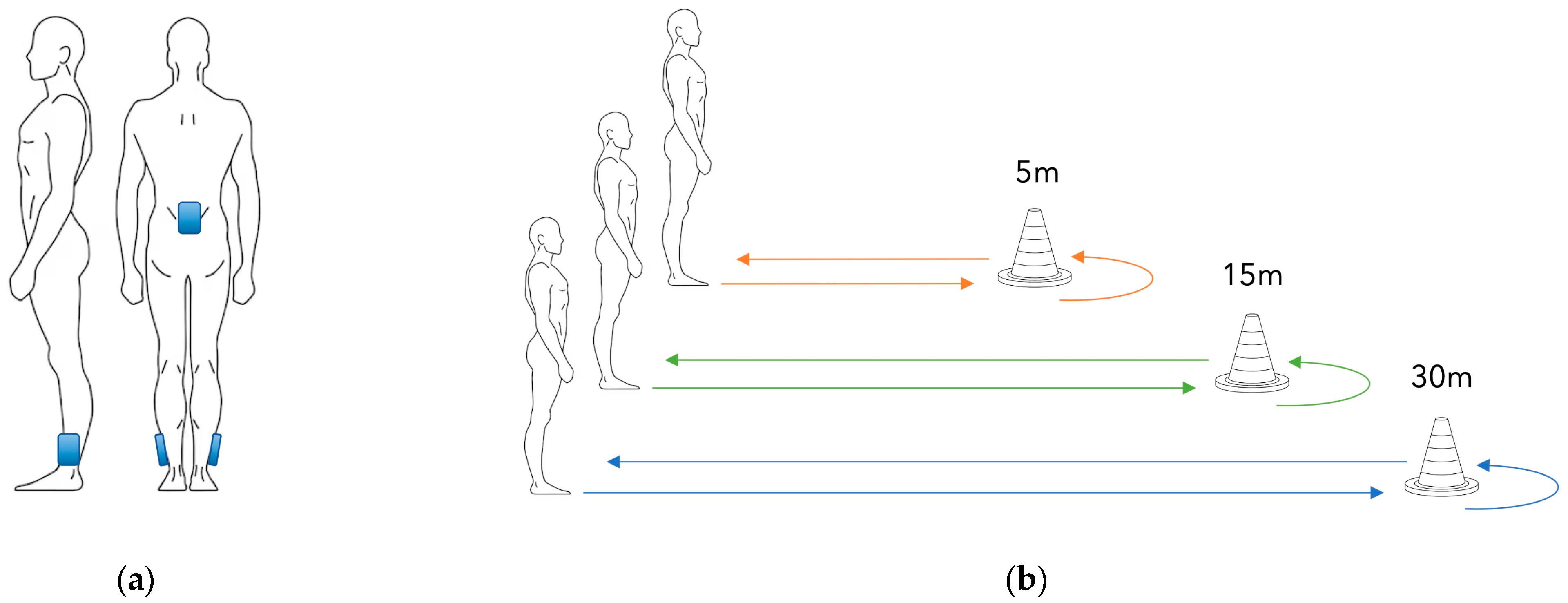
Figure 1. Illustration of the experimental setup: (a) schematic representation of the placement of the inertial sensors (Movit®, Captiks Srl, Rome, Italy) on both lateral distal tibiae and at the L5 level; (b) schematic representation of the walking paths used in the 2-min walk test (5 m, 15 m, and 30 m), with cones marking the turning points (180° turns).
Biomechanics 05 00082 g001
Table 1. The groups mean and standard deviation (SD) of each parameter for each straight-line walking length are reported. The p-value of each main effect is reported, and bold numbers indicate statistically significant differences (p < 0.05). Effect size is reported as Eta-squared (ηρ2), for parametric tests, or Kendall W (W), for non-parametric tests.
5 m 15 m 30 m
Mean±SDMean±SDMean±SDp-ValueEffect Size
Walking Distance (m)123.69 a,b14.36161.77 a,c14.67179.37 b,c19.42<0.001ηρ2 = 0.952
Walking Speed (m/s)1.62 a,b0.171.69 a,c0.161.76 b,c0.16<0.001W = 0.473
Stride Duration (s)1.05 a,b0.071.010.061.000.06<0.001ηρ2 = 0.625
Stance Time (s)0.610.060.600.050.590.050.01ηρ2 = 0.268
Swing Time (s)0.43 a,b0.030.410.020.410.02<0.001ηρ2 = 0.561
Single Support Time (s)0.42 a,b0.020.410.020.410.02<0.001ηρ2 = 0.508
Double Support Time (s)0.190.050.190.040.180.040.265ηρ2 = 0.067
Cadence (steps/min)115.48 a,b7.30119.056.66119.976.76<0.001ηρ2 = 0.658
a Significant difference from 30 m. b Significant difference from 15 m. c Significant difference from 5 m.
Table 2. The underestimation level of each parameter is reported as a difference expressed in their respective measurement units (left part of the table) and then expressed as a % difference (right part of the table). Bold values indicate measurements that were found to be statistically significant in Table 1.
Underestimation Level of:Difference (%)
5 m
Compared to
15 m
5 m
Compared to
30 m
15 m
Compared to
30 m
5 m
Compared to
15 m
5 m
Compared to
30 m
15 m
Compared to
30 m
Walking Distance (m)−38.07−55.68−17.61−23.5%−31.0%−9.8%
Walking Speed (m/s)−0.06−0.14−0.07−3.7%−7.8%−4.2%
Stride Duration (s)0.030.040.013.3%4.1%0.8%
Stance Time (s)0.010.020.012.1%3.1%1.0%
Swing Time (s)0.020.020.004.9%5.5%0.5%
Single Support Time (s)0.010.010.002.3%2.8%0.4%
Double Support Time (s)0.000.010.001.6%3.9%2.3%
Cadence (steps/min)−3.57−4.49−0.92−3.0%−3.7%−0.8%
Disclaimer/Publisher’s Note: The statements, opinions and data contained in all publications are solely those of the individual author(s) and contributor(s) and not of MDPI and/or the editor(s). MDPI and/or the editor(s) disclaim responsibility for any injury to people or property resulting from any ideas, methods, instructions or products referred to in the content.