Article

Can Thymol-Based Emulsion(s) Combat Intrahospital Multidrug-Resistant Isolates of Klebsiella pneumoniae?


by
Tatjana Vukotić
1,
Milica Nemoda
2,
Vladimir Živanović
1,
Filip Veljković
2,
Božana Petrović
2,
Marijana Janić
2,
Jelena Filipović Tričković
2,
Biljana Nikolić
3 and
Jelena Marinković
2,*
1
Clinical Hospital Center “Dr Dragiša Mišović—Dedinje”, Heroja Milana Tepića 1, 11 000 Belgrade, Serbia
2
Department of Physical Chemistry, “Vinča” Institute of Nuclear Sciences, National Institute of the Republic of Serbia, University of Belgrade, Mike Petrovića Alasa 12, 11 000 Belgrade, Serbia
3
Department of Microbiology, Faculty of Biology, University of Belgrade, Studentski Trg 16, 11 000 Belgrade, Serbia
*
Author to whom correspondence should be addressed.
Compounds 2026, 6(1), 22; https://doi.org/10.3390/compounds6010022
Submission received: 20 January 2026 / Revised: 25 February 2026 / Accepted: 11 March 2026 / Published: 13 March 2026

Abstract

Background: Increasing antimicrobial resistance has directed studies toward investigating the antimicrobial activity of thymol, as well as the antibiofilm and antioxidant potential of its emulsions (with Tween 80) against multidrug-resistant (MDR) K. pneumoniae isolates. Methods: A microdilution assay was used to estimate thymol’s antibacterial potential against 10 clinical isolates (labeled 1–10). The dynamic light scattering technique was used to measure the particle size diameter (Zavg) of formulated emulsions. The antibiofilm potential of emulsions was assessed in vitro using a crystal violet assay and ex vivo on a surgical drain through a colony-forming unit assay. Antioxidant activity was screened by using the DPPH assay. Results: The MIC values were ≤1.5 mg/mL for strains 1 and 7 and <0.5 mg/mL for the other strains. Emulsions E250:500, E250:750, E300:750, and E500:750 were stable and homogeneous, with a Zavg of approx. 200 nm (128.4 ± 0.8 nm for E250:750). These emulsions significantly reduced the biofilm biomass of strains 3 and 7 (50.6–74.32% and 34.60–59.8% of inhibition, respectively), with the strongest activity observed for E250:500 and E500:750. Antibiofilm potential was confirmed ex vivo, with E500:750 showing the highest efficacy (ΔLogCFU 2.60 and 2.68 for strains 3 and 7). E250:750 demonstrated the highest capacity to neutralize the DPPH• radical. Conclusions: Thymol and its emulsions exhibited antibacterial and antibiofilm activity against MDR K. pneumoniae isolates, along with the proven antioxidant properties of the emulsions.

1. Introduction

Healthcare-associated infections (HAIs) are infections that occur in patients during healthcare provision in a hospital or other healthcare facility and are absent at the time of admission or in the incubation period [1]. These infections, which typically manifest 48 h after admission or within 30 days after treatment [1], represent the most common adverse event during hospitalization [2]. According to data from the European Centre for Disease Prevention and Control (ECDC), over 3.5 million cases of HAIs occur annually in Europe, resulting in over 90,000 deaths [3]. The most common types of HAIs are urinary tract infections (UTIs), surgical site infections (SSIs), respiratory tract infections, bloodstream infections, and gastrointestinal infections [3]. Risk factors for HAIs include prolonged hospital stays, urinary and central venous catheters, and antibiotic therapy [4]. Presenting a significant global health concern, HAIs have prevalence rates that vary across regions. In Europe, a point prevalence survey estimated the prevalence of HAIs to be 6.5% in acute care hospitals and 3.9% in long-term care facilities [5]. A similar survey conducted in Serbia in 2017, following the ECDC framework, reported that 4.2% of adult patients had at least one HAI, with the highest prevalence observed in intensive care units (ICUs), at 15.9% [6]. This study identified UTIs and SSIs as the most common HAIs, occurring in 23.9% and 23% of cases, respectively, with Klebsiella spp. pointed out as the predominant pathogen responsible for 16.7% of total cases [6]. According to a study conducted by Despotović et al. [2], in ICUs in Serbia, Klebsiella spp. was the causative agent in 14% of HAIs, with 91.2% of Klebsiella isolates exhibiting a multidrug-resistant (MDR) pattern.
When MDR strains cause HAIs, properly selecting effective treatments and/or developing innovative therapeutic strategies to address resistant pathogens and avoid adverse outcomes is paramount. An alternative approach to combat MDR bacteria could involve the use of natural antimicrobial agents, such as essential oils (EOs) or their constituents [7]. Currently, there is no evidence of bacteria developing resistance to EOs or their constituents [8]. Among many different EO constituents, thymol has garnered significant interest among researchers [9,10]. It is a natural monoterpenoid phenol, colorless and crystalline, with a characteristic odor [11]. Thymol has shown various pharmacological properties, antibacterial, antiviral, antifungal, antiseptic, antioxidant, anti-inflammatory, and analgesic effects [12,13], making it useful in the pharmaceutical, cosmetic, food, and agronomic industries and especially in medicine [11].
Considering the literature data, thymol has demonstrated significant antimicrobial activity against Klebsiella pneumoniae reference and urine-originated strains. Moreover, its synergistic activity with commercial antibiotics in combating K. pneumoniae planktonic and biofilm forms was also previously suggested [9,10]. Recent pioneering studies have focused on exploring specific delivery systems to enhance thymol’s antibacterial activity through nanosizing. In this regard, Li et al. [14] and Bisso et al. (2022) [15] presented encouraging results, with the latter confirming the promising activity of the thymol nanoparticles against K. pneumoniae (urine isolates and reference strain). These results seem to motivate further exploration of properly delivered thymol particles against K. pneumoniae MDR isolates.
Bearing in mind that HAIs are associated with inflammation and oxidative stress, one additional valuable attribute of the novel therapeutic aid could be its antioxidant activity. This property has also been attributed to thymol [16], and previous studies have suggested that its nanoemulsification enhances its antioxidant potential [17,18]. Additionally, Deng et al. [19] demonstrated that the solubilization of thymol in Tween 80 micelles can enhance certain aspects of its antioxidant activity.
Taking into account the increasing prevalence of Klebsiella pneumoniae MDR isolates as a causative agent of HAIs and the desired antioxidant potential of novel antibacterial treatments for surgical wounds, this study aimed to investigate the antibacterial and antioxidant potential of thymol-based emulsions, with a particular focus on their antibiofilm properties, against biofilms formed both in vitro and ex vivo, i.e., on the surgical drain. A concentrated 10% povidone–iodine solution was included in the study as a control, as it is commonly used to treat surgical wounds.

2. Materials and Methods

2.1. Chemicals

Thymol (≥97%, FCC, FG), purchased from Sigma-Aldrich, St. Louis, MO, USA, and Polyoxyethylene sorbitan monooleate (Tween 80), purchased from Fisher Scientific, Geel, Belgium, were used in this study. A concentrated 10% solution of povidone–iodine (Hemofarm, Vršac, Serbia) was used as a positive control in the microdilution and antibiofilm assays and was also tested in the DPPH assay.

2.2. Bacterial Strains

All MDR Klebsiella pneumoniae isolates used in this study were not collected by the research team but were obtained directly from the hospital’s microbiology laboratory. These isolates had been routinely processed by the physicians (internists and surgeons) who are required, as part of standard diagnostic and therapeutic procedures, to collect swabs or other clinical materials from patients suspected of having hospital-acquired infections. All samples were therefore taken solely for clinical purposes, independently of this research, and in accordance with the approval of the Local Ethical Committee of University Hospital Center “Dr Dragiša Mišović−Dedinje”, Belgrade, Serbia [No. 15341/3-2025; 21 July 2025]. Upon completion of routine diagnostics, the microbiology specialist provided the research team exclusively with pure, identified bacterial isolates, without any patient identifiers or access to medical records. The research team had no involvement in patient sampling, clinical decision-making, or specimen collection procedures, nor did the microbiology specialist have insight into how swabs were taken, as this is not part of her professional responsibilities. Thus, only anonymized isolates, belonging to the hospital’s microbial flora, were used for experimental analyses. A total of ten isolates, labeled as K. pneumoniaen, where n is a digit in the range 1–10, were obtained: three from urine cultures, three from blood cultures, and four from pyocultures. All isolates were resistant to third- and fourth-generation cephalosporins, meropenem, and levofloxacin. However, two isolates from urine cultures and one from blood culture were sensitive to colistin, while one isolate from each culture type was sensitive to gentamicin (Table 1).

2.3. Antibacterial Potential of Thymol

The antibacterial efficacy of thymol on free-living planktonic cells of Klebsiella spp. was assessed in a microdilution assay similar to that of Marinković et al. [20]. Two-fold serial dilutions of thymol were performed in 96-well microtiter plates with TSB medium. The thymol was mixed with Tween 80 at a ratio of 1:1 (v/v), resulting in a 464 mg/mL stock solution. Two-fold serial dilutions were prepared in the rows of a microtiter plate, while the tested concentration range was 0.03–59 mg/mL. In addition, serial dilutions of Tween 80 were also tested, in order to estimate the possible solvent inhibitory effect. Bacterial inoculum was then added to achieve a final concentration of 105 CFU/mL in each well. Resazurin was used as a growth indicator at a concentration of 0.0675 mg/mL. The minimum inhibitory concentrations (MICs) were determined as the lowest concentrations of thymol that prevented bacterial growth, as indicated by the absence of a color change in the growth indicator. To determine the minimal bactericidal concentrations (MBCs), 15 μL from wells without visible growth was plated onto Mueller–Hinton Agar (MHA, Liofilchem, Italy). Moreover, 10% povidone–iodine was used as the positive control (tested concentrations: 1–127.25 mg/mL). The experiment was conducted in triplicate and repeated twice.

2.4. Preparation and Characterization of the Emulsions

2.4.1. Preparation of the Emulsions

Based on the obtained MIC values of thymol, several emulsions were formulated, each containing thymol and the emulsifier Tween 80 (T80) in different ratios (Table 2).
The emulsions were visually assessed immediately after preparation. The emulsification process involved 15 min of magnetic stirring, followed by 20 min of ultrasonic mixing at 30% power. Only emulsions deemed homogeneous after both processes were selected for further particle size analysis.

2.4.2. Characterization of Thymol-Based Emulsions

The dynamic light scattering (DLS) technique (Zetasizer Nano ZS, Malvern Panalytcal Ltd., Malvern, UK) was used to measure the particle sizes (Zavg) and polydispersity indices (PDIs) of the four emulsions (E250:500, E250:750, E300:750, and E500:750) identified as homogeneous. Intensity mean values were calculated by monitoring the time fluctuation of the laser beam intensity scattered from the sample at a 90° angle [21].

2.5. In Vitro Antibiofilm Activity of Thymol-Based Emulsions

Following the determination of MICs and the formulation of emulsions, four isolates (K. pneumoniae1, K. pneumoniae2, K. pneumoniae3, and K. pneumoniae7) with the lowest sensitivity to thymol were selected, and their biofilms were subjected to the emulsions. To promote biofilm formation, inoculum containing 2 × 10⁵ CFU/well in TSB medium, enriched with 2% glucose, was added to polystyrene 96-well flat-bottom microtiter plates and incubated at 37 °C for 7 days. After biofilms had formed, they were treated with thymol-based emulsions. The antibiofilm effects of the emulsions on pre-formed biofilms were assessed using the crystal violet assay in 96-well plates, as described previously in Marinković et al. [20]. After exposing the biofilms, the medium containing the test substances and planktonic cells was removed by rinsing twice with sterile distilled water. Subsequently, the plates were dried at room temperature, and the biofilm was stained with 0.1% crystal violet (Thermo Fisher Scientific, Horsham, UK). The wells were then washed, and the remaining stain incorporated into the biofilm was dissolved in 96% ethanol. Biofilm disruption was quantified spectrophotometrically at 570 nm, and the percentage of biofilm disruption (I) was calculated using the following formula:
I = (AC − AT)/AC × 100%
where AC and AT are absorbances at 570 nm of solvent control and treatment, respectively.
Tween 80 mixed with water in the same ratio as in the emulsion served as a negative control, in order to involve their possible effect in the calculation of the inhibitory potential of the active principle of the emulsion (thymol).

2.6. Ex Vivo Antibiofilm Potential of Thymol-Based Emulsion

Biofilms of selected isolates (K. pneumoniae3 and K. pneumoniae7) formed on a 10 mm long piece of a sterile surgical drain (18 FG) (Polymed Medical Devices, Brussels, Belgium) were used for the ex vivo screening of biofilm disruption potential, performed using the colony-forming unit (CFU) assay. Each sterile surgical drain piece was placed into an individual well of a 24-well flat-bottom plate under sterile conditions. To induce biofilm formation, 1 mL of inoculum, prepared in TSB with 2% glucose and adjusted to a concentration of 106 CFU/mL, was added to each well. The plates were incubated at 37 °C for 7 days, with medium replacement every 48 h. For each replacement, 500 µL of the used medium was aspirated and replaced with 500 µL of sterile TSB.
After seven days of biofilm formation, the surgical drain pieces were rinsed with 1 mL of sterile saline to remove planktonic bacterial cells and then divided into six treatment groups, each containing three drain pieces. All groups were subjected to treatments by immersion in each tested emulsion or sterile saline for 60 s. The tested emulsions were E250:500, E250:750, E300:750, and E500:750, while sterile saline was used as a negative control, and 10% povidone–iodine was used as a positive control.

2.7. CFU Assay

Following treatment, the drain pieces were transferred into microtubes containing 1 mL of sterile saline. The tubes were subjected to a 5-min ultrasonic bath (Sonorex, Bandelin Electronic, Berlin, Germany), followed by 2 min of vortexing, to disrupt the biofilm adhered to the drains and release cells into planktonic form, which could then be enumerated. Subsequently, aliquots (100 µL) of serially diluted test suspensions with decimal dilutions from −1 to −7 were spread onto MHA plates. After 48 h of incubation at 37 °C, CFU/mL values were determined as described by Marinković et al. [20]. Two independent experiments were conducted, each with quadruplicate samples.

2.8. DPPH Assay for Antioxidant Activity Evaluation

The ability of samples to neutralize the DPPH• (2,2-diphenyl-1-picrylhydrazyl) radical was determined using a spectrophotometric method [22], adapted for 96-well plates. The assay is based on monitoring the color change of a violet-colored solution of the stable nitrogen-centered DPPH• radical to a reduced, yellow-colored form, DPPH-H. Tested samples were prepared by two-fold dilution, with concentrations ranging from 0.09 to 6.08 mg/mL (samples E250:500, E250:750, and E500:750) and 0.11–7.30 mg/mL (sample E300:750). Povidone–iodine was tested in a series of different concentrations (0.16–10.0 mg/mL), while butylated hydroxytoluene (BHT, concentration range 0.62–40.0 μg/mL) was used as a positive control. Absorbance was measured on a microtiter plate reader (Sunrise, Tecan Group Ltd., Männedorf, Switzerland) after 60 min of incubation in the dark, at a wavelength of 515 nm. All tests were carried out in triplicate and the results were expressed as IC50 value (the concentration of the sample that neutralizes 50% of DPPH• radicals (μg/mL)).
The ability of the samples to neutralize DPPH• radicals was calculated based on the following formula:
[RSC]_(DPPH•) = (1 − A/(Acon − Ab)) × 100%
where Acon is the absorbance of the control and Ab is the absorbance of the blank, A is the final absorbance for each concentration of the sample and is calculated as A = Awp − Acor, Awp is the absorbance of the working probe, Acor is the absorbance of the correction, and RSC is the radical scavenging capacity. IC50 values were calculated using the OriginPro 8.0 program.

2.9. Statistical Analysis

The results are expressed as means ± standard deviations (SD). Group comparisons were conducted using a One-Way Analysis of Variance (ANOVA). Statistical analyses were performed with SPSS 20.0 (IBM Corporation, Armonk, NY, USA), and p-values below 0.05 were considered statistically significant.

3. Results

3.1. Antibacterial Potential of Thymol

Thymol showed moderate inhibitory potential against 8 isolates (MIC < 0.5 mg/mL), while its effect against K. pneumoniae1 and K. pneumoniae7 was weak (MIC 0.5–1.5 mg/mL). Bactericidal activity was also notable against all isolates except K. pneumoniae1 (Table 3). In comparison to the positive control (10% povidone–iodine) dilutions, thymol demonstrated significantly stronger antibacterial activity against all isolates.

3.2. Characterization of the Thymol-Based Emulsions

Nine emulsions were prepared in this study and among them, four (E250:500, E250:700, E300:750, and E500:750) were homogeneous and stable after both magnetic and ultrasonic stirring (Table 4). These emulsions were subjected to particle size analysis. The particle diameter analysis (Table 5) showed that in three out of four selected emulsions, particles with a diameter of 200 nm predominated, while particles in E250:750 were slightly smaller, measuring 128.4 ± 0.8 nm. These four emulsions were selected for further study of their antibiofilm capacity.

3.3. In Vitro Antibiofilm Activity of Thymol-Based Emulsions

After seven days of biofilm formation under in vitro conditions, the potential of thymol-based emulsions to reduce preformed biofilms was tested on two selected K. pneumoniae isolates. The strains were selected for this screening based on their previously determined poor sensitivity in the microdilution assay and their potential to form biofilm. All four emulsions significantly reduced the biofilm mass formed by K. pneumoniae3 (by 50.6–74.32%), with the strongest antibiofilm potential observed in emulsions E250:500 and E500:750. A slightly lower biofilm mass reduction was noted for K. pneumoniae7 (34.60–59.8%), with E500:750 once again proving to be the most effective. In both isolates, thymol-based emulsions achieved a higher degree of biofilm reduction than 10% povidone–iodine, which was used as a control (Figure 1).

3.4. Ex Vivo Antibiofilm Activity of the Selected Thymol-Based Emulsion

The results of the ex vivo experiment, showing the disruptive potential of biofilm performed on surgical drain pieces, highlighted the significant antibiofilm potential of all four emulsions, with the E500:750 emulsion being the most effective (ΔLogCFU 2.60 and 2.68 for K. pneumoniae7 and K. pneumoniae3, respectively). In addition, E300:750 is worth noting, as it showed almost the same effect against K. pneumoniae7 (ΔLogCFU 2.56) (Table 6).

3.5. Antioxidative Potential of Thymol-Based Emulsions

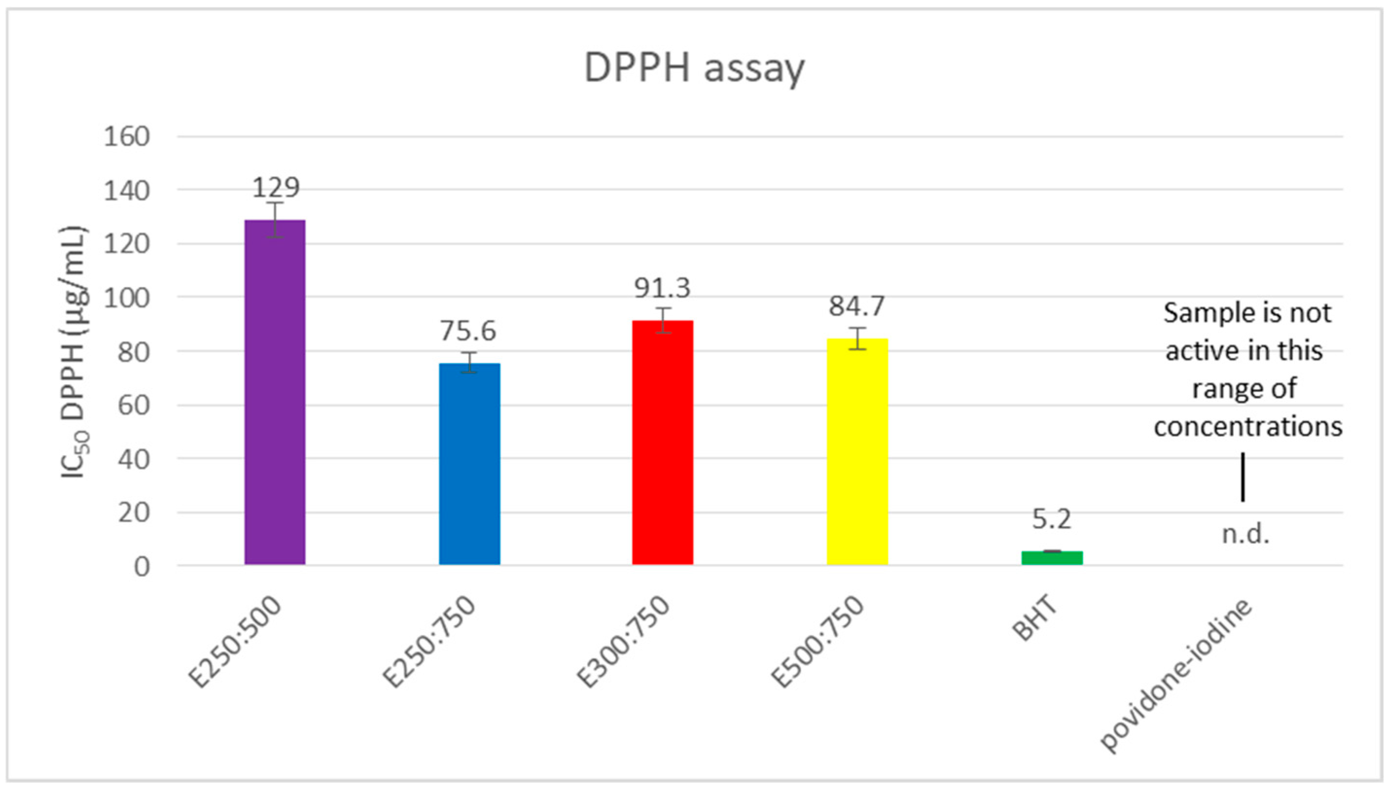
Concerning the antioxidant potential of the emulsions, the lowest IC50 and the highest antioxidant capacity to neutralize the DPPH• radical were observed in sample E250:750, while the lowest antioxidant activity was observed in sample E250:500 (75.6 and 129 μg/mL, respectively). In contrast, povidone–iodine showed no antioxidant capacity in the concentration range of 0.16–10.0 mg/mL, while the model antioxidant showed the lowest IC50 value, as expected (Figure 2).

4. Discussion

Klebsiella pneumoniae is one of the most common causative agents of HAIs, showing growing resistance to currently available antibiotics. Moreover, this strain belongs to the ESKAPE group, i.e., pathogens of utmost importance due to their high virulence and multidrug resistance [23,24]. Considering this, screening potential alternatives for antimicrobial therapy against Klebsiella pneumoniae is an urgent priority. In light of this, the fact that the antibacterial potential of thymol against various MDR K. pneumoniae isolates was screened for the first time in this study is extremely important. Moreover, based on these results, thymol was incorporated into (nano)emulsions with Tween 80, and the emulsions were subjected to evaluation of their antibiofilm potential against K. pneumoniae biofilms.
The first part of this study focused on evaluating the antibacterial potential of thymol against MDR K. pneumoniae isolates. In accordance with Aligiannis et al. [25], the obtained results (MIC values ranging from 0.060 to 1.2 mg/mL) showed that thymol exhibited notable antibacterial activity (MIC < 0.5 mg/mL) against the majority of isolates (80%). Previously, thymol demonstrated antimicrobial activity against K. pneumoniae strains of different origins and antibiotic sensitivities, with MIC values ranging from 64 μg/mL to 250 μg/mL [9,10], which generally aligns with the findings of this study. Mechanistically, thymol exerts antimicrobial activity through multiple complementary pathways, reducing the likelihood of resistance development [26]. It primarily disrupts bacterial membrane integrity by intercalating into the lipid bilayer, increasing permeability and causing leakage of intracellular components [27,28], followed by membrane depolarization and collapse of ATP synthesis [29]. It additionally interferes with energy metabolism [30], impairs DNA replication and transcription [31], and inhibits efflux pumps, thereby potentiating antibiotic activity [32]. In Gram-negative bacteria such as Klebsiella pneumoniae, molecular dynamics studies have shown that thymol can traverse the outer membrane and insert into the inner membrane with only a modest energy barrier [33]. Unlike iodine, whose antimicrobial action relies on a single, broadly oxidative mechanism that also affects host tissues [34], thymol’s multi-target strategy confers sustained antimicrobial activity, lower cytotoxicity, and shows no documented induction of resistance [35]. It is worth mentioning that thymol’s antibacterial activity was multifold higher than the positive control (povidone–iodine), making it a candidate for possible alternative therapies.
The next phase of this study was dedicated to formulating and characterizing thymol-based emulsions, following a previous suggestion that emulsification could enhance the pharmacokinetics and bioavailability of thymol [10]. Four stable emulsions with Tween 80 were designed and produced by means of magnetic and ultrasonic stirring. The emulsification process effectively addresses key challenges associated with thymol, such as its volatility, low stability, and high hydrophobicity, by improving its solubility, enhancing stability, extending its half-life, and enabling controlled release [36]. Particle size analysis indicated that both the reduction in thymol quantity and the increase in emulsifier Tween 80 led to a decrease in particle size. Although the classification of emulsions as nano- or micro-sized is borderline and depends on the authors’ classification, emulsions E250:750 and E300:750 could be considered nanosized (˂200 nm), while E250:500 and E500:750 could be considered microsized (˃200 nm) [37].
Although the anti-Klebsiella pneumoniae [9,35], antibiofilm [10,38], and antioxidant activities [39,40] of thymol are well established, its practical application is substantially limited by high volatility [41], poor water solubility [42], and chemical instability [43,44]. These intrinsic physicochemical constraints reduce its bioavailability and persistence in aqueous and biological environments [43].
Emulsion-based systems provide a rational technological solution to these limitations, as encapsulation within the dispersed phase protects thymol from evaporative loss, photodegradation, and oxidative degradation, while markedly improving its apparent solubility and dispersion in water [42]. Thymol emulsions have demonstrated preserved physicochemical stability and sustained antimicrobial activity during storage [45]. Critically, nanoemulsion formulations enable superior antimicrobial performance at lower concentrations compared to free thymol. MIC and MBC values for thymol nanoemulsions have been reported to be 2- to 4-fold lower than those of free thymol against a range of bacterial pathogens, including closely related Klebsiella species [46,47]. Due to their small droplet size and increased interfacial surface area, nanoemulsion systems facilitate improved interaction with and penetration into bacterial biofilms, which is highly relevant for resistant pathogens such as Klebsiella pneumoniae [48]. Furthermore, nanoemulsions formulated with Tween 80 have shown enhanced stability and lower MIC values compared to free thymol, indicating superior antimicrobial performance [49].
Changes in particle size can significantly influence the emulsion’s effectiveness, and previous studies have shown that nanosized emulsions (≤200 nm) offer better stability, while emulsions with larger droplets were suggested to be less stable [14]. The emulsions characterized as homogeneous and stable in this study had a particle size diameter of approx. 200 nm or lower.
The literature also suggests that thymol emulsification improves biofilm penetration and augments antibacterial activity against resistant pathogens [36]. The observed antibiofilm effect of the thymol based emulsions, being even higher than the effect of povidone–iodine, could be ascribed to potential of its active principle, thymol, to interfere with biofilm formation processes and affect several virulence factors of K. pneumoniae, such as the production of the biofilm extracellular matrix, with bacterial motility being important for initial biofilm establishment, and quorum sensing as a mechanism of bacterial communication, which coordinates biofilm formation, as shown previously [35].
However, it remains controversial whether the concentration of the active constituent or the reduced particle size diameter has more impact on the mentioned activity [14]. Accordingly, the following sections of the research focused on the comparative antibiofilm activity of stable emulsions E250:500, E250:750, E300:750, and E500:750, both in vitro and ex vivo. Investigating antibiofilm activity has become increasingly vital for preventing and treating biofilm-associated infections, particularly those linked to medical devices. This is especially relevant for K. pneumoniae residing in biofilms, which exhibit heightened resistance to antibiotics compared to planktonic cells [50,51]. Several factors contribute to the enhanced resistance of biofilms, including decreased metabolic activity and growth rates of cells within the biofilm, the horizontal transfer of resistance genes, increased expression of efflux pumps, and limited penetration of antibiotics through the biofilm structure [10]. Biofilm formation is a notable characteristic of Klebsiella pneumoniae, with a considerable percentage of clinical isolates exhibiting this trait. For example, one study found that 75% of K. pneumoniae isolates from various clinical samples could form biofilms, with 20% classified as strong biofilm producers [50]. Another investigation reported that among the isolated strains, 54% were strong biofilm producers, while 29% and 14% were moderate and weak producers, respectively [52]. Biofilm formation in Klebsiella pneumoniae is influenced by genetic differences among strains and environmental conditions during biofilm development. It progresses through distinct stages, adhesion, accumulation, maturation, and dispersion, with extracellular polysaccharides, eDNA, and proteins contributing to the biofilm matrix’s structural integrity and antimicrobial resistance [10,53]. Strain-specific variability impacts biofilm formation and antimicrobial sensitivity, with some strains producing more robust biofilms than others [51,54]. In line with this, not all isolates in the present study were biofilm producers; only two were used as biofilm-producing model isolates (K. pneumoniae3 and K. pneumoniae7).
Interestingly, all four emulsions showed notable biofilm biomass reduction under in vitro conditions, with the most efficient emulsions E250:500 against K. pneumoniae3 and E500:750 against biofilms of both isolates (K. pneumoniae3 and K. pneumoniae7). Although the reductive potential of the two other emulsions was slightly lower, all emulsions still showed notably higher antibiofilm potential compared to 10% povidone–iodine, a commercial disinfection solution used in this study as a positive control. Although slight variability in the emulsions’ activity can be observed, all of them, except E250:500 against K. pneumoniae7, reduced biofilm biomass by more than 50%. These findings align with previous studies demonstrating the antibiofilm potential of thymol formulations [15,55].
In this study, an ex vivo model was used to investigate biofilm formation on surgical drains, a common challenge in postoperative care. A similar effect was observed in the ex vivo model, where the most efficient emulsion was again the one with the highest amount of thymol, i.e., E500:750. As in the case of the in vitro model, thymol-based nanoemulsions again showed higher antibiofilm potential compared to the control (10% povidone–iodine).
For antibacterial and antibiofilm monitoring, a 10% povidone–iodine solution was used in this study as a positive control, as it is commonly employed as an antiseptic for various medical purposes, including the disinfection of surgical wounds. In addition, since there are indications that some iodine compounds could act both as oxidants and antioxidants [56], 10% povidone–iodine was also included as a control in the DPPH test. It is worth mentioning that although well known as an antiseptic, iodine’s effectiveness is diminished in the presence of organic matter [57]. The mechanism underlying the antimicrobial effect is multifaceted, affecting several cellular targets: (1) it binds to proteins and oxidizes S-H bonds within them, leading to protein denaturation, (2) it compromises membranes by reacting with C=C bonds within fatty acids, and (3) iodine also binds to purine and pyrimidine bases, preventing hydrogen bond formation between DNA strands. As a small molecule, iodine easily penetrates microbial cells and oxidizes proteins, fatty acids, and nucleotides [58]. However, iodine can also promote the oxidation of intracellular reductants within mammalian cells, such as nicotinamide adenine dinucleotide (phosphate) hydrogen (NAD(P)H) and reduced glutathione. Accordingly, it can negatively modulate cellular functions that are important for wound healing. Moreover, its cytotoxicity and potential to inhibit fibroblast proliferation, especially at higher concentrations, raise significant concerns. Additionally, some systemic side effects such as thyroid dysfunction, allergies, and psychological symptoms have also been noted. These concerns, especially in sensitive populations, highlight the need for alternative wound care treatments [57]. Considering the oxidative nature of iodine and the concerns associated with this feature, the search for antimicrobials that could also protect against oxidative stress seems to be a good strategy for simultaneously disinfecting and healing surgical wounds. In light of this, thymol-based emulsions were also screened for their antioxidative properties. While iodine, as expected, demonstrated no antioxidant activity, thymol-based emulsions exhibited a high antioxidant effect, in line with previous studies [17,19]. Thymol, as a single compound, exhibits potent antioxidant properties and effectively scavenges free radicals, including hydroxyl radicals, superoxide anions, and DPPH• radicals, as confirmed by numerous studies [12]. Due to its phenolic hydroxyl group, thymol directly neutralizes the free DPPH• radical [19]. In this study, we demonstrated that thymol-based emulsions, although containing low concentrations of thymol, possess strong DPPH radical-scavenging activity. Considering that infected wounds are characterized by inflammation accompanied by free radicals’ formation and subsequent oxidative tissue damage, therapeutics with combined antimicrobial and radical-scavenging activities—such as thymol-based emulsions formulated in this study—could be considered strong candidates for novel antiseptics with enhanced antioxidative properties.
Still, although the investigated emulsions demonstrated promising effects, the main limitation of this study is a lack of cytotoxicity assessment on human cell lines. Future research will therefore focus on comprehensive evaluation of their cytotoxicity and overall biocompatibility to further substantiate their safety profile.

5. Conclusions

This study demonstrated the strong antibacterial potential of thymol against the majority of clinical isolates of MDR Klebsiella pneumoniae strains. After the successful preparation of thymol (nano)emulsions using Tween 80, in vitro antibiofilm activity was shown by reducing the total biomass of pre-formed K. pneumoniae biofilms. Ex Vivo assays confirmed the antibiofilm potential of thymol’s emulsions and highlighted the decreased viability of the cells trapped within the biofilm. In addition, antioxidant, i.e., radical-scavenging, properties of the thymol-based emulsions were observed. The emulsions E250:500 and E500:750 exhibited the most potent antibiofilm effects, while the E250:750 emulsion demonstrated the highest antioxidant potential. In all conducted tests, thymol and its emulsions outperformed 10% povidone–iodine as a control mimicking conventional treatment of surgical wounds. Given that K. pneumoniae is one of the leading causes of HAIs, featuring growing resistance to antibiotics and notable biofilm formation properties, which are marked as the main culprits for the onset and persistence of hospital infections, the proven antimicrobial/antibiofilm efficacy of thymol/thymol-based emulsions, coupled with the antioxidant properties, highlight their potential as valuable tools in combating HAIs, including those caused by K. pneumoniae MDR strains. Although thymol-based emulsions could find a role in surgical wound treatments and the prevention of intrahospital infections, further studies are required to address these encouraging results.

Author Contributions

Conceptualization, J.M. and V.Ž.; methodology, T.V., M.N., F.V., B.P., M.J., J.F.T., B.N. and J.M.; software, T.V. and M.N.; validation, J.M., V.Ž. and B.N.; formal analysis, T.V., M.N., F.V. and B.P.; investigation, T.V., M.N., F.V. and B.P., resources, J.M.; data curation, T.V., M.N., F.V. and B.P.; writing-original draft, T.V., M.J., J.F.T. and B.P.; writing—review and editing, J.M., V.Ž. and B.N.; visualization, T.V., M.N., F.V., B.P., M.J. and J.F.T.; supervision, J.M., B.N. and V.Ž.; project administration, J.M.; funding acquisition, J.M. All authors have read and agreed to the published version of the manuscript.

Funding

This work was supported by the Ministry of Education, Science and Technological Development of the Republic of Serbia, grant numbers 451-03-68/2026-14/200017 and 451-03-65/2026-03/200178.

Institutional Review Board Statement

Approval was granted pursuant to Article 73 of the Law on Medicines and Medical Devices (“Official Gazette of RS”, No. 30/2010, 107/2012, 113/2017—other laws, and 105/2017—other laws) and Article 132 of the Law on Health Care (“Official Gazette of RS”, No. 25/2019). The request submitted by Stanislava Čukić, medical doctor specialist in microbiology, Department of Laboratory Diagnostics, for the provision of multidrug-resistant ESCAPE group isolates previously obtained through routine hospital procedures within the Department of Microbiology was reviewed and approved for use in scientific research in line with all relevant ethical and legal standards.

Informed Consent Statement

This study was approved by the Local Ethical Committee of University Hospital Center “Dr Dragiša Mišović–Dedinje”, Belgrade. As the collection of the study material originated from routine procedures, the requirement for informed consent was waived by the Ethical Committee (No. 15341/3-2025; 21 July 2025).

Data Availability Statement

The original contributions presented in this study are included in the article. Further inquiries can be directed to the corresponding author.

Acknowledgments

The authors are grateful to Marko Bošković, Vinča Institute of Nuclear Sciences, University of Belgrade, for providing DLS measurements. In addition, the authors are grateful to Marina Stojković and Stanislava Čukić, University Hospital Center “Dr Dragisa Misovic-Dedinje”, Belgrade, for the selection of MDR Klebsiella isolates.

Conflicts of Interest

The authors declare no conflicts of interest.

References

  1. Haque, M.; Sartelli, M.; McKimm, J.; Abu Bakar, M. Health care-associated infections—An overview. Infect. Drug Resist. 2018, 11, 2321–2333. [Google Scholar] [CrossRef] [Scilit]
  2. Despotovic, A.; Milosevic, B.; Milosevic, I.; Mitrovic, N.; Cirkovic, A.; Jovanovic, S.; Stevanovic, G. Hospital-acquired infections in the adult intensive care unit-Epidemiology, antimicrobial resistance patterns, and risk factors for acquisition and mortality. Am. J. Infect. Control 2020, 48, 1211–1215. [Google Scholar] [CrossRef] [Scilit]
  3. Healthcare-Associated Infections. European Centre for Disease Prevention and Control. Available online: https://www.ecdc.europa.eu/en/healthcare-associated-infections (accessed on 4 October 2024).
  4. Gentili, A.; Di Pumpo, M.; La Milia, D.I.; Vallone, D.; Vangi, G.; Corbo, M.I.; Berloco, F.; Cambieri, A.; Damiani, G.; Ricciardi, W.; et al. A Six-Year Point Prevalence Survey of Healthcare-Associated Infections in an Italian Teaching Acute Care Hospital. Int. J. Environ. Res. Public Health 2020, 17, 7724. [Google Scholar] [CrossRef] [Scilit]
  5. Suetens, C.; Latour, K.; Kärki, T.; Ricchizzi, E.; Kinross, P.; Moro, M.L.; Jans, B.; Hopkins, S.; Hansen, S.; Lyytikäinen, O.; et al. Prevalence of healthcare-associated infections, estimated incidence and composite antimicrobial resistance index in acute care hospitals and long-term care facilities: Results from two European point prevalence surveys, 2016 to 2017. Eurosurveillance 2018, 23, 1800516. [Google Scholar] [CrossRef] [Scilit]
  6. Ćirković, I.; Marković-Denić, L.; Bajčetić, M.; Dragovac, G.; Đorđević, Z.; Mioljević, V.; Urošević, D.; Nikolić, V.; Despotović, A.; Krtinić, G.; et al. Microbiology of Healthcare-Associated Infections: Results of a Fourth National Point Prevalence Survey in Serbia. Antibiotics 2022, 11, 1161. [Google Scholar] [CrossRef] [Scilit]
  7. Kon, K.V.; Rai, M.K. Plant essential oils and the ir constituents in coping with multidrug-resistant bacteria. Expert Rev. Anti-Infect. Ther. 2012, 10, 775–790. [Google Scholar] [CrossRef] [Scilit]
  8. Sienkiewicz, M.; Łysakowska, M.; Denys, P.; Kowalczyk, E. The antimicrobial activity of thyme essential oil against multidrug resistant clinical bacterial strains. Microb. Drug Resist. 2012, 18, 137–148. [Google Scholar] [CrossRef] [Scilit]
  9. Gan, C.; Langa, E.; Valenzuela, A.; Ballestero, D.; Pino-Otín, M.R. Synergistic Activity of Thymol with Commercial Antibiotics against Critical and High WHO Priority Pathogenic Bacteria. Plants 2023, 12, 1868. [Google Scholar] [CrossRef] [Scilit] [PubMed]
  10. Bisso Ndezo, B.; Tokam Kuaté, C.R.; Dzoyem, J.P. Synergistic Antibiofilm Efficacy of Thymol and Piperine in Combination with Three Aminoglycoside Antibiotics against Klebsiella pneumoniae Biofilms. Can. J. Infect. Dis. Med. Microbiol. 2021, 2021, 7029944. [Google Scholar] [CrossRef] [Scilit] [PubMed]
  11. Escobar, A.; Pérez, M.; Romanelli, G.; Blustein, G. Thymol bioactivity: A review focusing on practical applications. Arab. J. Chem. 2020, 13, 9243–9269. [Google Scholar] [CrossRef] [Scilit]
  12. Nagoor Meeran, M.F.; Javed, H.; Al Taee, H.; Azimullah, S.; Ojha, S.K. Pharmacological Properties and Molecular Mechanisms of Thymol: Prospects for Its Therapeutic Potential and Pharmaceutical Development. Front. Pharmacol. 2017, 8, 380. [Google Scholar] [CrossRef] [Scilit]
  13. Salehi, B.; Mishra, A.P.; Shukla, I.; Sharifi-Rad, M.; Contreras, M.D.M.; Segura-Carretero, A.; Fathi, H.; Nasrabadi, N.N.; Kobarfard, F.; Sharifi-Rad, J. Thymol, thyme, and other plant sources: Health and potential uses. Phytother. Res. 2018, 32, 1688–1706. [Google Scholar] [CrossRef] [Scilit]
  14. Li, J.; Chang, J.W.; Saenger, M.; Deering, A. Thymol nanoemulsions formed via spontaneous emulsification: Physical and antimicrobial properties. Food Chem. 2017, 232, 191–197. [Google Scholar] [CrossRef] [Scilit]
  15. Ndezo Bisso, B.; Tokam Kuaté, C.R.; Boulens, N.; Allémann, E.; Delie, F.; Dzoyem, J.P. Antibiofilm Synergistic Activity of Streptomycin in Combination with Thymol-Loaded Poly (Lactic-co-glycolic Acid) Nanoparticles against Klebsiella pneumoniae Isolates. Evid. Based Complement. Altern. Med. 2022, 2022, 1936165. [Google Scholar] [CrossRef] [Scilit]
  16. Braga, P.C.; Dal Sasso, M.; Culici, M.; Bianchi, T.; Bordoni, L.; Marabini, L. Anti-inflammatory activity of thymol: Inhibitory effect on the release of human neutrophil elastase. Pharmacology 2006, 77, 130–136. [Google Scholar] [CrossRef] [Scilit] [PubMed]
  17. Sedaghat Doost, A.; Van Camp, J.; Dewettinck, K.; Van der Meeren, P. Production of thymol nanoemulsions stabilized using Quillaja Saponin as a biosurfactant: Antioxidant activity enhancement. Food Chem. 2019, 293, 134–143. [Google Scholar] [CrossRef] [Scilit] [PubMed]
  18. da Silva, B.D.; do Rosário, D.K.A.; Neto, L.T.; Lelis, C.A.; Conte-Junior, C.A. Antioxidant, Antibacterial and Antibiofilm Activity of Nanoemulsion-Based Natural Compound Delivery Systems Compared with Non-Nanoemulsified Versions. Foods 2023, 12, 1901. [Google Scholar] [CrossRef] [Scilit]
  19. Deng, L.L.; Taxipalati, M.; Que, F.; Zhang, H. Physical characterization and antioxidant activity of thymol solubilized Tween 80 micelles. Sci. Rep. 2016, 6, 38160. [Google Scholar] [CrossRef] [Scilit] [PubMed]
  20. Marinković, J.; Ćulafić, D.M.; Nikolić, B.; Đukanović, S.; Marković, T.; Tasić, G.; Ćirić, A.; Marković, D. Antimicrobial potential of irrigants based on essential oils of Cymbopogon martinii and Thymus zygis towards in vitro multispecies biofilm cultured in ex vivo root canals. Arch. Oral. Biol. 2020, 117, 104842. [Google Scholar] [CrossRef] [Scilit]
  21. Balta, I.; Brinzan, L.; Stratakos, A.C.; Linton, M.; Kelly, C.; Pinkerton, L.; Corcionivoschi, N. Geraniol and linalool loaded nanoemulsions and their antimicrobial activity. Bull. Univ. Agric. Sci. Vet. Med. Cluj-Napoca Anim. Sci. Biotechnol. 2017, 74, 157. [Google Scholar] [CrossRef] [Scilit]
  22. Espín, J.C.; Soler-Rivas, C.; Wichers, H.J. Characterization of the total free radical scavenger capacity of vegetable oils and oil fractions using 2,2-diphenyl-1-picrylhydrazyl radical. J. Agric. Food Chem. 2000, 48, 648–656. [Google Scholar] [CrossRef] [Scilit]
  23. De Oliveira, D.M.P.; Forde, B.M.; Kidd, T.J.; Harris, P.N.A.; Schembri, M.A.; Beatson, S.A.; Paterson, D.L.; Walker, M.J. Antimicrobial Resistance in ESKAPE Pathogens. Clin. Microbiol. Rev. 2020, 33, e00181-19. [Google Scholar] [CrossRef] [Scilit]
  24. Miller, W.R.; Arias, C.A. ESKAPE pathogens: Antimicrobial resistance, epidemiology, clinical impact and therapeutics. Nat. Rev. Microbiol. 2024, 22, 598–616. [Google Scholar] [CrossRef] [Scilit]
  25. Aligiannis, N.; Kalpoutzakis, E.; Mitaku, S.; Chinou, I.B. Composition and antimicrobial activity of the essential oils of two Origanum species. J. Agric. Food Chem. 2001, 49, 4168–4170. [Google Scholar] [CrossRef] [Scilit]
  26. Kachur, K.; Suntres, Z. The antibacterial properties of phenolic isomers, carvacrol and thymol. Crit. Rev. Food Sci. Nutr. 2020, 60, 3042–3053. [Google Scholar] [CrossRef] [Scilit]
  27. Trombetta, D.; Castelli, F.; Sarpietro, M.G.; Venuti, V.; Cristani, M.; Daniele, C.; Saija, A.; Mazzanti, G.; Bisignano, G. Mechanisms of antibacterial action of three monoterpenes. Antimicrob. Agents Chemother. 2005, 49, 2474–2478. [Google Scholar] [CrossRef] [Scilit]
  28. Xu, J.; Zhou, F.; Ji, B.P.; Pei, R.S.; Xu, N. The antibacterial mechanism of carvacrol and thymol against Escherichia coli. Lett. Appl. Microbiol. 2008, 47, 174–179. [Google Scholar] [CrossRef] [Scilit]
  29. Tian, L.; Wang, X.; Liu, R.; Zhang, D.; Wang, X.; Sun, R.; Guo, W.; Yang, S.; Li, H.; Gong, G. Antibacterial mechanism of thymol against Enterobacter sakazakii. Food Control 2020, 123, 107716. [Google Scholar] [CrossRef] [Scilit]
  30. Al-Kandari, F.; Al-Temaimi, R.; Van Vliet, A.H.M.; Woodward, M.J. Thymol tolerance in Escherichia coli induces morphological, metabolic and genetic changes. BMC Microbiol. 2019, 19, 294. [Google Scholar] [CrossRef] [Scilit] [PubMed]
  31. Yin, L.; Liang, C.; Wei, W.; Huang, S.; Ren, Y.; Geng, Y.; Huang, X.; Chen, D.; Guo, H.; Fang, J.; et al. The Antibacterial Activity of Thymol Against Drug-Resistant Streptococcus iniae and Its Protective Effect on Channel Catfish (Ictalurus punctatus). Front. Microbiol. 2022, 13, 914868. [Google Scholar] [CrossRef] [Scilit] [PubMed]
  32. Miladi, H.; Zmantar, T.; Chaabouni, Y.; Fedhila, K.; Bakhrouf, A.; Mahdouani, K.; Chaieb, K. Antibacterial and efflux pump inhibitors of thymol and carvacrol against food-borne pathogens. Microb. Pathog. 2016, 99, 95–100. [Google Scholar] [CrossRef] [Scilit]
  33. Sharma, P.; Parthasarathi, S.; Patil, N.; Waskar, M.; Raut, J.S.; Puranik, M.; Ayappa, K.G.; Basu, J.K. Assessing Barriers for Antimicrobial Penetration in Complex Asymmetric Bacterial Membranes: A Case Study with Thymol. Langmuir 2020, 36, 8800–8814. [Google Scholar] [CrossRef] [Scilit] [PubMed]
  34. McDonnell, G.; Russell, A.D. Antiseptics and Disinfectants: Activity, action, and resistance. Clin. Microbiol. Rev. 1999, 12, 147–179. [Google Scholar] [CrossRef] [Scilit]
  35. Farhadi, K.; Rajabi, E.; Varpaei, H.A.; Iranzadasl, M.; Khodaparast, S.; Salehi, M. Thymol and carvacrol against Klebsiella: Anti-bacterial, anti-biofilm, and synergistic activities—A systematic review. Front. Pharmacol. 2024, 15, 1487083. [Google Scholar] [CrossRef] [Scilit]
  36. Hajibonabi, A.; Yekani, M.; Sharifi, S.; Nahad, J.S.; Dizaj, S.M.; Memar, M.Y. Antimicrobial activity of nanoformulations of carvacrol and thymol: New trend and applications. OpenNano 2023, 13, 100170. [Google Scholar] [CrossRef] [Scilit]
  37. Jaiswal, M.; Dudhe, R.; Sharma, P.K. Nanoemulsion: An advanced mode of drug delivery system. 3 Biotech 2015, 5, 123–127. [Google Scholar] [CrossRef] [Scilit]
  38. Kwiatkowski, P.; Sienkiewicz, M.; Pruss, A.; Łopusiewicz, Ł.; Arszyńska, N.; Wojciechowska-Koszko, I.; Kilanowicz, A.; Kot, B.; Dołęgowska, B. Antibacterial and Anti-Biofilm Activities of Essential Oil Compounds against New Delhi Metallo-β-Lactamase-1-Producing Uropathogenic Klebsiella pneumoniae Strains. Antibiotics 2022, 11, 147. [Google Scholar] [CrossRef] [Scilit]
  39. Sharopov, F.S.; Wink, M.; Setzer, W.N. Radical scavenging and antioxidant activities of essential oil components—An experimental and computational investigation. Nat. Prod. Commun. 2015, 10, 153–156. [Google Scholar] [CrossRef] [Scilit]
  40. Chen, X.; Shang, S.; Yan, F.; Jiang, H.; Zhao, G.; Tian, S.; Chen, R.; Chen, D.; Dang, Y. Antioxidant activities of essential oils and their major components in scavenging free radicals, inhibiting lipid oxidation and reducing cellular oxidative stress. Molecules 2023, 28, 4559. [Google Scholar] [CrossRef] [Scilit] [PubMed]
  41. Najafloo, R.; Behyari, M.; Imani, R.; Nour, S. A mini-review of Thymol incorporated materials: Applications in antibacterial wound dressing. J. Drug Deliv. Sci. Technol. 2020, 60, 101904. [Google Scholar] [CrossRef] [Scilit]
  42. Santos, D.L.S.D.; De Miranda, J.F.; Rodrigues, R.A.F.; Prata, A.S.; Silva, N.C.C. Evaluation of the stability and antimicrobial activity of emulsions loaded with thymol for the post-harvest sanitization of lettuce (Lactuca sativa L.). Food Biosci. 2025, 66, 106176. [Google Scholar] [CrossRef] [Scilit]
  43. Latorre, R.; Valerii, M.C.; Benati, M.; Lewis, R.E.; Spigarelli, R.; Bernacchi, A.; Lippi, G.; Spisni, E.; Gaibani, P. Lights and shadows of Essential Oil-Derived compounds: Antimicrobial and Anti-Inflammatory properties of eugenol, thymol, cinnamaldehyde, and Carvacrol. Curr. Issues Mol. Biol. 2025, 47, 915. [Google Scholar] [CrossRef] [Scilit] [PubMed]
  44. Soulaimani, B.; Abbad, I.; Dumas, E.; Gharsallaoui, A. Enhanced antimicrobial and biofilm disruption efficacy of the encapsulated Thymus pallidus and Lavandula stoechas essential oils and their mixture: A synergistic approach. Int. J. Pharm. 2024, 670, 125144. [Google Scholar] [CrossRef] [Scilit]
  45. Cai, Q.; Zhang, Y.; Fang, X.; Lin, S.; He, Z.; Peng, S.; Liu, W. Improving anti-listeria activity of thymol emulsions by adding lauric acid. Front. Nutr. 2022, 9, 859293. [Google Scholar] [CrossRef] [Scilit] [PubMed]
  46. Sepahvand, S.; Amiri, S.; Radi, M.; Akhavan, H.-R. Antimicrobial activity of thymol and Thymol-Nanoemulsion against three Food-Borne pathogens inoculated in a sausage model. Food Bioprocess Technol. 2021, 14, 1936–1945. [Google Scholar] [CrossRef] [Scilit]
  47. Doghish, A.; Shehabeldine, A.; El-Mahdy, H.; Hassanin, M.; Al-Askar, A.; Marey, S.; AbdElgawad, H.; Hashem, A.H. Thymus vulgaris oil nanoemulsion: Synthesis, characterization, antimicrobial and anticancer activities. Molecules 2023, 28, 6910. [Google Scholar] [CrossRef] [Scilit]
  48. Moghimi, R.; Ghaderi, L.; Rafati, H.; Aliahmadi, A.; McClements, D.J. Superior antibacterial activity of nanoemulsion of Thymus daenensis essential oil against E. coli. Food Chem. 2015, 194, 410–415. [Google Scholar] [CrossRef] [Scilit]
  49. Ozogul, Y.; Karsli, G.T.; Yazgan, H.; Kuley, E.; Oztop, H.M.; Ozogul, F.; Esatbeyoglu, T. Enhanced pathogen control through thymol and Carvacrol nanoemulsions: A microfluidization approach. Food Bioprocess Technol. 2025, 18, 5377–5387. [Google Scholar] [CrossRef] [Scilit]
  50. Karimi, K.; Zarei, O.; Sedighi, P.; Taheri, M.; Doosti-Irani, A.; Shokoohizadeh, L. Investigation of Antibiotic Resistance and Biofilm Formation in Clinical Isolates of Klebsiella pneumoniae. Int. J. Microbiol. 2021, 2021, 5573388. [Google Scholar] [CrossRef] [Scilit]
  51. Clegg, S.; Murphy, C.N. Epidemiology and Virulence of Klebsiella pneumoniae. Microbiol. Spectr. 2016, 4. [Google Scholar] [CrossRef] [Scilit]
  52. Li, Y.; Ni, M. Regulation of biofilm formation in Klebsiella pneumoniae. Front. Microbiol. 2023, 14, 1238482. [Google Scholar] [CrossRef] [Scilit]
  53. Oleksy-Wawrzyniak, M.; Junka, A.; Brożyna, M.; Paweł, M.; Kwiek, B.; Nowak, M.; Mączyńska, B.; Bartoszewicz, M. The In Vitro Ability of Klebsiella pneumoniae to Form Biofilm and the Potential of Various Compounds to Eradicate It from Urinary Catheters. Pathogens 2021, 11, 42. [Google Scholar] [CrossRef] [Scilit]
  54. Vuotto, C.; Longo, F.; Pascolini, C.; Donelli, G.; Balice, M.P.; Libori, M.F.; Tiracchia, V.; Salvia, A.; Varaldo, P.E. Biofilm formation and antibiotic resistance in Klebsiella pneumoniae urinary strains. J. Appl. Microbiol. 2017, 123, 1003–1018. [Google Scholar] [CrossRef] [Scilit] [PubMed]
  55. Mohamed, S.H.; Mohamed, M.S.M.; Khalil, M.S.; Azmy, M.; Mabrouk, M.I. Combination of essential oil and ciprofloxacin to inhibit/eradicate biofilms in multidrug-resistant Klebsiella pneumoniae. J. Appl. Microbiol. 2018, 125, 84–95. [Google Scholar] [CrossRef] [Scilit] [PubMed]
  56. Karbownik-Lewińska, M.; Stępniak, J.; Iwan, P.; Lewiński, A. Iodine as a potential endocrine disruptor-a role of oxidative stress. Endocrine 2022, 78, 219–240. [Google Scholar] [CrossRef] [Scilit] [PubMed]
  57. Cooper, R.A. Iodine revisited. Int. Wound J. 2007, 4, 124–137. [Google Scholar] [CrossRef] [Scilit]
  58. Lepelletier, D.; Maillard, J.Y.; Pozzetto, B.; Simon, A. Povidone Iodine: Properties, Mechanisms of Action, and Role in Infection Control and Staphylococcus aureus Decolonization. Antimicrob. Agents Chemother. 2020, 64, e00682-20. [Google Scholar] [CrossRef] [Scilit]
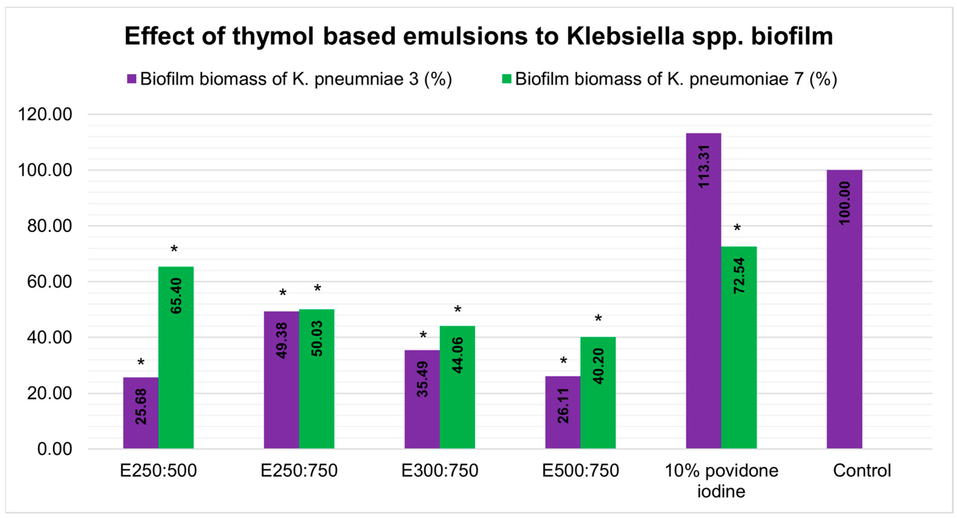
Figure 1. Effect of thymol-based emulsions on preformed Klebsiella pneumoniae biofilm. * Indicates statistical significance (p < 0.05).
Compounds 06 00022 g001
Figure 2. Antioxidant capacity of tested thymol-based emulsions.
Compounds 06 00022 g002
Table 1. Origin and sensitivity of MDR Klebsiella pneumoniae isolates.
IsolateSampleColistinCeftazidimeCefimMeropenemLevofloxacinGentamicin
1. K. pneumoniae1PyocultureR aRRRRS b
2. K. pneumoniae2Urine cultureRRRRRR
3. K. pneumoniae3Urine cultureSRRRRS
4. K. pneumoniae4Urine cultureSRRRRR
5. K. pneumoniae5Blood cultureSRRRRR
6. K. pneumoniae6Blood cultureRRRRRR
7. K. pneumoniae7Blood cultureRRRRRS
8. K. pneumoniae8PyocultureRRRRRR
9. K. pneumoniae9PyocultureRRRRRR
10. K. pneumoniae10PyocultureRRRRRR
a R—resistant; b S—sensitive.
Table 2. Composition of the thymol-based emulsions.
EmulsionThymol (μL)–Tween 80 (μL)–dH2O (μL)
E250:500250:500:9250
E250:750250:750:9000
E250:1000250:1000:8750
E500:250500:250:9250
E300:750300:750:8950
E500:500500:500:9000
E500:750500:750:8750
E500:1000500:1000:8500
E800:1000800:1000:8200
Table 3. Antibacterial activity of thymol against MDR Klebsiella spp. isolates.
ThymolPovidone–iodine b
K. pneumoniae IsolatesMICMBCMICMBC
mg/mL
1. K. pneumoniae11.25 ± 0.54 a3.75 ± 0.0063.62 ± 0.00>127.25
2. K. pneumoniae20.46 ± 0.000.93 ± 0.0063.62 ± 0.00>127.25
3. K. pneumoniae30.35 ± 0.150.93 ± 0.0063.62 ± 0.00>127.25
4. K. pneumoniae40.18 ± 0.080.24 ± 0.0063.62 ± 0.00>127.25
5. K. pneumoniae50.09 ± 0.040.24 ± 0.0047.72 ± 22.49>127.25
6. K. pneumoniae60.06 ± 0.000.24 ± 0.0063.62 ± 0.00>127.25
7. K. pneumoniae70.59 ± 0.500.94 ± 0.0063.62 ± 0.00>127.25
8. K. pneumoniae80.09 ± 0.040.48 ± 0.0063.62 ± 0.00>127.25
9. K. pneumoniae90.12 ± 0.000.24 ± 0.0063.62 ± 0.00>127.25
10. K. pneumoniae100.18 ± 0.090.48 ± 0.0063.62 ± 0.00>127.25
a The least sensitive strains, being the most important for further study, are shown in bold. b Commercially available solution of 10% povidone–iodine was used in this assay.
Table 4. Emulsions based on thymol—evaluated by means of visual inspection on the day of preparation.
Visual Evaluation of Prepared Emulsions Homogeneity
EmulsionThymol–Tween 80 RatioAfter the Magnetic StirringAfter the Additional Ultrasound Stirring
E250:500250:500NonhomogeneousHomogeneous a
E250:750250:750NonhomogeneousHomogeneous
E250:1000250:1000HomogeneousNonhomogeneous
E500:250500:250NonhomogeneousNonhomogeneous
E300:750300:750HomogeneousHomogeneous
E500:500500:500NonhomogeneousNonhomogeneous
E500:750500:750HomogeneousHomogeneous
E500:1000500:1000NonhomogeneousNonhomogeneous
E800:1000800:1000NonhomogeneousNonhomogeneous
a Homogeneous emulsions, after magnetic and additional ultrasonic stirring, were used in subsequent experiments.
Table 5. Characterization of thymol-based emulsions using dynamic light scattering (DLS).
Average Particle Size Diameter (Zavg)
E250:500209 ± 3
E250:750128.4 ± 0.8
E300:750196 ± 4
E500:750222 ± 3
Table 6. Ex vivo potential of thymol-based emulsions against K. pneumoniae biofilm.
ControlE250:500E250:750E300:750E500:75010% Povidone–iodine
K. pneumoniae7
logCFU9.077.667.46.516.478.44
(ΔLogCFU) 1.411.672.562.600.63
K. pneumoniae3
logCFU8.917.377.26.86.237.28
(ΔLogCFU) 1.541.712.112.681.63
Disclaimer/Publisher’s Note: The statements, opinions and data contained in all publications are solely those of the individual author(s) and contributor(s) and not of MDPI and/or the editor(s). MDPI and/or the editor(s) disclaim responsibility for any injury to people or property resulting from any ideas, methods, instructions or products referred to in the content.