Article

Investigation of the Colorimetric Transition in Polydiacetylene Nanovesicles Induced by α-Cyclodextrin and the Inhibitory Role of Triblock Copolymer Addition


by
Maria C. O. Rodrigues
1,
Maria E. F. R. Antunes
1,
Alex R. M. Alves
2,
Diego C. de Morais
3,
Frederico B. De Sousa
3,
Garbas A. S. Junior
2,
João P. C. Trigueiro
1,* and
Paulo F. R. Ortega
2,*
1
Departamento de Química, Centro Federal de Educação Tecnológica de Minas Gerais, Belo Horizonte 30421-169, MG, Brazil
2
Grupo de Estudos em Dispositivos de Armazenamento de Energia (GEDAE), Departamento de Química, Universidade Federal de Viçosa, Vicosa 36570-900, MG, Brazil
3
Laboratório de Sistemas Poliméricos e Supramoleculares (LSPS), Instituto de Física e Química, Universidade Federal de Itajubá (UNIFEI), Itajuba 37500-903, MG, Brazil
*
Authors to whom correspondence should be addressed.
Nanomanufacturing 2026, 6(3), 15; https://doi.org/10.3390/nanomanufacturing6030015
Submission received: 18 November 2025 / Revised: 1 February 2026 / Accepted: 15 April 2026 / Published: 24 June 2026
(This article belongs to the Special Issue Nanomanufacturing: Feature Papers 2025)

Abstract

Polydiacetylene (PDA) nanovesicles are widely recognized as versatile chromatic sensing platforms, exhibiting a visible blue-to-red colorimetric transition in response to external stimuli such as temperature, pH, and molecular recognition events. In contrast to the conventional goal of amplifying this chromatic response, this work presents a supramolecular approach to inhibit the α-cyclodextrin (α-CD)-induced colorimetric transition in PDA systems. α-CD is known to interact with PDA vesicles through host–guest inclusion at the vesicle interface, triggering the characteristic chromatic change. Here, we show that the incorporation of an EO–PO–EO triblock copolymer (L64) into PDA suspensions enables controlled modulation of the α-CD-induced chromatic response, leading to a progressive attenuation of the blue-to-red transition as the L64 concentration increases. Isothermal titration calorimetry reveals a stronger affinity of α-CD for L64 (K = 11,300) than for PDA vesicles (K = 4000), with both interactions being spontaneous (ΔG° ≈ −21 kJ mol−1) and predominantly entropy-driven. Copolymer aggregation and phase separation occur without compromising the PDA vesicles, indicating that the observed chromatic modulation arises from supramolecular competition. This study introduces a strategy to regulate PDA affinity chromism using biocompatible triblock copolymers, offering a tunable and robust pathway for the design of responsive and safe chromatic sensing platforms.

1. Introduction

Polydiacetylenes (PDAs) are regarded as highly promising polymers for sensing applications due to their ability to undergo chromatic transitions in the visible range in response to external stimuli, such as temperature [1,2,3], pH [4,5] and molecular interactions [6,7]. PDAs were first prepared by Wegner in 1969 [6]; however, it was the pioneering work of Charych and co-workers in 1993 that demonstrated the remarkable potential of these materials by enabling the detection of the influenza virus, paving the way for applications in diagnostics and therapeutics [8].
PDAs are obtained through the topochemical polymerization of diacetylene monomers that can be self-organized into different architectures, such as Langmuir–Blodgett/Langmuir–Schaefer films [9,10,11], crystalline solids [12,13], and lipid bilayers or vesicles in aqueous solution [14,15], among other structures. In these supramolecular arrangements, the monomers adopt a highly ordered geometry, with appropriate intermolecular distances between C≡C bonds and a packing angle of approximately 45°, allowing the 1,4-addition reaction to occur upon UV or γ irradiation, generating a polymeric backbone with alternating double and triple bonds (ene–yne) [16,17]. The chromatic transition of PDAs arises from environmental perturbations (such as temperature and pressure) or chemical interactions that are capable of altering the conformation of the carbon chains, thereby reducing the conjugation length of the π system and, consequently, increasing the energy gap between the frontier molecular orbitals [18]. As a result, the absorption band associated with the π–π transition shifts from approximately 640–660 nm to 540–560 nm, which manifests as a visible color change from blue to red.
With the understanding of these properties, numerous studies have been conducted to explore the thermochromism, mechanochromism, solvatochromism, and affinity-chromism of PDAs [16,17]. Moreover, the chemical modification of monomers or the formulation of composite systems has been extensively employed for the selective detection of metals [19], carcinogenic organic compounds [20], and even large and complex biomolecules (including proteins) [21]. In this context, investigating approaches not only to modulate but also to precisely control the sensitivity of the chromatic transition of these PDA-based structures becomes highly relevant.
In 2017, Ferreira et al. demonstrated that the preparation of PDA vesicles in aqueous solution containing EO–PO–EO triblock copolymers (EO = ethylene oxide, PO = propylene oxide), commercially referred to as Pluronics or Synperonics, results in structures exhibiting different sensitivities to chromatic transitions [22]. These authors showed that the use of copolymers with higher molar mass, at high concentrations, and with an increased number of propylene oxide segments leads to suspensions that undergo the color transition at progressively lower temperatures. This work was later extended by our group in 2021 [1], through the incorporation of triblock copolymers into PDA/poly(vinyl alcohol) hydrogels, enabling the modulation of thermochromic transitions in solid films, which are technologically more suitable for applications such as smart packaging.
In this work, we demonstrate for the first time that triblock copolymers can be employed with opposite effect: instead of increasing the sensitivity of the chromatic transition, these molecules can act as inhibitors, reducing the affinity-chromism of PDA structures in the presence of cyclodextrins, CDs, specifically α-cyclodextrin (α-CD). CDs are classified as biocompatible macrocycles, formed by glucose units linked by α (1–4) bonds, which results in a three-dimensional described as a truncated cone architecture, leading to a hydrophobic cavity and a hydrophilic outer surface [23]. The most commonly used CDs are: α-, β-, and γ-CD, formed by 6, 7, and 8 glucose units, respectively. CDs emerge as an invaluable supramolecular strategy applicable across different areas of science, including biomaterials [24], particularly in the pharmaceutical field [25], pollutant adsorption based on nanosponge materials [26], gas sensing, and chemical and physical sorption [27], due to their capability to form inclusion complexes (ICs) with a variety of guest molecules, including surfactants [28].
The chromatic transition of PDAs induced by α-CD was first investigated by Cho et al. in 2003 using polymerized diacetylene Langmuir–Schaefer films [9]. In 2009, Champaiboon et al. demonstrated that the chromatic transition of PDA films can be inhibited and consequently modulated through the addition of nitrophenolic compounds to the system, particularly 4-nitrophenol [29]. The inhibition of the colorimetric transition in PDAs reflects an increased resistance of the conjugated backbone to external perturbations. This enhanced stability is highly desirable for sensing applications in some cases, as it allows the preservation of the blue phase under non-specific stimuli, improving signal reliability and operational lifetime. Moreover, controlled inhibition enables fine-tuning of the PDA response threshold, which is essential for designing sensors that respond only to specific analytes or interactions rather than to background fluctuations. However, these nitrophenolic compounds are toxic to humans, causing methemoglobinemia [30], and are highly toxic to aquatic organisms, being considered environmental pollutants [31]. In contrast, Pluronic® triblock copolymers are regarded as non-toxic, biocompatible, and safe for a wide range of applications, including pharmaceutical and cosmetic uses [32].
Herein, we show that PDA vesicles undergo a chromatic transition upon interaction with α-CD, while the presence of varying concentrations of the triblock copolymer L64 inhibits this response through competitive inclusion complex formation. This inhibition strategy highlights the critical role of benign additives in stabilizing PDA assemblies without compromising environmental or biological safety. Moreover, the ability to suppress color transitions using biocompatible modifiers provides a rational pathway for developing robust PDA-based platforms for practical applications, including the visual detection of biomolecules, environmental monitoring of specific analytes, and smart packaging.

2. Materials and Methods

2.1. Materials

The diacetylene monomer 10,12-pentacosadiynoic acid (PCDA, 97% purity, wt%) and the triblock copolymer poly(ethylene glycol)-block-poly(propylene glycol)-block-poly(ethylene glycol) (L64, MW = 2900) were obtained from Sigma-Aldrich (São Paulo, SP, Brazil). Prior to use, PCDA was purified by dissolution in chloroform, followed by filtration through a 0.45 μm poly(vinylidene difluoride) membrane to remove any polymerized residues. All other reagents and solvents were of analytical or reagent grade, purchased from Sigma-Aldrich, and employed without further purification.

2.2. Preparation of PDA Vesicle and PDA/L64 Suspensions

PCDA monomers were dispersed in deionized water (initial pH 6.58 at 25 °C) to obtain a final monomer concentration of 1 mM. The resulting mixture was sonicated until a transparent dispersion was achieved and subsequently filtered through a 0.45 μm polyvinylidene difluoride membrane. As PCDA is a weak organic acid, its low concentration causes only a minor pH variation, and no buffer solution was required; the final pH of the dispersion was 6.36. The filtrate was stored at 4 °C for 12 h to promote lipid bilayer ordering. Polymerization was induced by UV irradiation at 254 nm for 5 min using a 15 W low-pressure mercury lamp Philips TUV 15W/G15T8 (São Paulo, Brazil). The suspension was irradiated in a 100 mL glass beaker (inner diameter 6.4 cm), corresponding to an average irradiance of approximately 470 mW cm−2 at the liquid surface, yielding a blue-colored suspension of PDA vesicles.
For the preparation of PDA/L64 suspensions, PCDA monomers were incorporated into aqueous L64 solutions at various polymer concentrations (0.5, 1.0, 2.0, 4.0, and 8.0 wt%). The mixtures were subjected to the same sonication, filtration, cooling, and photopolymerization procedures described for the pure PDA vesicle suspensions.

2.3. Preparation of PDA/L64/α-CD Mixtures and Image Analysis

An aqueous α-cyclodextrin (α-CD) solution (50 mM) was freshly prepared, and aliquots were added to PDA/L64 dispersions containing 0–8.0 wt% L64 to obtain final α-CD concentrations ranging from 0 to 7 mM. The resulting samples were transferred to a 96-well microtiter plate comprising 90 individual solutions and positioned against a uniform white background at a fixed distance of 20 cm from the camera lens.
Photographs were acquired under controlled indoor conditions using a Samsung Galaxy S24 FE (São Paulo, SP, Brazil) smartphone equipped with a 13-megapixel rear camera (f/1.8 aperture, 26 mm equivalent focal length). All images were captured in a standard laboratory environment at a temperature of 25 °C, illuminated exclusively by overhead fluorescent ceiling lights (4000 K color temperature, approximately 500 lux illuminance). No additional light sources, optical filters, or external imaging setups were employed. All photographs were taken 30 min after the addition of the α-CD solutions to the PDA/L64 suspensions, a time sufficient to reach equilibrium conditions and, consequently, stabilize the colorimetric response.
The camera was operated in Auto mode, with white balance set to Auto, ISO fixed at 100, exposure compensation set to 0 EV, and image stabilization enabled. Image framing was standardized using a 9:16 aspect ratio (vertical orientation), ensuring consistent sample positioning and composition across all measurements.
All RGB data presented in Figure 1 were extracted from a single photograph of the microtiter plate, in which all 90 samples were imaged simultaneously. This approach ensured identical lighting conditions, camera settings, and environmental parameters for all samples, thereby minimizing variability arising from external factors. Red and blue channel intensities were extracted using ImageJ® software (1.54g) RGB values were measured within regions of interest corresponding to approximately 60% of the area, with six independent points analyzed for each sample. UV–Vis absorption spectra of the PDA/L64/α-CD suspensions were also recorded using a UV–Vis spectrophotometer Bel Photonics M51 (Brazil).

2.4. Isothermal Titration Calorimetry Experiments

Isothermal titration calorimetry (ITC) experiments were performed using a VP-ITC microcalorimeter (Microcal®, Cambridge, MA, USA). Thermodynamic parameters were obtained by placing either PDA vesicles (without L64) or an aqueous L64 solution in the reaction cell, while the α-CD solution was loaded into the titration syringe. The α-CD concentration in the syringe was fixed at 12.0 mM, and the L64 concentration in the cell was 2.0 mM. For the PDA system, freshly prepared PDA vesicle suspensions were used without further modification, and α-CD was titrated at a concentration of 10.0 mM.
All titrations were performed in duplicate, consisting of 25 successive injections of the aqueous α-CD solution into either the L64 solution or the PDA vesicle suspension. Experiments were conducted at 298.15 K with a spacing of 300 s between injections. The first injection (1.0 μL) was discarded to minimize dispersion effects, and all subsequent injections were 10.0 μL each. The reference cell was filled with Milli-Q® water (Merck, Boston, MA, USA).
Complementary dilution experiments were carried out under identical conditions to account for dilution and mixing effects. These included titrations of α-CD solution into Milli-Q® water, as well as titrations of Milli-Q® water into L64 solutions or PDA vesicle suspensions. The corresponding heat contributions were subtracted from the experimental titration data prior to analysis.
Calorimetric data were analyzed using ORIGIN 7.0 software for ITC, employing a nonlinear least-squares fitting procedure based on the Wiseman isotherm. From the fits, the stoichiometric coefficient (n), association constant (K), and standard enthalpy change (ΔH°) were obtained. The standard Gibbs free energy (ΔG°) and entropy term (TΔS°) were calculated using the fundamental thermodynamic relationships given in Equations (1) and (2).
∆G° = −RT ln K
∆G° = ∆H° − T∆S°

3. Results and Discussion

Figure 1 presents photographs of a well plate containing PDA vesicle suspensions prepared with increasing concentrations of L64 (arranged vertically) and the effect of adding α-CD at increasing concentrations (arranged horizontally). A clear colorimetric transition is observed by naked eye, with the suspensions evolving from blue to purple and ultimately to orange as the α-CD concentration increases. Importantly, this transition is progressively inhibited with increasing L64 content, such that higher α-CD concentrations are required to induce the purple and orange states in suspensions containing larger amounts of copolymer.
Colorimetric transitions in PDA systems are commonly quantified using UV–Vis absorption spectra through ratiometric parameters, defined as [1,2,3]:
CR% = [(PB0 − PB1)/PB0] × 100
PB = Ablue/(Ablue + Ared)
where Ablue and Ared correspond to the absorbance at approximately 650 nm and 540 nm, respectively. PB0 represents the value for unperturbed PDA suspensions, whereas PB1 refers to the value obtained after perturbation by an analyte at a given concentration.
In the present system, however, α-CD addition not only induces the PDA colorimetric transition but also promotes aggregation of the L64 copolymer chains, as discussed later. This aggregation generates intense light scattering along the optical path, severely compromising the accuracy of CR% values derived from UV–Vis measurements. The absorption spectra of all suspensions (Figure S1, Supplementary Materials) display elevated and non-flat baselines, peak broadening, and loss of spectral definition, rendering ratiometric analysis unreliable.
In addition to visual inspection, the colorimetric transition can also be investigated through variations in RGB coordinates extracted from the photographs. However, the first limitation of this approach arises from the fact that suspensions prepared with different L64 concentrations exhibit distinct molar absorptivities. Increasing the L64 concentration favors the self-organization of PCDA monomers and, consequently, the formation of the π-conjugated PDA backbone [22]. Figure S2 (Supplementary Materials) shows the UV–Vis spectra of PDA/L64 vesicle suspensions in the absence of α-CD, clearly evidencing significant differences in absorbance among the samples. This effect is also evident to the naked eye: in the well-plate photographs, the coloration is much more intense for PDA/L64 (8.0 wt%) suspensions, whereas it is considerably weaker for suspensions containing lower L64 contents. Photographs of solutions arranged in tubes with a longer optical path facilitate visual observation and are shown in Figure S3 (Supplementary Materials).
Figure S4 (Supplementary Materials) displays the variations in the red and blue (RB) signals upon α-CD addition to PDA and PDA/L64 vesicle suspensions. Despite the scattering effects caused by polymer aggregation and the substantial differences in molar absorptivity among the samples, the visual changes observed in Figure 1 can still be qualitatively correlated with the RB signal values. For compositions in which the colorimetric transition has progressed extensively and the suspensions appear orange, the RB coordinates show a higher red (R) signal relative to blue (B). Nevertheless, it is important to emphasize that quantitative assessment of the colorimetric response based on RGB coordinates is not rigorous, as it does not correlate well with UV–Vis spectra and precludes the accurate determination of CR% values. This limitation is particularly evident when analyzing the blue (B) coordinate. During a typical PDA colorimetric transition, the absorbance at the blue band (~650 nm) decreases progressively as the red band (~540 nm) increases. By contrast, the RB coordinates are not sufficiently sensitive to capture this effect, as the B signal exhibits only small variations. To illustrate this point, Figure S5 (Supplementary Materials) presents UV–Vis spectra of PDA vesicles undergoing a pH-induced colorimetric transition, in which scattering effects are negligible.
Therefore, in the present study, the colorimetric transitions were not quantified using CR values, and the RB coordinates should be interpreted only qualitatively. Nevertheless, the inhibitory effect of the L64 copolymer on the α-CD-induced colorimetric transition is clearly discernible to the naked eye, which is particularly relevant for the practical applications of PDA-based systems.
To understand the inhibitory effect of L64 addition to the suspensions, it is essential to consider the structure and physicochemical properties of the triblock copolymers and their interaction with α-CD [33,34]. These copolymers are amphiphilic macromolecules that self-assemble in aqueous media to form micelles featuring a hydrophobic core composed of PO segments and a hydrophilic shell formed by EO units [35,36]. Moreover, the L64 copolymer can also self-associate below its critical micelle concentration (CMC), forming small, short-lived oligomers [37].
The interaction between cyclodextrins (CDs) and EO–PO–EO triblock copolymers has been previously investigated by Pradal et al., particularly focusing on the viscoelastic properties of the resulting hydrogels [38]. The association between these molecules leads to the formation of host-guest inclusion complexes that assemble into supramolecular structures known as pseudopolyrotaxanes. This process occurs due to the size compatibility between the polymer cross-section and the internal cavity diameter of α- or β-CD, but not γ-CD. Moreover, it is well established that β-CD preferentially forms inclusion complexes with the hydrophobic PO segments [39], whereas α-CD selectively interacts with the hydrophilic EO segments [40].
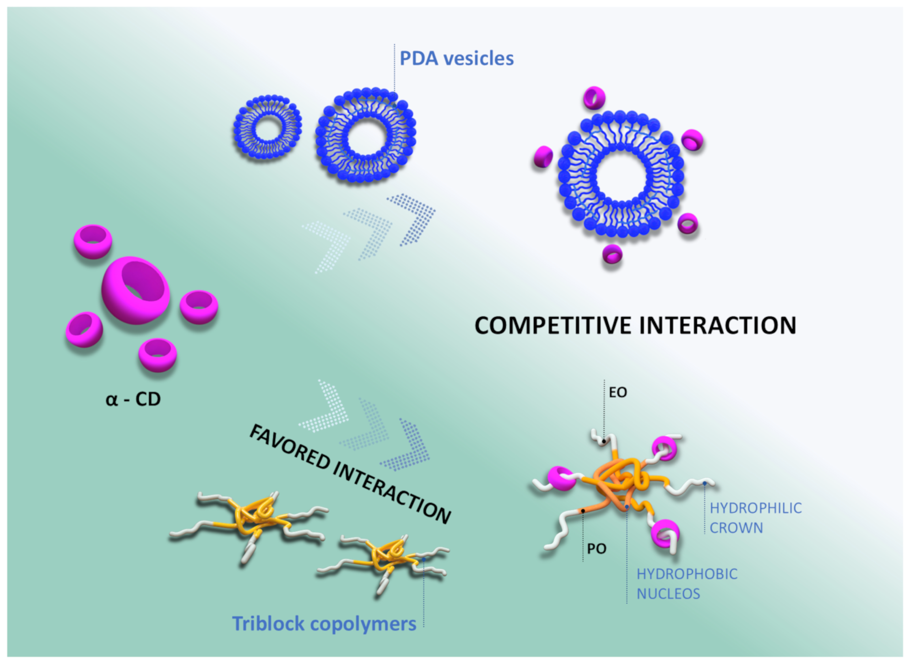
Therefore, in the PDA/L64 mixtures, α-CD molecules encounter two competing chemical environments for inclusion complex formation. In PDA vesicles, the hydrophilic headgroups containing acidic moieties can be included within the host cavity, inducing conformational rearrangements in the monomers that constitute the nanostructure and, consequently, triggering the color transition. Upon the addition of the copolymer, however, the EO segments can compete with PDA for the inclusion complex formation. Figure 2 schematically illustrates these two possible inclusion pathways.
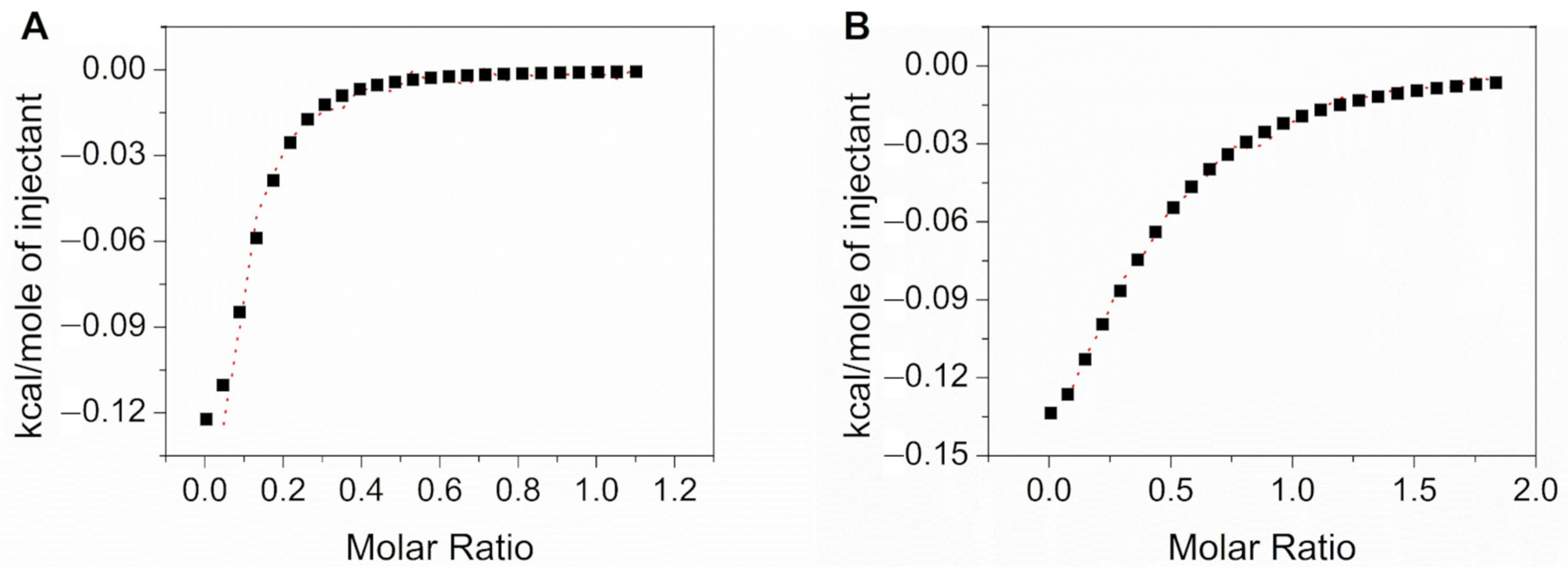
Based on the chromatic transition inhibition results, it can be inferred that the inclusion of the EO segments of the copolymer is thermodynamically more favorable than that of the PDA monomers. This preference arises because the inner cavity of α-CD is hydrophobic, and the EO segments (–CH2CH2O–) are comparatively more hydrophobic than the PDA monomer segments containing acidic groups (–CH2CH2COOH), which can establish strong hydrogen bonds with surrounding water molecules. In this sense, the thermodynamic parameters for the supramolecular interaction between α-CD with L64 and α-CD with PDA vesicles were investigated by ITC. Table 1 summarizes the thermodynamic data for both supramolecular systems, while Figure 3A and Figure 3B show the final ITC curves for the α-CD/L64 solution and the α-CD/PDA vesicle system, respectively.
At 298.15 K, both systems exhibit negative standard Gibbs free energy changes (ΔG° ≈ −21.78 kJ mol−1 for α-CD/L64 and −21.54 kJ mol−1 for α-CD/PDA), indicating spontaneous complexation under the experimental conditions. However, the binding constant for α-CD/L64 (K ≈ 11,300 ± 1250) exceeds that for α-CD/PDA (K ≈ 4000 ± 353), suggesting a markedly stronger affinity of α-CD for the L64 copolymer. The K values are comparable to those reported for other supramolecular systems involving surfactants [41].
Interestingly, both systems display only marginally exothermic enthalpy changes (ΔH° = −0.73 kJ mol−1 and −0.94 kJ mol−1 for α-CD/L64 and α-CD/PDA, respectively), accompanied by substantial positive entropy contributions (TΔS° ≈ +21.05 kJ mol−1 for α-CD/L64 and +19.60 kJ mol−1 for α-CD/PDA). From a thermodynamic standpoint, this indicates that while enthalpic contributions are nearly negligible, the driving force for complexation is dominated by entropic gain, most plausibly arising from the release of water molecules structured around hydrophobic moieties, and possibly from multiple supramolecular configurations coexisting in solution [38].
For the α-CD/L64 system, the significantly larger binding constant points to an optimal alignment of inclusion geometry and binding-site accessibility. The central polypropylene oxide (PPO) block of L64 is flanked by polyethylene oxide (PEO) segments [38], and α-CD can thread and stack along the PEO chains, forming “molecular necklace” structures. Incorporation of the PPO block may create hydrophobic–hydrophilic transitions that further favor α-CD inclusion. The higher K suggests that α-CD not only engages with the PEO domains but may also interact at the hydrophilic–hydrophobic interface of L64 in either micellar or non-micellar form. By contrast, in the α-CD/PDA vesicle system, the lower K and similar thermodynamic signature indicate a less favorable or less accessible binding environment, as the vesicular architecture of PDA may sterically hinder α-CD access to the hydrophobic domains.
Taken together, the thermodynamic profiles of both systems strongly suggest that α-CD complexation is predominantly entropy-driven and weakly exothermic, but that the local microenvironment and polymer architecture are crucial. The L64 system provides a dynamic and readily accessible hydrophilic–hydrophobic interface, along with sufficient PEO segment length, to enable stronger host–guest interactions, whereas the PDA vesicles impose a more constrained binding environment, thereby limiting the magnitude of the entropic contribution.
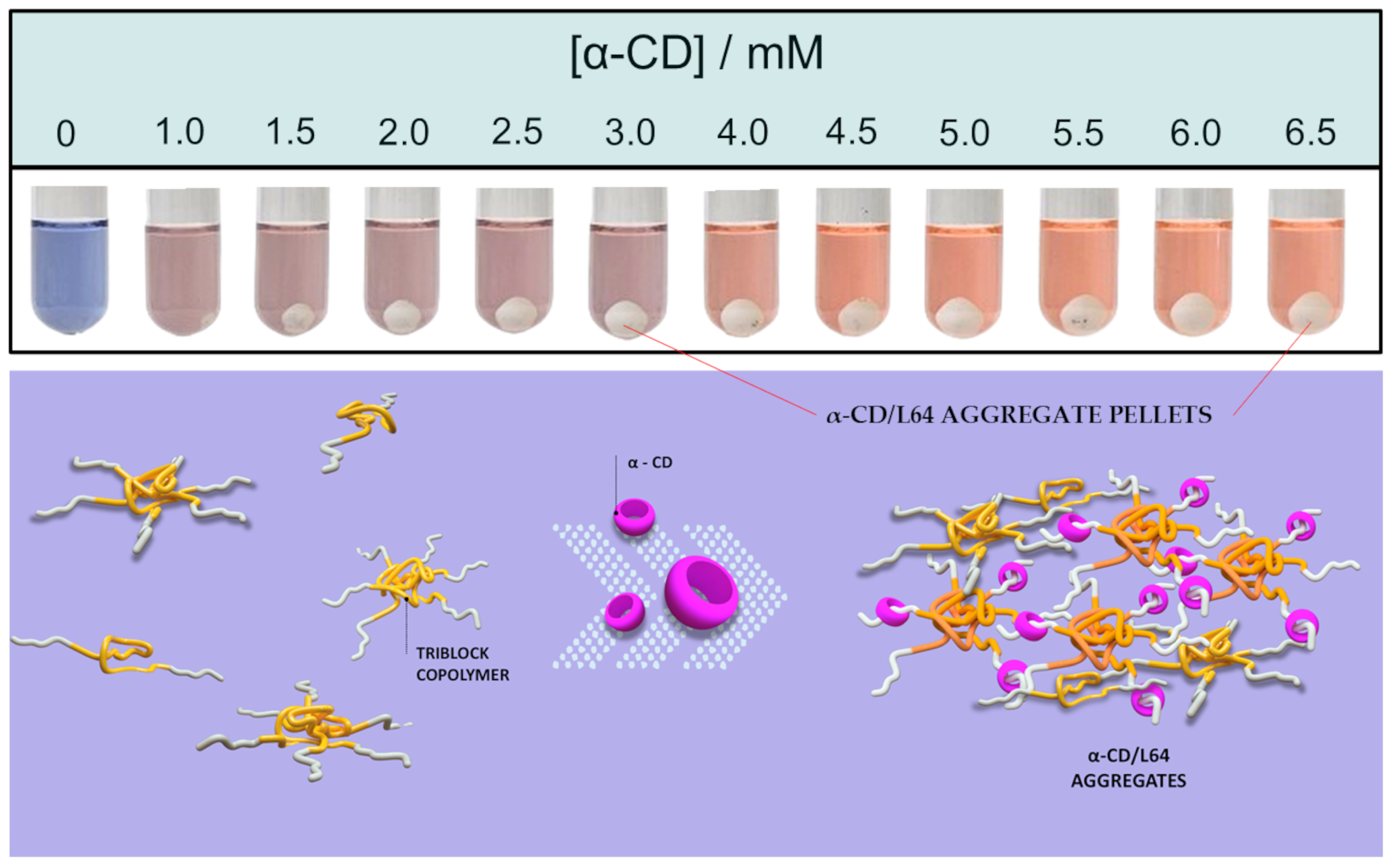
As previously discussed, the addition of α-CD increases the turbidity of the solution, which can be explained by the formation of α-CD/L64 inclusion complexes. The incorporation of EO segments into the hydrophobic cavities of α-CD promotes aggregation or interpenetration of oligomers and micelles, consequently favoring phase separation. To evaluate this effect, tubes containing 0.5 wt% L64 were centrifuged after the addition of different α-CD concentrations. Figure 4 presents images of the tubes after centrifugation, where the formation of a white precipitate at the bottom, corresponding to the α-CD–L64 complex, becomes increasingly evident with increasing α-CD concentrations. In the absence of α-CD, no sediment was observed.
It is also important to emphasize that the PDA-containing phase did not collapse under the applied centrifugation conditions. This observation suggests that during the self-assembly of the diacetylene monomers, PDA vesicles are not organized over L64 chains and are therefore not structurally connected. If such association had occurred, the precipitation of the copolymer would have resulted in a clear supernatant. From a technological perspective, this structural independence is particularly advantageous, as it ensures that the sedimentation of L64, either upon α-CD addition or during analyte exposure does not interfere with the PDA vesicular phase responsible for the chromatic response. In other words, the optical functionality of the PDA domain remains intact, even when the copolymer undergoes phase separation.

4. Conclusions

This study demonstrates, for the first time, that L64 triblock copolymer can act as effective inhibitors of the affinity-chromism of PDA vesicles in the presence of α-CD. The colorimetric response was progressively suppressed as the L64 concentration increased. Thermodynamic analysis by ITC revealed that α-CD interacts more strongly with the L64 copolymer (K ≈ 11,300) than with PDA vesicles (K = 4000), with both complexation processes being spontaneous (ΔG° = −21 kJ mol−1) and predominantly entropy-driven. The higher affinity of α-CD for L64 arises from the favorable alignment and accessibility of its EO segments, which compete with PDA headgroups for host–guest interactions. In addition, centrifugation experiments confirmed the formation of α-CD/L64 inclusion aggregates and phase separation without collapse of the PDA phase. This structural independence between the optical and copolymer domains ensures that the chromatic sensing function of the PDA vesicles remains intact.
Finally, these findings reveal a new supramolecular strategy for controlling the chromatic response of PDA-based sensors through competitive complexation with biocompatible triblock copolymers. Future studies may further optimize this system by exploring copolymers with different molecular weights and degrees of hydrophilicity, enabling fine control over concentration windows for colorimetric response. Such an approach may also help mitigate aggregation-induced light scattering, which currently limits quantitative colorimetric analysis. This tunability opens promising perspectives for the rational design of robust and adjustable chromatic materials for biosensing, environmental monitoring, and smart packaging applications.

Supplementary Materials

The following supporting information can be downloaded at: https://www.mdpi.com/article/10.3390/nanomanufacturing6030015/s1, Figure S1: UV–vis spectral evolution upon addition of α-CD (0–7 mM) to PDA vesicles (A) and PDA/L64 mixtures containing 0.5 (B), 1.0 (C), 2.0 (D), 4.0 (E), and 8.0 (F) wt% of L64. The blue and pink shaded regions are centered at 650 and 540 nm, corresponding to the absorption maxima of the blue and red phases of the PDA/L64 suspensions, respectively, whose intensities are modulated by α-CD addition. Increasing α-CD concentration leads to a pronounced rise in light scattering at shorter wavelengths and a progressive baseline shift, indicating the formation of larger aggregates and increased turbidity; Figure S2: UV–vis spectra of PDA/L64 suspensions prepared with different L64 concentrations, in the absence of α-CD. As the L64 content increases, the suspensions exhibit higher molar absorptivity, despite being prepared with the same PCDA concentration. This behavior evidences the beneficial role of the block copolymer in promoting PDA self-organization and the formation of extended π-conjugated systems. The spectrum of PDA vesicles prepared without L64 is shown in the inset for improved visualization; Figure S3: Photographs illustrating the colorimetric transition induced by the addition of α-CD to PDA/L64 suspensions in test tubes with increased optical path length. Increasing α-CD concentration (highlighted horizontally) promotes the visible colorimetric transition. As the L64 content increases, the blue-to-orange transition observable to the naked eye occurs at progressively higher α-CD concentrations, confirming the inhibitory effect of the copolymer on the colorimetric response; Figure S4: Variations in the red (R) and blue (B) colorimetric signals upon α-CD addition to PDA/L64 suspensions. The R and B coordinates were extracted from the photograph shown in Figure 1, with all compositions arranged in the same well plate to ensure consistent optical conditions. Upon increasing α-CD concentration, the B signal shows a gradual decrease with relatively limited variation, whereas significantly larger changes are observed in the R coordinate; Figure S5: UV–vis spectra of PDA vesicles undergoing a pH-induced colorimetric transition (A) and variations in the red and blue signals as a function of pH (B). The R and B coordinates were extracted from photographs of the suspensions acquired in a well plate. A markedly larger variation is observed for the R coordinate compared to B, together with a behavior that differs from the trends observed in the UV–vis spectra, highlighting the distinct sensitivities of optical absorption and image-based colorimetric analyses.

Author Contributions

M.C.O.R.: methodology, formal analysis, investigation; M.E.F.R.A.: methodology, formal analysis, investigation; A.R.M.A.: investigation, formal analysis, data curation; D.C.d.M.: investigation, formal analysis, data curation; F.B.D.S.: supervision, conceptualization, funding acquisition; G.A.S.J.: conceptualization, formal analysis, data curation, writing—review and editing; J.P.C.T.: data curation, writing—original draft preparation, supervision, funding acquisition, project administration, writing—review and editing; P.F.R.O.: data curation, writing—original draft preparation, supervision, funding acquisition, project administration, writing—review and editing. All authors have read and agreed to the published version of the manuscript.

Funding

This research was funded by The CONSELHO NACIONAL DE DESENVOLVIMENTO CIENTÍFICO E TECNOLÓGICO-CNPq (grant numbers 407585/2023-0 and 303311/2024-0), FUNDAÇÃO DE AMPARO À PESQUISA DO ESTADO DE MINAS GERAIS–FAPEMIG (grant numbers APQ-01211-24, APQ-01313-24, APQ-03785-25, APQ-04537-22, RED-00045-23, RED-00200-23, APQ-00144-24, and APD-00076-25) and COORDENAÇÃO DE APERFEIÇOAMENTO DE PESSOAL DE NÍVEL SUPERIOR (88887.202956/2025-000).

Data Availability Statement

The data adopted in support of the findings in this work are available upon request from the corresponding author.

Acknowledgments

J.P.C Trigueiro and F.B Sousa are a recipient of a fellowship from CNPq (grant number 303712/2025-2 and 303311/2024-0, respectively). M.C.O.R., M.E.F.R.A., and A.R.M.A. are grateful to FAPEMIG, CNPq and CAPES for the scholarships granted.

Conflicts of Interest

The authors declare no conflicts of interest. The funders had no role in the design of the study; in the collection, analyses, or interpretation of data; in the writing of the manuscript; or in the decision to publish the results.

Abbreviations

The following abbreviations are used in this manuscript:
PDAsPolydiacetylenes
UVUltraviolet
EOEthylene oxide
POPropylene oxide
CDCyclodextrin
ICsInclusion complexes
L64Poly(ethylene glycol)-block-poly(propylene glycol)-block-poly(ethylene glycol)
MWMolecular weight
PCDA10,12-pentacosadiynoic acid
RGBRed–Green–Blue color model
ROIsRegions of interest
UV–VisUltraviolet–Visible spectroscopy
ITCIsothermal titration calorimetry
nStoichiometric coefficients
KAssociation constant
VP-ITCVariable-Pressure Isothermal Titration Calorimeter
CRColorimetric response
CMCCritical micelle concentration
PPOPolypropylene oxide
PEOPolyethylene oxide

References

  1. Ortega, P.F.R.; Galvao, B.R.L.; de Oliveira, P.S.C.; Bastos, G.A.A.; Bernardes, M.R.F.; Lavall, R.L.; Trigueiro, J.P.C. Thermochromism in Polydiacetylene/Poly(vinyl alcohol) Hydrogels Obtained by the Freeze-Thaw Method: A Theoretical and Experimental Study. Ind. Eng. Chem. Res. 2021, 60, 13243–13252. [Google Scholar] [CrossRef]
  2. Huo, J.; Hu, Z.; He, G.; Hong, X.; Yang, Z.; Luo, S.; Ye, X.; Li, Y.; Zhang, Y.; Zhang, M.; et al. High temperature thermochromic polydiacetylenes: Design and colorimetric properties. Appl. Surf. Sci. 2017, 423, 951–956. [Google Scholar] [CrossRef]
  3. Wilk-Kozubek, M.; Potaniec, B.; Gazinska, P.; Cybinska, J. Exploring the Origins of Low-Temperature Thermochromism in Polydiacetylenes. Polymers 2024, 16, 2856. [Google Scholar] [CrossRef] [PubMed]
  4. Singh, Y.; Jayaraman, N. Visual Detection of pH and Biomolecular Interactions at Micromolar Concentrations Aided by a Trivalent Diacetylene-Based Vesicle. Macromol. Chem. Phys. 2017, 218, 1700039. [Google Scholar] [CrossRef]
  5. Beliktay, G.; Shaikh, T.; Koca, E.; Cingil, H.E. Effect of UV Irradiation Time and Headgroup Interactions on the Reversible Colorimetric pH Response of Polydiacetylene Assemblies. ACS Omega 2023, 8, 37213–37224. [Google Scholar] [CrossRef] [PubMed]
  6. Weston, M.; Tjandra, A.D.; Chandrawati, R. Tuning chromatic response, sensitivity, and specificity of polydiacetylene-based sensors. Polym. Chem. 2020, 11, 166–183. [Google Scholar] [CrossRef]
  7. Kim, T.; Moon, D.; Park, J.H.; Yang, H.; Cho, S.; Park, T.H.; Ahn, D.J. Visual detection of odorant geraniol enabled by integration of a human olfactory receptor into polydiacetylene/lipid nano-assembly. Nanoscale 2019, 11, 7582–7587. [Google Scholar] [CrossRef] [PubMed]
  8. Charych, D.; Nagy, J.; Spevak, W.; Bednarski, M. Direct colorimetric detection of a receptor-ligand interaction by a polymerized bilayer assembly. Science 1993, 261, 585–588. [Google Scholar] [CrossRef] [PubMed]
  9. Cho, J.; Woo, S.; Ahn, D.; Ahn, K.; Lee, H.; Kim, J. Cyclodextrin-induced color changes in polymerized diacetylene Langmuir-Schaefer films. Chem. Lett. 2003, 32, 282–283. [Google Scholar] [CrossRef]
  10. Seto, K.; Hosoi, Y.; Furukawa, Y. Raman spectra of Langmuir-Blodgett and Langmuir-Schaefer films of polydiacetylene prepared from 10,12-pentacosadiynoic acid. Chem. Phys. Lett. 2007, 444, 328–332. [Google Scholar] [CrossRef]
  11. Geiger, E.; Hug, P.; Keller, B. Chromatic transitions in polydiacetylene Langmuir-Blodgett films due to molecular recognition at the film surface studied by spectroscopic methods and surface analysis. Macromol. Chem. Phys. 2002, 203, 2422–2431. [Google Scholar] [CrossRef]
  12. Iimori, Y.; Onodera, T.; Kasai, H.; Mitsuishi, M.; Miyashita, T.; Oikawa, H. Fabrication of pseudo single crystalline thin films composed of polydiacetylene nanofibers and their optical properties. Opt. Mater. Express 2017, 7, 2218–2223. [Google Scholar] [CrossRef]
  13. Velarde, M.G.; Chetverikov, A.P.; Ebeling, W.; Wilson, E.G.; Donovan, K.J. On the electron transport in polydiacetylene crystals and derivatives. Europhys. Lett. 2014, 106, 27004. [Google Scholar] [CrossRef]
  14. Lebegue, E.; Farre, C.; Jose, C.; Saulnier, J.; Lagarde, F.; Chevalier, Y.; Chaix, C.; Jaffrezic-Renault, N. Responsive Polydiacetylene Vesicles for Biosensing Microorganisms. Sensors 2018, 18, 599. [Google Scholar] [CrossRef] [PubMed]
  15. Miller, J.S.; Finney, T.J.; Ilagan, E.; Frank, S.; Chen-Izu, Y.; Suga, K.; Kuhl, T.L. Fluorogenic Biosensing with Tunable Polydiacetylene Vesicles. Biosensors 2025, 15, 27. [Google Scholar] [CrossRef] [PubMed]
  16. Yu, Z.; MuYu, C.; Xu, H.; Zhao, J.; Yang, G. Recent progress in the design of conjugated polydiacetylenes with reversible thermochromic performance: A review. Polym. Chem. 2023, 14, 2266–2290. [Google Scholar] [CrossRef]
  17. Jelinek, R.; Ritenberg, M. Polydiacetylenes—Recent molecular advances and applications. RSC Adv. 2013, 3, 21192–21201. [Google Scholar] [CrossRef]
  18. Nguyen, L.H.; Oveissi, F.; Chandrawati, R.; Dehghani, F.; Naficy, S. Naked-Eye Detection of Ethylene Using Thiol-Functionalized Polydiacetylene-Based Flexible Sensors. ACS Sens. 2020, 5, 1921–1928. [Google Scholar] [CrossRef] [PubMed]
  19. Guo, J.; Yang, L.; Zhu, L.; Chen, D. Selective detection of metal ions based on nanocrystalline ionochromic polydiacetylene. Polymer 2013, 54, 743–749. [Google Scholar] [CrossRef]
  20. Tjandra, A.D.; Chandrawati, R. Polydiacetylene/copolymer sensors to detect lung cancer breath volatile organic compounds. RSC Appl. Polym. 2024, 2, 1043–1056. [Google Scholar] [CrossRef]
  21. Jang, H.; Jeon, J.; Shin, M.; Kang, G.; Ryu, H.; Kim, S.M.; Jeon, T.-J. Polydiacetylene (PDA) Embedded Polymer-Based Network Structure for Biosensor Applications. Gels 2025, 11, 66. [Google Scholar] [CrossRef] [PubMed]
  22. Dias Ferreira, G.M.; Dias Ferreira, G.M.; Hespanhol, M.C.; Rezende, J.P.; Pires, A.C.S.; Ortega, P.F.R.; Mendes da Silva, L.H. A simple and inexpensive thermal optic nanosensor formed by triblock copolymer and polydiacetylene mixture. Food Chem. 2018, 241, 358–363. [Google Scholar] [CrossRef]
  23. Morais, D.C.; Vieira, B.B.M.; Carvalho, M.C.; Miguez, F.B.; Lopes, J.F.; Sousa, F.B.D. Thermodynamic investigation of biperiden hydrochloride and cyclodextrins supramolecular systems. Chem. Phys. Lett. 2024, 851, 141500. [Google Scholar] [CrossRef]
  24. Passos, J.J.; De Sousa, F.B.; Mundim, I.M.; Bonfim, R.R.; Melo, R.; Viana, A.F.; Stolz, E.D.; Borsoi, M.; Rates, S.M.K.; Sinisterra, R.D. Double continuous injection preparation method of cyclodextrin inclusion compounds by spray drying. Chem. Eng. J. 2013, 228, 345–351. [Google Scholar] [CrossRef]
  25. Passos, J.J.; De Sousa, F.B.; Mundim, I.M.; Bonfim, R.R.; Melo, R.; Viana, A.F.; Stolz, E.D.; Borsoi, M.; Rates, S.M.K.; Sinisterra, R.D. In vivo evaluation of the highly soluble oral β-cyclodextrin–Sertraline supramolecular complexes. Int. J. Pharm. 2012, 436, 478–485. [Google Scholar] [CrossRef] [PubMed]
  26. Utzeri, G.; Matias, P.M.C.; Murtinho, D.; Valente, A.J.M. Cyclodextrin-Based Nanosponges: Overview and Opportunities. Front. Chem. 2022, 10, 859406. [Google Scholar] [CrossRef] [PubMed]
  27. Roy, I.; Stoddart, J.F. Cyclodextrin Metal-Organic Frameworks and Their Applications. Acc. Chem. Res. 2021, 54, 1440–1453. [Google Scholar] [CrossRef] [PubMed]
  28. Valente, A.J.M.; Soderman, O. The formation of host–guest complexes between surfactants and cyclodextrins. Adv. Colloid Interface Sci. 2014, 205, 156–176. [Google Scholar] [CrossRef] [PubMed]
  29. Champaiboon, T.; Tumcharern, G.; Potisatityuenyong, A.; Wacharasindhu, S.; Sukwattanasinitt, M. A polydiacetylene multilayer film for naked eye detection of aromatic compounds. Sens. Actuators B Chem. 2009, 139, 532–537. [Google Scholar] [CrossRef]
  30. Martínez, M.; Ballesteros, S.; Almarza, E.; de la Torre, C.; Búa, S. Acute nitrobenzene poisoning with severe associated methemoglobinemia: Identification in whole blood by GC-FID and GC-MS. J. Anal. Toxicol. 2003, 27, 221–225. [Google Scholar] [CrossRef] [PubMed]
  31. Tchieno, F.M.M.; Tonle, I.K. p-Nitrophenol determination and remediation: An overview. Rev. Anal. Chem. 2018, 37, 20170019. [Google Scholar] [CrossRef]
  32. de Castro, K.C.; Coco, J.C.; dos Santos, E.M.; Ataide, J.A.; Martinez, R.M.; Monteiro do Nascimento, M.H.; Prata, J.; Lopes da Fonte, P.R.M.; Severino, P.; Mazzola, P.G.; et al. Pluronic® triblock copolymer-based nanoformulations for cancer therapy: A 10-year overview. J. Control. Release 2023, 353, 802–822. [Google Scholar] [CrossRef] [PubMed]
  33. Liu, D.; Yang, M.; Wang, D.; Jing, X.; Lin, Y.; Feng, L.; Duan, X. DPD Study on the Interfacial Properties of PEO/PEO-PPO-PEO/PPO Ternary Blends: Effects of Pluronic Structure and Concentration. Polymers 2021, 13, 2866. [Google Scholar] [CrossRef] [PubMed]
  34. Mayer, B.; Klein, C.; Topchieva, I.; Köhler, G. Selective assembly of cyclodextrins on poly(ethylene oxide)-poly(propylene oxide) block copolymers. J. Comput.-Aided Mol. Des. 1999, 13, 373–383. [Google Scholar] [CrossRef] [PubMed]
  35. Almgren, M.; Brown, W.; Hvidt, S. Self-aggregation and phase-behavior of poly(ethylene oxide) poly(propylene oxide) poly(ethylene oxide) block-copolymers in aqueous solution. Colloid Polym. Sci. 1995, 273, 2–15. [Google Scholar] [CrossRef]
  36. Mata, J.; Majhi, P.; Guo, C.; Liu, H.; Bahadur, P. Concentration, temperature, and salt-induced micellization of a triblock copolymer Pluronic L64 in aqueous media. J. Colloid Interface Sci. 2005, 292, 548–556. [Google Scholar] [CrossRef] [PubMed]
  37. Marinov, G.; Michels, B.; Zana, R. Study of the state of the triblock copolymer poly(ethylene oxide) poly(propylene oxide) poly(ethylene oxide) L64 in aqueous solution. Langmuir 1998, 14, 2639–2644. [Google Scholar] [CrossRef]
  38. Pradal, C.; Jack, K.S.; Grondahl, L.; Cooper-White, J.J. Gelation kinetics and viscoelastic properties of Pluronic and α-cyclodextrin-based pseudopolyrotaxane hydrogels. Biomacromolecules 2013, 14, 3780–3792. [Google Scholar] [CrossRef] [PubMed]
  39. Tsai, C.-C.; Zhang, W.-B.; Wang, C.-L.; Van Horn, R.M.; Graham, M.J.; Huang, J.; Chen, Y.; Guo, M.; Cheng, S.Z.D. Evidence of formation of site-selective inclusion complexation between β-cyclodextrin and poly(ethylene oxide)-block-poly(propylene oxide)-block-poly(ethylene oxide) copolymers. J. Chem. Phys. 2010, 132, 204903. [Google Scholar] [CrossRef] [PubMed]
  40. Yang, C.; Ni, X.; Li, J. Synthesis of polyrotaxanes consisting of multiple α-cyclodextrin rings threaded on reverse Pluronic PPO-PEO-PPO triblock copolymers based on block-selected inclusion complexation. Eur. Polym. J. 2009, 45, 1570–1579. [Google Scholar] [CrossRef]
  41. Meira, L.H.R.; Soares, G.A.B.; Bonomini, H.I.M.; Lopes, J.F.; De Sousa, F.B. Thermodynamic compatibility between cyclodextrin supramolecular complexes and surfactant. Int. J. Pharm. 2018, 544, 203–212. [Google Scholar] [CrossRef] [PubMed]
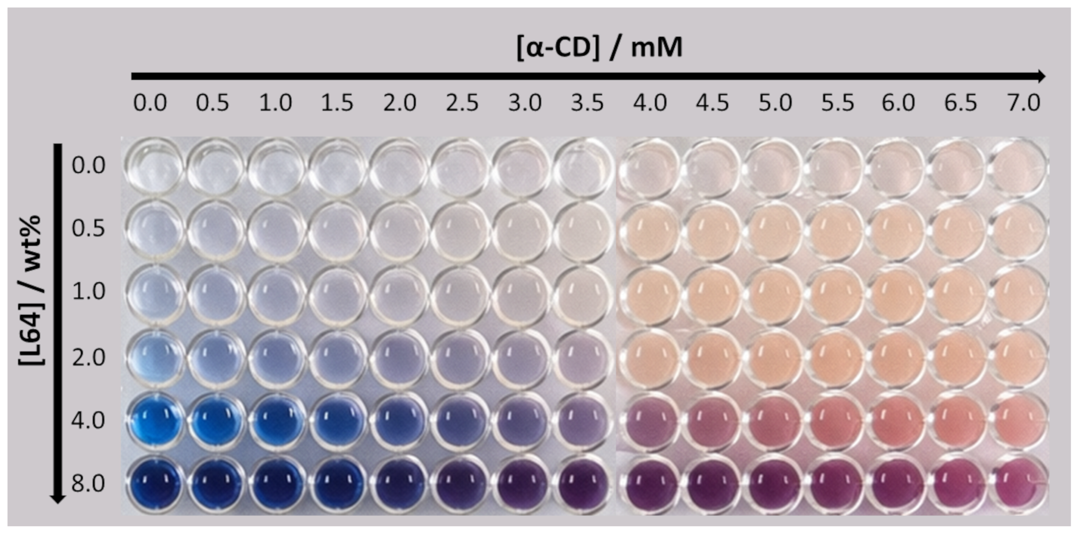
Figure 1. Photographs of PDA/L64 suspensions arranged in a well-plate format, illustrating the colorimetric response as a function of composition. L64 concentration increases vertically, while the α-CD concentration added to the suspensions increases horizontally. The progressive color change reflects the α-CD–induced chromatic transition and its modulation by the presence of the triblock copolymer.
Nanomanufacturing 06 00015 g001
Figure 2. Schematic representation of the competitive host–guest interactions between α-CD molecules and PDA nanostructures organized as vesicles, as well as with triblock copolymers. The preferential complexation of α-CD with the EO segments of the copolymer competes with its interaction at the PDA vesicle interface, thereby modulating the chromatic response of the system.
Nanomanufacturing 06 00015 g002
Figure 3. ITC titration curves at 298.15 K for α-CD titrated into (A) L64 solution and (B) PDA vesicle suspension.
Nanomanufacturing 06 00015 g003
Figure 4. Photographs of tubes containing PDA/L64 (4.0 wt%) suspensions after the addition of increasing α-CD concentrations. After centrifugation (4000 rpm, 5 min), α-CD/L64 aggregate pellets are observed at the bottom of the tubes, as indicated. Increasing α-CD concentration leads to the formation of larger pellet volumes. A schematic representation of the α-CD/L64 aggregate formation is also included.
Nanomanufacturing 06 00015 g004
Table 1. Thermodynamic parameters of the α-CD/L64 and α-CD/PDA vesicle supramolecular interactions at 298.15 K.
Supramolecular SystemKΔHo/kJ mol−1TΔSo/kJ mol−1ΔGo/kJ mol−1
α-CD with L6411 300 ± 1.250−0.7321.05−21.78
α-CD with PDA4 000 ± 353−0.9419.60−21.54
Disclaimer/Publisher’s Note: The statements, opinions and data contained in all publications are solely those of the individual author(s) and contributor(s) and not of MDPI and/or the editor(s). MDPI and/or the editor(s) disclaim responsibility for any injury to people or property resulting from any ideas, methods, instructions or products referred to in the content.