Review

Electrospun Nanofibers for Antibiotic Release and Antibacterial Performance: A Nanomanufacturing Perspective


by
Jorge A. Ornelas-Guillén
1,
Lisbeth Daniela Mora-González
1,
Estefanía Reyes-Mercado
1,
Mario Valle-Sánchez
2,3,
Erick Cuevas-Yáñez
2,3,
J. Betzabe González-Campos
1,* and
Alejandra Pérez-Nava
3,4,*
1
Instituto de Investigaciones Químico-Biológicas, Universidad Michoacana de San Nicolás de Hidalgo, Gral. Francisco J. Múgica S/N, Ciudad Universitaria, Morelia 58030, Michoacán, Mexico
2
Facultad de Química, Universidad Autónoma del Estado de México, P.º Colón S/N, Residencial Colón y Col. Ciprés, Toluca de Lerdo 50120, Mexico, Mexico
3
Centro Conjunto de Investigación en Química Sustentable UAEMéx-UNAM, Km 14.5 Carretera Toluca—Atlacomulco, Unidad San Cayetano, Toluca de Lerdo 50200, Mexico, Mexico
4
Instituto de Química, Universidad Nacional Autónoma de México, Circuito Exterior S/N, Circuito de la Investigación Científica, Ciudad Universitaria, Ciudad de México 04510, Mexico, Mexico
*
Authors to whom correspondence should be addressed.
Nanomanufacturing 2026, 6(2), 11; https://doi.org/10.3390/nanomanufacturing6020011
Submission received: 13 December 2025 / Revised: 23 January 2026 / Accepted: 20 April 2026 / Published: 19 May 2026

Abstract

Electrospun composites are desirable materials for drug delivery applications. Regarding microbial infections as a case study, the antibacterial effect is enhanced by physical attributes of electrospun meshes, namely, a high surface area-to-volume ratio and porosity, 3D topography, and customized surface functions. Beyond mimicking nanostructured fibers, the delivery of antibiotics from such composites enhances antibacterial efficacy, sustained release kinetics, and reduced wound infection while minimizing side effects. Concern over antibiotic resistance and the insufficient availability of pharmaceutical agents for effective infection treatment is increasing worldwide. A significant number of publications have reported the fabrication of electrospun composites to mitigate bacterial pathogenesis. However, from a structural and morphological perspective, the implications of electrospinning approaches for antibiotic delivery have not been reviewed. This proposal presents a comparative study of the different assemblies induced by electrospinning, enabling the development of platforms for administering antibacterial agents. The primary objective is to conduct a comprehensive examination of the considerations involved in electrospinning-based manufacturing of drug delivery systems and antibiotic loading, ensuring a thorough design process that accounts for composite processability, monitoring methods for kinetic behavior analysis and modeling, and biological considerations for pre-clinical in vitro characterization.

1. Introduction

The terms antimicrobial, antibacterial, and antibiotic are often used interchangeably in the scientific literature, even though they represent distinct pharmacological and functional concepts, which can lead to conceptual ambiguity and misinterpretation of experimental outcomes. Antibacterial agents are compounds that target bacterial organisms, whether of natural or synthetic origin, and are often characterized by their bacteriostatic or bactericidal properties [1]. In contrast, the term antibiotic traditionally denotes compounds produced by microorganisms or derived from them that exert activity against bacteria. However, its use has broadened clinically to include related synthetic or semi-synthetic agents directed against bacterial infections [2]. In this review, we adopt and apply these definitions—antimicrobial as the overarching category, antibacterial as bacteria-specific agents, and antibiotic as the subclass of naturally derived or related agents—to ensure clarity and scientific precision throughout the manuscript.
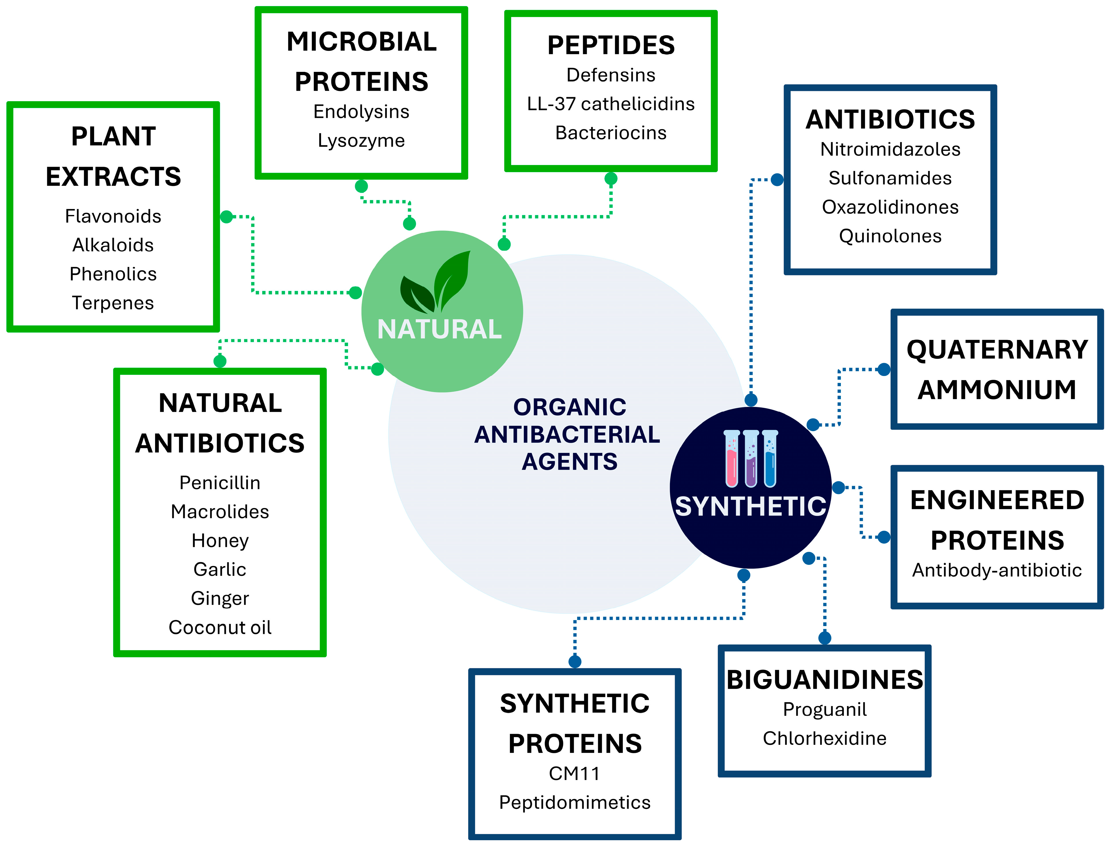
Antibiotics are among the most prescribed drugs in human and animal medicine, serving as powerful tools for treating bacterial infections; however, they are also misused to treat viral diseases. Repeated exposure to these drugs, especially at low doses, contributes to the spread of antibiotic-resistant bacteria, making infections more challenging to treat. Organic antibacterial agents can be classified by nature (natural or synthetic), as shown in Figure 1.
Antibiotic resistance has necessitated the development of new and improved antibacterial agents. Improving the precision of existing antibiotic therapies by optimizing dosing and therapy duration can reduce the risk of toxicity and minimize the development of drug resistance. Low bioavailability, side effects, poor penetration, and accumulation at the site of infection are other factors that contribute to antibiotic resistance and the clinical failure of antibiotic therapy [3]. In addition, the development of new antibiotics could be based on the chemical structures of existing ones; however, their mechanisms of action are the same as those to which bacteria have already developed resistance. Therefore, approaches to the stewardship of existing antibiotics are needed [3].
In this context, the use of plant-derived antibacterial compounds (such as alkaloids, coumarins, essential oils, flavonoids, lectins, phenolics, polypeptides, terpenoids, and tannins) [4] is a promising alternative for controlling bacterial infections and mitigating the emergence of antibiotic resistance, since natural products have high potential against resistant infections due to their multimodal mechanism of action [5].
Investigations related to the development of new and efficient antibiotic delivery systems include siderophores, antibodies, bacteriophages, short cell-penetrating peptides, host innate antimicrobial peptides, and nanotechnology-based delivery systems [3]. Nanotechnology-based delivery systems could improve treatment for drug-resistant bacteria and reduce side effects by using nanocarriers such as liposomes, polymer nanoparticles, polymer nanofibers, and nanogels. These are some examples of targeted delivery designs based on nanotechnology, improving solubility and controlled release, enhancing bioavailability, bearing the entry into bacterial cells and biofilms, and thereby improving drug efficacy [6].
Nanogels are composed of soft hydrogel nanoparticles that can encapsulate and deliver antibiotics more effectively while protecting them from degradation [7]. Antibiotics released from nanogels maintain therapeutic levels over time and reduce the frequency of dosing [8]. Drug penetration and efficacy are enhanced by nanogels’ high-water content, biocompatibility, tunable particle size, and stimulus-responsive properties. Scalability, reproducibility, biosafety, immunogenicity, and stability in complex biological environments are concerns that hinder clinical translation [7,9,10].
Polymer nanoparticles loaded with antibiotics can increase drug concentration and reduce systemic side effects. They can be engineered to target specific pathogens or infected cells, offering potential advantages in treating bacterial infections, especially those caused by multidrug-resistant bacteria. Biocompatible and biodegradable polymer nanoparticles protect drugs from degradation, can improve their solubility, enhance their penetration into target tissues, and facilitate controlled release at the site of infection [11,12]. They can be delivered via different routes, including lung drug-delivery; however, biodegradability and nanoparticles’ safety are concerns on this delivery route [12]. Some limitations of these delivery systems are related to particle stability and aggregation, premature drug release before reaching the target, and potential toxicity of the polymers and the used solvents, in addition to difficulties in scalability, regulatory compliance, and controlling drug release kinetics to prevent leakage or ensure sustained delivery [13,14,15].
On the other hand, electrospun polymeric nanofibers loaded with antibiotics can fight against bacteria by effectively delivering drug cargo to the target site. Topographic and unique physicochemical properties, including bacteria-like nano-scale features, are key factors in enhancing bacterial attachment and inhibition. A large surface area-to-volume ratio facilitates the encapsulation of antimicrobial therapeutic agents and allows high interconnected porosity, which enables higher loading of antimicrobial agents, increases bacterial attachment to their surfaces, promotes oxygen and nutrient exchange, ensures sustained drug release, and enhances wound healing by promoting cell proliferation. Surface chemistry also influences their therapeutic activity and offers tailored antimicrobial therapy. Their nanometric-scale structure resembles the native extracellular matrix (ECM) architecture and provides an excellent environment for cell migration, adhesion, differentiation, and regeneration [16]. Nanofibers have a remarkable drug-loading efficiency and adaptability in enabling efficient drug administration.
Ultrafine polymer nanofibers are commonly produced by melt blowing [17], phase separation [18], centrifugal spinning [1], template synthesis [19], and electrospinning [20]. Among them, electrospinning is the simplest and most effective method for their manufacture. Due to the versatility of the electrospinning setup, the morphological features of polymer nanofibers can be tailored by tuning polymer solution parameters or processing parameters. The final characteristics related to size, diameter, porosity, surface wettability, and surface charge of nanofibers are crucial to induce a bactericidal effect [16].
Due to the high potential of nanofibers as drug-delivery systems for antimicrobial agents, this review compares various electrospun assemblies used to fabricate platforms for the delivery of natural and synthetic antibacterial agents. Beyond electrospinning implications, a perspective for kinetic analysis and in vitro procedures is also outlined.

2. Nanofiber Manufacturing

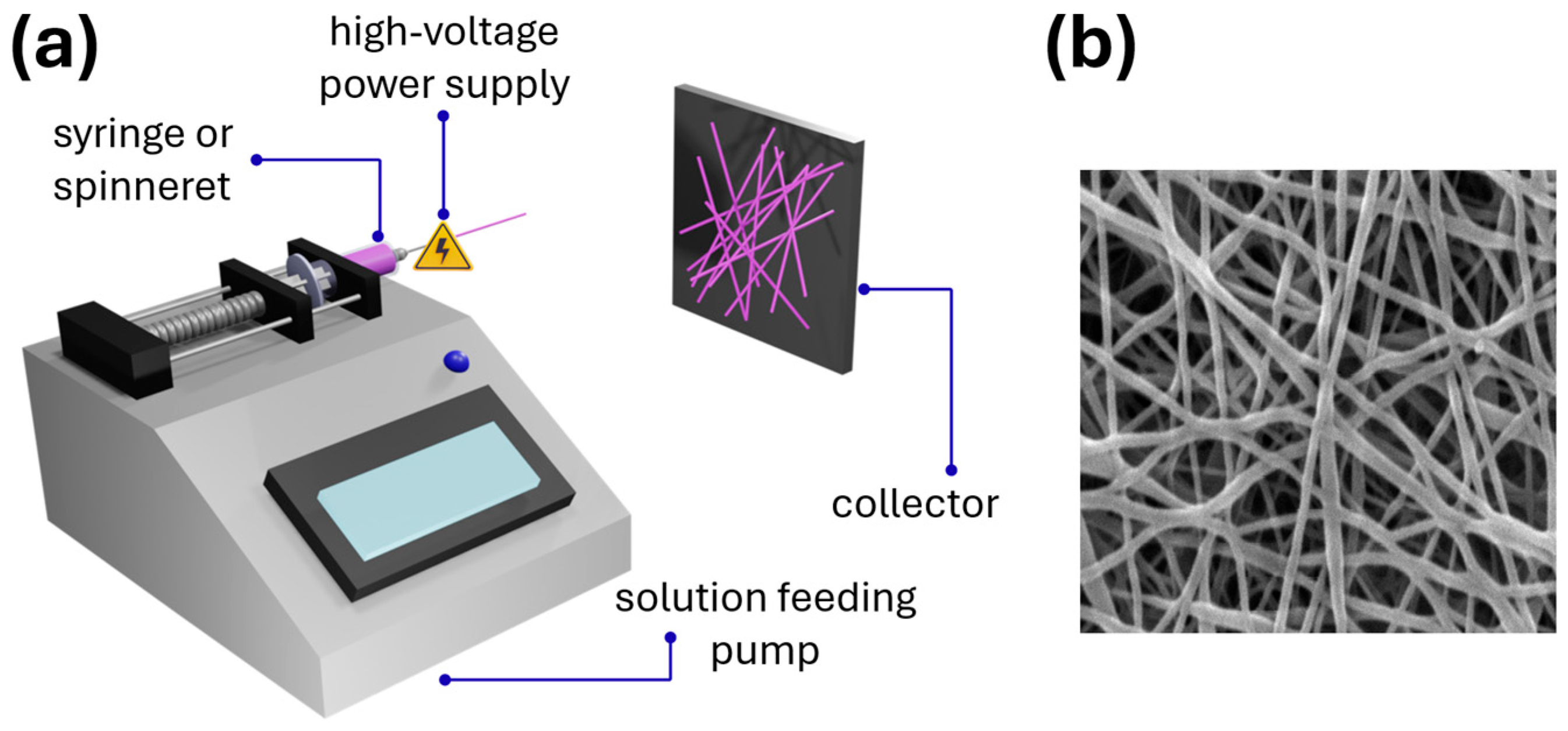
Electrospinning is a voltage-driven process for producing micro- and nanofiber collections from polymeric solutions. The typical electrospinning setup (shown in Figure 2) includes four main components: a high-voltage power supply, a syringe pump, a spinneret, and a collector. The polymer solution is charged in a syringe and pumped at a determined flow rate. An electrostatic force is induced between the needle tip and the target ground electrode (the collector) with a sufficient voltage to overcome the solution surface tension, forming the Taylor cone and elongating the jet, which travels from the needle to the collector, reducing its diameter and increasing its length. The use of a volatile solvent in the polymer solution is significant for this process, as it enables solvent evaporation. In contrast, the polymer solidifies and deposits as fibers on the collector.
The control of nanofiber morphology and diameter relies mainly on process parameters, such as voltage, distance between the needle and the collector, solution flow rate, collector type, and ambient conditions, which affect fiber diameter and bead formation. Voltage is crucial for drawing the polymer solution into a jet and subsequently forming nanofibers [21,22]. Precise control of the polymer solution flow rate ensures a consistent, controllable electrospinning process [23]. The spinneret, which is typically a metallic needle, acts as a nozzle through which the polymer is ejected [22,24], and the collector can influence orientation and morphology [24,25]. Other solution parameters, such as concentration, viscosity, molecular weight, surface tension, conductivity, and solvent type, can, in addition to affecting fiber diameter and the production of good-quality nanofibers without beads, lead to no fiber formation [20].
Electrospinning is recognized as a versatile technique for creating random or aligned nanofiber mats from diverse materials, with controlled microstructures and morphologies, including solid, porous, or hollow structures. Single-phase, embedded with nanoparticles or active compounds, surface-porous, multiaxial, or hollow nanofibers can be produced. This diversity is achieved by modifying the polymer solution or the electrospinning setup. Nanofiber mats possess a high surface area-to-volume ratio and a high degree of interconnected porosity. Control of fiber diameter and alignment [26] enables potential functionalization [26,27], compositional versatility, and customizable properties by adjusting the material system and processing parameters [28,29]. Thus, their unique microstructure makes them excellent candidates for use as drug carriers in drug delivery systems (DDS).
DDS and drug carriers are crucial for enhancing drug efficacy and safety. Key functions of DDS include controlled release, target delivery, enhanced bioavailability, drug protection, and overcoming biological barriers.

2.1. Drug Delivery Systems (DDS) Based on Nanofibers

DDS are formulations or technologies for the safe and effective transportation of active pharmaceutical ingredients (API) or drugs into the body to enhance the desired therapeutic effect [30]. Key functions of DDS include controlled release, target delivery, enhanced bioavailability, drug protection, and overcoming biological barriers [31]. These systems can influence a drug’s pharmacological activity by modulating its release from the carrier. In addition, incorporating a drug into a DDS can reduce the frequency of administration and toxicity [32].
DDS based on nanofiber mats can encapsulate and release drugs in a controlled manner; furthermore, they can be designed in different forms for administration via topical, oral, and transdermal routes [32,33,34,35]. The drug-loading technique depends on the drug solubility in the polymer solution and is crucial to the release process. Drugs can be encapsulated into nanofibers using a single-phase electrospinning process, chemically immobilized on fiber surfaces, or loaded by physical adsorption via electrostatic, hydrophobic, hydrogen-bonding, van der Waals forces, and fiber-surface interactions [36].
Incorporating antibiotics into DDS could improve bioavailability and target efficiency to address antimicrobial resistance, focusing on improved treatment strategies [37]. The nanofiber structure promotes surface interactions and enhances bacterial inhibition [38]. Antibiotic drugs are commonly incorporated into polymer nanofibers through simple immersion of the nanofiber membrane into the corresponding antibiotic solution [39], by mixing components into the solution before electrospinning [40,41,42] or by covalent bond to the nanofiber surface [43]. The resulting antibiotic-loaded nanofibers can also be used as coatings for metallic implants to prevent and treat bone infections [44].

2.2. Electrospun Polymers for Antibiotic Loading

Electrospinning’s versatility also stems from the ability to produce antimicrobial electrospun nanofibers from natural or synthetic polymers. Standard polymeric matrices for antibiotic drug delivery include natural polymers (such as polysaccharides and proteins), synthetic polymers, and, to improve properties, natural/synthetic mixtures.

2.2.1. Polysaccharides

Chitosan (CS) [45] is an antibacterial, biocompatible, non-toxic, and biodegradable biopolymer [46]. Pristine chitosan nanofibers showed promise for wound dressings; however, they have limitations in broad-spectrum antibacterial activity and mechanical properties [47]. It is a poorly electrospinnable polymer due to increased repulsive forces between polymer ion groups during electrospinning, which leads to the formation of bead-like particles [38]; therefore, it is commonly blended with a secondary electrospinnable biocompatible synthetic polymer, such as polyvinyl alcohol (PVA) [48], polyvinylpyrrolidone (PVP), or polyethylene oxide (PEO) [49] to improve the spinnability and mechanical properties of nanofiber mats. By electrospinning, chitosan-based nanofibers can be produced using binary solvent systems of acetic acid/water [50], acetic acid/1,1,1,3,3,3-hexafluoroisopropanol (HFIP) [51], and trifluoroacetic acid/dichloromethane (DCM) [52], resulting in a fiber diameter of 200–400 nm [50,51].
Cellulose and its derivatives are another family of electrospinnable polysaccharides. Cellulose is not intrinsically antimicrobial, so it must be chemically modified to turn it into an antimicrobial agent, e.g., by increasing the alkyl chain length of the aminoalkyl groups or the number of amino groups. Chemical modifications are performed on the surface of cellulose fibrils by reacting through the hydroxyl groups of cellulose with carboxylic acids, amines, or aldehydes [53]. Cellulose and its derivatives can be electrospun from their corresponding solutions in binary solvent systems of dimethyl carbonate (DMC) and cyclopentanone (CPO) [54], dimethylacetamide (DMAc) and dimethylformamide (DMF) [55], and superbase-derived ionic liquid (SIL) [56] to produce non-woven mats of diameters of 100–500 nm [57], depending on the composite components.
Hyaluronic acid (HA) is another polysaccharide that has been loaded with antimicrobial agents due to its biocompatibility and biodegradability. In addition, HA has been shown to possess intrinsic antimicrobial properties, thereby increasing its biomedical value [58]. It is implicated in physiological functions, including maintaining the extracellular space to regulate osmotic pressure, lubrication, and promoting cell repair [59]. HA is commonly electrospun using distilled water as a solvent, rendering fiber diameters of 100–300 nm [60], and encapsulation capacity of bioactive molecules ranging from 70 to >90% [60] as typical attributes.
To a minor extent, sodium alginate has been incorporated into nanofibers loaded with essencial oils, such as those from oregano [61] cinnamon, clove, and lavender [62]. This polysaccharide is useful due to its hydrophilic nature, which provides rapid tissue granulation and re-epithelialization under moist environments [63]. The fabrications of sodium alginate nanofibers often implies blending with synthetic and natural polymers, such as PEO [64,65], PVA [66], and chitosan, to produce vancomycin [64], or levofloxacin-loaded systems [67]. Sodium alginate electrospinning from distilled water [64] has been reported to produce a fiber diameter of around 100–900 nm [65,66].
Pullulan is another natural polysaccharide produced by Aureobasidium pullulans in starch crops that can form electrospun nanofibers [67]. Pullulan-based nanofibers have been loaded with tetracycline [68], cinnamaldehyde [69], and different antimicrobial peptides [70,71,72], showing bactericidal activity against S. Typhimurium, L. monocytogenes, and L. mesenteroides [72]. From aqueous solutions, pullulan can be processed to obtain fibers with diameters of 200–600 nm [73,74].

2.2.2. Proteins

Zein [42], poly (L-lysine) [75,76], whey protein [77], and casein [78,79] are examples of proteins used to craft biocompatible nanofibers. However, due to their low spinnability, these materials are commonly blended with synthetic polymers to improve the mechanical properties of the resulting nanofibers.
Gelatin nanofiber electrospun dressings containing ε-polylysine crosslinked with polydopamine have been successfully tested for treating second-degree burns in a porcine model, promoting wound closure, reducing hypertrophic scarring, and decreasing bacterial bioburden in porcine models [80]. The co-electrospun zein and collagen nanofiber mat has been used as a dressing to cover full-thickness skin wounds in mice, promoting rapid tissue regeneration [81].

2.2.3. Synthetic Polymers

Synthetic nanofiber mats based on biocompatible polymers enriched with antibiotics have also been produced [82,83]. The most used synthetic polymers have been polycaprolactone (PCL), PVA, PEO, poly (D,L-lactic acid-co-glycolic acid) (PLGA), poly(L-lactide-co-D,L-lactide) (PDLLA), and polylactic acid (PLA); some of them have been applied to prevent bone infections and used as coating for implants [84].
In general, synthetic polymers have also been enriched with essential oils to develop antimicrobial agents [85,86], most of which are used for food packaging applications [87] or even for cancer treatment [86].

2.2.4. Natural/Synthetic Polymer Combinations

Nanofiber composites combining natural and synthetic polymers have been commonly produced and tested for bacterial inhibition, as is the case with the chitosan/PEO/bacterial cellulose nanofibers composite [88], which has shown reduced antibacterial activity against E. coli and S. aureus, ascribed to the high hydrophilicity and crystalline structure of bacterial cellulose. PCL/gelatin nanofiber mats loaded with metronidazole and used as subcutaneous implants in rabbits for 8 months have exhibited good biocompatibility, and increasing the gelatin proportion enhances cell adhesion and proliferation [89]. Polyurethane/cellulose acetate/zein composite mats have been investigated as wound dressings; to prevent common clinical infections, incorporating streptomycin sulfate into electrospun nanofibers that demonstrated antibacterial activity against either Gram-positive and Gram-negative bacteria [90].
Zein/PCL electrospun matrices loaded with tetracycline hydrochloride have efficiently inhibited the growth of S. aureus, displaying sustained antibiotic release to treat and prevent bacterial infections [79]. The sustained release of tetracycline from these matrices, in addition to efficient in vitro destruction of S. aureus, has also demonstrated antibacterial activity against ATCC 25923 in an ex vivo pig skin model [91]. Core–shell nanofibers composed of PCL in the shell surrounding the zein core containing tetracycline hydrochloride have shown cytocompatibility, enhanced fibroblast cell attachment, and antimicrobial activity against E. coli and S. aureus [92].
Using poly(L-lactide-co-ε-caprolactone) (PLCL) as the base biomaterial to produce electrospun nanofibers, which have been surface modified with tannic acid and polylysine to yield a dressing with exceptional cytocompatibility and a synergistic antibacterial-anti-inflammatory effect for infected wound healing [75].
Casein/PVA blends containing the antimicrobial compounds, octenidine and polyhexanide have been used to produce nanofiber mats for multifunctional antimicrobial wound dressings with potent activity against S. aureus, S. pyogenes, E. coli, and C. albicans [78]. While casein/PEO electrospun mats incorporating tannic acid inhibit the growth of S. aureus and E. coli [79] and PEO/casein nanofibers loaded with thymol and β-cyclodextrin inclusion complexes have presented long-term antimicrobial activity for chilled beef preservation [93].
Most antibiotic-loaded DDS based on random nanofiber mats have been studied for wound-dressing applications and implant-associated infections. They are based on polymer mixtures of natural and synthetic polymers and exhibit antibacterial activity against Gram-positive and Gram-negative bacteria (Table 1).
Although antibacterial nanomaterials have multiple mechanisms of action (e.g., physical membrane damage, ROS generation, and ion release), scientific evidence indicates that bacteria have a high adaptive capacity. Recent studies suggest that repeated exposure to nanostructures can induce defensive mechanisms, such as efflux pumps, changes in membrane permeability, the production of protective biopolymers, and the formation of denser biofilms, thereby reducing their susceptibility. These adaptations do not always equate to classic resistance, but they do represent induced tolerance to prolonged selective pressure [113,114].
Furthermore, it has been documented that the mechanisms of adaptation to nanomaterials share similarities with antibiotic resistance, including the activation of the SOS response and the genetic regulation of stress proteins. Systematic reviews have shown that sublethal exposure to metal nanoparticles and metal oxides can select strains with greater tolerance, suggesting a real evolutionary risk if these technologies are used extensively and without control [114,115].
In the specific case of nanofibers, there are no clinical reports of bacterial resistance directly induced by these systems. However, because some nanofibers release active agents gradually, they can generate subinhibitory gradients like those of conventional antibiotics, favoring adaptive processes. Therefore, although no widespread clinical phenomenon is currently observed, the literature warns that chronic selective pressure could induce long-term microbial tolerance [113,116].

3. Antibiotic Loading Mechanisms

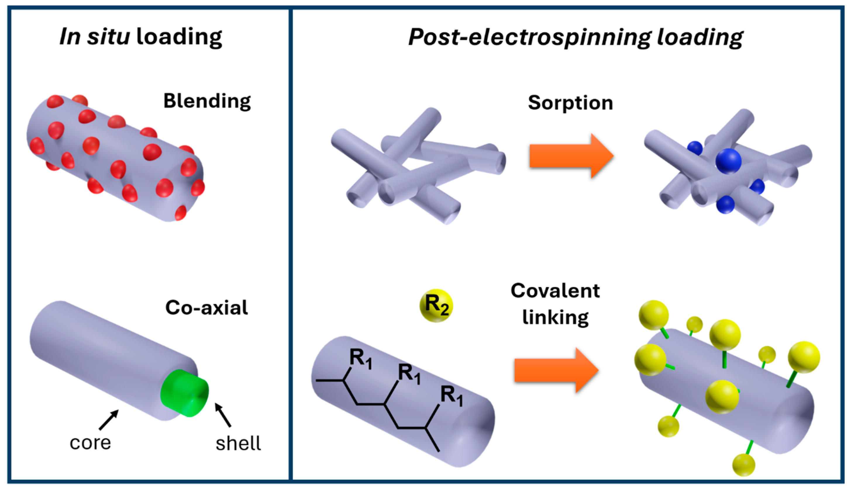
In general, polymeric electrospun nanofibers are excellent platforms for drug delivery purposes [36,44,117,118,119,120]. The most popular strategies for loading drugs can be divided into two categories: (i) in situ loading during electrospinning, and (ii) post-electrospinning loading. In situ loading of antibiotics involves electrospinning solutions composed of a polymer and the bioactive compound, or coaxial fabrication of composite nanofibers.
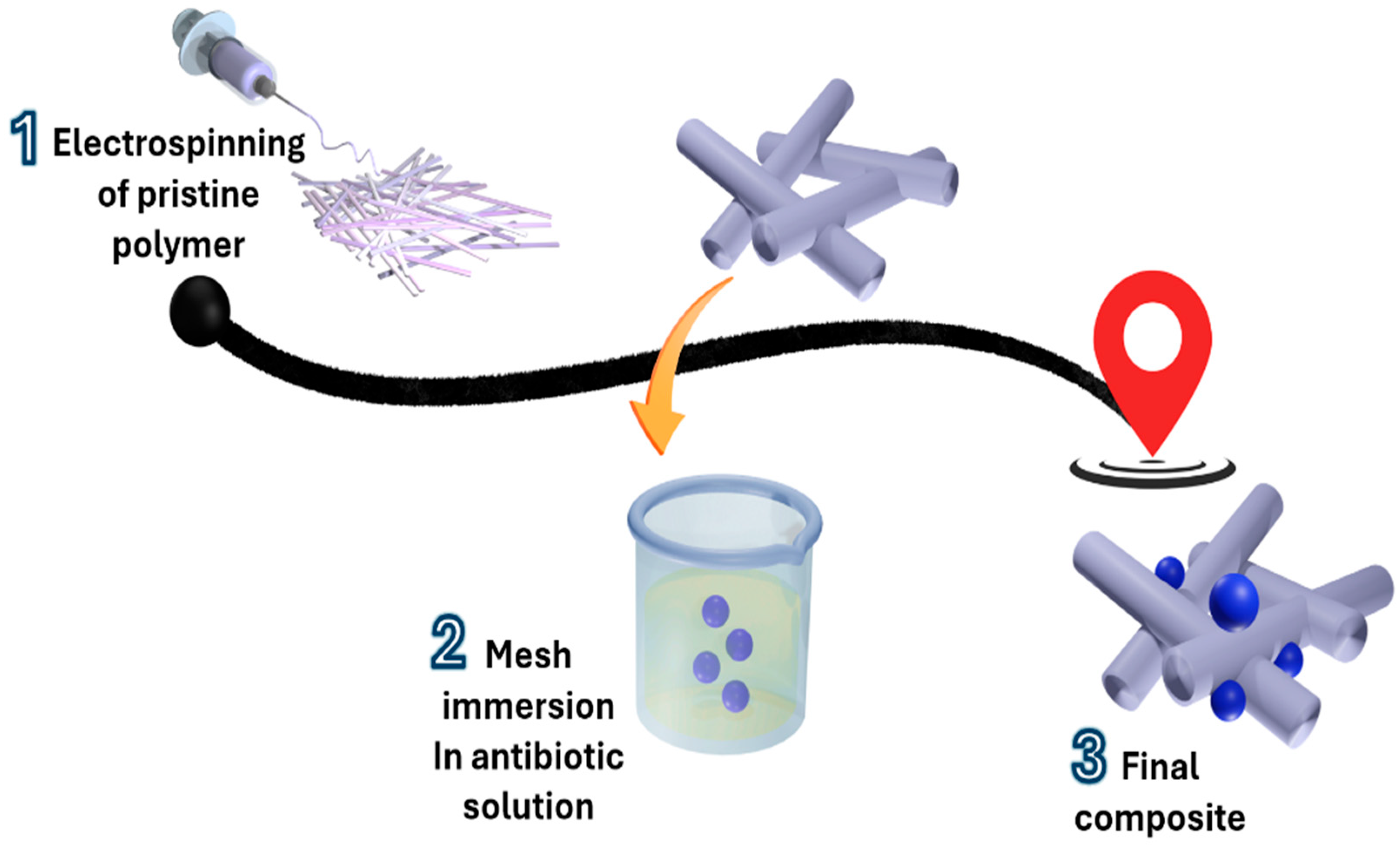
Meanwhile, the post-electrospinning loading of antibiotics is a two-step procedure: electrospun mesh is produced at the first stage, and the mesh is then functionalized by either passive absorption or by covalent linking of bioactive drugs through immersion of native electrospun meshes in solutions of therapeutic molecules [117,118,119]. In general, post-electrospinning loading approaches are alternative methods to incorporate bioactive molecules that decompose under the electrostatic field or that can reduce their therapeutic potential when dissolved. Figure 3 summarizes the primary drug-loading methods, which are suitable approaches for a wide variety of bioactive compounds, including antibiotics.

3.1. Blended Fibers

The basic equipment for electrospinning requires a programmable pump and a feeding system to supply the polymer flow, a high-voltage generator, and a collector. The random orientation distinguishes the most typical assembly of as-spun nanofibers, since manufacturing via a basic electrospinning setup promotes the superposition of subsequent fibers [25], which arise from classical features such as porosity and interconnectivity.
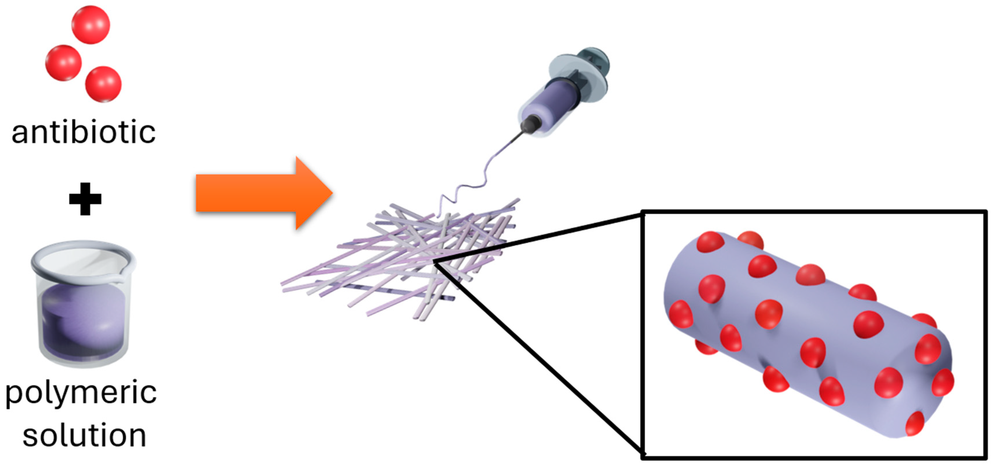
By the electrospinning of polymeric/antibiotic emulsions (Figure 4), different concentrations of antibiotics can be incorporated into the polymeric solution, including mixtures of components. Features such as intrinsic porosity, absorptive capacity, and high surface area made nanofibers an ideal substrate for the design of antimicrobial wound-healing materials [119,121]. The effectiveness of antibacterial polymer/antibiotic composites for drug delivery is driven by the degradation rate of the polymeric matrix and, consequently, by drug diffusion. In this stage, the nature of the polymer and the composite morphology are crucial factors [122].
Following this strategy, Abdoli et al. investigated the incorporation of gum tragacanth (GT), an ideal drug carrier that cannot form nanofibers on its own, into a polyvinyl alcohol (PVA)–graphene oxide (GO) blend, a combination of polymers that possess biocompatibility and drug-delivery properties [121]. A composite fiber of PVA-GT-GO has been prepared, mixed with tetracycline hydrochloride (TCH) and GO, and used to generate electrospun structures for transdermal drug delivery. From this manufacturing, the PVA/GT/TCH and PVA/GT/GO/TCH composites have reached 98% and 82% drug delivery, respectively, at 24 h. During the antibiogram test, both composites have shown bactericidal activity against 12 bacterial strains, with inhibition zones of 23 and 27 millimeters (mm) for E. coli and 25 and 30 mm for S. aureus in PVA/GT/TCH and PVA/GT/GO/TCH, respectively [121].
Similarly, Qi et al. have studied the preparation of poly(lactic acid-co-glycolic) (PLGA) nanofibers loaded with TCH and halloysite nanotubes [122]. In their study, the composite was found to sustain TCH release for 42 days, inhibiting S. aureus growth to a greater extent (80.3%) than the TCH control (81.1%).
Ho et al. have studied the encapsulation of amoxicillin (AMX) in PDLLA before electrospinning composite mixtures or treatment [110]. The composed fibers PDLLA-AMX delivered antibiotics in a sustained manner, inhibiting bacterial growth and reducing periodontal inflammation; moreover, they promoted cellular proliferation along the fiber direction. The in vitro release resulted in a sustained kinetic reaching 81.6% of AMX after 28 days of installation.
Can-Suner et al. used a tricomponent mixture of vancomycin (VAN), ceftriaxone (CFT), and cefpodoxime (CFD) for electrospinning of polylactic acid (PLA) to manufacture wound dressing materials [83]. Regarding the release, the composite produced inhibition halos of 11.66–20.66 mm and 19–27 mm against S. aureus, depending on the CFD and VAN content, respectively. On the other hand, for the E. coli strain, CFT produced superior inhibition, with inhibition zones of 32.33–36.66 mm at concentrations of 5–10%.
Polyvinylpyrrolidone (PVP) nanofibers have been used in combination with the drug metronidazole (MET) for the treatment of bacterial vaginosis due to their mucoadhesive, hydrophilic, and biocompatible properties. In their study, Tuğcu-Demiröz et al. have investigated the bactericidal effect of MET (5 mg/mL), achieving 95% delivery within the first 10 min and an ex vivo permeability flux of 5.4–8.1 μg/cm2h [123].
In this context, electrospinning equipment is particularly well-suited for the fabrication of antibiotic delivery platforms, wound dressings, and other antibacterial biomaterials. In terms of advantages, this type of material is ideal for use as a dressing capable of slowing bacterial growth and promoting cell proliferation due to its mimicry, in terms of structure and composition, of the extracellular matrix at the site of skin injury, avoiding bacterial infection; high interconnectivity and porosity are key factors responsible for these highlights. However, the main disadvantage is the amount of antibiotic loaded into the polymeric solution; depending on its nature (viscosity, dielectric constant, surface tension), a high antibiotic concentration can reduce spinnability, leading to beads and irregular structures. Additionally, the kinetic behavior of randomly oriented fibers can be inconveniently rapid due to the fast degradation rate of the polymeric matrix. Another general consideration is the miscibility of the solvents used, given the polymer and antibiotic solubilities, as heterogeneous structures can result from electrostatic-induced phase separation during electrospinning.

3.2. Co-Axial Electrospinning

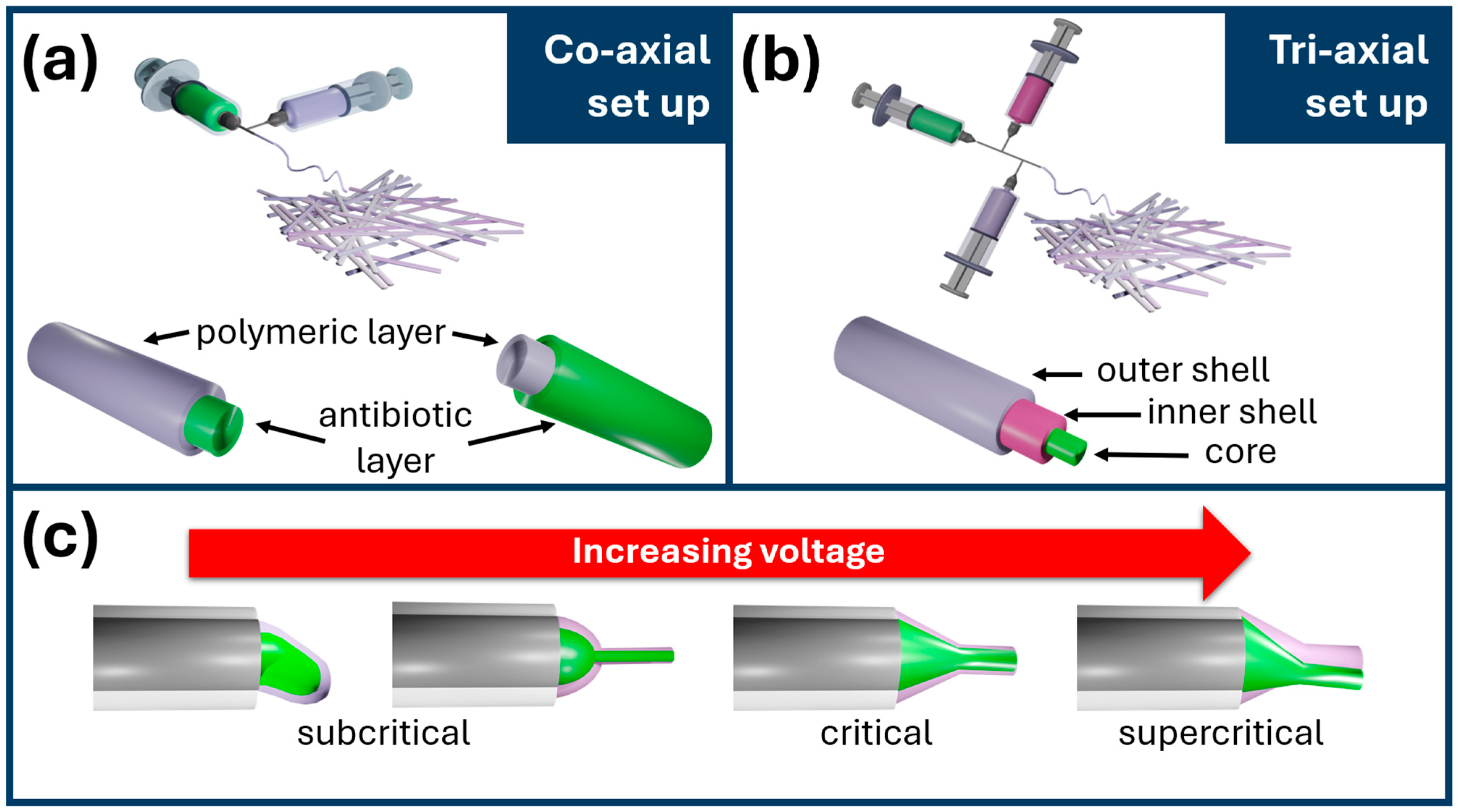
The crafting of core–shell nanofibers is commonly achieved via coaxial electrospinning, an approach first reported by Sun et al. in 2003 [124]. In principle, coaxial electrospinning involves the simultaneous processing of two polymeric or composite solutions (Figure 5a) and, with the triaxial setup [125], even three (Figure 5b). The solution supplied in the inner needle is commonly referred to as the core, while the solution provided through the outer needle forms the shell of the final composite fibers. Coaxial electrospinning permits the processing of a spinnable polymeric solution as the shell and a second fluid, spinnable or not, as the core [126]. For producing uniform core–shell electrospun fibers, the critical voltage is a key factor (Figure 5c) [127].
Using the coaxial electrospinning approach, Ali et al. reported the fabrication of coaxial fibers using poly(vinyl alcohol) (PVA)/superabsorbent polymer (sodium polyacrylate) (SAP) embedded with betel leaf extract (BLE) as the germicidal component [128]. In their experimental design, composites comprising PVA/BLE@SAP/BLE/PVA showed inhibition halos of 14–27 mm for S. aureus and 12–24 mm for E. coli, depending on the BEL content of the coaxial fiber core.
Comparatively, Zhou et al. have analyzed the kinetic and antibacterial performance of ciprofloxacin (CIP) loaded on cellulose acetate (CA) nanofibers from blended fibers and coaxial nanohybrids [126]. In their study, CA/CIP coaxial nanofibers reached 78.4% release at 1 h and 90% of the loaded drug after 1.6 h, providing faster release than the one-step method, 38.2% release during the first hour and 90% after 8.6 h., demonstrating that coaxial electrospinning loading improves pulsatile rather than sustained release. In accordance, the in vitro bacterial inhibition produced inhibition zones of 40–54 mm against S. aureus, E. coli, P. gingivalis, and F. nucleatum for the coaxial CA/CIP composite, and 29–44 mm halos for the blended CA/CIP composites.
This electrospinning variation offers several advantages, such as protecting the drug, for example, for the encapsulation of molecules sensitive to organic solvents, increasing their release time, and improving mechanical properties. However, it is a more complex process that requires specialized equipment and is challenging to scale up to an industrial level. Moreover, applying a critical voltage can be challenging for producing uniform core–shell nanofibers.

3.3. Passive Loading

Passive loading of therapeutic drugs occurs due to physical immobilization on electrospun nanofibers, often via sorption [36]. This approach is highlighted for its simplicity in terms of instrumental requirements: it is a two-step method in which the electrospun substrate is fabricated during the first stage, followed by the direct immersion of the as-spun mesh in the drug solution (Figure 6).
According to this protocol, Cheng et al. evaluated the bacterial adhesion onto electrospun polytetrafluoroethylene (ePTFE), collagen, and glycolide fibrous membranes loaded with tetracycline (TTC) and amoxicillin (AMX) [129]. The authors concluded that the loading method does not affect the primary morphology, preserving the microarchitecture to a high degree; however, bacterial adhesion was significantly reduced.
Khampieng et al. loaded doxycycline (Doxy) onto poly(acrylic acid) (PAA) nanofibers [130]. The incorporation of Doxy was carried out by direct immersion of PAA nanofibers in aqueous Doxy solutions of variable concentration (125–1000 μg/mL) overnight. The PAA/Doxy composite released 1600 μg/mL in a sustained manner for 75 days, resulting in inhibition zones of approximately 8, 18, and 20 mm against P. aeruginosa, S. aureus, and S. agalactiae, respectively.

3.4. Covalent and Coordinative Loading

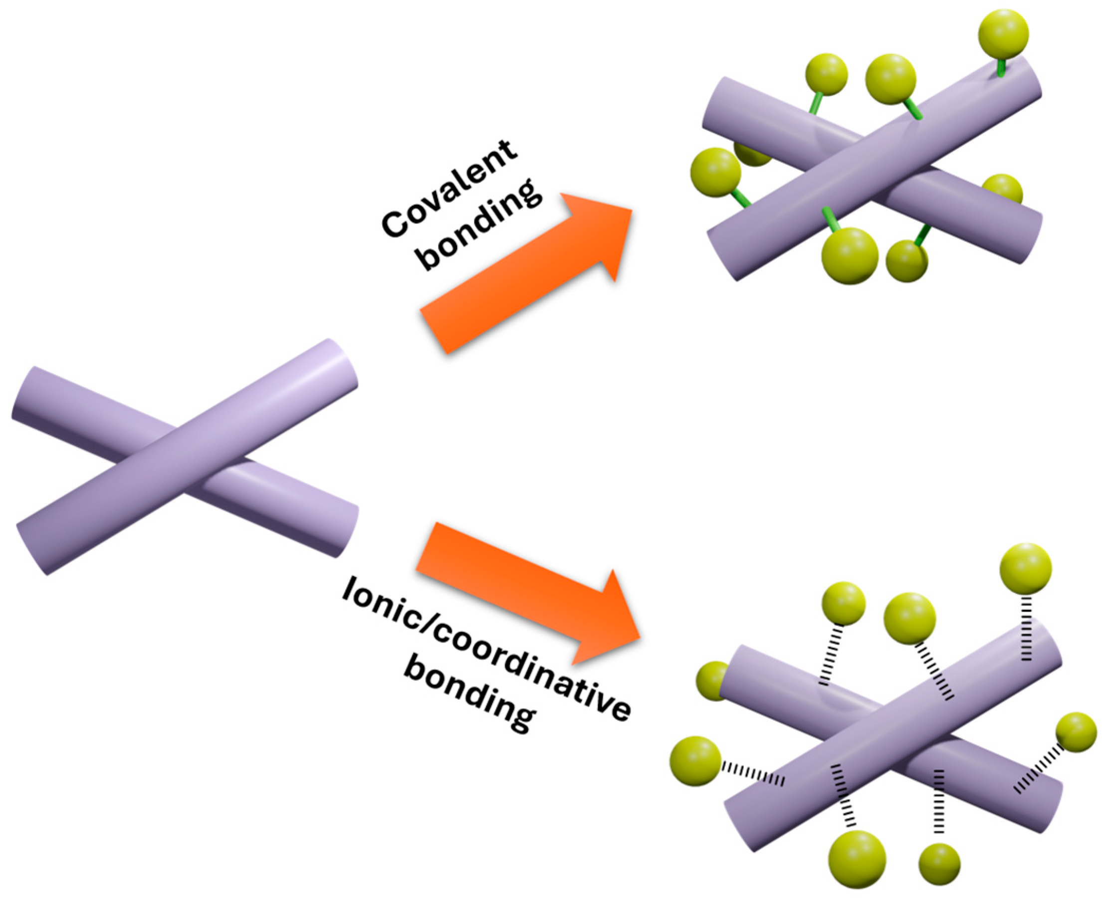
Post-electrospinning functionalization of nanofibrous mats is an alternative that allows overcoming solubility issues and electrostatic decomposition of labile components, thereby enhancing advanced applications [26,27], and antibiotic loading is not an exception [131,132]. Following a two-step approach, electrospinning is the first stage, and antibacterial agent incorporation can be achieved via covalent linking (Figure 7). These post-electrospinning drug incorporations induce modifications in the secondary morphology through chemical bonding. Shi et al. [132] reported the incorporation of metronidazole (MNA) through esterification on PCL, taking advantage of the capacity of cholesterol esterase to hydrolyze the ester bonds selectively, triggering the release of 25.3 μg of MNA per 1 cm2 and resulting in bacteria viability of 34.0–36.9%, considering H. pylori as the model of study.
Bueno et al. incorporated Doxy onto electrospun membranes composed of polymer blend (MMA)1-co-(HEMA)1 and (MA)3-co-(HEA)2 [131]. In their study, a polymeric electrospun membrane was treated with a sodium carbonate buffer solution to promote the hydrolysis of carboxyl and ester bonds on its surface, and then Doxy was chemically conjugated. The conjugated membrane prevents bacterial attachment, reducing the colony-forming units (CFU) of biofilm 1.28 × 10⁶ versus 2.59 × 10⁸ in the control at 72 h of incubation.

4. Aligned Nanofibers

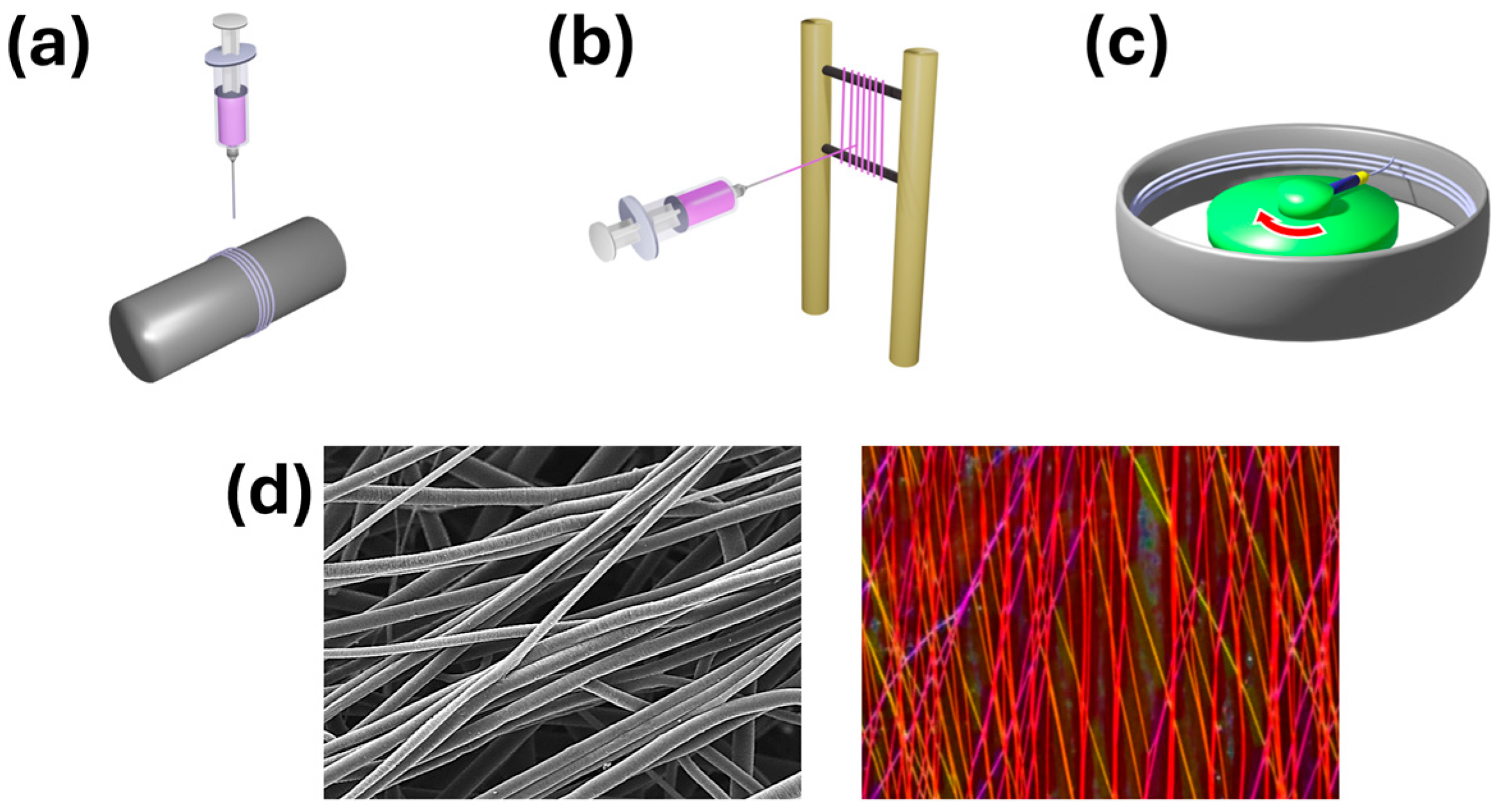
Highly oriented fibers are associated with better kinetic performance, enabling sustained release over extended periods compared with randomly oriented nonwoven meshes. Several methods have been reported for producing aligned nanofibers using a special collector system, such as a high-speed rotating mandrel or disks, and parallel electrodes to induce nanofiber alignment [25,133]. The typical set-up variations are presented in Figure 8.

4.1. Mechanical Induction

Using a rotative collector, uniaxially aligned nanofibers can be generated when the collector’s rotation speed is well controlled (Figure 8a) [25,133,134]. Mechanical induction of nanofiber orientation is the most widely used protocol for manufacturing aligned nanofibers for antibiotic delivery. By mechanical induction, Liu et al. produced aligned PCL nanofibers loaded with ampicillin, which produced inhibition halos of 53 and 26 mm in S. aureus and K. pneumoniae, respectively [135]. The kinetic study showed a mixed release mechanism involving small-molecule diffusion and polymer degradation, reaching 95% of ampicillin delivered within 24 h.
Li et al. [28] studied the wound-healing application of composite membranes composed of aligned PCL and aligned polyurethane (TPU) loaded with TCH. Mechanical induction enabled the deposition of aligned patterns capable of releasing 100% of TCH at 120 h and forming inhibition zones of approximately 18 mm in E. coli and S. aureus strains.
Azithromycin (AZ) has also been included in mechanically aligned gelatin methacrylate (GelMa) electrospun composites by Ayoub et al. [136]. In the kinetic monitoring, GelMa-AZ exhibited the highest release capacity (19.4 μg/mL) on day 3. At this time, the antibacterial test showed inhibition zones of 7 and 10 mm for A. naeslundii and A. actinomycetemcomitans, respectively.
Ranjbar-Mohammadi et al. demonstrated that mechanical alignment also benefits the development of antibacterial suture yarns [137]. In their work, they presented the simultaneous electrospinning of poly(vinyl alcohol)/chitosan blend solution and poly(lactic acid) (PLA)/TCH composite solution. The aligned constructions inhibit the growth of E. coli and S. aureus to 20 and 17 mm, respectively.
Nonetheless, the principal disadvantage is often related to the challenge of aligning nanofibers regardless of rotation speed. Another critical concern is the combination of high rotation speed, which can induce fiber fracture.

4.2. Air Gap

The air-gap electrospinning concept was proposed by Li et al. in 2003, resulting in a successful approach for producing uniaxially aligned nanofibers up to several centimeters in length [138]. Since then, much effort has been devoted to increasing the length of aligned nanofibers. However, the length is currently still typically limited to 10 cm [139].
This method consists of arranging two longitudinally parallel electrodes connected to ground. The electrostatic field lines are thus arranged vertically, and the nanofibers charged with opposite polarity to the electrodes, when they reach the gap between them, have no preferred direction in which to organize themselves, since the field distribution exerts equal force on both sides in the direction perpendicular to the electrodes. Therefore, the nanofibers in this area are arranged according to that perpendicular direction (Figure 8b). On the other hand, the deposited nanofibers retain their charge and therefore repel new nanofibers that reach the collector, preventing them from growing thick [25].
In terms of antibiotic delivery, air-gap electrospinning is rarely reported. Ornelas-Guillén et al. proposed the loading of TTC on PVA-aligned fibers following the air gap electrospinning protocol, aided by the choline chloride:urea eutectic mixture [140]. In terms of bacterial inhibition, composites loaded with 23.5 μg produced inhibition halos of 20, 18, and 16 mm against S. aureus, K. pneumoniae, and E. coli, respectively, surpassing the performance of standard TTC disks (30 μg). From the kinetic point of view, the aligned composite undergoes an initial burst, delivering 25–30% of TTC, followed by the sustained release up to 50 h.
However, while air-gap electrospinning has demonstrated multiple benefits for producing aligned nanofibers with superior properties, it presents several limitations that must be carefully considered for its application in antibacterial biomedical environments. These disadvantages are mainly related to aspects of scalability, process control, reproducibility, technical limitations, and potential risks in clinical translation. The main challenges documented in scientific literature are discussed below.

4.3. Centrifugal Electrospinning

Another advanced electrospinning setup is the centrifugal electrospinning. By this method, fiber alignment is promoted by the interaction of the electrostatic field and centrifugal force. In this setup, the polymeric solution is supplied through a rotating spinneret, and the oriented nanofibers are collected in a stationary collector (Figure 8c) [141,142].
Wang et al. studied the layering of aligned PVP nanofibers for the delivery of TCH, achieving at least 98.9% drug entrapment through centrifugal electrospinning [143]. In comparison, randomly oriented nanofibers delivered 87%, monolayered aligned PVP reached 70%, and multilayer aligned composite reached 64% at 5 min. Therefore, the reduced fiber surface area resulting from multiple stacking reduces the release rate—likewise, Hou et al. processed ethyl cellulose via centrifugal electrospinning for the delivery of TCH. After 48 h, the composite delivered 80% inhibition zones of 28.7 mm versus the E. coli strain and 27.3 mm against the S. aureus strain [144].
The highlights of centrifugal electrospinning include its scalability, which is adequate for large-scale fabrication and compatible with a wide range of polymers and composites; in addition, multicompartment spinnerets improve drug loading [145]. However, the intrinsic combination of high voltage and high rotational speed constitutes the most crucial concern from a manufacturing perspective.

5. Patterned Nanofibers

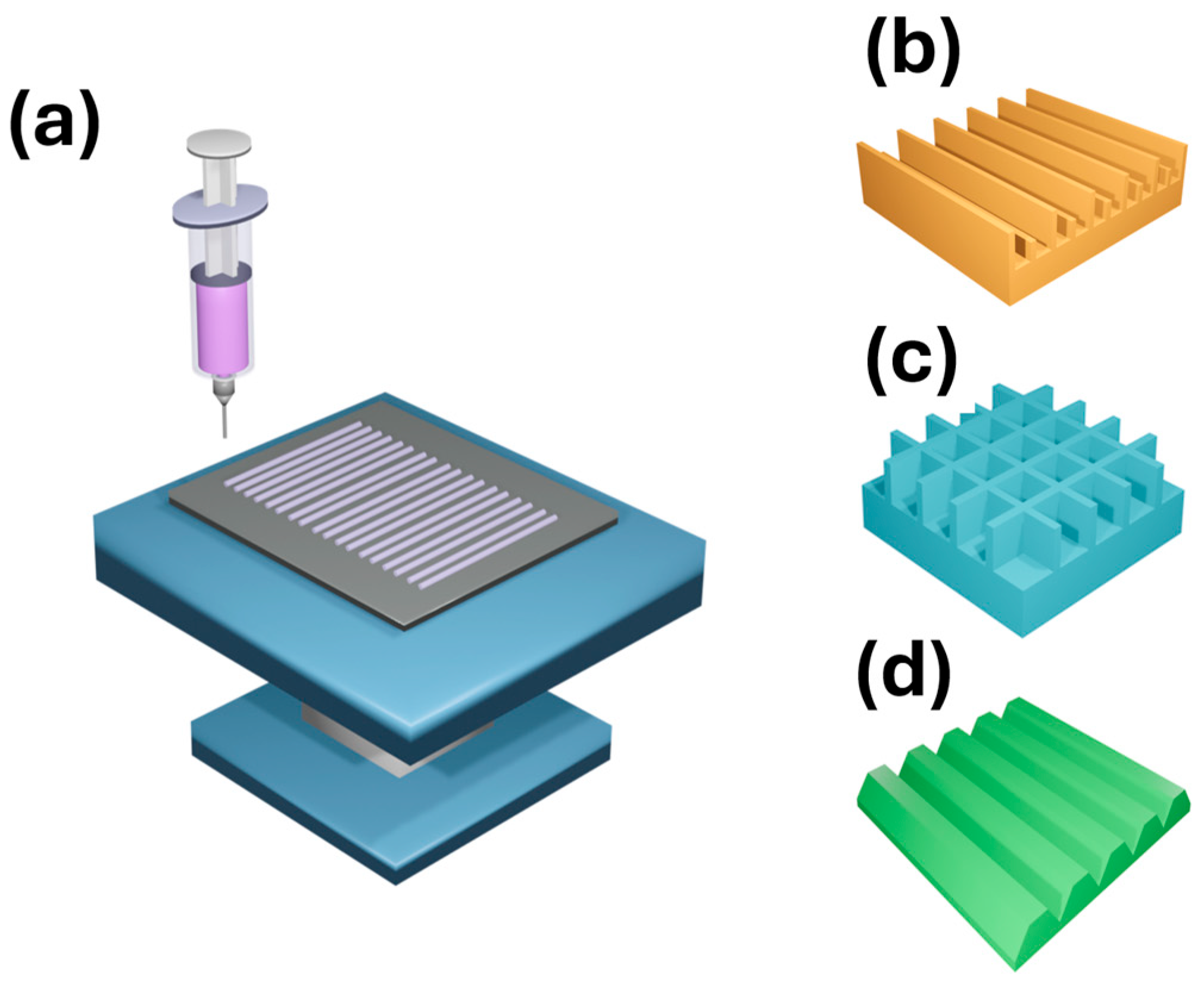
The fabrication of patterned microarrays via electrospinning is primarily associated with near-field electrospinning (NFES). This approach involves performing electrospinning at reduced needle-to-collector distances. Compared with the standard electrospinning setup, the main difference in NFES is the use of X-Y-Z or X-Y mobile platforms to collect the as-spun fibers (Figure 9a), enabling controlled deposition of linear and curved patterns [146,147,148].
Alternatively, 3D printing technology has a significant impact on the manufacturing of patterned electrospun mats, permitting the design and printing of highly organized, complex frames that can be used as collectors to promote nanofiber assembly [149,150].

5.1. Near-Field Electrospinning

Considering NFES as the manufacturing method for antibiotic-delivery scaffolds, Mai et al. reported an NFES configuration for the electrospinning of well-defined patterned composite materials composed of a mixture of polycaprolactone/collagen loaded with the natural antibacterial drug usnic acid (UA) [151]. The well-organized arrangement (produced at 0.4 mL/h, 2.1 kV, 2 mm, and 150 mm/s in hexafluoroisopropanol (HFIP)) exhibited sustained in vitro release of 84% of UA. In their study, it was shown that 1%wt of UA loaded into a PCL/collagen matrix could inhibit the growth of Gram-negative and Gram-positive bacteria, using E. coli and S. thermophilus as model organisms.
Following a similar protocol, Li et al. prepared polycaprolactone-collagen nanofibers loaded with the antibiotic erythromycin (ERY) and analyzed the patterned patches as wound dressings [152]. The composite membranes could prevent the growth of E. coli and S. thermophilus and sustain ERY release at 80%. The bactericidal effect was achieved at concentrations of 1 to 5%wt of ERY, producing inhibition zones ranging from 15.17 to 18.61 mm and from 16.22 to 18.17 mm for E. coli and S. thermophilus, respectively. Moreover, due to the morphological properties conferred by the NFES (produced at 0.4 mL/h, 2 kV, 2 mm, and 80 mm/s in HFIP), the dressing material presented good air permeability and desirable mechanical properties.
NFES patterned assemblies highlighted by high porosity, strong mechanical behavior, and improved aqueous interactions, benefiting their application as DDS. The approach is versatile for electrospinning polymeric composite solutions loaded with antibiotics. However, essential considerations for NFES in the fabrication of drug delivery materials include reducing the length and thickness of the meshes, due to process-specific constraints (e.g., reduced working distance, low flow rate, and highly volatile solvents). Because of the typical reduced needle-to-collector distance, nanofiber fabrication occurs at lower voltages; additionally, highly volatile solvents are required to achieve well-defined nanofiber deposition.

5.2. Patterned Collectors

Using customized 3D printed collectors, Zdraveva et al. reported the fabrication of PCL loaded with cefuroxime (CFU) for dual scaffolding and antibacterial purposes [153]. The 3D-printed collectors allow for tailored topologies, inducing hexagonal arrangements and increasing porosity to up to 90%. Structural features of the electrospun material improved mechanical strength, cell distribution, and confluence compared with random fibers. 3D printed collectors and silicon molds, composed of parallel bars [154,155], perpendicular bars [154], and parallel trapezoidal prisms [156], are reported for the practical fabrication of electrospun mats and are illustrated in Figure 9b–d.
Grgurić et al. compared the use of ribbed and mesh 3D-printed substrates (represented in Figure 9b,c) for the electrospinning of the same PCL/CFU formulation [154]. Both geometries were studied to improve cell adhesion and proliferation. In their study, the authors demonstrate that the geometry of collectors can induce fiber arrangement. Continuing this strategy, Trcin et al. used the ribbed collector for the electrospinning of antimicrobial scaffolding materials based on a PCL/CFU mixture [155]. With the CFU concentration ranging from 5 to 25%wt, the bactericidal effect resulted in inhibition halos of 27 to 40 mm in P. aeruginosa and 31 to 44 mm in S. aureus.
Regarding the drug delivery efficacy, PCL/CFU 5%wt and PCL/CFU 25% could release a maximum of 14% and 9% of CFU, respectively, during the first 4 days; after this period, a slow and sustained release (<10%) was observed, lasting up to 14 days. Moreover, PCL/CFU 5% showed superior performance in limbal stem cell growth and corneal epithelial differentiation; possible cytotoxicity may be related to higher CFU concentrations. The main advantages of using 3D-printed collectors are their rapid, customized fabrication, high compatibility with the electrospinning approach, and the high organization of controllable shapes and geometries induced in the electrospun meshes. On the other hand, the main disadvantage is the specialized equipment required for 3D printing. Other authors also suggest that electrospun nanofibers collected in 3D-printed templates lack sufficient mechanical properties for biomedical applications [149].

6. Hierarchical Structures Combining Electrospinning and 3D Printing

The fusion of electrospinning and 3D printing technologies is a valuable approach to fabricating personalized multilayer materials. From the perspective of antimicrobial resistance, administering antibiotics from multilayered substrates is an innovative alternative that enables overcoming bacterial resistance. The paramount convenience of combining emerging technologies such as 3D printing and electrospinning relies in (i) the versatility of incorporating a wide range of components, (ii) the feasibility of fabricating complex microstructures of high surface to volume ratio and high degree of interconnected porosity, and (iii) the mimicry of extracellular matrix, which is particularly important for the regeneration of infected or damaged tissue [157].
Following this strategy, Song et al. fabricated a bilayer skin scaffold loaded with amoxicillin (AMX) [158]. Their proposed protocol consisted of two stages: the fabrication of a PCL/AMX outer layer by electrospinning and the production of a sodium alginate-gelatine-epidermal growth factor (SA-Gel-rhEGF) inner layer by 3D printing. The resulting bilayer composite PCL/AMX@SG-rhEGF facilitated skin healing while simultaneously preventing bacterial infection; the composite suppressed bacterial proliferation, producing inhibition zones of 2.85 and 3.27 mm against E. coli and S. aureus, respectively. Considering kinetic behavior, the composite released 35.59 μm/mL after 240 h; during the first 24 h, it reached the maximum release concentration (35 μm/mL), thereby establishing sterile conditions in the wound environment. After that lapse, a sustained release remains constant.
A complex bilayer patch PCL-silicon oxide (SiO2) covered by PCL gelatine methacrylate/sodium alginate and vancomycin (PCL@GelMa/SA-Van), designed to prevent abdominal wall damage, was produced by Hu et al. [159]. At the first stage of patch fabrication, PCL was electrospun; in the second stage, a PCL-SiO2 coating was deposited to enhance hydrophobicity. Finally, the third stage consisted of performing coaxial 3D printing on the surface of an antiadhesive PCL-SiO2 layer to construct core–shell-like coatings based on PCL as the core and a mixture of GelMa/SA-Van as the antibacterial layer. The composite patches exerted an antiproliferative effect, reducing the numbers of E. coli (6-times) and S. aureus (4-times) after 24 h compared with the PCL control. The release of Van from composite patches reached 55 μg/mL rapidly after 36 h; thereafter, Van delivery was sustained up to 240 h, preventing post-operative infection in animal models.
In a three-layer wound dressing design, Mirhaj et al. proposed mupirocin (Mup), an alternative antibiotic against methicillin-resistant S. aureus, as the bioactive component added with AgNO3 [160]. The fabrication of the antibacterial three-layered composite was carried out in three main steps: (i) the construction of the top layer of polyurethane (PU) nanofibrous mesh, (ii) a middle layer composed of pluronic F127-quaternized chitosan-AgNO3 nanoparticles (F127-QCS-AgNO3), and (iii) the inner layer composed of core–shell nanofibers of F127-Mup/pectin-keratin (F127-Mup/Pec-Kr). The three-layered composite reached 80% release of Mup at 7 h, producing inhibition zones of at least 3 mm and 2 mm with S. aureus and E. coli strains, respectively.
Using a two-step method, Santos et al. fabricated bilayered membranes for treating periodontitis via a dual-drug delivery approach [161]. A honeycomb substrate composed of polylactic acid/zein/curcumin (PLA/Zein/Curc) was 3D-printed and used as the basis for the electrospun layer. The electrospun coating was achieved by coaxial electrospinning, using a mixture of poly(ethylene oxide)/curcumin/tetracycline hydrochloride (PEO/Curc/TH) as the core and a zein/poly(ε-caprolactone)/β-glycerolphosphate (Zein/PCL/β/β-GP) mixture as the shell. The composite was capable of continuous release of both Curc and TH for up to 8 days, reaching cumulative release of 75% and 65%, respectively, within this time frame. On the other hand, the influence of microstructure on bactericidal capacity was tested; considering PCL/Zein/Curc/TH composites fabricated at different zein concentrations (10, 20 and 30%wt) by electrospinning or 3D printing, as a result electrospun composite exhibited slightly larger (~1.15 fold) inhibition zones than 3D printed composite against clinical strains extracted from periodontal subgingival pocket of chronic periodontitis patients. This result highlights that the superior porosity of electrospun fibers enables higher efficacy in inhibiting bacteria.
Vancomycin and ceftazidime (CAZ) have also been included in multilayer composites produced by coaxial electrospinning on 3D-printed substrates. Yu et al. reported the 3D printing of PCL mesh-like scaffold used for the electrospinning of core–shell poly(lactic-co-glycolic acid)/bone morphogenetic protein (PLGA/BMP-2) and PLGA/Van/CAZ [162]. From the point of view of release, composites released 86% and 84% of Van and CAZ, respectively. By coupling 3D printing and electrospinning, each layer displayed a specific role. First, the PCL mesh fabricated by 3D printing resulted in the structural basis of an artificial bone graft, being the scaffolding component to promote osteocyte healing; secondly, the PLGA/Van/CAZ/BMP-2 electrospun coating prevents bacterial infection due to the sustained release of the antibiotics during a long-term period under in vitro and in vivo conditions.
The combination of 3D printing and electrospinning is suitable for constructing bilayer composites on metallic substrates. Maver et al. deposited two bioactive layer coatings based on carboxymethylcellulose (CMC) and clindamycin (CLIN) on TiAlV and AISI 316LVM surgical substrates [163]. The multilayer composite was fabricated by electrospinning CMC/polyethylene oxide (PEO)/CLIN solutions onto both metallic surgical substrates. Further, six layers (1 mm in total height) of an alginate/CMC and cellulose nanofibrils (NFC) suspension loaded with CLIN were 3D-printed. During the in vitro delivery, the multilayered composites exhibited an initial burst in the first 60 min, reaching 82% and 83% of the CLIN cumulative release after 24 h for TiAlV and AISI 316LVM, respectively.
The combination of 3D printing and electrospinning offers, as a principal advantage, the suitability to achieve complex patterned composites; the precise control of microstructure permits improving mimicry. On the other hand, integrating a wide variety of bioactive components and customizing the structure and properties (bioactive or physical) of each layer are key features that enhance the versatility of this approach. However, the additional cost implied in the 3D printing setup can be considered the limiting factor.

7. General Considerations Regarding Primary Morphology

Unlike randomly oriented fibers, aligned and patterned nanofibers demonstrate better mechanical properties. The production of nanofiber networks with an aligned pattern offers important properties to biomaterials in fields such as tissue engineering, where naturally occurring extracellular matrix biostructures inherently exhibit these anisotropic patterns, including nerve, heart, tendon, blood vessel, muscle, and cartilage tissues. Therefore, the correct architecture of biopolymeric tissues is necessary to mimic the properties of the extracellular matrix correctly [164,165]. On the other hand, patterned and randomly oriented fibers provide interconnectivity and porosity, thereby improving wound-healing capacity [166].

7.1. Equipment Requirements

Beyond the basic electrospinning setup, specialized equipment is required for producing aligned and patterned nanofibers. To promote fiber alignment, additional costs may be considered to install advanced setups such as air-gap, near-field electrospinning, or rotating collectors. The precise configuration of these elements is essential for obtaining aligned and patterned collections with the desired properties for biomedical applications [118,167,168].
Over the last two decades, multiple experimental variants have been developed, ranging from simple designs with two parallel electrodes to complex collectors such as origami, rotating cores, and 3D assemblies, which have enabled exploration of applications in tissue engineering, nerve guides, biomimetic scaffolds, and matrices for the controlled release of antibacterial agents. At the same time, innovative collector designs have been proposed, such as the origami system described by Jha et al. [169], which offers a low-cost alternative for producing highly aligned fibers in far-field conditions. This simplified design avoids the need for high-speed rotation or expensive components, demonstrating that relatively simple geometric modifications to the collector can improve fiber deposition.

7.2. Aligned, Patterned, or Random Nanofibers

Alignment and highly oriented patterning significantly enhance the scaffold’s anisotropy, which is critical for applications such as tissue regeneration (i.e., muscle, nerves, tendons) where cell direction, elongation, and migration depend on the aligned topography of the support. Recent studies have shown that alignment improves targeted cell proliferation, organized extracellular matrix production, and the orientation of collagen fibers secreted by cells, thereby limiting bacterial adhesion or guiding their colonization in a less invasive way when combined with antibacterial agents [118,170].

7.3. Specific Surface Area

Highly controlled frames and aligned fibers tend to have more uniform, often smaller diameters than randomly oriented fibers, resulting in a higher surface area-to-volume ratio. Allowing more efficient functionalization with antibacterial agents (injection, immersion, coating) enhances their controlled release and increases surface exposure for biological interaction and response [170].

7.4. Mechanical Properties and Functional Anisotropy

The orientation of nanofibers contributes to greater mechanical strength, elastic modulus, elongation in the preferred direction, and structural rigidity along the alignment or pattern direction. The orientation is essential for maintaining the integrity of biomedical devices when implanted in areas subject to stress, such as tendons, ligaments, or as coatings for metal implants, where the scaffold must not collapse or deform under physiological forces. In addition, a proper mechanical structure can help resist bacterial attack by reducing deformations that promote fluid or biofilm accumulation in folds [25].

7.5. Permeability, Interconnected Porosity, and Nutrient Transport

Depending on pore diameter, aligned, patterned, or randomly oriented scaffolds can promote the formation of interconnected, more uniform-sized pores, facilitating (at a customized level) the diffusion of nutrients and oxygen and the elimination of metabolic waste. In living tissues, this is critical, as necrosis or bacterial infection can arise in regions where diffusive transport is deficient. Porosity creates virtual “channels” that enhance this transport, improving cell viability, vascular integration, and the effectiveness of diffusion-dependent antibacterial materials, such as sustained release of metal ions. Biomimetic scaffolds with aligned structures and high porosity efficiently support cardiac cell colonization through interconnected pores [118,167].

7.6. Guided Cell Behavior

Cells respond not only to the chemistry of scaffolds, but also strongly to topography and spatial order. In aligned scaffolds, cells such as fibroblasts, nerve cells, and progenitor cells (to name a few) tend to align, which improves their physiological function (i.e., conduction, muscle contraction, nerve regeneration) [164]. On the other hand, skin [171] and bone [172] scaffolds support a higher rate of healing. The orientation can also help reduce unwanted bacterial colonization, or at least guide how biofilm forms, enabling designs to incorporate accessible barriers or antibacterial agents, and even modulating breathability.

7.7. Release of Antimicrobial Agents

Thanks to their ordered geometry, greater surface exposure, and uniform diameters, nanofibrous scaffolds can enable more homogeneous packaging of antimicrobial agents or nanoparticles (e.g., silver nanoparticles, copper nanoparticles, peptide agents) and more predictable sustained release [173]. Homogeneity is essential for antibacterial applications, as irregular release can cause resistance, cytotoxicity, or loss of efficacy. Combining polymers with antibacterial agents in electrospun nanofibers controls morphology, thereby improving release profiles [174].
Electrospun nanofiber morphology, particularly fiber diameter, plays a critical role in determining the degradation behavior of biodegradable polymeric scaffolds and, consequently, the release kinetics of encapsulated drugs. Nanofibers with smaller diameters possess a higher surface-to-volume ratio, enabling more rapid penetration of aqueous media and facilitating polymer chain hydrolysis or enzymatic attack. This increased interaction with the surrounding fluid accelerates polymer degradation, leading to faster drug release profiles than with thicker fibers. For example, studies with paclitaxel-loaded PLGA fibers have shown that submicron-diameter fibers exhibit higher degradation rates and greater drug release than micron-sized fibers, due to enhanced matrix hydrolysis and greater relative surface area. These observations underscore the importance of fiber diameter as a tunable parameter for controlling biodegradation and release dynamics in drug-eluting nanofiber systems [175].
In addition to diameter, other architectural factors such as porosity, core–shell structures, and fiber morphology have been shown to influence degradation pathways and drug release. Tailored porous fibers created via phase separation or coaxial electrospinning can produce distinct degradation environments within the fiber by introducing microchannels or distinct polymer phases that degrade at different rates. For instance, porous nanofibers with core–shell architecture exhibit a synergistic release mechanism in which the outer shell may initially slow the ingress of water, controlling early drug diffusion. At the same time, subsequent matrix degradation governs sustained release over the long term. This interplay between diffusion and degradation control is a central design consideration in fabricating advanced nanofiber carriers for sustained drug delivery, where controlled degradation supports predictable release profiles suited for biomedical applications [176].
Notably, the impact of degradation on drug release is also observed ex situ by comparing different polymer systems under controlled hydrolytic conditions. In electrospun PDLLA systems, the specific alignment between fiber diameter and drug localization influences not only the release rate but also the onset of degradation-mediated release, with smaller diameters favoring quicker onset and larger diameters supporting extended kinetics due to slower matrix breakdown. These results emphasize that fiber dimension is a primary tunable factor not only for diffusion control but also for orchestrating degradation-driven release mechanisms. This insight informs rational design of electrospun nanofibers for antibacterial and other biomedical therapies where time-dependent release is critical [177].

7.8. Compatibility with Multiple Polymers and Functionalization Methods

All electrospinning setups can be adapted to a wide range of natural or synthetic polymers, hybrid blends, copolymers, nanoparticle-containing compounds, and bioactive coatings, among others, allowing the design of customized scaffolds to adjust degradation rate, compatibility, mechanics, and antimicrobial functionalization. Polymers such as PCL, PLA, gelatin, and chitosan have been used to form hybrid nanofibers for wounds, implants, and tissue regeneration [167,178].

7.9. Potential to Reduce Biofilm Formation and Improve Material Hygiene

Although not all studies have directly evaluated biofilm on nanofibers, the high surface-to-volume ratio, porosity, and interconnected structure of nanofibers reduce cumulative surface irregularities, folds, or voids where microorganisms can hide [114,165] compared to non-nanostructured materials. A smoother, more uniform scaffold can be cleaned more easily and, when combined with antibacterial treatments, can reduce colonization. In addition, the localized release of antimicrobial agents from an optimized nanofibrillar structure can achieve greater efficacy with lower doses, reducing systemic adverse effects [179]. Some recent studies of hybrid nanofibers report strong anti-S. aureus and P. aeruginosa activity when controlled release from nanofibers is used [118,174].

7.10. Esthetic Improvements

In biomedical implant or aftercare applications, not only does function matter, but also integration with surrounding tissue, controlled degradation, avoiding permanent residues, reduction in adverse immune responses, and visual surface appearance [180]. Nanofibers can replicate extracellular matrix patterns, giving a more natural appearance, which may promote tissue acceptance. Biodegradable polymers used in aligned scaffolds have shown degradation rates compatible with tissue regeneration [118,167].

8. Equipment and Operating Parameters Considerations

Unlike conventional electrospinning with flat or rotating collectors, advanced electrospinning setups require precise control of nanofiber orientation through the needle-to-collector distance and voltage, depending on the collector features. These requirements increase experimental complexity and error susceptibility, especially in laboratories without prior experience with specialized equipment design. Minimal variation in environmental conditions (humidity, temperature, air flow) can also significantly alter fiber morphology and alignment [118,165,181].

8.1. Limitations on Mesh Length

A recurring disadvantage is that aligned fibers obtained with air gaps or patterned by near-field electrospinning are often restricted in length, which prevents the manufacture of continuous meshes or large scaffolds without additional bonding or transfer processes. This limitation restricts their applicability in the industrial-scale manufacture of medical devices and in coatings for large surfaces, such as prostheses or antibacterial membranes [118,167].

8.2. Limited Thickness

The accumulation of patterned and aligned nanofibers is restricted by the electrostatic repulsion effect between previously deposited fibers and new nanofibers arriving at the collector, and the length of the collector. This results in membranes with reduced thicknesses, which are unsuitable for applications requiring robust three-dimensional structures or high volumetric density, such as bone regeneration implants or multilayer matrices with prolonged release of antibacterial agents [118,170,182].

8.3. Restricted Compatibility with Specific Polymers and Bioactive Fillers

Some polymers with high viscosity, rapid crystallization, or poor stability under high voltage are challenging to process into aligned or patterned structures. Likewise, the incorporation of antibacterial agents (such as metal nanoparticles or antibiotics sensitive to electrical discharge) can be compromised by chemical instability, loss of bioactivity, or non-uniform distribution in fragile fibers [183]. The solubility and dielectric properties may be considered when selecting the most suitable approach and the most convenient antibiotic-loading method.

8.4. Sensitivity to Process Environment

The surrounding air plays a crucial role in electrospinning; variations in relative humidity or temperature directly affect jet stability and the final morphology. In uncontrolled environments, nanofiber formation can result in defective, collapsed, or poorly aligned structures [184] the disadvantages of facilities without environmentally controlled infrastructure.

8.5. Three-Dimensional Integration

Although nanofibers offer advantages in 2D, integrating them into complex 3D structures remains a challenge. The difficulty of stacking multiple layers with stable morphology limits their usefulness in advanced three-dimensional scaffolds that mimic anatomic zones, which are necessary for the regeneration of complex tissues (bone, cartilage, liver). This limitation requires hybrid strategies or combinations with bioprinting techniques to generate true antibacterial 3D scaffolds. However, additional equipment is a prerequisite [185].

8.6. Relatively High Energy Consumption

The need to apply high voltages and maintain stable environmental conditions for prolonged periods can increase the process’s energy consumption [186,187], which is a disadvantage compared to other polymer processing methods that are more efficient in terms of sustainability and cost, which is especially relevant considering the growing demand for green manufacturing and scalable nanofabrication processes in biomaterials [181].

9. Quantification of Antibiotic Release

Quantifying the levels of active compounds in nanofibers is a fundamental step toward understanding their release kinetics, antibacterial efficacy, and overall performance in biomedical applications. Accurate quantification ensures that nanofibers release therapeutic agents at the desired rate and concentration, which is essential for achieving the intended biological effect. To this end, several analytical techniques are employed, each with its own advantages and limitations. Among the most widely used are high-performance liquid chromatography (HPLC), ultraviolet-visible spectroscopy (UV-Vis), mass spectrometry (MS), Fourier-transform infrared spectroscopy (FTIR), and fluorescence spectroscopy [188,189,190,191]. Their principal advantages and drawbacks for quantifying the release of active compounds from nanofibers are summarized in Table 2.

10. Kinetic Characterization and Modeling Methodologies

The kinetic characterization and modeling of drug release from electrospun nanofibers are essential for understanding the release behavior of antibacterial agents and optimizing their performance in biomedical applications. Release kinetics determine the rate at which active compounds reach the target site, directly impacting their therapeutic efficacy [192,193]. This section provides a comprehensive analysis of methodologies, equations, and computational tools used for kinetic characterization and modeling, with particular emphasis on their application in electrospun nanofibers for antibacterial purposes.

10.1. Methodology for Kinetic Characterization

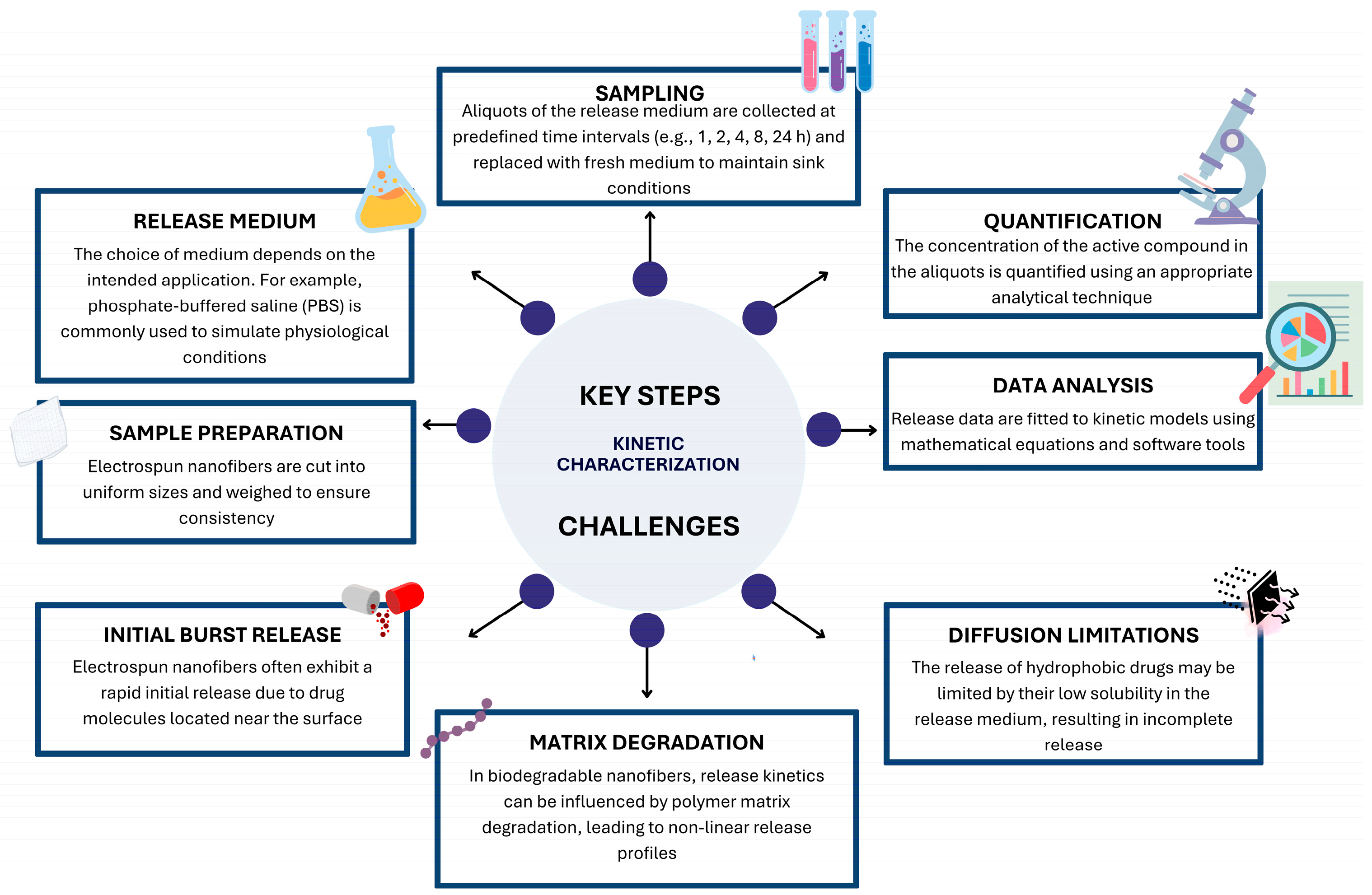
The kinetic characterization of drug release from electrospun nanofibers typically involves in vitro release studies, in which nanofibers are immersed in a release medium (e.g., phosphate-buffered saline or simulated body fluid) under controlled conditions [194]. Samples of the release medium are collected at regular intervals, and the concentration of the active compound is quantified using analytical techniques such as HPLC, UV-Vis, or MS. The resulting data are then fitted to various kinetic models to elucidate the release mechanism [193]. Figure 10 summarizes crucial aspects in kinetic characterization.

10.2. Kinetic Models

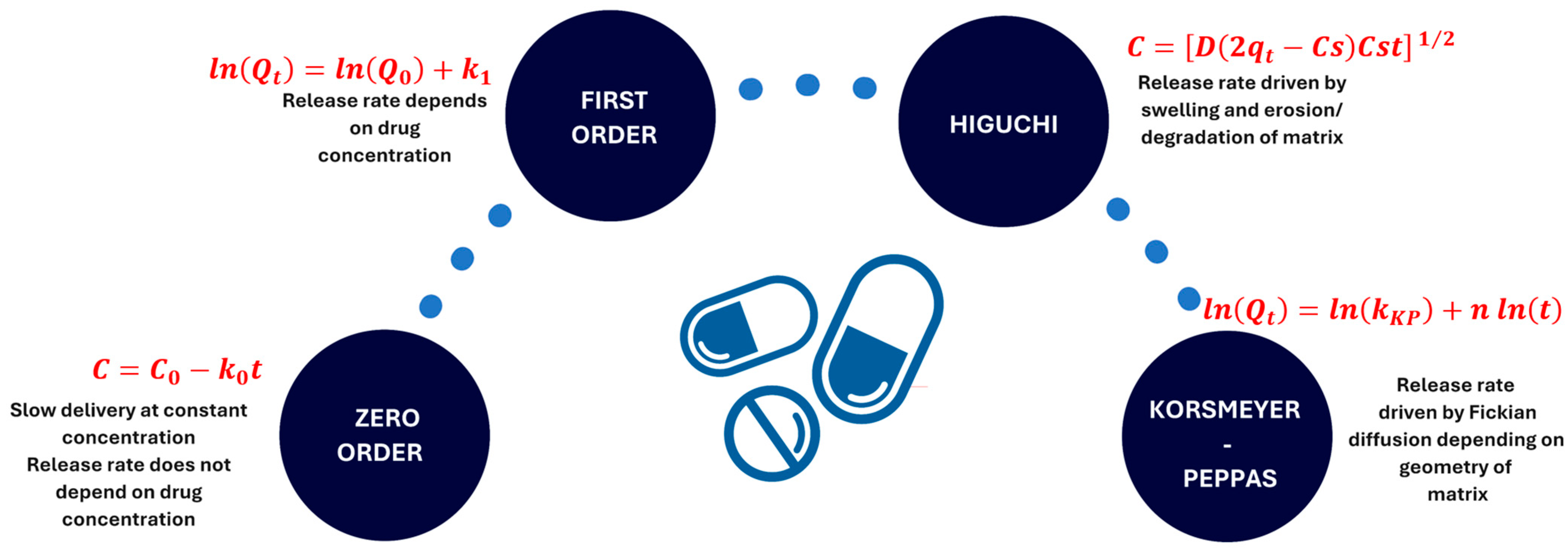
Data obtained from in vitro studies can be fitted to various kinetic models to understand the release mechanism. The most used models include the zero-order, first-order, Higuchi, and Korsmeyer–Peppas models. Each model is based on different assumptions regarding the release mechanism and is described by a specific set of equations [193,194,195]. Equations and generalities are summarized in Figure 11.
The zero-order model describes slow release at a constant concentration and is an ideal kinetic model, in which the drug concentration remains constant during the release process in physiological media and plasma because the release rate does not depend on the drug concentration. The first-order models assume that the amount of drug released over time depends exclusively on the drug concentration loaded into the matrix [196].
The Higuchi model involves two mechanisms responsible for the release rate: swelling and erosion/degradation. Water-solubility and low solubility of a solid or semi-solid matrix promote the drug coverage and decrease the delivery rate [197,198].
The Korsmeyer–Peppas model describes the release mechanism from polymeric nanostructures driven by a Fickian pathway. This model has a diffusion exponent (m) that can be adjusted to distinguish among other mechanisms, while accounting for matrix geometry [199].

10.3. Software Tools for Kinetic Modeling

Kinetic characterization and modeling are essential for understanding the release behavior of antibacterial agents from electrospun nanofibers. Various software tools and add-ons, such as MATLAB, Origin, Excel Solver, R, and Python, provide different advantages and limitations for kinetic modeling. MATLAB and Python offer high flexibility and advanced statistical capabilities but require programming knowledge. Origin and Excel Solver are more accessible and user-friendly, though they may lack flexibility for complex models. R provides a balance between flexibility and accessibility, with strong community support. The choice of tool depends on the specific requirements of the study, including model complexity, dataset size, and the researcher’s programming proficiency [200,201,202].
R is a free, open-source programming language widely used in scientific research for data analysis and modeling. It offers a wide range of packages for kinetic modeling, including nls for nonlinear least squares fitting and deSolve for solving differential equations [200].
Python, also free and open source, is widely used in scientific research for data analysis, modeling, and visualization. Its libraries for kinetic modeling include SciPy for optimization and lmfit for nonlinear least squares fitting [202].

11. In Vitro and Pre-Clinical Studies

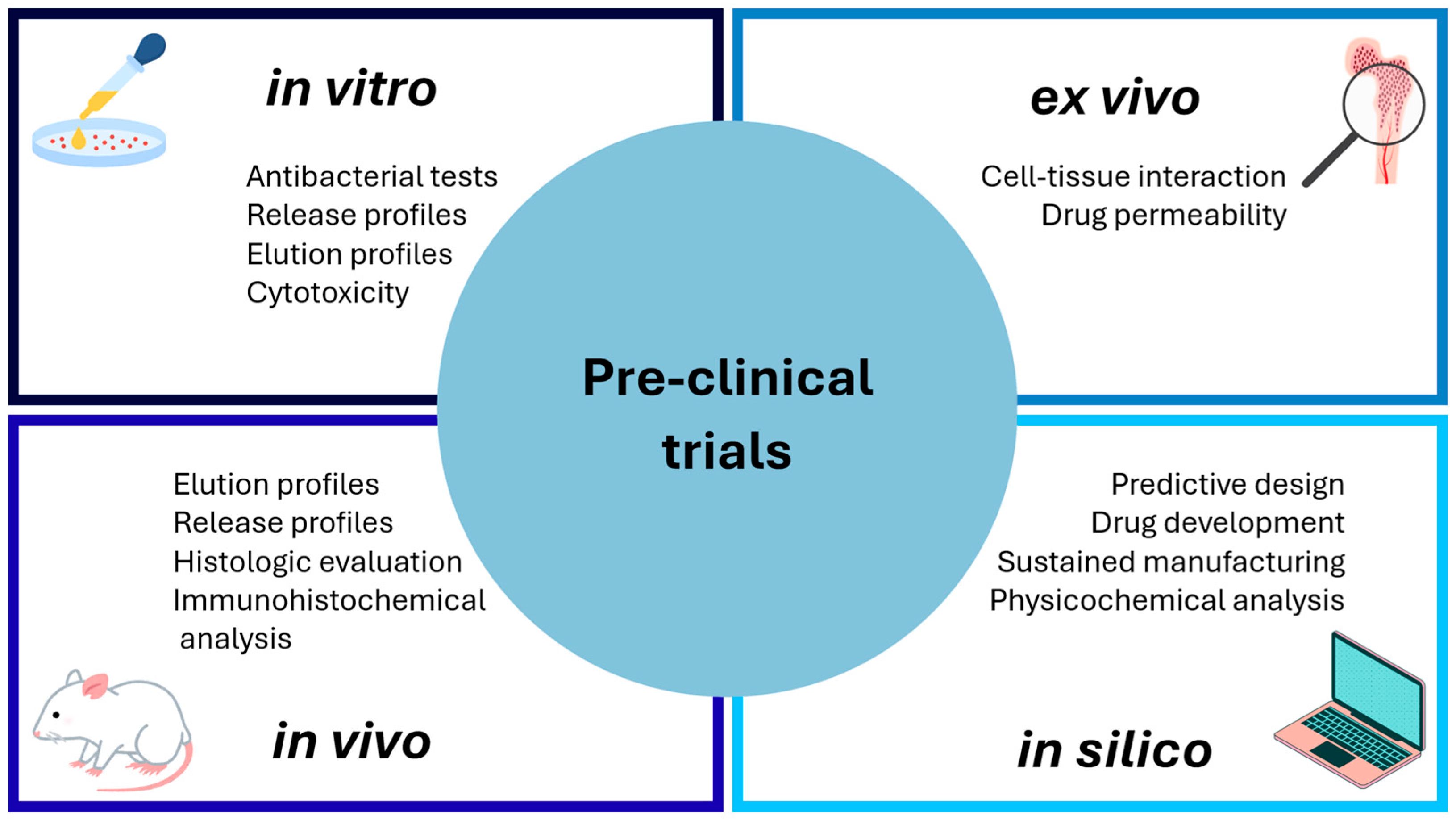
In the development of general drug delivery systems, pre-clinical tests are intended to assess safe administration, effectiveness, and dosage efficacy, analyze pharmacokinetic features, and validate non-toxicity. Common preclinical trials include in vitro [203,204,205], in vivo [162,206,207], ex vivo [208,209], and in silico procedures [207,210,211]. The analysis of antibiotic-loaded nanofibers often involves the evaluation of release [123,203,206,212,213] and elution profiles [162,206], bacterial inhibition tests [130,151,205,213], cytotoxicity [121], histologic evaluation [162,214], hemocompatibility [208], immunohistochemical analysis [162], tissue regeneration [119,215], wound healing [49,216], and degradation [204,209]. Figure 12 presents its classification depending on testing conditions. In addition to kinetic analysis, bacterial inhibition and cytotoxicity are among the most widely used approaches for designing nanofibers for antibiotic delivery.

11.1. Bacterial Inhibition Tests

Demonstrating bacterial inhibition is intrinsic to the concept of antibiotic delivery from electrospun nanofibers. The evaluation of the antibacterial activity of nanomaterials requires standardized microbiological tests that allow the effect to be quantified and results to be compared across laboratories. Among the most commonly used methods are the disk diffusion test (Kirby–Bauer), broth dilution tests (micro- and macro-dilution to determine the minimum inhibitory concentration (MIC), and minimum bactericidal concentration (MBC), well diffusion tests, the E-test method, dynamic contact tests (e.g., ASTM E2149), and standardized tests for textile substrates or surfaces (e.g., ISO 20743, ASTM E2149) [217]. Each technique provides complementary information—inhibition zones, MIC/MBC, time-kill curves, dynamic contact capacity, or activity on surfaces—and its choice depends both on the nature of the material (solid, coating, colloidal suspension) and the expected mechanism of action (ionic release, direct contact, photoactivation) [218].
The Kirby–Bauer disk diffusion assay is a widely standardized qualitative and semi-quantitative method for soluble antibiotics that measures the diameter of the inhibition zone on inoculated agar. Its strength lies in its operational simplicity, low cost, and standardization by bodies such as the Clinical and Laboratory Standards Institute (CLSI) and the European Committee on Antimicrobial Susceptibility Testing (EUCAST), which facilitates clinical comparisons. However, it has intrinsic limitations for nanomaterials: diffusion in agar depends on the mobility of the active agent (large nanoparticles do not diffuse), and many nanomaterials exert their effect by releasing ions or through contact interaction, so the absence or small size of an inhibition zone does not necessarily imply a lack of activity. Consequently, disks or wells may underestimate the activity of nanoparticles whose action does not depend on significant diffusion in the solid medium [219]. Broth dilution tests (micro- or macro-dilution) allow the MIC to be determined and, through subculture, the MBC; these quantitative metrics are considered the gold standard for characterizing inhibitory potency. In the context of nanomaterials, microdilution is advantageous because it measures the effect in suspension and therefore captures the activity mediated by released ions or colloidal interactions; it also facilitates the generation of concentration-response curves and calculations of pharmacodynamic parameters. However, the presence of nanoparticles can interfere with optical readings (turbidity) or adsorb dyes/indicators, requiring adaptations (e.g., subculture readings, adenosine triphosphate (ATP) measurements, or alternative colorimetric assays) to avoid false positives/negatives. Therefore, CLSI standards for microdilution are the starting point, but the literature abounds with modifications to manage interference from nanomaterials [220].
Gradient-based methods, such as the E-test, combine diffusion and quantification principles to obtain MICs directly in agar and are widely used in clinical microbiology. However, their applicability to nanomaterials is limited because they depend on diffusion in the solid medium, which is challenging when the antibacterial mechanism requires direct contact or involves slow release of ions with low mobility. Although the E-test can correlate with the MIC obtained by microdilution, its use with nanomaterials requires prior validation and specific controls due to differences in mobility, kinetics, and suspension stability [221].
On the other hand, standardized dynamic contact tests such as ASTM E2149 (Standard Test Method for Determining the Antimicrobial Activity of Immobilized Antimicrobial Agents Under Dynamic Contact Conditions) or ISO 20743 [217] (Determination of antibacterial activity) are better suited for nanomaterials incorporated into surfaces, as they simulate mechanical contact and are less affected by nanomaterial release. They are better suited for nanomaterials incorporated into surfaces, fibers, or coatings, as they quantify bacterial reduction without requiring diffusion through agar. Their strength lies in their practical relevance for solid materials. However, results can vary significantly depending on experimental conditions such as agitation, volume, inoculum, or surface-to-volume ratio, requiring a detailed description to ensure comparability [217].
Complementary methods, such as time–kill, add essential kinetic information for slow-release systems, allowing differentiation between bacteriostatic and bactericidal effects; however, they require a greater experimental workload and strict controls due to optical or colloidal interference inherent in nanoparticulate suspensions, so they are often combined with plate counting [222].
The antibacterial evaluation of nanomaterials presents specific challenges due to inherent analytical interferences, such as light scattering or absorption, dye absorption, and effects on fluorometric readings, which can bias methods based on turbidity or fluorescence. In addition, phenomena such as agglomeration, colloidal stabilization, and the presence of biomolecules can modulate the availability of the active agent, especially in ion-release systems. Therefore, the recent literature recommends combining multiple assays (diffusion, dilution, dynamic contact, time-kill, or biofilm formation) and reporting physicochemical controls, such as size, ζ-potential, or ion concentration, to ensure interpretability and reproducibility and to avoid erroneous conclusions [223].
In operational terms, the Kirby–Bauer disk diffusion assay is rapid and cost-effective; however, it may underestimate the antibacterial activity of materials with limited diffusivity, including nanomaterials and poorly soluble compounds, since its outcome depends on radial diffusion through agar media [224,225]. Conversely, broth microdilution assays allow direct interaction between bacteria and antimicrobial agents, enabling accurate determination of minimum inhibitory concentration (MIC), although these methods can be affected by nanoparticle aggregation and optical interference during turbidity measurements [223,226]. ASTM/ISO standards are suitable for solid surfaces but depend on experimental parameters that affect comparability. In contrast, time-kill and biofilm assays provide kinetic and ecological information, though they require greater experimental complexity. Due to this variability, recent organizations and reviews emphasize the need to harmonize and standardize methodologies by following guidelines from CLSI, EUCAST, ASTM, or ISO, and to rigorously report experimental conditions to enable replication and facilitate comparative analyses for clinical or technological applications [218].

11.2. Cytotoxicity

In addition to the bacterial inhibition assay, the design of antibiotic delivery systems based on nanofibrous mats often involves cytotoxicity analysis. Given the scope of applying nanostructured DDS, cell viability is a crucial aspect. According to ISO 10993-5 and Food and Drug Administration (FDA) guidelines [227,228,229,230], the cytotoxic effects of implantable devices can be assessed primarily using the [3-(4,5-dimethylthiazol-2-yl)-2,5-diphenyltetrazolium bromide (MTT) assay, lactate dehydrogenase (LDH) release, trypan blue, and neutral red uptake [227,228,229,230]. Among them, the MTT is the most widely used protocol for determining cell viability/toxicity in vitro. The effectiveness of MTT assays depends on measuring mitochondrial metabolic activity. This approach refers to the use of MTT. This yellow salt undergoes a color change upon metabolic reduction by the NADH enzyme, yielding the insoluble purple formazan, which is retained by metabolically viable cells [227,228]. Thus, the MTT approach measures mitochondrial integrity, with the amount of formazan proportional to the surviving cell population.
From the point of view of the cell line, rodent and human cell lines are alternatives for cytotoxicity testing, including Jurkat human T-cell leukemia cells, human embryonic kidney HEK-293 cells, human neuroblastoma SH-SY5Ym cells, mouse embryonic NIH-3T3 cells, mouse neuroblastoma N2a cells, and rat hepatoma H4-II-E cells. Nonetheless, the cytotoxicity testing upon cancer-derived cell lines can result in metabolic abnormalities [229].

12. Development of Nanosystems

Over time, pathogenic germs become resistant, particularly to antimicrobial drugs that are overused. Chemists, pharmacists, and physicists conduct research on the development of novel antibacterial agents and on medical education and preventive care. Over the past few years, researchers have focused on the creation and study of polymeric materials with antimicrobial properties to reduce environmental toxicity, and the short half-life typical of low-molecular-weight antimicrobial agents [231]. Enormous efforts to modify existing drugs and design delivery systems have been made over the last three decades, with the sole goal of overcoming antimicrobial resistance. From encapsulation in micelles, the use of polymers, and the control of the micro (nano) architecture to ralentize drug diffusion are some of the tested features for a better therapeutic outcome. Among these nanosystems, electrospun nanofiber meshes represent an advanced evolution, offering superior surface area, porosity, and local retention for topical antibacterial therapy. With advances in combinatorial chemistry and high-throughput screening, most drug candidates selected for development are highly hydrophobic and have negligible water solubility. Because of their poor solubility or slow dissolution in the human gastrointestinal tract, many medications have limited oral absorption. An assortment of formulation methods, including salt formation, complexation, particle size reduction, prodrugs, micellization, and solid dispersions, is now being widely studied in the pharmaceutical field to enhance the oral absorption of poorly water-soluble drugs by increasing their dissolution rates [232].
Metal nanoparticles, emulsion-based nanosystems, nanovesicles, lipid nanoparticles, polymeric nanoparticles, and, recently, nanofiber meshes were among the various categories in which the preclinical safety profile and antibacterial activity of topical nanoformulations were thoroughly examined. Because of their strong antibacterial efficacy and tolerability, topical composites are expected to replace traditional topical formulations in the treatment of skin and soft tissue infections (SSTIs) [233]. Notably, nanofiber-based topical systems provide prolonged residence time and controlled release compared to conventional gels and creams.
As a matter of fact, the drug-delivery electrospun fiber mat is an innovative formulation with potential future clinical applications. Anticancer, antibacterial, and antiviral medicines were used to investigate the effects of drug solubility and compatibility in the drug/polymer/solvent system on encapsulation [234]. Electrospun nanofibers have demonstrated relevance in antibiotic delivery, as exemplified by the following studies. According to Kenawy and colleagues, tetracycline was released from electrospun poly(ethylene vinyl acetate) (PEVA)-PLA (1:1) mats, and PEVA mats provided relatively steady drug release over approximately 5 days [235]. To increase the production of tenofovir (TFV) fibers and assess essential characteristics of the final products, including fiber form, drug crystallinity, and drug loading and release kinetics, Krogstad’s team published a description of the free-surface electrospinning technique in 2014. Employing a nozzle-free manufacturing-scale electrospinning device, PVA containing up to 60 weight percent TFV was effectively electrospun into fibers. The pharmaceutical manufacturing of fiber-based medical textiles for clinical application may be significantly impacted by these discoveries, which establish crucial solution and processing parameters for the scale-up production of TFV drug-eluting fibers by electrospinning [236].
Electrospinning was used to create PCL loaded with ofloxacin (OFL) and PCL: poly(butylene succinate) PBS fibers as a drug delivery method for the treatment of ocular infections. Fiber morphology and size were examined explicitly in relation to several formulation variables, such as the polymer/drug ratio (9:1, 8:2, and 7:3 w/w), solvent systems such as dichloromethane (DCM), N,N-dimethylformamide (DMF), N,N-dimethylacetamide (DMAc), and dimethylsulfoxide (DMSO), and polymer blends of PCL:PBS at 80:20, 60:40, and 40:60 ratios. P. aeruginosa, S. epidermidis, S. aureus, and E. coli strains served to evaluate the microbiological activity of the modified formulation. Its results clearly showed that freely released OFL from fibers prevented the development of the tested bacteria. The activity of the medication included in the fibers was unaffected by the electrospinning process [237]. This method was employed in recent research to facilitate the incorporation of loaded nanofibers into tablet formulations of the analgesic meloxicam, yielding quick- or sustained-release dosage forms. The tablets can provide the dose in accordance with the anticipated release behavior, and the dominant release mechanism of the tablets was examined using zero-order, first-order, Higuchi, Korsmeyer–Peppas, and Hixon–Crowell kinetics models [238]. According to Soroush and colleagues, mats containing amphotericin B (AMB) and VAN are antibacterial and antifungal agents [239]. Upon the addition of medicines and mucoadhesive agents, SEM images showed a slight reduction in fiber diameter. The Higuchi model was used to fit the in vitro release profile of VAN, whereas zero-order equations were used to fit the AMB release profile. The outcome demonstrated that although other factors influenced AMB release, biodegradable mucoadhesive nanofibrous membranes produced large amounts of VAN within the first 24 h.
In addition to their well-established role as controlled drug-delivery vehicles, electrospun nanofiber matrices can serve as effective platforms for activating prodrugs in response to endogenous stimuli, particularly enzymatic activity and pH changes at infection sites. In this context, prodrugs are designed to remain pharmacologically dormant until they encounter specific biochemical cues characteristic of pathological environments, such as elevated levels of enzymes secreted by bacteria or host immune cells, or the acidic microenvironment typical of infected or inflamed tissues. For example, inflammation-responsive prodrugs synthesized with ester linkages have been successfully integrated into poly(ε-caprolactone)/gelatin nanofibers, where cholesterol esterase secreted by activated macrophages catalyzes ester hydrolysis, triggering the release of the active drug only in the presence of the enzyme; in the absence of enzymatic activity, minimal drug is released, preserving stability and reducing off-target exposure. Such enzyme-responsive strategies demonstrate how nanofiber scaffolds can be tailored to exploit local biochemical triggers, ensuring that the therapeutic payload is liberated predominantly in disease-associated microenvironments, thereby enhancing site-specific activation and efficacy while limiting systemic toxicity [240,241,242].
Beyond enzyme triggers, the pH responsiveness of nanofiber systems enables selective activation of prodrugs in acidic infection milieus. Electrospun nanofiber–hydrogel hybrid dressings have been developed where antibiotic release is governed by pH changes associated with wound infection; these systems remain relatively inert at physiological pH but undergo conformational or chemical changes under acidic conditions, leading to accelerated release of the active agent specifically where bacteria proliferate. This pH-triggered release mechanism is particularly advantageous in chronic or biofilm-associated infections where localized acidity is a hallmark, as it minimizes premature drug liberation and enhances therapeutic concentrations at the site of pathology [243]. Together, enzyme- and pH-responsive nanofiber prodrug platforms represent a versatile toolkit for innovative antibacterial therapy, improving drug stability during delivery, reducing systemic side effects, and maximizing local drug activation precisely where it is needed most.
On the other hand, modifications and repurposing of proven antibiotics, attachment of siderophores, membrane-targeting peptides, and other drug-delivery systems have also been considered adequate vehicles for delivering antimicrobials to pathogenic microbes. Among them, the conjugation of anti-infectious agents to antibodies and nanocarriers has increasingly attracted researchers’ interest [244].
Novel metronidazole analogs, such as ruthenium- and Schiff base-based derivatives, have been developed to address the rise in antimicrobial resistance [245]. These next-generation agents exhibit broader antimicrobial coverage, alternative mechanisms of action, reduced cytotoxicity, improved efficacy against resistant strains, and potential applications beyond traditional infectious disease contexts. Nonetheless, this drug remains the best option for treating Blastocystis infection, even compared with the 1,3-bis-(4-phenyl-[1,2,3] triazole-1-il)2-propanol analog [246].
As repurposed non-antibiotics, phenothiazines enhance the effectiveness of antibiotics by inhibiting efflux pumps. It was noted that thioridazine decreased the ethambutol MIC for M. avium from 8 µg/mL to 2 µg/mL, indicating synergism. According to one study, chlorpromazine decreased the norfloxacin MIC in S. aureus RN4220 from 4 µg/mL to 1 µg/mL, which was interpreted as a combined effect due to efflux inhibition. However, no checkerboard synergic studies were conducted, and FIC values were not determined [247].
In addition to inhibiting biofilm formation and other pathogenicity, the NSAID-antibiotic combination reduced the expression of the efflux pump gene in certain bacterial strains.
As a bio-friendly alternative, loading antimicrobial peptides (AMPs) into topical preparations might prolong their skin contact time. Scalability and affordability encourage the pharmaceutical industry to manufacture and market them; immunogenicity and long-term biological fate, which are the primary barriers to clinical applications; and proper animal disease models and reliable statistical analyses to confirm their therapeutic potential [248]. Synthetic peptide–polymers and their mimics, and in particular structurally nano-engineered antimicrobial peptide–polymers (SNAPPs), are antimicrobial materials with clinical potential as novel therapeutics to combat antimicrobial resistance due to their inherent biodegradability, biocompatibility, and tunable cytocompatibility. Macromolecular design, in conjunction with rational monomer composition, can direct their architecture, self-assembly, and chemical behavior, ultimately guiding the choice of appropriate applications within the biomedical field [249].
In the context of polymicrobial illnesses and biofilms, as with antibiotics and their analogs or metal complexes and NPs, the antibacterial activity of AMP-coated biofilms should be investigated; the materials’ effectiveness should be comparable to or better than that of antibiotics.

13. Regulatory and Scalability Challenges

Although electrospinning has challenges, it stands out for relatively simple adjustments in parameters, namely electrode distance, voltage, concentration, flow rate, and environmental conditions, to reproduce results. Recent studies have shown that even modified setups can enable continuous production of nanofibrous mats at an industrial scale [118,170].
In a biomedical context, technical limitations translate into greater regulatory hurdles for approving devices manufactured using this technique. Morphological variability, the potential cytotoxicity of solvent residues, and the complexity of the process make it difficult to ensure reproducible, consistent clinical standards, delaying the translation of academic prototypes into commercial products in the antibacterial field [187].
The industrial translation of antibacterial nanomaterials poses significant challenges in reproducibility, batch-to-batch consistency, and regulatory compliance, particularly in controlling particle size, surface chemistry, and drug-loading efficiency [250]. These challenges arise from the inherent complexity of controlling physicochemical properties at scale, maintaining reproducibility and uniformity across batches, and navigating regulatory requirements for biomedical applications. For example, reproducibility in size distribution, surface chemistry, and drug-loading efficiency is critical for ensuring consistent therapeutic performance [251,252,253].
Beyond physical and chemical parameters, regulatory and quality requirements for biomedical nanomaterials further complicate scale-up. Regulatory agencies such as the U.S. Food and Drug Administration (FDA) and the European Medicines Agency (EMA) require extensive characterization of nanoscale products, including particle size distribution, surface chemistry, sterility, and stability. These requirements are essential for ensuring safety but demand robust analytical methods and validated quality controls that are challenging to implement in high-throughput environments. Compared with other technologies, many nanomedicine products have successfully reached the market, yet this success is often contingent on early consideration of manufacturability and regulatory alignment during development [254].
Cost considerations also play a significant role in industrial translation. High costs of specialty polymers and lipids, combined with the need for specialized equipment and energy-intensive processing, can render specific nanomaterial manufacturing strategies economically unfeasible without further process optimization. Continuous manufacturing approaches are being explored to reduce operational costs and improve scale-up feasibility by enhancing control and reducing waste. These continuous systems can achieve consistent product quality, but they introduce new engineering challenges related to system integration and control stability [255].
Finally, environmental, health, and safety (EHS) concerns associated with large-scale nanomaterial production must be addressed during industrial translation. The use of volatile organic solvents and the potential use of nanomaterials in the environment necessitate comprehensive lifecycle assessments (LCAs) and implementation of solvent recovery and waste management strategies. Moreover, the long-term impacts of engineered nanostructures on human health and ecosystems remain an active area of research, underscoring the importance of sustainable manufacturing practices that minimize emissions and exposure risks throughout the product’s life cycle [250].
Likewise, translating electrospinning technologies from laboratory-scale research to industrial nanomanufacturing remains a significant challenge. Although electrospinning is widely recognized as a versatile technique capable of producing fibers with controlled diameter and morphology, conventional capillary systems suffer from intrinsically low throughput, typically in the range of 0.01–1 g h−1, which severely restricts their industrial implementation [256,257]. This limited productivity is insufficient for biomedical devices, functional textiles, and filtration applications that require mass production [183]. Moreover, technical limitations related to jet instability and process reproducibility further complicate scale-up [258,259].
One of the principal challenges in industrial translation is balancing productivity and product quality. Increasing production rates often compromise fiber uniformity and morphology due to factors such as inadequate solvent evaporation, jet instability, and interactions among multiple jets in multi-needle setups. For example, while multi-needle electrospinning configurations have been developed to increase production capacity, electrostatic field interference between needles can result in inconsistent fiber deposition and morphological defects, ultimately affecting the reproducibility of fiber properties. Additionally, needle clogging remains a persistent operational issue, which not only disrupts continuous operation but also increases maintenance complexity and costs [260].
Alternative strategies to address scale-up limitations include free-surface and needleless electrospinning, in which multiple jets are generated from an open liquid surface without discrete spinnerets. These approaches can enhance output by enabling the simultaneous formation of several jets; however, they introduce challenges, such as increased demands on process control, higher voltage requirements, and issues with solvent management under high-throughput conditions. Free surface systems, while mitigating clogging, often require precise control over environmental factors such as humidity and temperature to maintain fiber consistency, thereby increasing equipment and operational complexity [260].
Another significant consideration for industrial nanomanufacturing is environmental, health, and safety (EHS) compliance. Many electrospinning processes rely on volatile organic solvents for polymer solution preparation, which present ecological and occupational hazards at larger scales due to solvent vapor emissions and worker exposure unless effective recovery and mitigation systems are implemented. These environmental concerns, including residual solvent toxicity and regulatory VOC compliance, are emerging barriers to large-scale adoption of electrospinning technology [186].
Additionally, the high voltage required for electrospinning poses operational safety risks, necessitating rigorous insulation, grounding, and control systems to protect personnel and equipment from electrical hazards.
Environmental control and process parameter sensitivity (e.g., temperature and humidity) also influence fiber morphology and consistency, posing challenges for maintaining uniform fiber diameter distribution and nanostructure quality during scaled-up production. To address some of these concerns, green electrospinning strategies using biologically benign solvents have been proposed to reduce solvent toxicity and environmental impact, representing a promising step toward safer, scalable production systems [261]. Quality control and consistency across large production batches are essential for end-use applications such as drug delivery systems, wound dressings, or filtration membranes; therefore, advanced process monitoring, environmental control, and real-time feedback systems are necessary to adjust parameters and detect deviations in fiber morphology dynamically, but such technologies are still under active development [259].
Additionally, achieving cost-effective scalability requires not only equipment innovation but also process standardization and integration with downstream manufacturing steps. For nanofiber applications in pharmaceuticals, for instance, integrating electrospinning with sterile filling, coating, and packaging processes under Good Manufacturing Practice (GMP) conditions represents a substantial challenge that extends beyond fiber production itself. There are relatively few documented industrial case studies in the literature that demonstrate end-to-end solutions from polymer dissolution to finished biomedical products, underscoring the need for collaborative efforts between academia, industry, and regulatory bodies to fill this gap [262].
Emerging approaches seek to mitigate these obstacles through innovative spinneret designs, the incorporation of secondary forces (e.g., centrifugal and magnetic forces), and the development of hybrid processes that combine electrospinning with complementary manufacturing techniques such as centrifugal spinning or blow spinning. While these strategies show promise for enhancing throughput and fiber quality, further research is needed to validate their scalability, cost-effectiveness, and compatibility with regulatory requirements for biomedical nanofiber products. Consequently, the full industrial potential of electrospinning remains constrained by technological, economic, and regulatory challenges, and realizing this potential will require sustained innovation in nanomanufacturing science and engineering [263].
In summary, the successful industrial scaling and commercial translation of antibacterial nanomaterials requires a multifaceted approach that integrates process engineering, quality control, economic analysis, regulatory foresight, and environmental stewardship. While laboratory-scale research continues to innovate new compositions and functionalities, bridging the gap to industrial production remains a critical challenge that demands collaboration across disciplines and sustained effort to align technological innovation with practical manufacturing realities.

14. Progress and Potential

The incorporation of antibiotics into nanostructured polymeric materials is a promising alternative for overcoming bacterial infections. The suitability of electrospinning for the versatile fabrication of polymeric nanofibrous templates stands out. Moreover, variability in antibiotic loading via different approaches improves kinetic and pre-clinical antibacterial performance. Bactericidal or bacteriostatic activity can be modulated by combining the loading mechanism with the structural features of electrospun meshes, demonstrating the versatility of electrospinning as a manufacturing tool for antibiotic release and other antibacterial aims. The principal advantage of combining nanofibers and antibiotics lies in the enhanced efficacy of bacterial inhibition enabled by nanostructured platforms.
Considering comparative aims, efforts to adopt conventional methods and ensure reasonable discussion are required, especially regarding inhibition results and release efficacy (i.e., mm versus Colony-Forming Units (UFC) and μg/mg versus %, respectively). The use of standardized guidelines and procedures orients this aspect, but there is still a long way to go.
Currently, there is no solid clinical evidence demonstrating that antibacterial nanoformulations (including nanofibers, metallic nanoparticles, or polymeric nanoparticles) are directly associated with emerging infectious diseases or induced pathologies in humans. Most available data come from in vitro studies and animal models, which mainly evaluate antimicrobial efficacy and toxicity, without long-term epidemiological follow-up. Systematic reviews on nanomedicine indicate that clinical evidence remains limited, as many of these technologies remain in preclinical phases or in exploratory trials. In the case of nanofibers for wound healing, studies report a significant reduction in bacterial load without clinical adverse effects attributable to the material, supporting its preliminary safety.
However, experts warn that the extensive use of nanomaterials in consumer products and in uncontrolled environments could expose human and environmental microbiomes to sublethal concentrations, favoring bacterial selection. Although there is still no direct clinical evidence, environmental studies suggest co-selection of genes conferring resistance to both antibiotics and nanomaterials, posing a potential long-term concern and, at the same time, offering a research opportunity.

Author Contributions

J.A.O.-G.: Writing—original draft, Visualization, Investigation. L.D.M.-G.: Writing—original draft, Investigation. E.R.-M.: Writing—original draft. M.V.-S.: Conceptualization, Writing—original draft, Supervision, Investigation. E.C.-Y.: Writing—original draft, Writing-reviewing and editing, Investigation. J.B.G.-C.: Conceptualization, Writing—original draft, Writing-reviewing and editing, Investigation. A.P.-N.: Conceptualization, Investigation, Supervision, Visualization, Writing—original draft, Writing-reviewing and editing. All authors have read and agreed to the published version of the manuscript.

Funding

This research received no external funding.

Data Availability Statement

Data sharing is not applicable to this article.

Acknowledgments

The authors acknowledge financial support from SECIHTI through Graduate Grants awarded to J.A.O.-G. (CVU 1187100) and L.D.M.-G. (CVU 2080005), and Postdoctoral Fellowships granted to M.V.-S. (CVU 593548) and A.P.-N. (CVU 593576).

Conflicts of Interest

The authors declare no conflicts of interest.

References

  1. Ishak, A.; Mazonakis, N.; Spernovasilis, N.; Akinosoglou, K.; Tsioutis, C. Bactericidal versus Bacteriostatic Antibacterials: Clinical Significance, Differences and Synergistic Potential in Clinical Practice. J. Antimicrob. Chemother. 2025, 80, 1–17. [Google Scholar] [CrossRef] [Scilit]
  2. Seifert, R.; Schirmer, B. Problems Associated with the Use of the Term “Antibiotics”. Naunyn-Schmiedeberg’s Arch. Pharmacol. 2021, 394, 2153–2166. [Google Scholar] [CrossRef] [Scilit]
  3. Kharga, K.; Jha, S.; Vishwakarma, T.; Kumar, L. Current Developments and Prospects of the Antibiotic Delivery Systems. Crit. Rev. Microbiol. 2025, 51, 44–83. [Google Scholar] [CrossRef] [Scilit]
  4. Guedes, B.N.; Krambeck, K.; Durazzo, A.; Lucarini, M.; Santini, A.; Oliveira, M.B.P.P.; Fathi, F.; Souto, E.B. Natural Antibiotics against Antimicrobial Resistance: Sources and bioinspired Delivery Systems. Braz. J. Microbiol. 2024, 55, 2753–2766. [Google Scholar] [CrossRef] [Scilit]
  5. Kaushik, H.; Sharma, R.; Kumar, A. Natural Products against Resistant Bacterial Infections: A Systematic Literature Review. Microbe 2025, 6, 100247. [Google Scholar] [CrossRef] [Scilit]
  6. Obeid, M.A.; Alyamani, H.; Alenaizat, A.; Tunç, T.; Aljabali, A.A.A.; Alsaadi, M.M. Nanomaterial-Based Drug Delivery Systems in Overcoming Bacterial Resistance: Current Review. Microb. Pathog. 2025, 203, 107455. [Google Scholar] [CrossRef] [Scilit]
  7. Keskin, D.; Zu, G.; Forson, A.M.; Tromp, L.; Sjollema, J.; van Rijn, P. Nanogels: A Novel Approach in Antimicrobial Delivery Systems and Antimicrobial Coatings. Bioact. Mater. 2021, 6, 3634–3657. [Google Scholar] [CrossRef] [Scilit]
  8. Delgado-Pujol, E.J.; Martínez, G.; Casado-Jurado, D.; Vázquez, J.; León-Barberena, J.; Rodríguez-Lucena, D.; Torres, Y.; Alcudia, A.; Begines, B. Hydrogels and Nanogels: Pioneering the Future of Advanced Drug Delivery Systems. Pharmaceutics 2025, 17, 215. [Google Scholar] [CrossRef] [Scilit]
  9. Vashist, A.; Perez Alvarrez, G.; Andion Camargo, V.; Raymond, A.D.; Yndart Arias, A.; Kolishetti, N.; Vashist, A.; Manickam, P.; Aggarwal, S.; Nair, M. Recent Advances in Nanogels for Drug Delivery and biomedical Applications. Biomater. Sci. 2024, 12, 6006–6018. [Google Scholar] [CrossRef] [Scilit] [PubMed]
  10. Mura, P.A.; Cirri, M.; Rossetti, A.; Allemandi, D.A.; Paredes, A.J.; Palma, S.D. Preparation of Glyburide Nanocrystals with Improved Dissolution Properties by Dry-Ball- and Wet-Bead- Milling: Systematic Comparison by Experimental Design of the Performance of the Two Methods. J. Drug Deliv. Sci. Technol. 2024, 91, 105222. [Google Scholar] [CrossRef] [Scilit]
  11. Bhardwaj, H.; Jangde, R.K. Current Updated Review on Preparation of Polymeric Nanoparticles for Drug Delivery and Biomedical Applications. Next Nanotechnol. 2023, 2, 100013. [Google Scholar] [CrossRef] [Scilit]
  12. Rahman Sabuj, M.Z.; Islam, N. Inhaled Antibiotic-Loaded Polymeric Nanoparticles for the Management of Lower Respiratory Tract Infections. Nanoscale Adv. 2021, 3, 4005–4018. [Google Scholar] [CrossRef] [Scilit]
  13. Eltaib, L. Polymeric Nanoparticles in Targeted Drug Delivery: Unveiling the Impact of Polymer Characterization and Fabrication. Polymers 2025, 17, 833. [Google Scholar] [CrossRef] [Scilit]
  14. Zielinska, A.; Carreiró, F.; Oliveira, A.M.; Neves, A.; Pires, B.; Nagasamy Venkatesh, D.; Durazzo, A.; Lucarini, M.; Eder, P.; Silva, A.M.; et al. Polymeric Nanoparticles: Production, Characterization, Toxicology and Ecotoxicology. Molecules 2020, 25, 3731. [Google Scholar] [CrossRef] [Scilit]
  15. Naik, G.; Alias, R.R.; Roy, A.A.; Mutalik, S.; Dhas, N. Unleashing the Power of Polymeric Nanoparticles—Creative Triumph against Antibiotic Resistance: A Review. Int. J. Biol. Macromol. 2024, 278, 134977. [Google Scholar] [CrossRef] [Scilit]
  16. Hamdan, N.; Yamin, A.; Hamid, S.A.; Khodir, W.K.W.A.; Guarino, V. Functionalized Antimicrobial Nanofibers: Design Criteria and Recent Advances. J. Funct. Biomater. 2021, 12, 59. [Google Scholar] [CrossRef] [Scilit]
  17. Sun, G.; Han, W.; Wang, Y.; Xin, S.; Yang, J.; Zou, F.; Wang, X.; Xiao, C. Overview of the Fiber Dynamics during Melt Blowing. Ind. Eng. Chem. Res. 2022, 61, 1004–1021. [Google Scholar] [CrossRef] [Scilit]
  18. Chatterjee, A.; Yadav, A.; Sen, S.; Ghosh, S.; Choudhary, N.; Dey, N.; Mishra, S.; Maji, P.K. Polymer-Modified Nonwovens: Functional Surfaces for Separation, Sensing, Biomedical and Smart Textile Applications. J. Mater. Chem. A 2025, 14, 3210–3252. [Google Scholar] [CrossRef] [Scilit]
  19. Ding, M.; Yang, X.; Liu, Y.; Zeng, S.; Duan, G.; Huang, Y.; Liang, Z.; Zhang, P.; Ji, J.; Jiang, S. A Review of Advanced Helical Fibers: Formation Mechanism, Preparation, Properties, and Applications. Mater. Horiz. 2024, 11, 5843–5873. [Google Scholar] [CrossRef] [Scilit] [PubMed]
  20. Ahmadi Bonakdar, M.; Rodrigue, D. Electrospinning: Processes, Structures, and Materials. Macromol 2024, 4, 58–103. [Google Scholar] [CrossRef] [Scilit]
  21. Banitaba, S.N.; Ehrmann, A. Application of Electrospun Nanofibers for Fabrication of Versatile and Highly Efficient Electrochemical Devices: A Review. Polymers 2021, 13, 1741. [Google Scholar] [CrossRef] [Scilit]
  22. Cho, Y.; Won Baek, J.; Sagong, M.; Ahn, S.; Nam, J.S.; Kim, I.-D. Electrospinning and Nanofiber Technology: Fundamentals, Innovations, and Applications. Adv. Mater. 2025, 37, e2500162. [Google Scholar] [CrossRef] [Scilit]
  23. Leach, M.K.; Feng, Z.Q.; Tuck, S.J.; Corey, J.M. Electrospinning Fundamentals: Optimizing Solution and Apparatus Parameters. J. Vis. Exp. 2010, 47, 2494. [Google Scholar] [CrossRef] [Scilit]
  24. Tan, S.M.; Teoh, X.Y.; Le Hwang, J.; Khong, Z.P.; Sejare, R.; Almashhadani, A.Q.; Assi, R.A.; Chan, S.Y. Electrospinning and Its Potential in Fabricating Pharmaceutical Dosage form. J. Drug Deliv. Sci. Technol. 2022, 76, 103761. [Google Scholar] [CrossRef] [Scilit]
  25. Robinson, A.J.; Pérez-Nava, A.; Ali, S.C.; González-Campos, J.B.; Holloway, J.L.; Cosgriff-Hernandez, E.M. Comparative Analysis of Fiber Alignment Methods in Electrospinning. Matter 2021, 4, 821–844. [Google Scholar] [CrossRef] [Scilit]
  26. Pérez-Nava, A.; Bedolla-Guzmán, S.; García-Bassoco, D.; Cuevas-Yáñez, E.; Frontana-Uribe, B.A.; Chacón-García, L.; Valle-Sánchez, M.; González-Campos, J.B. Dipyrromethanes Grafting on a Poly (Vinyl Alcohol) Nanofibrous Mat as Naked-Eye Sensor/Receptor for Detection and Removal of Ionic Pollutants from Water. Chem. Eng. Process.-Process Intensif. 2024, 197, 109688. [Google Scholar] [CrossRef] [Scilit]
  27. Pérez-Nava, A.; Espino-Saldaña, A.E.; Pereida-Jaramillo, E.; Hernández-Vargas, J.; Martínez-Torres, A.; Vázquez-Lepe, M.O.; Mota-Morales, J.D.; Frontana Uribe, B.A.; Betzabe González-Campos, J. Surface Collagen Functionalization of Electrospun Poly(Vinyl Alcohol) Scaffold for Tissue Engineering. Process Biochem. 2023, 126, 1–14. [Google Scholar] [CrossRef] [Scilit]
  28. Wang, J.; Yao, S.; Yu, S.; Huang, Y.; Liu, W.; Zhu, M. Polymer Templates Effects on Microstructure and Mechanical Properties of Electrospun Mullite Nanofibers. Ceram. Int. 2022, 48, 5787–5794. [Google Scholar] [CrossRef] [Scilit]
  29. Ekram, B. Review: Functionalization of Biopolymer-Based Electrospun Nanofibers for Wound Healing. J. Mater. Sci. 2025, 60, 8308–8342. [Google Scholar] [CrossRef] [Scilit]
  30. Mirchandani, Y.; Patravale, V.B.; Brijesh, S. Solid Lipid Nanoparticles for Hydrophilic Drugs. J. Control. Release 2021, 335, 457–464. [Google Scholar] [CrossRef] [Scilit]
  31. Ezike, T.C.; Okpala, U.S.; Onoja, U.L.; Nwike, C.P.; Ezeako, E.C.; Okpara, O.J.; Okoroafor, C.C.; Eze, S.C.; Kalu, O.L.; Odoh, E.C.; et al. Advances in Drug Delivery Systems, Challenges and Future Directions. Heliyon 2023, 9, e17488. [Google Scholar] [CrossRef] [Scilit] [PubMed]
  32. Rekha, M.R.; Sharma, C.P. Nanoparticle Mediated Oral Delivery of Peptides and Proteins. In Peptide and Protein Delivery; Academic Press: Cambridge, MA, USA, 2011. [Google Scholar]
  33. Ghasemiyeh, P.; Mohammadi-Samani, S.; Nokhodchi, A. An Overview of Nanofibers and Microfibers for Improved Oral Delivery of Medicines: Challenges and Advances. J. Drug Deliv. Sci. Technol. 2024, 91, 105235. [Google Scholar] [CrossRef] [Scilit]
  34. Busra, M.F.M.; Xian, D.L.J.; Lokanathan, Y.; Idrus, R.H. Functional Bio-Based Materials for Regenerative Medicine: From Bench to Bedside (Part 2); Bentham Science Publishers: Sharjah, United Arab Emirates, 2024. [Google Scholar]
  35. Goyal, R.; Macri, L.K.; Kaplan, H.M.; Kohn, J. Nanoparticles and Nanofibers for Topical Drug Delivery. J. Control. Release 2016, 240, 77–92. [Google Scholar] [CrossRef] [Scilit]
  36. Singh, B.; Kim, K.; Park, M.H. On-Demand Drug Delivery Systems Using Nanofibers. Nanomaterials 2021, 11, 3411. [Google Scholar] [CrossRef] [Scilit]
  37. Zong, T.X.; Silveira, A.P.; Morais, J.V.; Sampaio, M.C.; Muehlmann, L.A.; Zhang, J.; Jiang, C.S.; Liu, S.K. Recent Advances in Antimicrobial Nano-Drug Delivery Systems. Nanomaterials 2022, 12, 1855. [Google Scholar] [CrossRef] [Scilit] [PubMed]
  38. Li, J.; Liu, Y.; Zhao, X. Latest Research Progress on Antibacterial Properties of Chitosan-Based Nanofibers. Chem. Eng. J. 2025, 517, 163776. [Google Scholar] [CrossRef] [Scilit]
  39. Altinbasak, I.; Jijie, R.; Barras, A.; Golba, B.; Sanyal, R.; Bouckaert, J.; Drider, D.; Bilyy, R.; Dumych, T.; Paryzhak, S.; et al. Reduced Graphene-Oxide-Embedded Polymeric Nanofiber Mats: An “on-Demand” Photothermally Triggered Antibiotic Release Platform. ACS Appl. Mater. Interfaces 2018, 10, 41098–41106. [Google Scholar] [CrossRef] [Scilit] [PubMed]
  40. Arafat, M.T.; Mahmud, M.M.; Wong, S.Y.; Li, X. PVA/PAA Based Electrospun Nanofibers with PH-Responsive Color Change Using Bromothymol Blue and on-Demand Ciprofloxacin Release Properties. J. Drug Deliv. Sci. Technol. 2021, 61, 102297. [Google Scholar] [CrossRef] [Scilit]
  41. Miranda-Calderon, L.; Yus, C.; Landa, G.; Mendoza, G.; Arruebo, M.; Irusta, S. Pharmacokinetic Control on the Release of Antimicrobial Drugs from PH-Responsive Electrospun Wound Dressings. Int. J. Pharm. 2022, 624, 122003. [Google Scholar] [CrossRef] [Scilit]
  42. Aytac, Z.; Ipek, S.; Durgun, E.; Tekinay, T.; Uyar, T. Antibacterial Electrospun Zein Nanofibrous Web Encapsulating Thymol/Cyclodextrin-Inclusion Complex for Food Packaging. Food Chem. 2017, 233, 117–124. [Google Scholar] [CrossRef] [Scilit]
  43. Monteiro, N.; Martins, M.; Martins, A.; Fonseca, N.A.; Moreira, J.N.; Reis, R.L.; Neves, N.M. Antibacterial Activity of Chitosan Nanofiber Meshes with Liposomes Immobilized Releasing Gentamicin. Acta Biomater. 2015, 18, 196–205. [Google Scholar] [CrossRef] [Scilit] [PubMed]
  44. Lopes Gama e Silva, G.; Sato de Souza de Bustamante Monteiro, M.; Lopes Dias, M.; Machado Costa, A.; Malta Rossi, A.; Paula dos Santos Matos, A.; Santos-Oliveira, R.; Ricci-Junior, E. Antibiotics-Loaded Nanofibers Fabricated by Electrospinning for the Treatment of Bone Infections. Arab. J. Chem. 2023, 16, 104392. [Google Scholar] [CrossRef] [Scilit]
  45. Antaby, E.; Klinkhammer, K.; Sabantina, L. Electrospinning of Chitosan for Antibacterial Applications—Current Trends. Appl. Sci. 2021, 11, 11937. [Google Scholar] [CrossRef] [Scilit]
  46. Huang, S.; Fu, X. Naturally Derived Materials-Based Cell and Drug Delivery Systems in Skin Regeneration. J. Control. Release 2010, 142, 149–159. [Google Scholar] [CrossRef] [Scilit]
  47. Ehrmann, A.; Skorik, Y.A.; Ali, S.H.; Mahammed, M.A.; Yasin, S.A. Characterization of Electrospinning Chitosan Nanofibers Used for Wound Dressing. Polymers 2024, 16, 1984. [Google Scholar] [CrossRef] [Scilit]
  48. Liu, Q.; Ouyang, W.C.; Zhou, X.H.; Jin, T.; Wu, Z.W. Antibacterial Activity and Drug Loading of Moxifloxacin-Loaded Poly(Vinyl Alcohol)/Chitosan Electrospun Nanofibers. Front. Mater. 2021, 8, 643428. [Google Scholar] [CrossRef] [Scilit]
  49. Amiri, N.; Ajami, S.; Shahroodi, A.; Jannatabadi, N.; Amiri Darban, S.; Fazly Bazzaz, B.S.; Pishavar, E.; Kalalinia, F.; Movaffagh, J. Teicoplanin-Loaded Chitosan-PEO Nanofibers for Local Antibiotic Delivery and Wound Healing. Int. J. Biol. Macromol. 2020, 162, 645–656. [Google Scholar] [CrossRef] [Scilit]
  50. Scheidt, D.T.; Pellá, M.C.G.; Breitenbach, G.L.; Simões, M.R.; Caetano, J.; Martins, C.V.B.; Rossin, A.R.d.S.; Muniz, E.C.; Dragunski, D.C. Blend Composition Effect on the Diameter of Electrospun Chitosan (CHT)/Poly(Ethylene Oxide) (PEO) Nanofibers. Colloids Surf. A Physicochem. Eng. Asp. 2023, 670, 131516. [Google Scholar] [CrossRef] [Scilit]
  51. Pérez-Nava, A.; Reyes-Mercado, E.; González-Campos, J.B. Production of Chitosan Nanofibers Using the HFIP/Acetic Acid Mixture as Electrospinning Solvent. Chem. Eng. Process.-Process Intensif. 2022, 173, 108849. [Google Scholar] [CrossRef] [Scilit]
  52. Pérez-Nava, A.; Mota-Morales, J.D.; García-Carvajal, Z.Y.; Herrera-Rodríguez, S.E.; Arrevillaga-Boni, G.; Azanza-Ricardo, C.L.; Vazquez-Lepe, M.O.; Betzabe Gonzalez-Campos, J. Eco-Friendly Production of Metallic Nanoparticles in Polymeric Solutions and Their Processing into Biocompatible Composites. Fibers Polym. 2018, 19, 156–169. [Google Scholar] [CrossRef] [Scilit]
  53. Rashki, S.; Shakour, N.; Yousefi, Z.; Rezaei, M.; Homayoonfal, M.; Khabazian, E.; Atyabi, F.; Aslanbeigi, F.; Safaei Lapavandani, R.; Mazaheri, S.; et al. Cellulose-Based Nanofibril Composite Materials as a New Approach to Fight Bacterial Infections. Front. Bioeng. Biotechnol. 2021, 9, 732461. [Google Scholar] [CrossRef] [Scilit]
  54. Oldal, D.G.; Topuz, F.; Holtzl, T.; Szekely, G. Green Electrospinning of Biodegradable Cellulose Acetate Nanofibrous Membranes with Tunable Porosity. ACS Sustain. Chem. Eng. 2023, 11, 994–1005. [Google Scholar] [CrossRef] [Scilit]
  55. Refate, A.; Mohamed, Y.; Mohamed, M.; Sobhy, M.; Samhy, K.; Khaled, O.; Eidaroos, K.; Batikh, H.; El-Kashif, E.; El-Khatib, S.; et al. Influence of Electrospinning Parameters on Biopolymers Nanofibers, with Emphasis on Cellulose & Chitosan. Heliyon 2023, 9, e17051. [Google Scholar] [CrossRef] [Scilit]
  56. Wang, X.; You, T.; Zheng, W.; Li, X.; Chen, S.; Xu, F. Efficient Fabrication of Cellulose Nanofibers with Novel Superbase-Derived Ionic Liquid/Co-Solvents: Rapid Cellulose Dissolution and Improved Solution Electrospinnability. Chem. Eng. J. 2024, 483, 148841. [Google Scholar] [CrossRef] [Scilit]
  57. Norrrahim, M.N.F.; Ariffin, H.; Yasim-Anuar, T.A.T.; Ghaemi, F.; Hassan, M.A.; Ibrahim, N.A.; Ngee, J.L.H.; Yunus, W.M.Z.W. Superheated Steam Pretreatment of Cellulose Affects Its Electrospinnability for Microfibrillated Cellulose Production. Cellulose 2018, 25, 3853–3859. [Google Scholar] [CrossRef] [Scilit]
  58. Zamboni, F.; Wong, C.K.; Collins, M.N. Hyaluronic Acid Association with Bacterial, Fungal and Viral Infections: Can Hyaluronic Acid Be Used as an Antimicrobial Polymer for Biomedical and Pharmaceutical Applications? Bioact. Mater. 2023, 19, 458–473. [Google Scholar] [CrossRef] [Scilit] [PubMed]
  59. Yang, Q.; Xie, Z.; Hu, J.; Liu, Y. Hyaluronic Acid Nanofiber Mats Loaded with Antimicrobial Peptide towards Wound Dressing Applications. Mater. Sci. Eng. C 2021, 128, 112319. [Google Scholar] [CrossRef] [Scilit]
  60. Bardoňová, L.; Kotzianová, A.; Skuhrovcová, K.; Židek, O.; Vágnerová, H.; Kulhánek, J.; Hanová, T.; Knor, M.; Starigazdová, J.; Mamulová Kutláková, K.; et al. Effects of Emulsion, Dispersion, and Blend Electrospinning on Hyaluronic Acid Nanofibers with Incorporated Antiseptics. Int. J. Biol. Macromol. 2022, 194, 726–735. [Google Scholar] [CrossRef] [Scilit]
  61. Lu, H.; Butler, J.A.; Britten, N.S.; Venkatraman, P.D.; Rahatekar, S.S. Natural Antimicrobial Nano Composite Fibres Manufactured from a Combination of Alginate and Oregano Essential Oil. Nanomaterials 2021, 11, 2062. [Google Scholar] [CrossRef] [Scilit]
  62. Rafiq, M.; Hussain, T.; Abid, S.; Nazir, A.; Masood, R. Development of Sodium Alginate/PVA Antibacterial Nanofibers by the Incorporation of Essential Oils. Mater. Res. Express 2018, 5, 035007. [Google Scholar] [CrossRef] [Scilit]
  63. Abid, S.; Wang, L.; Haider, M.K.; Mayakrishnan, G.; Lakshminarayanan, R.; Kim, K.O.; Ullah, A.; Kim, I.S. Investigating Alginate and Chitosan Electrospun Nanofibers as a Potential Wound Dressing: An in Vitro Study. Nanocomposites 2024, 10, 242–255. [Google Scholar] [CrossRef] [Scilit]
  64. Hao, J.; Yan, S.; Yuan, H.; Du, C.; Tan, Y. High-Strength Alginate Fibers Wet-Spun from Pre-Crosslinked Sodium Alginate Solutions. Carbohydr. Polym. 2024, 342, 122386. [Google Scholar] [CrossRef] [Scilit]
  65. Orisawayi, A.O.; Koziol, K.; Hao, S.; Tiwari, S.; Rahatekar, S.S. Development of Hybrid Electrospun Alginate-Pulverized Moringa Composites. RSC Adv. 2024, 14, 8502–8512. [Google Scholar] [CrossRef] [Scilit]
  66. Penton, K.E.; Kinler, Z.; Davis, A.; Spiva, J.A.; Hamilton, S.K. Electrospinning Drug-Loaded Alginate-Based Nanofibers towards Developing a Drug Release Rate Catalog. Polymers 2022, 14, 2773. [Google Scholar] [CrossRef] [Scilit] [PubMed]
  67. Poudel, D.; Swilley-Sanchez, S.; O’keefe, S.; Matson, J.; Long, T.; Fernández-Fraguas, C. Novel Electrospun Pullulan Fibers Incorporating Hydroxypropyl-β-Cyclodextrin: Morphology and Relation with Rheological Properties. Polymers 2020, 12, 2558. [Google Scholar] [CrossRef] [Scilit]
  68. Hsiung, E.; Celebioglu, A.; Chowdhury, R.; Kilic, M.E.; Durgun, E.; Altier, C.; Uyar, T. Antibacterial Nanofibers of Pullulan/Tetracycline-Cyclodextrin Inclusion Complexes for Fast-Disintegrating Oral Drug Delivery. J. Colloid Interface Sci. 2022, 610, 321–333. [Google Scholar] [CrossRef] [Scilit]
  69. Celebioglu, A.; Lawson, C.W.; Hsiung, E.Z.; Chowdhury, R.; Altier, C.; Uyar, T. Antibacterial Nanofibrous Mat of Pullulan/Cinnamaldehyde-Cyclodextrin Inclusion Complexes as a Potential Cloth Mask Layer with Long-Term Storage Stability and Facile Disposal Property. ACS Sustain. Chem. Eng. 2023, 11, 11269–11280. [Google Scholar] [CrossRef] [Scilit]
  70. Román, J.T.; Fuenmayor, C.A.; Dominguez, C.M.Z.; Clavijo-Grimaldo, D.; Acosta, M.; García-Castañeda, J.E.; Fierro-Medina, R.; Rivera-Monroy, Z.J. Pullulan Nanofibers Containing the Antimicrobial Palindromic Peptide LfcinB (21–25)Pal Obtained: Via Electrospinning. RSC Adv. 2019, 9, 20432–20438. [Google Scholar] [CrossRef] [Scilit]
  71. Rodríguez-Sánchez, I.J.; Rivera-Monroy, Z.J.; García-Castañeda, J.E.; Clavijo-Grimaldo, D.; Fuenmayor, C.A.; Zuluaga-Domínguez, C.M. Multilayer Polycaprolactone—Pullulan Nanofiber Mats Incorporated with the Antimicrobial Palindromic Peptide LfcinB (21–25)Pal as a Potential Application in Active Packaging. Food Packag. Shelf Life 2023, 38, 101110. [Google Scholar] [CrossRef] [Scilit]
  72. Soto, K.M.; Hernández-Iturriaga, M.; Loarca-Piña, G.; Luna-Bárcenas, G.; Mendoza, S. Antimicrobial Effect of Nisin Electrospun Amaranth: Pullulan Nanofibers in Apple Juice and Fresh Cheese. Int. J. Food Microbiol. 2019, 295, 25–32. [Google Scholar] [CrossRef] [Scilit]
  73. Xiao, Q.; Lim, L.T. Pullulan-Alginate Fibers Produced Using Free Surface Electrospinning. Int. J. Biol. Macromol. 2018, 112, 809–817. [Google Scholar] [CrossRef] [Scilit]
  74. Li, R.; Tomasula, P.; de Sousa, A.M.M.; Liu, S.C.; Tunick, M.; Liu, K.; Liu, L. Electrospinning Pullulan Fibers from Salt Solutions. Polymers 2017, 9, 32. [Google Scholar] [CrossRef] [Scilit]
  75. Xu, W.; Xu, T.; Yu, L.; Ning, X.; Zhang, C.; Yi, B.; Dai, W.; Zhu, Z.; Zhao, H. Nanofibrous Dressings Incorporating a Synergistic Antibacterial-Anti-Inflammatory Effect for Infected Wound Healing. Mater. Today Bio 2025, 34, 102155. [Google Scholar] [CrossRef] [Scilit]
  76. Chen, M.; Li, L.; Xia, L.; Kong, Y.; Jiang, S.; Chen, X.; Wang, H. Preparation of PLGA/ε-Polylysine Nanofibers and Their Application for Pork Preservation. Food Packag. Shelf Life 2023, 35, 101031. [Google Scholar] [CrossRef] [Scilit]
  77. Pino, P.; Ronchetti, S.; Mollea, C.; Sangermano, M.; Onida, B.; Bosco, F. Whey Proteins–Zinc Oxide Bionanocomposite as Antibacterial Films. Pharmaceutics 2021, 13, 1426. [Google Scholar] [CrossRef] [Scilit]
  78. Nobre, D.; Ekoh, E.; Silva, D.C.; Branco, A.C.; Pais, V.; Almeida, A.; Barroso, H.; Salema-Oom, M.; Fangueiro, R.; Colaço, R.; et al. Antimicrobial Casein/Poly(Vinyl Alcohol) Electrospun Nanofibers-Based Dressings. Int. J. Pharm. 2025, 686, 126320. [Google Scholar] [CrossRef] [Scilit] [PubMed]
  79. Flores-Nieves, M.M.; Castellanos-Espinoza, R.; Estevez, M.; Baldenegro-Pérez, L.A.; Trejo, J.F.G.; García, M.E.; Cano, B.M.; Soto-Zarazúa, G.M.; España-Sánchez, B.L. Electrospun Casein Fibers Obtained from Revalued Milk with Mechanical and Antibacterial Properties. Arab. J. Chem. 2022, 15, 104201. [Google Scholar] [CrossRef] [Scilit]
  80. Mayandi, V.; Wen Choong, A.C.; Dhand, C.; Lim, F.P.; Aung, T.T.; Sriram, H.; Dwivedi, N.; Periayah, M.H.; Sridhar, S.; Fazil, M.H.U.T.; et al. Multifunctional Antimicrobial Nanofiber Dressings Containing ϵ-Polylysine for the Eradication of Bacterial Bioburden and Promotion of Wound Healing in Critically Colonized Wounds. ACS Appl. Mater. Interfaces 2020, 12, 15989–16005. [Google Scholar] [CrossRef] [Scilit] [PubMed]
  81. Zhang, Y.; Cui, L.; Chen, Y.; Zhang, H.; Zhong, J.; Sun, Y.; Shi, N.; Li, C.; Kong, W. Zein-Based Nanofibres for Drug Delivery: Classes and Current Applications. Curr. Pharm. Des. 2015, 21, 3199–3207. [Google Scholar] [CrossRef] [Scilit]
  82. Maliszewska, I.; Czapka, T. Electrospun Polymer Nanofibers with Antimicrobial Activity. Polymers 2022, 14, 1661. [Google Scholar] [CrossRef] [Scilit] [PubMed]
  83. Can Suner, S.; Yildirim, Y.; Yurt, F.; Ozel, D.; Oral, A.; Ozturk, I. Antibiotic Loaded Electrospun Poly (Lactic Acid) Nanofiber Mats for Drug Delivery System. J. Drug Deliv. Sci. Technol. 2022, 71, 103263. [Google Scholar] [CrossRef] [Scilit]
  84. Gilchrist, S.E.; Lange, D.; Letchford, K.; Bach, H.; Fazli, L.; Burt, H.M. Fusidic Acid and Rifampicin Co-Loaded PLGA Nanofibers for the Prevention of Orthopedic Implant Associated Infections. J. Control. Release 2013, 170, 64–73. [Google Scholar] [CrossRef] [Scilit]
  85. Gungorertugral, T.; In Coş Kun, Y.; Oral, A.; Ulusoy, S. Preparation and Characterization of Poly(Lactic Acid)-Based Poly(Ethylene Glycol) and Daphne Essential Oil-Loaded Smart Nanofibers for Thermal Protection. ACS Omega 2024, 9, 42679–42686. [Google Scholar] [CrossRef] [Scilit] [PubMed]
  86. Ercin, E.; Kecel-Gunduz, S.; Gok, B.; Aydin, T.; Budama-Kilinc, Y.; Kartal, M. Laurus Nobilis L. Essential Oil-Loaded PLGA as a Nanoformulation Candidate for Cancer Treatment. Molecules 2022, 27, 1899. [Google Scholar] [CrossRef] [Scilit]
  87. Ataei, S.; Azari, P.; Hassan, A.; Pingguan-Murphy, B.; Yahya, R.; Muhamad, F. Essential Oils-Loaded Electrospun Biopolymers: A Future Perspective for Active Food Packaging. Adv. Polym. Technol. 2020, 2020, 9040535. [Google Scholar] [CrossRef] [Scilit]
  88. Cetin, F.S.; Avci, T.; Uygur, E.; Ilhan, E.; Kaya, E.; Bosgelmez Tinaz, G.; Duta, L.; Dogan, C.; Gunduz, O. Development and Characterization of Antimicrobial Chitosan/Polyethylene Oxide/Bacterial Cellulose Nanofibers. Polymers 2025, 17, 693. [Google Scholar] [CrossRef] [Scilit] [PubMed]
  89. Xue, J.; Shi, R.; Niu, Y.; Gong, M.; Coates, P.; Crawford, A.; Chen, D.; Tian, W.; Zhang, L. Fabrication of Drug-Loaded Anti-Infective Guided Tissue Regeneration Membrane with Adjustable Biodegradation Property. Colloids Surf. B Biointerfaces 2015, 135, 846–854. [Google Scholar] [CrossRef] [Scilit]
  90. Unnithan, A.R.; Gnanasekaran, G.; Sathishkumar, Y.; Lee, Y.S.; Kim, C.S. Electrospun Antibacterial Polyurethane–Cellulose Acetate–Zein Composite Mats for Wound Dressing. Carbohydr. Polym. 2014, 102, 884–892. [Google Scholar] [CrossRef] [Scilit] [PubMed]
  91. Alhusein, N.; Blagbrough, I.S.; Beeton, M.L.; Bolhuis, A.; De Bank, P.A. Electrospun Zein/PCL Fibrous Matrices Release Tetracycline in a Controlled Manner, Killing Staphylococcus Aureus Both in Biofilms and Ex Vivo on Pig Skin, and Are Compatible with Human Skin Cells. Pharm. Res. 2016, 33, 237–246. [Google Scholar] [CrossRef] [Scilit]
  92. Martin, A.; Cai, J.; Schaedel, A.L.; van der Plas, M.; Malmsten, M.; Rades, T.; Heinz, A. Zein-Polycaprolactone Core–Shell Nanofibers for Wound Healing. Int. J. Pharm. 2022, 621, 121809. [Google Scholar] [CrossRef] [Scilit]
  93. Dai, J.; Hu, W.; Yang, H.; Li, C.; Cui, H.; Li, X.; Lin, L. Controlled Release and Antibacterial Properties of PEO/Casein Nanofibers Loaded with Thymol/β-Cyclodextrin Inclusion Complexes in Beef Preservation. Food Chem. 2022, 382, 132369. [Google Scholar] [CrossRef] [Scilit]
  94. Tort, S.; Acartürk, F.; Beşikci, A. Evaluation of Three-Layered Doxycycline-Collagen Loaded Nanofiber Wound Dressing. Int. J. Pharm. 2017, 529, 642–653. [Google Scholar] [CrossRef] [Scilit]
  95. Abid, S.; Hussain, T.; Nazir, A.; Zahir, A.; Ramakrishna, S.; Hameed, M.; Khenoussi, N. Enhanced Antibacterial Activity of PEO-Chitosan Nanofibers with Potential Application in Burn Infection Management. Int. J. Biol. Macromol. 2019, 135, 1222–1236. [Google Scholar] [CrossRef] [Scilit]
  96. Lin, J.; Li, C.; Zhao, Y.; Hu, J.; Zhang, L.M. Co-Electrospun Nanofibrous Membranes of Collagen and Zein for Wound Healing. ACS Appl. Mater. Interfaces 2012, 4, 1050–1057. [Google Scholar] [CrossRef] [Scilit] [PubMed]
  97. Alhusein, N.; Blagbrough, I.S.; de Bank, P.A. Zein/Polycaprolactone Electrospun Matrices for Localised Controlled Delivery of Tetracycline. Drug Deliv. Transl. Res. 2013, 3, 542–550. [Google Scholar] [CrossRef] [Scilit]
  98. Shi, R.; Geng, H.; Gong, M.; Ye, J.; Wu, C.; Hu, X.; Zhang, L. Long-Acting and Broad-Spectrum Antimicrobial Electrospun Poly (ε-Caprolactone)/Gelatin Micro/Nanofibers for Wound Dressing. J. Colloid Interface Sci. 2018, 509, 275–284. [Google Scholar] [CrossRef] [Scilit] [PubMed]
  99. Gaidau, C.; Râpă, M.; Stanca, M.; Tanase, M.L.; Olariu, L.; Constantinescu, R.R.; Lazea-Stoyanova, A.; Alexe, C.A.; Tudorache, M. Fish Scale Gelatin Nanofibers with Helichrysum Italicum and Lavandula Latifolia Essential Oils for Bioactive Wound-Healing Dressings. Pharmaceutics 2023, 15, 2692. [Google Scholar] [CrossRef] [Scilit]
  100. Li, H.; Williams, G.R.; Wu, J.; Lv, Y.; Sun, X.; Wu, H.; Zhu, L.M. Thermosensitive Nanofibers Loaded with Ciprofloxacin as Antibacterial Wound Dressing Materials. Int. J. Pharm. 2017, 517, 135–147. [Google Scholar] [CrossRef] [Scilit]
  101. Zhang, J.; Li, Y.; Wu, H.; Wang, C.; Salleh, K.M.; Li, H.; Zakaria, S. Thermally Treated Berberine-Loaded SA/PVA/PEO Electrospun Microfiber Membranes for Antibacterial Wound Dressings. Polymers 2022, 14, 4473. [Google Scholar] [CrossRef] [Scilit] [PubMed]
  102. Hussein, Y.; El-Fakharany, E.M.; Kamoun, E.A.; Loutfy, S.A.; Amin, R.; Taha, T.H.; Salim, S.A.; Amer, M. Electrospun PVA/Hyaluronic Acid/L-Arginine Nanofibers for Wound Healing Applications: Nanofibers Optimization and in Vitro Bioevaluation. Int. J. Biol. Macromol. 2020, 164, 667–676. [Google Scholar] [CrossRef] [Scilit]
  103. Foroozandeh, A.; Shakiba, M.; Zamani, A.; Tajiki, A.; Sheikhi, M.; Pourmadadi, M.; Pahnavar, Z.; Rahmani, E.; Aghababaei, N.; Amoli, H.S.; et al. Electrospun Nylon 6/Hyaluronic Acid/Chitosan Bioactive Nanofibrous Composite as a Potential Antibacterial Wound Dressing. J. Biomed. Mater. Res. B Appl. Biomater. 2024, 112, e35370. [Google Scholar] [CrossRef] [Scilit]
  104. Kazeminava, F.; Javanbakht, S.; Nouri, M.; Adibkia, K.; Ganbarov, K.; Yousefi, M.; Ahmadi, M.; Gholizadeh, P.; Kafil, H.S. Electrospun Nanofibers Based on Carboxymethyl Cellulose/Polyvinyl Alcohol as a Potential Antimicrobial Wound Dressing. Int. J. Biol. Macromol. 2022, 214, 111–119. [Google Scholar] [CrossRef] [Scilit] [PubMed]
  105. El-Aassar, M.R.; El-Beheri, N.G.; Agwa, M.M.; Eltaher, H.M.; Alseqely, M.; Sadik, W.S.; El-Khordagui, L. Antibiotic-Free Combinational Hyaluronic Acid Blend Nanofibers for Wound Healing Enhancement. Int. J. Biol. Macromol. 2021, 167, 1552–1563. [Google Scholar] [CrossRef] [Scilit]
  106. Ahire, J.J.; Robertson, D.D.; van Reenen, A.J.; Dicks, L.M.T. Polyethylene Oxide (PEO)-Hyaluronic Acid (HA) Nanofibers with Kanamycin Inhibits the Growth of Listeria Monocytogenes. Biomed. Pharmacother. 2017, 86, 143–148. [Google Scholar] [CrossRef] [Scilit] [PubMed]
  107. Zhang, L.; Yan, J.; Yin, Z.; Tang, C.; Guo, Y.; Li, D.; Wei, B.; Xu, Y.; Gu, Q.; Wang, L. Electrospun Vancomycin-Loaded Coating on Titanium Implants for the Prevention of Implant-Associated Infections. Int. J. Nanomed. 2014, 9, 3027–3036. [Google Scholar] [CrossRef] [Scilit]
  108. Gao, J.; Huang, G.; Liu, G.; Liu, Y.; Chen, Q.; Ren, L.; Chen, C.; Ding, Z. A Biodegradable Antibiotic-Eluting PLGA Nanofiber-Loaded Deproteinized Bone for Treatment of Infected Rabbit Bone Defects. J. Biomater. Appl. 2016, 31, 241–249. [Google Scholar] [CrossRef] [Scilit]
  109. Xue, J.; He, M.; Niu, Y.; Liu, H.; Crawford, A.; Coates, P.; Chen, D.; Shi, R.; Zhang, L. Preparation and in Vivo Efficient Anti-Infection Property of GTR/GBR Implant Made by Metronidazole Loaded Electrospun Polycaprolactone Nanofiber Membrane. Int. J. Pharm. 2014, 475, 566–577. [Google Scholar] [CrossRef] [Scilit]
  110. Ho, M.-H.; Claudia, J.C.; Tai, W.-C.; Huang, K.-Y.; Lai, C.-H.; Chang, C.-H.; Chang, Y.-C.; Wu, Y.-C.; Kuo, M.Y.-P.; Chang, P.-C. The Treatment Response of Barrier Membrane with Amoxicillin-Loaded Nanofibers in Experimental Periodontitis. J. Periodontol. 2020, 92, 886–895. [Google Scholar] [CrossRef] [Scilit]
  111. Ashbaugh, A.G.; Jiang, X.; Zheng, J.; Tsai, A.S.; Kim, W.S.; Thompson, J.M.; Miller, R.J.; Shahbazian, J.H.; Wang, Y.; Dillen, C.A.; et al. Polymeric Nanofiber Coating with Tunable Combinatorial Antibiotic Delivery Prevents Biofilm-Associated Infection in Vivo. Proc. Natl. Acad. Sci. USA 2016, 113, E6919–E6928. [Google Scholar] [CrossRef] [Scilit]
  112. Eren Boncu, T.; Uskudar Guclu, A.; Catma, M.F.; Savaser, A.; Gokce, A.; Ozdemir, N. In Vitro and in Vivo Evaluation of Linezolid Loaded Electrospun PLGA and PLGA/PCL Fiber Mats for Prophylaxis and Treatment of MRSA Induced Prosthetic Infections. Int. J. Pharm. 2020, 573, 118758. [Google Scholar] [CrossRef] [Scilit]
  113. Kamat, S.; Kumari, M. Emergence of Microbial Resistance against Nanoparticles: Mechanisms and Strategies. Front. Microbiol. 2023, 14, 1102615. [Google Scholar] [CrossRef] [Scilit]
  114. Suchánková, L.; Kvítek, L.; Kolář, M.; Panáček, A. Emerging Strategies of Bacterial Adaptation Mechanisms to Silver and Metal Oxide Nanomaterials. FEMS Microbiol. Rev. 2026, 50, fuaf060. [Google Scholar] [CrossRef] [Scilit]
  115. Mouzakis, A.; Panagopoulos, P.; Papazoglou, D.; Petrakis, V. A Comprehensive Review of Nanoparticles in the Fight Against Antimicrobial Resistance. Pathogens 2025, 14, 1090. [Google Scholar] [CrossRef] [Scilit] [PubMed]
  116. Thakur, A.; Ganesan, R.; Ray Dutta, J. Nanomaterials against Antimicrobial Resistance and beyond: Toward Mitigating Bacterial Resuscitation. Total Environ. Microbiol. 2026, 2, 100052. [Google Scholar] [CrossRef] [Scilit]
  117. Shangguan, W.; Li, S.; Cao, L.; Wei, M.; Wang, Z.; Xu, H. Electrospinning and Nanofibers: Building Drug Delivery Systems and Potential in Pesticide Delivery. Mater. Today Commun. 2022, 33, 104399. [Google Scholar] [CrossRef] [Scilit]
  118. Luraghi, A.; Peri, F.; Moroni, L. Electrospinning for Drug Delivery Applications: A Review. J. Control. Release 2021, 334, 463–484. [Google Scholar] [CrossRef] [Scilit]
  119. Toledano-Osorio, M.; Vallecillo, C.; Vallecillo-Rivas, M.; Manzano-Moreno, F.J.; Osorio, R. Antibiotic-Loaded Polymeric Barrier Membranes for Guided Bone/Tissue Regeneration: A Mini-Review. Polymers 2022, 14, 840. [Google Scholar] [CrossRef] [Scilit] [PubMed]
  120. Mouro, C.; Gouveia, I.C. Electrospun Wound Dressings with Antibacterial Function: A Critical Review of Plant Extract and Essential Oil Incorporation. Crit. Rev. Biotechnol. 2024, 44, 641–659. [Google Scholar] [CrossRef] [Scilit]
  121. Abdoli, M.; Sadrjavadi, K.; Arkan, E.; Zangeneh, M.M.; Moradi, S.; Zangeneh, A.; Shahlaei, M.; Khaledian, S. Polyvinyl Alcohol/Gum Tragacanth/Graphene Oxide Composite Nanofiber for Antibiotic Delivery. J. Drug Deliv. Sci. Technol. 2020, 60, 102044. [Google Scholar] [CrossRef] [Scilit]
  122. Qi, R.; Guo, R.; Zheng, F.; Liu, H.; Yu, J.; Shi, X. Controlled Release and Antibacterial Activity of Antibiotic-Loaded Electrospun Halloysite/Poly(Lactic-Co-Glycolic Acid) Composite Nanofibers. Colloids Surf. B Biointerfaces 2013, 110, 148–155. [Google Scholar] [CrossRef] [Scilit]
  123. Tuğcu-Demiröz, F.; Saar, S.; Tort, S.; Acartürk, F. Electrospun Metronidazole-Loaded Nanofibers for Vaginal Drug Delivery. Drug Dev. Ind. Pharm. 2020, 46, 1015–1025. [Google Scholar] [CrossRef] [Scilit] [PubMed]
  124. Sun, Z.; Zussman, E.; Yarin, A.L.; Wendorff, J.H.; Greiner, A. Compound Core-Shell Polymer Nanofibers by Co-Electrospinning. Adv. Mater. 2003, 15, 1929–1932. [Google Scholar] [CrossRef] [Scilit]
  125. Wang, Y.; Liu, L.; Zhu, Y.; Wang, L.; Yu, D.G.; Liu, L.Y. Tri-Layer Core–Shell Fibers from Coaxial Electrospinning for a Modified Release of Metronidazole. Pharmaceutics 2023, 15, 2561. [Google Scholar] [CrossRef] [Scilit]
  126. Zhou, K.; Wang, M.; Zhou, Y.; Sun, M.; Xie, Y.; Yu, D.G. Comparisons of Antibacterial Performances between Electrospun Polymer@drug Nanohybrids with Drug-Polymer Nanocomposites. Adv. Compos. Hybrid Mater. 2022, 5, 907–919. [Google Scholar] [CrossRef] [Scilit]
  127. Rathore, P.; Schiffman, J.D. Beyond the Single-Nozzle: Coaxial Electrospinning Enables Innovative Nanofiber Chemistries, Geometries, and Applications. ACS Appl. Mater. Interfaces 2021, 13, 48–66. [Google Scholar] [CrossRef] [Scilit]
  128. Ali, A.; Hossain, M.F.; Bhuiyan, M.A.R.; Mohebbullah, M.; Khan, N.M.M.U.; Wang, L. Coaxial Electrospun PVA-SAP Functional Nanofibers Embedded with Betel Leaf Extract for Enhanced Germicidal Activity and Breathability. Ind. Crops Prod. 2024, 213, 118428. [Google Scholar] [CrossRef] [Scilit]
  129. Cheng, C.F.; Wu, K.M.; Chen, Y.T.; Hung, S.L. Bacterial Adhesion to Antibiotic-Loaded Guided Tissue Regeneration Membranes—A Scanning Electron Microscopy Study. J. Formos. Med. Assoc. 2015, 114, 35–45. [Google Scholar] [CrossRef] [Scilit] [PubMed]
  130. Khampieng, T.; Wnek, G.E.; Supaphol, P. Electrospun DOXY-h Loaded-Poly(Acrylic Acid) Nanofiber Mats: In Vitro Drug Release and Antibacterial Properties Investigation. J. Biomater. Sci. Polym. Ed. 2014, 25, 1292–1305. [Google Scholar] [CrossRef] [Scilit]
  131. Bueno, J.; Sánchez, M.C.; Toledano-Osorio, M.; Figuero, E.; Toledano, M.; Medina-Castillo, A.L.; Osorio, R.; Herrera, D.; Sanz, M. Antimicrobial Effect of Nanostructured Membranes for Guided Tissue Regeneration: An in Vitro Study. Dent. Mater. 2020, 36, 1566–1577. [Google Scholar] [CrossRef] [Scilit]
  132. Shi, R.; Ye, J.; Li, W.; Zhang, J.; Li, J.; Wu, C.; Xue, J.; Zhang, L. Infection-Responsive Electrospun Nanofiber Mat for Antibacterial Guided Tissue Regeneration Membrane. Mater. Sci. Eng. C 2019, 100, 523–534. [Google Scholar] [CrossRef] [Scilit] [PubMed]
  133. Han, W.H.; Wang, M.Q.; Yuan, J.X.; Hao, C.C.; Li, C.J.; Long, Y.Z.; Ramakrishna, S. Electrospun Aligned Nanofibers: A Review. Arab. J. Chem. 2022, 15, 104193. [Google Scholar] [CrossRef] [Scilit]
  134. Katta, P.; Alessandro, M.; Ramsier, R.D.; Chase, G.G. Continuous Electrospinning of Aligned Polymer Nanofibers onto a Wire Drum Collector. Nano Lett. 2004, 4, 2215–2218. [Google Scholar] [CrossRef] [Scilit]
  135. Liu, H.; Leonas, K.K.; Zhao, Y. Antimicrobial Properties and Release Profile of Ampicillin from Electrospun Poly(ε-Caprolactone) Nanofiber Yarns. J. Eng. Fibers Fabr. 2010, 5, 155892501000500402. [Google Scholar] [CrossRef] [Scilit]
  136. Ayoub, A.A.; Mahmoud, A.H.; Ribeiro, J.S.; Daghrery, A.; Xu, J.; Fenno, J.C.; Schwendeman, A.; Sasaki, H.; Dal-Fabbro, R.; Bottino, M.C. Electrospun Azithromycin-Laden Gelatin Methacryloyl Fibers for Endodontic Infection Control. Int. J. Mol. Sci. 2022, 23, 13761. [Google Scholar] [CrossRef] [Scilit]
  137. Ranjbar-Mohammadi, M.; Sa’di, V.; Moezzi, M.; Saghafi, R. Fabrication and Characterization of Antibacterial Suture Yarns Containing PLA/Tetracycline Hydrochloride-PVA/Chitosan Nanofibers. Fibers Polym. 2022, 23, 1538–1547. [Google Scholar] [CrossRef] [Scilit]
  138. Li, D.; Wang, Y.; Xia, Y. Electrospinning of Polymeric and Ceramic Nanofibers as Uniaxially Aligned Arrays. Nano Lett. 2003, 3, 1167–1171. [Google Scholar] [CrossRef] [Scilit]
  139. Cai, X.; Zhu, P.; Lu, X.; Liu, Y.; Lei, T.; Sun, D. Electrospinning of Very Long and Highly Aligned Fibers. J. Mater. Sci. 2017, 52, 14004–14010. [Google Scholar] [CrossRef] [Scilit]
  140. Ornelas-Guillén, J.A.; Pérez-Nava, A.; García-Morales, C.; Figueroa-Ochoa, E.B.; González-Campos, J.B. Deep Eutectic Solvent-Assisted Air Gap Electrospinning of the Poly(Vinyl Alcohol)-Tetracycline System: The Influence on Nanofibers Alignment and Antibacterial Activity. J. Drug Deliv. Sci. Technol. 2025, 107, 106795. [Google Scholar] [CrossRef] [Scilit]
  141. Chen, J.; Yu, Z.; Li, C.; Lv, Y.; Hong, S.; Hu, P.; Liu, Y. Review of the Principles, Devices, Parameters, and Applications for Centrifugal Electrospinning. Macromol. Mater. Eng. 2022, 307, 2200057. [Google Scholar] [CrossRef] [Scilit]
  142. Xu, H.; Yagi, S.; Ashour, S.; Du, L.; Hoque, M.E.; Tan, L. A Review on Current Nanofiber Technologies: Electrospinning, Centrifugal Spinning, and Electro-Centrifugal Spinning. Macromol. Mater. Eng. 2023, 308, 2200502. [Google Scholar] [CrossRef] [Scilit]
  143. Wang, L.; Chang, M.W.; Ahmad, Z.; Zheng, H.; Li, J.S. Mass and Controlled Fabrication of Aligned PVP Fibers for Matrix Type Antibiotic Drug Delivery Systems. Chem. Eng. J. 2017, 307, 661–669. [Google Scholar] [CrossRef] [Scilit]
  144. Hou, T.; Li, X.; Lu, Y.; Zhou, J.; Zhang, X.; Liu, S.; Yang, B. Fabrication of Hierarchical Porous Ethyl Cellulose Fibrous Membrane by Electro-Centrifugal Spinning for Drug Delivery Systems with Excellent Integrated Properties. Int. J. Biol. Macromol. 2023, 242, 125141. [Google Scholar] [CrossRef] [Scilit]
  145. Wang, L.; Ahmad, Z.; Huang, J.; Li, J.S.; Chang, M.W. Multi-Compartment Centrifugal Electrospinning Based Composite Fibers. Chem. Eng. J. 2017, 330, 541–549. [Google Scholar] [CrossRef] [Scilit]
  146. Calzado-Delgado, M.; Guerrero-Pérez, M.O.; Yeung, K.L. A New Versatile x–y–z Electrospinning Equipment for Nanofiber Synthesis in Both Far and near Field. Sci. Rep. 2022, 12, 4872. [Google Scholar] [CrossRef] [Scilit] [PubMed]
  147. He, X.X.; Zheng, J.; Yu, G.F.; You, M.H.; Yu, M.; Ning, X.; Long, Y.Z. Near-Field Electrospinning: Progress and Applications. J. Phys. Chem. C 2017, 121, 8663–8678. [Google Scholar] [CrossRef] [Scilit]
  148. Huang, Y.; Li, Y.; Zhang, Y.; Yu, H.; Tan, Z. Near-Field Electrospinning for 2D and 3D Structuring: Fundamentals, Methods, and Applications. Mater. Today Adv. 2024, 21, 100461. [Google Scholar] [CrossRef] [Scilit]
  149. Yang, D.L.; Faraz, F.; Wang, J.X.; Radacsi, N. Combination of 3D Printing and Electrospinning Techniques for Biofabrication. Adv. Mater. Technol. 2022, 7, 2101309. [Google Scholar] [CrossRef] [Scilit]
  150. Rogers, C.M.; Morris, G.E.; Gould, T.W.A.; Bail, R.; Toumpaniari, S.; Harrington, H.; Dixon, J.E.; Shakesheff, K.M.; Segal, J.; Rose, F.R.A.J. A Novel Technique for the Production of Electrospun Scaffolds with Tailored Three-Dimensional Micro-Patterns Employing Additive Manufacturing. Biofabrication 2014, 6, 035003. [Google Scholar] [CrossRef] [Scilit]
  151. Mai, Z.; Liu, Q.; Bian, Y.; Wang, P.; Fu, X.; Lin, D.; Kong, N.; Huang, Y.; Zeng, Z.; Li, D.; et al. PCL/Collagen/UA Composite Biomedical Dressing with Ordered Microfiberous Structure Fabricated by a 3D Near-Field Electrospinning Process. Polymers 2023, 15, 223. [Google Scholar] [CrossRef] [Scilit] [PubMed]
  152. Zanetti, M.; Li, D.; Lin, D.; Li, Y.; Xu, S.; Cao, Q.; Zhou, W. Preparation and Characterization of Novel Multifunctional Wound Dressing by Near-Field Direct-Writing Electrospinning and Its Application. Polymers 2024, 16, 1573. [Google Scholar] [CrossRef] [Scilit]
  153. Zdraveva, E.; Mijović, B.; Govorčin Bajsić, E.; Slivac, I.; Holjevac Grgurić, T.; Tomljenović, A.; Zubin Ferri, T.; Ujčić, M. Electrospun PCL/Cefuroxime Scaffolds with Custom Tailored Topography. J. Exp. Nanosci. 2019, 14, 41–55. [Google Scholar] [CrossRef] [Scilit]
  154. Holjevac Grgurić, T.; Mijović, B.; Zdraveva, E.; Govorčin Bajsić, E.; Slivac, I.; Ujčić, M.; Dekaris, I.; Tominac Trcin, M.; Vuković, A.; Kuzmić, S.; et al. Electrospinning of PCL/CEFUROXIM® Fibrous Scaffolds on 3D Printed Collectors. J. Text. Inst. 2020, 111, 1288–1299. [Google Scholar] [CrossRef] [Scilit]
  155. Trcin, M.T.; Zdraveva, E.; Dolenec, T.; Zimić, I.V.; Mihica, M.B.; Batarilo, I.; Dekaris, I.; Blažević, V.; Slivac, I.; Grgurić, T.H.; et al. Poly(ε-Caprolactone) Titanium Dioxide and Cefuroxime Antimicrobial Scaffolds for Cultivation of Human Limbal Stem Cells. Polymers 2020, 12, 1758. [Google Scholar] [CrossRef] [Scilit]
  156. Norzain, N.A.; Yu, Z.W.; Lin, W.C.; Su, H.H. Micropatterned Fibrous Scaffold Produced by Using Template-Assisted Electrospinning Technique for Wound Healing Application. Polymers 2021, 13, 2821. [Google Scholar] [CrossRef] [Scilit]
  157. Caracciolo, P.C.; Abraham, G.A.; Battaglia, E.S.; Bongiovanni Abel, S. Recent Progress and Trends in the Development of Electrospun and 3D Printed Polymeric-Based Materials to Overcome Antimicrobial Resistance (AMR). Pharmaceutics 2023, 15, 1964. [Google Scholar] [CrossRef] [Scilit]
  158. Song, Y.; Hu, Q.; Liu, S.; Wang, Y.; Zhang, H.; Chen, J.; Yao, G. Electrospinning/3D Printing Drug-Loaded Antibacterial Polycaprolactone Nanofiber/Sodium Alginate-Gelatin Hydrogel Bilayer Scaffold for Skin Wound Repair. Int. J. Biol. Macromol. 2024, 275, 129705. [Google Scholar] [CrossRef] [Scilit]
  159. Hu, Q.; Zhang, Y.; Song, Y.; Shi, H.; Yang, D.; Zhang, H.; Gu, Y. 3D Printing/Electrospinning of a Bilayered Composite Patch with Antibacterial and Antiadhesive Properties for Repairing Abdominal Wall Defects. J. Mater. Chem. B 2024, 12, 10054–10067. [Google Scholar] [CrossRef] [Scilit] [PubMed]
  160. Mirhaj, M.; Varshosaz, J.; Labbaf, S.; Emadi, R.; Marcus Seifalian, A.; Sharifianjazi, F. An Antibacterial Multi-Layered Scaffold Fabricated by 3D Printing and Electrospinning Methodologies for Skin Tissue Regeneration. Int. J. Pharm. 2023, 645, 123357. [Google Scholar] [CrossRef] [Scilit]
  161. Dos Santos, D.M.; De Annunzio, S.R.; Carmello, J.C.; Pavarina, A.C.; Fontana, C.R.; Correa, D.S. Combining Coaxial Electrospinning and 3D Printing: Design of Biodegradable Bilayered Membranes with Dual Drug Delivery Capability for Periodontitis Treatment. ACS Appl. Bio Mater. 2022, 5, 146–159. [Google Scholar] [CrossRef] [Scilit] [PubMed]
  162. Yu, Y.H.; Lee, D.; Hsu, Y.H.; Chou, Y.C.; Ueng, S.W.N.; Chen, C.K.; Liu, S.J. A Three-Dimensional Printed Polycaprolactone Scaffold Combined with Co-Axially Electrospun Vancomycin/Ceftazidime/Bone Morphological Protein-2 Sheath-Core Nanofibers for the Repair of Segmental Bone Defects during the Masquelet Procedure. Int. J. Nanomed. 2020, 15, 913–925. [Google Scholar] [CrossRef] [Scilit]
  163. Maver, T.; Mastnak, T.; Mihelič, M.; Maver, U.; Finšgar, M. Clindamycin-Based 3D-Printed and Electrospun Coatings for Treatment of Implant-Related Infections. Materials 2021, 14, 1464. [Google Scholar] [CrossRef] [Scilit] [PubMed]
  164. Li, Y.; Ge, Z.; Liu, Z.; Li, L.; Song, J.; Wang, H.; Tian, F.; Lei, P.; Li, L.; Xue, J. Integrating Electrospun Aligned Fiber with Bovine Serum Albumin-basic Growth Factor nanoparticles to Promote Tendon Regeneration. J. Nanobiotechnol. 2024, 22, 799. [Google Scholar] [CrossRef] [Scilit] [PubMed]
  165. Venmathi Maran, B.A.; Jeyachandran, S.; Kimura, M. A Review on the Electrospinning of Polymer Nanofibers and Its Biomedical Applications. J. Compos. Sci. 2024, 8, 32. [Google Scholar] [CrossRef] [Scilit]
  166. Akhmetova, A.; Heinz, A. Electrospinning Proteins for Wound Healing Purposes: Opportunities and Challenges. Pharmaceutics 2021, 13, 4. [Google Scholar] [CrossRef] [Scilit]
  167. Zulkifli, M.Z.A.; Nordin, D.; Shaari, N.; Kamarudin, S.K. Overview of Electrospinning for Tissue Engineering Applications. Polymers 2023, 15, 2418. [Google Scholar] [CrossRef] [Scilit]
  168. Linder, H.R.; Glass, A.A.; Day, D.E.; Sell, S.A. Manipulating Air-Gap Electrospinning to Create Aligned Polymer Nanofiber-Wrapped Glass Microfibers for Cortical Bone Tissue Engineering. Bioengineering 2020, 7, 165. [Google Scholar] [CrossRef] [Scilit]
  169. Jha, B.S.; Colello, R.J.; Bowman, J.R.; Sell, S.A.; Lee, K.D.; Bigbee, J.W.; Bowlin, G.L.; Chow, W.N.; Mathern, B.E.; Simpson, D.G. Two Pole Air Gap Electrospinning: Fabrication of Highly Aligned, Three-Dimensional Scaffolds for Nerve Reconstruction. Acta Biomater. 2011, 7, 203–215. [Google Scholar] [CrossRef] [Scilit]
  170. Jose, M.S.; Sumathi, S. A Review of Electrospun Polymeric Fibers as Potential Drug Delivery Systems for Tunable Release Kinetics. J. Sci. Adv. Mater. Devices 2025, 10, 100933. [Google Scholar] [CrossRef] [Scilit]
  171. Keirouz, A.; Chung, M.; Kwon, J.; Fortunato, G.; Radacsi, N. 2D and 3D Electrospinning Technologies for the Fabrication of Nanofibrous Scaffolds for Skin Tissue Engineering: A Review. Wiley Interdiscip. Rev. Nanomed. Nanobiotechnol. 2020, 12, e01626. [Google Scholar] [CrossRef] [Scilit] [PubMed]
  172. Shin, S.H.; Purevdorj, O.; Castano, O.; Planell, J.A.; Kim, H.W. A Short Review: Recent Advances in Electrospinning for Bone Tissue Regeneration. J. Tissue Eng. 2012, 3, 2041731412443530. [Google Scholar] [CrossRef] [Scilit] [PubMed]
  173. Li, H.; Chen, X.; Lu, W.; Wang, J.; Xu, Y.; Guo, Y. Application of Electrospinning in Antibacterial Field. Nanomaterials 2021, 11, 1822. [Google Scholar] [CrossRef] [Scilit]
  174. Le, Y.-C.; Su, W.-T. Aligned Electrospun PCL/PLA Nanofibers Containing Green-Synthesized CeO2 Nanoparticles for Enhanced Wound Healing. Int. J. Mol. Sci. 2025, 26, 6087. [Google Scholar] [CrossRef] [Scilit] [PubMed]
  175. Erickson, A.; Chiarelli, P.A.; Huang, J.; Levengood, S.L.; Zhang, M. Electrospun Nanofibers for 3-D Cancer Models, Diagnostics, and Therapy. Nanoscale Horiz. 2022, 7, 1279–1298. [Google Scholar] [CrossRef] [Scilit]
  176. Lao, M.; Li, X.; Wang, Y.; Li, J.; Tian, Z.; Zhang, J.; Yin, S.; Deng, X. Electrospun Porous Nanofibers for Sustained Drug Delivery: Degradation-Controlled Release through Architectural Design. Colloids Surf. B Biointerfaces 2026, 257, 115126. [Google Scholar] [CrossRef] [Scilit] [PubMed]
  177. Chou, S.F.; Carson, D.; Woodrow, K.A. Current Strategies for Sustaining Drug Release from Electrospun Nanofibers. J. Control. Release 2015, 220, 584–591. [Google Scholar] [CrossRef] [Scilit]
  178. Tamilarasi, G.P.; Sabarees, G.; Manikandan, K.; Gouthaman, S.; Alagarsamy, V.; Solomon, V.R. Advances in Electrospun Chitosan Nanofiber Biomaterials for Biomedical Applications. Mater. Adv. 2023, 4, 3114–3139. [Google Scholar] [CrossRef] [Scilit]
  179. Di Salle, A.; Viscusi, G.; Di Cristo, F.; Valentino, A.; Gorrasi, G.; Lamberti, E.; Vittoria, V.; Calarco, A.; Peluso, G. Antimicrobial and Antibiofilm Activity of Curcumin-Loaded Electrospun Nanofibers for the Prevention of the Biofilm-Associated Infections. Molecules 2021, 26, 4866. [Google Scholar] [CrossRef] [Scilit]
  180. Ganguly, K.; Randhawa, A.; Deb Dutta, S.; Park, H.; Mohammad Hossein Pour, M.; Kim, H.; Acharya, R.; Patil, T.V.; Shin, B.-S.; Hyun Kim, D.; et al. Ultrathin, Stimuli-Responsive, Antimicrobial, Self-Cleaning, Reusable, and Biodegradable, Micro/Nanofibrous Electrospun Mat as an Efficient Face Mask Filter for Airborne Disease Prevention. Nano Lett. 2025, 25, 7641–7650. [Google Scholar] [CrossRef] [Scilit]
  181. Ramakrishna, S.; Fujihara, K.; Teo, W.E.; Yong, T.; Ma, Z.; Ramaseshan, R. Electrospun Nanofibers: Solving Global Issues. Mater. Today 2006, 9, 40–50. [Google Scholar] [CrossRef] [Scilit]
  182. De Laporte, L.; Lei Yan, A.; Shea, L.D. Local Gene Delivery from ECM-Coated Poly(Lactide-Co-Glycolide) Multiple Channel Bridges after Spinal Cord Injury. Biomaterials 2009, 30, 2361–2368. [Google Scholar] [CrossRef] [Scilit]
  183. Huang, Z.M.; Zhang, Y.Z.; Kotaki, M.; Ramakrishna, S. A Review on Polymer Nanofibers by Electrospinning and Their Applications in Nanocomposites. Compos. Sci. Technol. 2003, 63, 2223–2253. [Google Scholar] [CrossRef] [Scilit]
  184. Teo, W.E.; Ramakrishna, S. Electrospun Fibre Bundle Made of Aligned Nanofibres over Two Fixed Points. Nanotechnology 2005, 16, 1878–1884. [Google Scholar] [CrossRef] [Scilit]
  185. Dalton, P.D.; Vaquette, C.; Farrugia, B.L.; Dargaville, T.R.; Brown, T.D.; Hutmacher, D.W. Electrospinning and Additive Manufacturing: Converging Technologies. Biomater. Sci. 2013, 1, 171–185. [Google Scholar] [CrossRef] [Scilit] [PubMed]
  186. Malara, A. Environmental Concerns on the Use of the Electrospinning Technique for the Production of Polymeric Micro/Nanofibers. Sci. Rep. 2024, 14, 8293. [Google Scholar] [CrossRef] [Scilit] [PubMed]
  187. Forgie, J.R.P.; Leclinche, F.; Dréan, E.; Dolez, P.I. Electrospinning of High-Performance Nanofibres: State of the Art and Insights into the Path Forward. Appl. Sci. 2023, 13, 12476. [Google Scholar] [CrossRef] [Scilit]
  188. Selcuk, O.; Demir, Y.; Erkmen, C.; Yıldırım, S.; Uslu, B. Analytical Methods for Determination of Antiviral Drugs in Different Matrices: Recent Advances and Trends. Crit. Rev. Anal. Chem. 2022, 52, 1662–1693. [Google Scholar] [CrossRef] [Scilit]
  189. Domingo, C.; Saurina, J. An Overview of the Analytical Characterization of Nanostructured Drug Delivery Systems: Towards Green and Sustainable Pharmaceuticals: A Review. Anal. Chim. Acta 2012, 744, 8–22. [Google Scholar] [CrossRef] [Scilit] [PubMed]
  190. da Silva Leite, J.M.; Barros Araújo, C.B.; Alves, L.P.; Bezerra Pereira, M.R.; Guedes, G.G.; de Carvalho Moreira, L.M.C.; Rocha, B.P.; Borges, J.C.; de Azevedo, E.P.; Alencar Fernandes, F.H.; et al. Trends and Application of Analytical Methods for the Identification and Quantification of Dexamethasone in Drug Delivery System. Curr. Pharm. Anal. 2022, 19, 1–19. [Google Scholar] [CrossRef] [Scilit]
  191. Salem, H.; Abo Elsoud, F.A.; Magdy, A.; Heshmat, D.; Elkady, A.; Wissa, K.; Hassan, K.; Abdelrady, R. Spectroscopic Methods for Analysis of Nano Drug Distribution System. Pharm. Anal. Acta 2020, 11, 619. [Google Scholar] [CrossRef] [Scilit]
  192. Wening, K.W.; Herdyastuti, N. Immobilization of Papain Enzyme with Silica Mesoporous and Carrageenan as Support Material Kuala. Front. Neurosci. 2021, 10, 268–279. [Google Scholar] [CrossRef] [Scilit]
  193. Askarizadeh, M.; Esfandiari, N.; Honarvar, B.; Sajadian, S.A.; Azdarpour, A. Kinetic Modeling to Explain the Release of Medicine from Drug Delivery Systems. ChemBioEng Rev. 2023, 10, 1006–1049. [Google Scholar] [CrossRef] [Scilit]
  194. Sadri, M.; Mohammadi, A.; Hosseini, H. Drug Release Rate and Kinetic Investigation of Composite Polymeric Nanofibers. Nanomed. Res. J. 2016, 1, 112–121. [Google Scholar] [CrossRef]
  195. D’Ambrosio, A.; Scialla, S.; Piemonte, V.; Moscatelli, D.; Mauri, E. Drug Delivery from Nanogel Formulations: Mathematical Modeling of Different Release Profiles and Conditions. Mol. Pharm. 2025, 22, 6611–6622. [Google Scholar] [CrossRef] [Scilit]
  196. Shaikh, H.K.; Kshirsagar, R.V.; Patil, S.G. Mathematical Models for Drug Release Characterization: A Review. World J. Pharm. Pharm. Sci. 2015, 4, 324–338. [Google Scholar]
  197. Siepmann, J.; Peppas, N.A. Mathematical Modeling of Controlled Drug Delivery. Adv. Drug Deliv. Rev. 2001, 48, 137–138. [Google Scholar] [CrossRef] [Scilit] [PubMed]
  198. Higuchi, T. Mechanism of Sustained-action Medication. Theoretical Analysis of Rate of Release of Solid Drugs Dispersed in Solid Matrices. J. Pharm. Sci. 1963, 52, 1145–1149. [Google Scholar] [CrossRef] [Scilit]
  199. Brunner, E. Reaktionsgeschwindigkeit in Heterogenen Systemen. Z. Phys. Chem. 1904, 92, 227–237. [Google Scholar] [CrossRef] [Scilit]
  200. Badenhorst, M.; Barry, C.J.; Swanepoel, C.J.; van Staden, C.T.; Wissing, J.; Rohwer, J.M. Workflow for Data Analysis in Experimental and Computational Systems Biology: Using Python as ‘Glue’. Processes 2019, 7, 460. [Google Scholar] [CrossRef] [Scilit]
  201. Zhang, Y.; Huo, M.; Zhou, J.; Zou, A.; Li, W.; Yao, C.; Xie, S. DDSolver: An Add-In Program for Modeling and Comparison of Drug Dissolution Profiles. AAPS J. 2010, 12, 263–271. [Google Scholar] [CrossRef] [Scilit] [PubMed]
  202. Newville, M.; Ingargiola, A.; Stensitzki, T.; Allen, D.B. LMFIT: Non-Linear Least-Square Minimization and Curve-Fitting for Python; record ascl:1606.014; Astrophysics Source Code Library: Online, 2014. [Google Scholar]
  203. Pour Khalili, N.; Parsa, M.; Moradi, R. Polyvinyl Alcohol Nanofibers Encompass Chitosan/Tripolyphosphate Nanogels for Controlled Release of Gemifloxacin Antibiotic. Mater. Today Proc. 2022, 65, 2920–2925. [Google Scholar] [CrossRef] [Scilit]
  204. Nadri, S.; Rahmani, A.; Hosseini, S.H.; Habibizadeh, M.; Araghi, M.; Mostafavi, H. Prevention of Peritoneal Adhesions Formation by Core-Shell Electrospun Ibuprofen-Loaded PEG/Silk Fibrous Membrane. Artif. Cells Nanomed. Biotechnol. 2022, 50, 40–48. [Google Scholar] [CrossRef] [Scilit]
  205. Abasalta, M.; Zibaseresht, R.; Yousefi Zoshk, M.; Foroutan Koudehi, M.; Irani, M.; Hami, Z. Simultaneous Loading of Clarithromycin and Zinc Oxide into the Chitosan/Gelatin/Polyurethane Core–Shell Nanofibers for Wound Dressing. J. Dispers. Sci. Technol. 2023, 44, 2664–2674. [Google Scholar] [CrossRef] [Scilit]
  206. Liu, K.S.; Kao, C.W.; Tseng, Y.Y.; Chen, S.K.; Lin, Y.T.; Lu, C.J.; Liu, S.J. Assessment of Antimicrobial Agents, Analgesics, and Epidermal Growth Factors-Embedded Anti-Adhesive Poly(Lactic-Co-Glycolic Acid) Nanofibrous Membranes: In Vitro and in Vivo Studies. Int. J. Nanomed. 2021, 16, 4471–4480. [Google Scholar] [CrossRef] [Scilit] [PubMed]
  207. Balusamy, B.; Senthamizhan, A.; Uyar, T. In Vivo Safety Evaluations of Electrospun Nanofibers for Biomedical Applications. In Electrospun Materials for Tissue Engineering and Biomedical Applications: Research, Design and Commercialization; Woodhead Publishing: Cambridge, UK, 2017; pp. 101–113. [Google Scholar] [CrossRef] [Scilit]
  208. Heidari, F.; Raoufi, Z.; Abdollahi, S.; Asl, H.Z. Antibiotic Delivery in the Presence of Green AgNPs Using Multifunctional Bilayer Carrageenan Nanofiber/Sodium Alginate Nanohydrogel for Rapid Control of Wound Infections. Int. J. Biol. Macromol. 2024, 277, 134109. [Google Scholar] [CrossRef] [Scilit]
  209. Liu, K.S.; Lee, C.H.; Lee, D.; Liu, M.; Tsai, F.C.; Tseng, Y.Y. Sustained Local Delivery of High-Concentration Vancomycin from a Hybrid Biodegradable, Antibiotic-Eluting, Nanofiber-Loaded Endovascular Prosthesis for Treatment of Mycotic Aortic Aneurysms. J. Vasc. Surg. 2018, 68, 597–606. [Google Scholar] [CrossRef] [Scilit]
  210. Ahadian, S.; Obregón, R.; Ramón-Azcón, J.; Salazar, G.; Ramalingam, M. Clinical/Preclinical Aspects of Nanofiber Composites. In Nanofiber Composites for Biomedical Applications; Woodhead Publishing: Cambridge, UK, 2017; pp. 507–528. [Google Scholar] [CrossRef] [Scilit]
  211. Bae, H.; Ji, H.; Konstantinov, K.; Sluyter, R.; Ariga, K.; Kim, Y.H.; Kim, J.H.; Bae, H.; Konstantinov, K.; Kim, J.H.; et al. Artificial Intelligence-Driven Nanoarchitectonics for Smart Targeted Drug Delivery. Adv. Mater. 2025, 37, e10239. [Google Scholar] [CrossRef] [Scilit] [PubMed]
  212. Topuz, F.; Kilic, M.E.; Durgun, E.; Szekely, G. Fast-Dissolving Antibacterial Nanofibers of Cyclodextrin/Antibiotic Inclusion Complexes for Oral Drug Delivery. J. Colloid Interface Sci. 2021, 585, 184–194. [Google Scholar] [CrossRef] [Scilit] [PubMed]
  213. Baghali, M.; Ziyadi, H.; Faridi-Majidi, R. Fabrication and Characterization of Core–Shell TiO2-Containing Nanofibers of PCL-Zein by Coaxial Electrospinning Method as an Erythromycin Drug Carrier. Polym. Bull. 2022, 79, 1729–1749. [Google Scholar] [CrossRef] [Scilit]
  214. Minooei, F.; Gilbert, N.M.; Zhang, L.; Sarah NeCamp, M.; Mahmoud, M.Y.; Kyser, A.J.; Tyo, K.M.; Watson, W.H.; Patwardhan, R.; Lewis, W.G.; et al. Rapid-Dissolving Electrospun Nanofibers for Intra-Vaginal Antibiotic or Probiotic Delivery. Eur. J. Pharm. Biopharm. 2023, 190, 81–93. [Google Scholar] [CrossRef] [Scilit]
  215. Wibowo, A.; Vyas, C.; Cooper, G.; Qulub, F.; Suratman, R.; Mahyuddin, A.I.; Dirgantara, T.; Bartolo, P. 3D Printing of Polycaprolactone-Polyaniline Electroactive Scaffolds for Bone Tissue Engineering. Materials 2020, 13, 512. [Google Scholar] [CrossRef] [Scilit]
  216. Bahadori Zade, M.; Abdollahi, S.; Raoufi, Z.; Zare Asl, H. Synergistic Antibacterial and Wound Healing Effects of Chitosan Nanofibers with ZnO Nanoparticles and Dual Antibiotics. Int. J. Pharm. 2024, 666, 124767. [Google Scholar] [CrossRef] [Scilit]
  217. Campos, M.D.; Zucchi, P.C.; Phung, A.; Leonard, S.N.; Hirsch, E.B. The Activity of Antimicrobial Surfaces Varies by Testing Protocol Utilized. PLoS ONE 2016, 11, e0160728. [Google Scholar] [CrossRef] [Scilit]
  218. Salam, M.A.; Al-Amin, M.Y.; Pawar, J.S.; Akhter, N.; Lucy, I.B. Conventional Methods and Future Trends in Antimicrobial Susceptibility Testing. Saudi J. Biol. Sci. 2023, 30, 103582. [Google Scholar] [CrossRef] [Scilit] [PubMed]
  219. Hudzicki, J. Kirby-Bauer Disk Diffusion Susceptibility Test Protocol; American Society for Microbiology: Washington, DC, USA, 2012; Volume 15, pp. 1–23. [Google Scholar]
  220. Parvekar, P.; Palaskar, J.; Metgud, S.; Maria, R.; Dutta, S. The Minimum Inhibitory Concentration (MIC) and Minimum Bactericidal Concentration (MBC) of Silver Nanoparticles against Staphylococcus Aureus. Biomater. Investig. Dent. 2020, 7, 105–109. [Google Scholar] [CrossRef] [Scilit]
  221. Kolesnik-Goldmann, N.; Seth-Smith, H.M.B.; Haldimann, K.; Imkamp, F.; Roloff, T.; Zbinden, R.; Hobbie, S.N.; Egli, A.; Mancini, S. Comparison of Disk Diffusion, E-Test, and Broth Microdilution Methods for Testing In Vitro Activity of Cefiderocol in Acinetobacter Baumannii. Antibiotics 2023, 12, 1212. [Google Scholar] [CrossRef] [Scilit] [PubMed]
  222. Khan, S.; Rukayadi, Y.; Jaafar, A.H.; Ahmad, N.H. Antibacterial Potential of Silver Nanoparticles (SP-AgNPs) Synthesized from Syzygium Polyanthum (Wight) Walp. against Selected Foodborne Pathogens. Heliyon 2023, 9, e22771. [Google Scholar] [CrossRef] [Scilit] [PubMed]
  223. Duval, R.E.; Gouyau, J.; Lamouroux, E. Limitations of Recent Studies Dealing with the Antibacterial Properties of Silver Nanoparticles: Fact and Opinion. Nanomaterials 2019, 9, 1775. [Google Scholar] [CrossRef] [Scilit]
  224. Balouiri, M.; Sadiki, M.; Ibnsouda, S.K. Methods for in Vitro Evaluating Antimicrobial Activity: A Review. J. Pharm. Anal. 2016, 6, 71–79. [Google Scholar] [CrossRef] [Scilit]
  225. Wiegand, I.; Hilpert, K.; Hancock, R.E.W. Agar and Broth Dilution Methods to Determine the Minimal Inhibitory Concentration (MIC) of Antimicrobial Substances. Nat. Protoc. 2008, 3, 163–175. [Google Scholar] [CrossRef] [Scilit]
  226. Kourmouli, A.; Valenti, M.; van Rijn, E.; Beaumont, H.J.E.; Kalantzi, O.I.; Schmidt-Ott, A.; Biskos, G. Can Disc Diffusion Susceptibility Tests Assess the Antimicrobial Activity of Engineered Nanoparticles? J. Nanoparticle Res. 2018, 20, 62. [Google Scholar] [CrossRef] [Scilit] [PubMed]
  227. Goud, N.S. Biocompatibility Evaluation of Medical Devices. In A Comprehensive Guide to Toxicology in Nonclinical Drug Development; Academic Press: Cambridge, MA, USA, 2017; pp. 825–840. [Google Scholar] [CrossRef] [Scilit]
  228. Sukumaran, A.K.; Sweety, V.; Vikas, B.; Joseph, B. Cytotoxicity and Cell Viability Assessment of Biomaterials. In Cytotoxicity—Understanding Cellular Damage and Response; IntechOpen: London, UK, 2023. [Google Scholar] [CrossRef] [Scilit]
  229. Çetin, Y. Cytotoxicity: A Crucial Toxicity for In Vitro Experiments. In Cytotoxicity—A Crucial Toxicity Test for In Vitro Experiments; Erkekoğlu, P., Ed.; IntechOpen: London, UK, 2025. [Google Scholar] [CrossRef] [Scilit]
  230. ISO 10993-5:2009; Biological Evaluation of Medical Devices. Part 5: Tests for In Vitro Cytotoxicity. International Organization for Standardization: Geneva, Switzerland, 2009.
  231. Grigoras, A.G. Polymeric Antimicrobials with Quaternary Ammonium Moieties. In Sustainable Agriculture Reviews; Springer: Cham, Switzerland, 2021; pp. 123–170. [Google Scholar] [CrossRef] [Scilit]
  232. Ignatious, F.; Sun, L.; Lee, C.-P.; Baldoni, J. Electrospun Nanofibers in Oral Drug Delivery. Pharm. Res. 2010, 27, 576–588. [Google Scholar] [CrossRef] [Scilit] [PubMed]
  233. Teo, M.Z.Y.; Loo, H.L.; Goh, B.H.; Chuah, L.H. Progress in Topical Nanoformulations against Bacterial Skin and Soft Tissue Infections– Current Trends. Drug Deliv. Transl. Res. 2025, 15, 4141–4186. [Google Scholar] [CrossRef] [Scilit]
  234. Zeng, J.; Yang, L.; Liang, Q.; Zhang, X.; Guan, H.; Xu, X.; Chen, X.; Jing, X. Influence of the Drug Compatibility with Polymer Solution on the Release Kinetics of Electrospun Fiber Formulation. J. Control. Release 2005, 105, 43–51. [Google Scholar] [CrossRef] [Scilit]
  235. Kenawy, E.-R.; Bowlin, G.L.; Mansfield, K.; Layman, J.; Simpson, D.G.; Sanders, E.H.; Wnek, G.E. Release of Tetracycline Hydrochloride from Electrospun Poly(Ethylene-Co-Vinylacetate), Poly(Lactic Acid), and a Blend. J. Control. Release 2002, 81, 57–64. [Google Scholar] [CrossRef] [Scilit]
  236. Krogstad, E.A.; Woodrow, K.A. Manufacturing Scale-up of Electrospun Poly(Vinyl Alcohol) Fibers Containing Tenofovir for Vaginal Drug Delivery. Int. J. Pharm. 2014, 475, 282–291. [Google Scholar] [CrossRef] [Scilit]
  237. Karataş, A.; Hilal Algan, A.; Pekel-Bayramgil, N.; Turhan, F.; Altanlar, N. Ofloxacin Loaded Electrospun Fibers for Ocular Drug Delivery: Effect of Formulation Variables on Fiber Morphology and Drug Release. Curr. Drug Deliv. 2016, 13, 433–443. [Google Scholar] [CrossRef] [Scilit]
  238. Friuli, V.; Pisani, S.; Conti, B.; Bruni, G.; Maggi, L. Tablet Formulations of Polymeric Electrospun Fibers for the Controlled Release of Drugs with PH-Dependent Solubility. Polymers 2022, 14, 2127. [Google Scholar] [CrossRef] [Scilit]
  239. Karimi, S.; Moradipour, P.; Azandaryani, A.H.; Arkan, E. Amphotericin-B and Vancomycin-Loaded Chitosan Nanofiber for Antifungal and Antibacterial Application. Braz. J. Pharm. Sci. 2019, 55, e17115. [Google Scholar] [CrossRef] [Scilit]
  240. Penm, J.; Chaar, B.; Moles, R.J. Use of the International Pharmaceutical Federation’s Basel Statements to Assess and Advance Hospital Pharmacy Practice: A Scoping Review. Can. J. Hosp. Pharm. 2016, 69, 131–137. [Google Scholar] [CrossRef] [Scilit] [PubMed]
  241. Zavaleta-Monestel, E.; Villalobos-Madriz, A.; Fallas-Mora, A.; Martínez-Vargas, E.; Munich-Calvo, A.; Teixeira, V.; Dalmaso-Neto, S.; Sánchez, C.; Morales-Vallespín, J.; Milano-Gil, A.; et al. Aplicación Del Cuestionario de Autoevaluación de Las Declaraciones de Basilea En 8 Hospitales de América Latina. Farm. Hosp. 2026; in press. [CrossRef] [Scilit]
  242. Yan, S.; Qian, Y.; Haghayegh, M.; Xia, Y.; Yang, S.; Cao, R.; Zhu, M. Electrospun Organic/Inorganic Hybrid Nanofibers for Accelerating Wound Healing: A Review. J. Mater. Chem. B 2024, 12, 3171–3190. [Google Scholar] [CrossRef] [Scilit]
  243. Wang, J.; Chen, X.Y.; Zhao, Y.; Yang, Y.; Wang, W.; Wu, C.; Yang, B.; Zhang, Z.; Zhang, L.; Liu, Y.; et al. PH-Switchable Antimicrobial Nanofiber Networks of Hydrogel Eradicate Biofilm and Rescue Stalled Healing in Chronic Wounds. ACS Nano 2019, 13, 11686–11697. [Google Scholar] [CrossRef] [Scilit] [PubMed]
  244. Pham, T.-N.; Loupias, P.; Dassonville-Klimpt, A.; Sonnet, P. Drug Delivery Systems Designed to Overcome Antimicrobial Resistance. Med. Res. Rev. 2019, 39, 2343–2396. [Google Scholar] [CrossRef] [Scilit] [PubMed]
  245. Suvarna, V.; Murahari, M.; Pawar, S. Metronidazole—An Old Drug for Structure Optimization and Repurposing. Chem. Biodivers. 2025, 22, e03389. [Google Scholar] [CrossRef] [Scilit]
  246. García-Flores, L.; Santillán-Benítez, J.; Cuevas-Yáñez, E.; Caballero-Vásquez, P.; Zamudio-Chávez, S.; Morales-Ávila, E. Evaluation of the Effect of 1,3-Bis-(4-Phenyl-[1,2,3] Triazole-1-Il)2-Propanol in Comparison with Metronidazole in an in Vitro Culture of Blastocystis in Samples of Patients with Irritable Bowel Syndrome. J. Parasit. Dis. 2019, 43, 506–512. [Google Scholar] [CrossRef] [Scilit] [PubMed]
  247. Tiwana, G.; Cock, I.E.; Taylor, S.M.; Cheesman, M.J. Beyond Antibiotics: Repurposing Non-Antibiotic Drugs as Novel Antibacterial Agents to Combat Resistance. Int. J. Mol. Sci. 2025, 26, 9880. [Google Scholar] [CrossRef] [Scilit]
  248. Rai, A.; Ferrão, R.; Palma, P.; Patricio, T.; Parreira, P.; Anes, E.; Tonda-Turo, C.; Martins, M.C.L.; Alves, N.; Ferreira, L. Antimicrobial Peptide-Based Materials: Opportunities and Challenges. J. Mater. Chem. B 2022, 10, 2384–2429. [Google Scholar] [CrossRef] [Scilit]
  249. Shabani, S.; Hadjigol, S.; Li, W.; Si, Z.; Pranantyo, D.; Chan-Park, M.B.; O’Brien-Simpson, N.M.; Qiao, G.G. Synthetic Peptide Branched Polymers for Antibacterial and Biomedical Applications. Nat. Rev. Bioeng. 2024, 2, 343–361. [Google Scholar] [CrossRef] [Scilit]
  250. Paliwal, R.; Babu, R.J.; Palakurthi, S. Nanomedicine Scale-up Technologies: Feasibilities and Challenges. Ageing Int. 2014, 15, 1527–1534. [Google Scholar] [CrossRef] [Scilit]
  251. Caggiano, N.J.; Armstrong, M.S.; Georgiou, J.S.; Rawal, A.; Wilson, B.K.; White, C.E.; Priestley, R.D.; Prud’homme, R.K. Formulation and Scale-up of Delamanid Nanoparticles via Emulsification for Oral Tuberculosis Treatment. Mol. Pharm. 2023, 20, 4546–4558. [Google Scholar] [CrossRef] [Scilit] [PubMed]
  252. Soares, S.; Sousa, J.; Pais, A.; Vitorino, C. Nanomedicine: Principles, Properties, and Regulatory Issues. Front. Chem. 2018, 6, 360. [Google Scholar] [CrossRef] [Scilit] [PubMed]
  253. Feng, J.; Markwalter, C.E.; Tian, C.; Armstrong, M.; Prud’Homme, R.K. Translational Formulation of Nanoparticle Therapeutics from Laboratory Discovery to Clinical Scale. J. Transl. Med. 2019, 17, 200. [Google Scholar] [CrossRef] [Scilit]
  254. Thapa, R.K.; Kim, J.O. Nanomedicine-Based Commercial Formulations: Current Developments and Future Prospects. J. Pharm. Investig. 2023, 53, 19–33. [Google Scholar] [CrossRef] [Scilit]
  255. Herdiana, Y.; Wathoni, N.; Shamsuddin, S.; Muchtaridi, M. Scale-up Polymeric-Based Nanoparticles Drug Delivery Systems: Development and Challenges. OpenNano 2022, 7, 100048. [Google Scholar] [CrossRef] [Scilit]
  256. Persano, L.; Camposeo, A.; Tekmen, C.; Pisignano, D. Industrial Upscaling of Electrospinning and Applications of Polymer Nanofibers: A Review. Macromol. Mater. Eng. 2013, 298, 504–520. [Google Scholar] [CrossRef] [Scilit]
  257. Teo, W.E.; Ramakrishna, S. A Review on Electrospinning Design and Nanofibre Assemblies. Nanotechnology 2006, 17, R89–R106. [Google Scholar] [CrossRef] [Scilit]
  258. Greiner, A.; Wendorff, J.H. Electrospinning: A Fascinating Method for the Preparation of Ultrathin Fibers. Angew. Chem. Int. Ed. 2007, 46, 5670–5703. [Google Scholar] [CrossRef] [Scilit] [PubMed]
  259. Omer, S.; Forgách, L.; Zelkó, R.; Sebe, I. Scale-up of Electrospinning: Market Overview of Products and Devices for Pharmaceutical and Biomedical Purposes. Pharmaceutics 2021, 13, 286. [Google Scholar] [CrossRef] [Scilit]
  260. Li, G.; Jankowski, W.; Kujawa, J.; Yalcinkaya, B.; Yalcinkaya, F.; Balogh-Weiser, D.; Tóth, G.; Ender, F.; Sepsik, N.; Kujawski, W. Recent Advances in the Preparation and Applications in Separation Processes of Electrospun Nanofiber-Based Materials. J. Environ. Chem. Eng. 2025, 13, 115174. [Google Scholar] [CrossRef] [Scilit]
  261. Mosher, C.Z.; Brudnicki, P.A.P.; Gong, Z.; Childs, H.R.; Lee, S.W.; Antrobus, R.M.; Fang, E.C.; Schiros, T.N.; Lu, H.H. Green Electrospinning for Biomaterials and Biofabrication. Biofabrication 2021, 13, 035049. [Google Scholar] [CrossRef] [Scilit] [PubMed]
  262. Fadil, F.; Affandi, N.D.N.; Misnon, M.I.; Bonnia, N.N.; Harun, A.M.; Alam, M.K. Review on Electrospun Nanofiber-Applied Products. Polymers 2021, 13, 2087. [Google Scholar] [CrossRef] [Scilit] [PubMed]
  263. Liu, Y.; Hao, M.; Zhou, C.; Yang, B.; Jiang, S.; Huang, J.; Chen, Z.; Liu, Y.; Ramakrishna, S. Scale-up Strategies for Electrospun Nanofiber Production. In Electrospun and Nanofibrous Membranes: Principles and Applications; Elsevier: Amsterdam, The Netherlands, 2022. [Google Scholar] [CrossRef] [Scilit]
Figure 1. General classification of organic antibacterial agents.
Nanomanufacturing 06 00011 g001
Figure 2. (a) Basic electrospinning setup and (b) its typical primary morphology.
Nanomanufacturing 06 00011 g002
Figure 3. Popular mechanisms for the loading of antibiotics on electrospun nanofibers in situ during electrospinning and post-fabrication.
Nanomanufacturing 06 00011 g003
Figure 4. Schematic representation of the in situ incorporation of antibiotics during electrospinning via processing blended composites.
Nanomanufacturing 06 00011 g004
Figure 5. Schematic representation of loading mechanisms of antibiotics by (a) coaxial and (b) triaxial electrospinning; (c) illustrative representation of the viscoelastic deformation of a polymeric solution caused by electrostatic induction during the electrospinning process.
Nanomanufacturing 06 00011 g005
Figure 6. Schematic representation of passive loading of antibiotics after post-electrospinning promoted by direct immersion of electrospun nanofibers in the antibiotic solution.
Nanomanufacturing 06 00011 g006
Figure 7. Schematic representation of post-electrospinning loading by bonding of antibiotics.
Nanomanufacturing 06 00011 g007
Figure 8. Schematic representation of electrospinning setups for fiber alignment: (a) mechanical induction; (b) air gap electrospinning; (c) centrifugal electrospinning, and (d) representative primary morphology.
Nanomanufacturing 06 00011 g008
Figure 9. (a) NFES setup; representative primary morphology produced by NFES and customized patterned collectors fabricated by 3D printing, (b) parallel bars, (c) perpendicular bars, and (d) trapezoidal prism bars.
Nanomanufacturing 06 00011 g009
Figure 10. Key steps and challenges in kinetic characterization.
Nanomanufacturing 06 00011 g010
Figure 11. Principal theoretical models for kinetic study regarding the delivery of antibiotics from electrospun nanofibrous meshes. Where Zero order C = drug concentration, C0 = initial drug concentration in the testing media, k0 = order cero constant, t = time; First order Qt = percentage of released drug at time, Q0 = initial concentration of drug in the testing media, k1= first order constant, t = time; Higuchi C = cumulative released drug per matrix unit, D = diffusion coefficient, qt= total amount of released drug per matrix volume, Cs = dimensional solubility of drug in polymeric matrix, t = time; and Korsmeyer–Peppas Qt = percentage of released drug at time, kKP = constant of Korsmeyer–Peppas, n = release exponent, t = time.
Nanomanufacturing 06 00011 g011
Figure 12. Classification of principal pre-clinical tests performed on polymer/antibiotic composites.
Nanomanufacturing 06 00011 g012
Table 1. Antibiotics-loaded nanofibers for wound dressing applications and implant-associated infections.
Wound Dressing Applications
Polymer MatrixType of
Microstructure
Antibiotic/
Antibacterial
AgainstRef.
SA/CS
Core: PCL/collagen
Shell: doxycycline/PEO
Three-layer/alignedDoxycyclineNot reported[94]
CS/SARandom matsLevofloxacinS. aureus, P. earuginosa[63]
CS/PEORandom matsCiprofloxacinS. aureus, E. coli[95]
CS/PEORandom matsTeicoplaninS.aureus[49]
GelatinRandom matsε-polylysineP. aueruginosa and S. aureus[80]
Zein/collagenRandom matsBerberineS. aureus and E. coli[96]
Zein/PCLRandom matsTetracyclineS. aureus[97]
Zein/PCL and Zein/PEOCore–shellTetracyclineS. aureus and E. coli[92]
Casein/PVARandom matsOctiset® and polyhexanideS. aureus, S. pyogenes, E. coli and C. albicanis[78]
PCL/gelatinRandom matsTrimethoxysilylpropyl octadecyldimethyl ammonium chlorideS.aureus, P. earuginosa[98]
Fish Scale GelatinRandom matsHelichrysum italicum and Lavandula latifoliaS. aureus, E. coli and C. albicanis[99]
PDEGMA/P(LLA-CL)Random matsCiprofloxacinS. aureus, E. coli[100]
SA/PVA/PEORandom matsBerberineE. coli[101]
PVA/HA/cellulose nanocrystals Random matsL-arginineK. pneumonia[102]
Nylon 6/HA/CS Random matsnoneS. aureus, E. coli[103]
CMC/PVA Random matsColistinS.aureus, E. coli, K. pneumonia, P. earuginosa[104]
HARandom matsε-polylysine
Cinnamon essential oil
S. aureus, E. coli
S. aureus
[59]
[105]
PEO/HARandom matsKanamycinL. monocytogenes[106]
PU/cellulose acetate/zeinRandom matsStreptomycin sulfateS. typhimurium, E. coli, V. vulnificus, S. aureus, B. subtilis[90]
PLCLRandom matsTannic acid and polylysineS. aureus[75]
Implant-associated infections
PLGARandom matsFusidic acid/rifampicinS. aureus, S epidermides, MRSA and MRSA (Newman)[84]
PLGARandomVancomycinS. aureus[107]
PGLA-loaded deproteinized boneRandomVancomycinS. aureus[108]
PCLRandomMetronidazoleF. nucleatum[109]
PDLLARandomAmoxicillinS. sanguinis and P. gingivalis[110]
PLGA/PCLRandomVancomycin/Rifampicin/Linezolid/DaptomycinS. aureus[111]
PLGA/PCLRandomLinezolidS. aureus[112]
CMC: carboxymethyl cellulose, CS: chitosan, HA: hyaluronic acid, PVA: polyvinyl alcohol, PCL: polycaprolactone, PEO: polyethylene oxide, PU: polyurethane, PLCL: Poly(L-lactide-co-ε-caprolactone), PDEGMA: poly(di(ethylene glycol) methyl ether methacrylate), P(LLA-CL) and poly(l-lactic acid-co-ε-caprolactone), SA: sodium alginate, PLGA: D,L-lactic acid-co-glycolic acid.
Table 2. Advantages and disadvantages of analytical techniques towards drug quantification.
TechniqueAdvantagesDisadvantages
HPLCHigh sensitivity, selectivity, and reproducibility; versatileExpensive, complex, time-consuming sample preparation
UV-VisSimple, cost-effective, rapid, non-destructiveLimited sensitivity and selectivity; requires chromophores
MSHigh sensitivity and selectivity; provides structural informationExpensive, complex, matrix effects
FTIRNon-destructive, provides chemical informationLimited sensitivity, quantitative limitations
FluorescenceHigh sensitivity, selective for fluorescent compoundsLimited applicability, interference, and photobleaching
Disclaimer/Publisher’s Note: The statements, opinions and data contained in all publications are solely those of the individual author(s) and contributor(s) and not of MDPI and/or the editor(s). MDPI and/or the editor(s) disclaim responsibility for any injury to people or property resulting from any ideas, methods, instructions or products referred to in the content.