Case Report

What If It Is Not Cochliomyia hominivorax (Screwworm)? An Unexpected Case of Nasal Myiasis Caused by Lucilia sericata


by
Juan Pablo Ramirez-Hinojosa
1,
Nora Denice Cuevas-Obispo
1,
Luis Antonio Cortes-Islas
1,
Lirio Nathali Valverde-Ramos
1,
Priscila Mishelle Bartolo-Gomez
1,
Nancy Rivas
2,
Yessenia Montes-Vergara
2,
Mirza Romero-Valdovinos
1,
Guiehdani Villalobos
3,
Ricardo Alejandre-Aguilar
2,
Pablo Maravilla
1,* and
Fernando Martinez-Hernandez
1,*
1
Hospital General “Dr. Manuel Gea González”, Secretaría de Salud, Calzada de Tlalpan 4800, Col. Seccion XVI, Tlalpan, Mexico City 14080, Mexico
2
Laboratorio de Entomología, Escuela Nacional de Ciencias Biológicas, Instituto Politécnico Nacional, Carpio y Plan de Ayala s/n, Col. Casco de Santo Tomas, Mexico City 11340, Mexico
3
Departamento de Produccion Agricola y Animal, Universidad Autonoma Metropolitana, Mexico City 04960, Mexico
*
Authors to whom correspondence should be addressed.
Parasitologia 2026, 6(4), 37; https://doi.org/10.3390/parasitologia6040037
Submission received: 20 April 2026 / Revised: 12 June 2026 / Accepted: 29 June 2026 / Published: 3 July 2026

Abstract

In Mexico, since June 2025, human cases of myiasis caused by Cochliomyia hominivorax (the screwworm) have been identified, prompting health authorities to issue a surveillance alert for potential infections by this fly. Here we report the identification and interdisciplinary management of a case of nasal myiasis caused by Lucilia sericata. In September 2025, a 40-year-old Hispanic man was admitted to the emergency room due to an altered mental status; after a 17-day hospital stay, nasal larvae were detected. The larvae were submitted for morphological identification by microscopy and molecular identification by polymerase chain reaction, using ITS-2 and 16S loci as nuclear and mitochondrial markers, respectively; the amplicons were purified and sequenced, and a Bayesian phylogenetic analysis was performed. Morphological analysis showed that they were L. sericata larvae at different developmental stages (1st to 3rd instars). Molecular analysis confirmed the morphological result, as phylogenetic inferences showed a clear grouping of the sequences within the L. sericata cluster. While remaining vigilant for possible human cases of myiasis caused by C. hominivorax, here, we confirmed by morphological and molecular analysis the identification of an unexpected case of nasal myiasis caused by L. sericata, demonstrating the importance of care in all aspects of thorough epidemiological surveillance and the interdisciplinary management to address these alerts and any related cases.

1. Introduction

Human myiasis is a neglected tropical disease, especially endemic in some Sub-Saharan African and Latin American regions, due to infestation by dipterous larvae of the genera Dermatobia, Cordylobia, Chrysomyia, Oestrus, Gasterophilus, Hypoderma, Lucilia, Wohlfahrtia, and Cochliomyia, among others [1,2,3]. Although Dermatobia hominis is the fly that causes many cases of human myiasis in Latin America, affecting both local and foreign tourists and travelers [1,3], the larvae of Cochliomyia hominivorax (screwworm) are of great interest, under surveillance, and are a concern to governments and health authorities, as they infest livestock, generating significant economic losses in many regions of the Americas [3,4]. In late 2025, the Mexican Ministry of Health issued an epidemiological alert, reporting the first case of human screwworm myiasis, following a previous increase in cases of this disease in livestock and companion animals [5]. During the first two months of 2026, this epidemiological alert remained in effect, since human cases of C. hominivorax continued to be reported [6].
On the other hand, Lucilia sericata belongs to the family Calliphoridae within the order Diptera and is considered a synanthropic fly (ecologically associated with humans). Its larvae are facultative parasites, unable to ingest or damage healthy human tissue, and are more commonly found on dead flesh; however, infestations of living hosts by Lucilia do occur, and these are commonly found in animals and are not usually associated with human infestations [4,7]. However, there are three reports of human nasal myiasis due to L. sericata that have been previously documented [8,9,10]; in all cases, the patients were unconscious, and two of them required respiratory assistance [8,10]. In addition, one case corresponds to a nosocomial infection documented in the intensive care unit (ICU) of a Mexican hospital [10].
Diptera, with over 125,000 described species, undoubtedly constitute a megadiverse order; however, because their immature forms exhibit considerable morphological homogeneity, their identification can be challenging, even for expert entomologists [11]. Therefore, molecular methods can be an alternative to morphology-based identification and are useful when only scarce specimen fragments are available, especially in cases of human myiasis [12]. Identification using DNA barcoding, with the cytochrome c oxidase subunit I (COI) and 16S genes, has proven useful for the rapid and accurate identification of fly larvae; however, these genes sometimes fail to reliably distinguish between some recently diverged species, raising doubts about their usefulness for delimiting species of forensic importance. Thus, the use of the nuclear locus ITS-2 as a second marker has increased identification accuracy [13].
Here, we report L. sericata identification by morphological, mitochondrial, and nuclear markers as 16S and ITS-2 loci, respectively, as well as the interdisciplinary management of an unexpected case of human nasal myiasis.

2. Materials and Methods

2.1. Case Presentation

In September 2025, a Hispanic 40-year-old male, living in Mexico City with a history of untreated systemic hypertension, obesity, and 27 years of cocaine use, arrived at the emergency room after being found by family members with an altered mental status. Upon arrival, he presented coma with a Glasgow Scale score of 3, blood pressure of 180/30 mmHg, and an oxygen saturation of 70%, leading to his admission with a diagnosis of hypertensive emergency with suspicion of intracranial bleeding requiring orotracheal intubation and advanced airway management. During intubation, bad oral hygiene and gingivitis were noted. Serological tests for HIV, Hepatitis B and C, and syphilis were negative. A toxicological examination was done with a positive test for cocaine.
The patient remained hospitalized in the emergency department under deep sedation and invasive mechanical ventilation for 13 days. During this period, he developed ventilation-associated bacterial pneumonia with isolation of pan-susceptible Escherichia coli and Staphylococcus aureus and received systemic antimicrobial treatment with Ceftriaxone and trimethoprim-sulfamethoxazole. On his seventh day of hospitalization, an increase in volume within the cervical region was noted, and a case of Ludwig’s angina was diagnosed, which required surgical drainage and antibiotic therapy adjustment. Subsequently, a tracheostomy was performed. He was transferred to the ICU (Intensive Care Unit) for management, where he continued on mechanical ventilation.
Sixteen days after admission to the hospital, multiple motile larvae were identified in the right nasal cavity. Physical examination revealed active larvae in the nasal cavity, consistent with myiasis, with no immediate clinical signs of active bleeding or presence of necrosis. The larvae were mechanically removed, and a single dose of 6 mg ivermectin was administered. The larvae were collected and sent to the research laboratory for morphological and molecular identification. Two days later, the patient was finally extubated, but 48 h after this event, he reported dyspnea and sudden asystolia, which did not reverse despite cardio-pulmonary resuscitation.

2.2. Morphological and Molecular Identification

Larvae extracted from the patient were placed in 70% ethanol and examined morphologically for identification. Five specimens were immersed in 10% KOH to clear the structures. All samples were analyzed with a Nikon SMZ1500 stereoscopic microscope with a Nikon DS-Fi1 camera (Nikon Instruments Inc, Melville, NY, USA); analysis was mainly focused on the morphology of the cephaloskeleton, anterior spiracles, and peritreme plaque structures as suggested by Szpila et al. [14,15], for the classification of the genus Lucilia, and the identification was verified by two entomologists.
On the other hand, for molecular identification, two specimens were used, and their DNAs were processed as a pooled sample. DNA was isolated using a phenol–chloroform technique and then stored at −20 °C until use [16]; oligonucleotides and PCR cycling conditions to amplify 16S and ITS-2 loci were described previously [17,18]. Briefly, the oligonucleotides used to amplify 16S were Td16Sd 5′-GTA AAA ATT TAA AGGTC GAA CAG ACC-3′ and Td16Sr 5′-AAC TCG GCA AAT TTA GTG TTC GCC TG-3′ which amplified a 600 bp fragment, while for ITS-2 they were 5′-CTA AGC GGT GGA TCA CTC GG-3′ and 5′-GCA CTA TCA AGC AAC ACG ACT C-3′, which amplified a 625 bp segment. For both markers, PCR conditions were established in a final volume of 25 μL. The amplification conditions for 16S were: one cycle at 94 °C for 5 min; 35 cycles, including denaturation at 94 °C for 30 s, annealing at 55 °C, and extension at 72 °C for 30 s; and a final extension step at 72 °C for 7 min. For ITS2, the initial denaturation was 94 °C for 5 min; followed by 35 cycles of 94 °C for 30 s, 55 °C for 30 s, and 68 °C for 2 min; followed by a final extension step of 68 °C for 5 min. All amplicons were detected by electrophoresis on a 1.5% of agarose gel with 0.5 mg/mL ethidium bromide and visualized under ultraviolet light.
Amplicons were purified using an AxyPrep PCR clean-up kit (Axigen Biosciences, Union City, CA, USA) and submitted for sequencing to a commercial supplier. The sequences of 16S and ITS-2 were subjected to BLAST TN (https://blast.ncbi.nlm.nih.gov/Blast.cgi, accessed on 19 April 2026) searches in the GenBank database to examine their identity among different fly species (Supplementary Material). Then, the amplified sequences were submitted to the GenBank database (PZ052118 and PZ053815). Multiple alignments were performed for all sequences available in the Lucilia GenBank for both genes, using the Clustal W and Muscle algorithms included in the software MEGA v 10.1.8 [19,20,21], with the GTR + G + I model as the best fit for nucleotide substitution. Phylogenetic reconstruction was conducted using a Bayesian approach with Mr. Bayes v 3.2 [22]. The analysis was performed over 2,000,000 generations, with trees sampled every 100 generations. Trees with scores lower than those at the stationary phase (‘burn-in’) were dis-carded, and trees that reached the stationary phase were collected and used to build majority consensus trees. All Lucilia sequences available in GenBank for both genes were analyzed and other sequences from different fly species of both markers were obtained from GenBank and used as subtype references.

3. Results

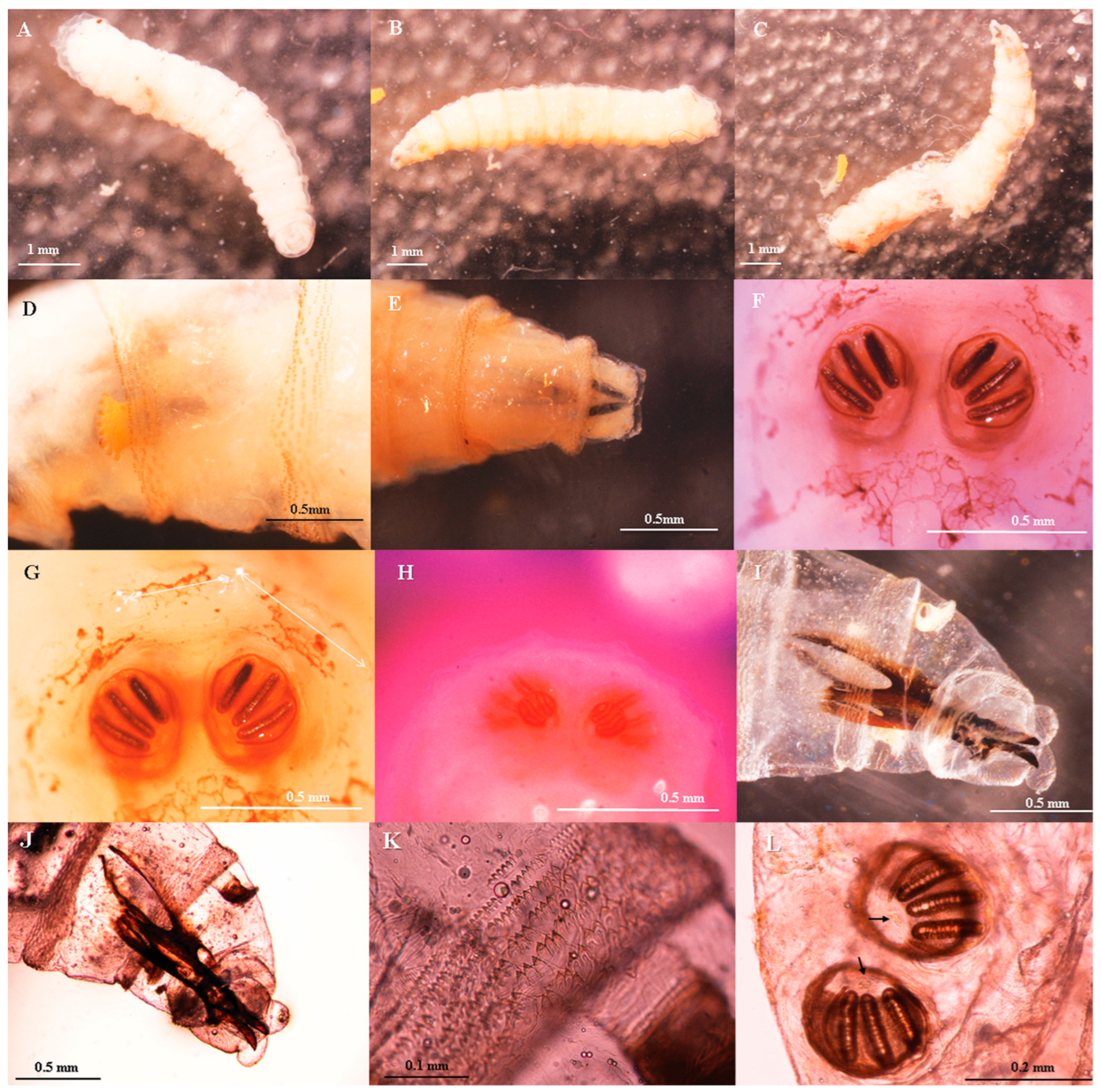
Sixteen larvae were removed from the patient; they were highly mobile and ranged in length from 0.5 to 0.8 mm (0.67 ± 0.16 mm). For morphological identification, five specimens were used, two first-instar larvae and two second-instar larvae, all in the process of molt, with the new respiratory spiracles visible below the current ones. One third-instar larva had molted recently and was soft and slightly sclerotized. The samples were morphologically identified as Lucilia sericata, with an absence of the cephalopharyngeal skeleton’s accessory sclerite, and the anterior spiracles each had eight branches. The posterior spiracles were complete with a peritreme and a button was present, but not evident, with three slits (Figure 1).
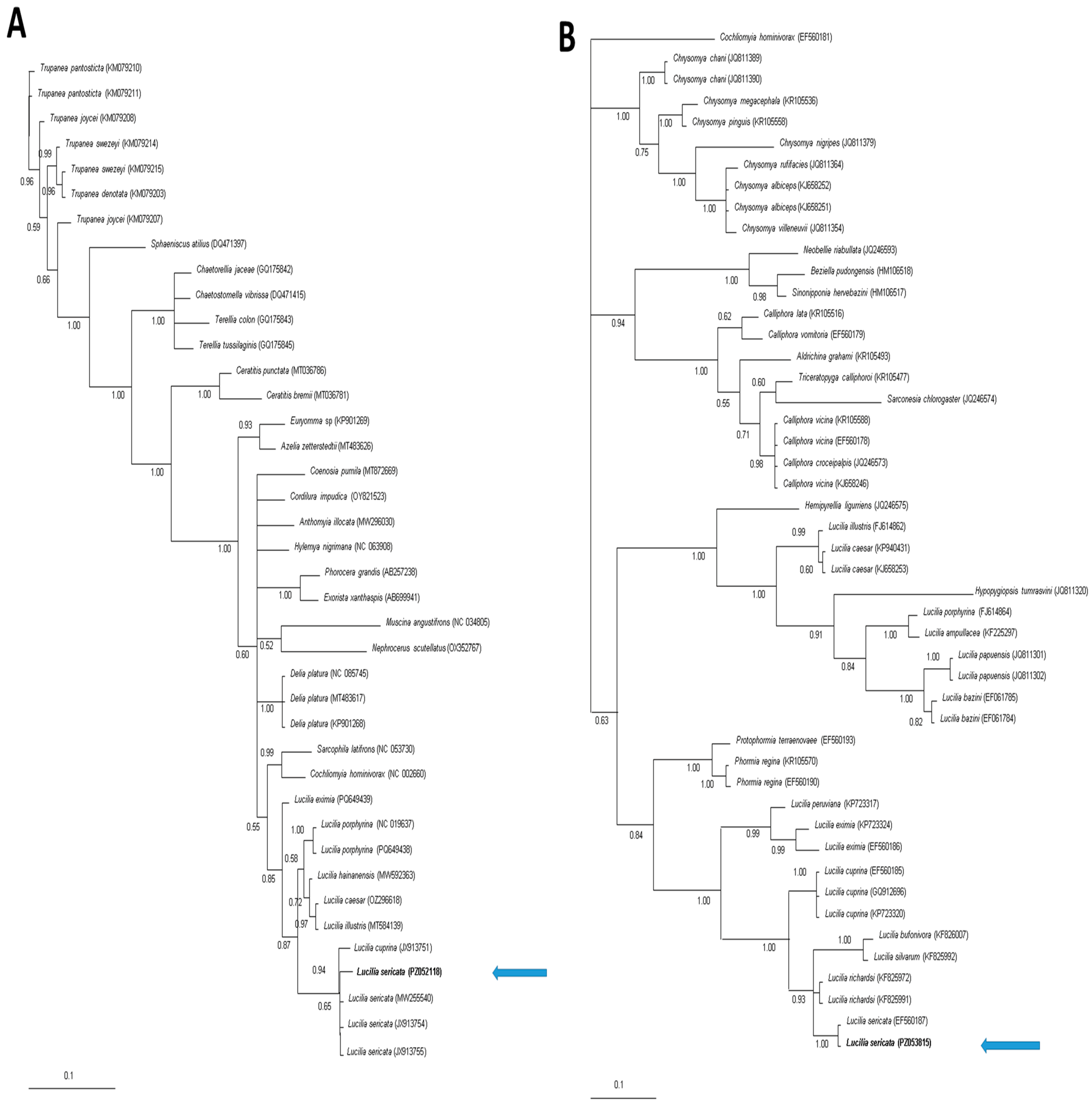
Genetic identity analysis showed values of 99.73% and 100% for PZ052118 and PZ053815, respectively (sequences obtained in the present study), for Lucilia sericata as determined by BLAST (Supplementary Material). The phylogenetic inference for the 16S tree showed a monophyletic profile for the Lucilia genus; the sequence from the patient (PZ052118) was concentrated inside an L. sericata cluster, exhibiting a discreet separation from L. cuprina (Figure 2A). In contrast, ITS-2 showed a paraphyletic profile, sorting among different species by clades with high values of posterior probability support; then, the sequence of L. sericata obtained in the present study (PZ053815) was clustered with other sequences belonging to the same species (EF560187), and grouped near the clade of species belonging to the Luciliiae subfamily (Figure 2B).

4. Discussion

L. sericata is a cosmopolitan synanthropic fly whose larvae are essentially necrophagous and are frequently associated with decomposing tissue; however, there are also reports that it can cause humans and livestock myiasis. In addition, L. sericata larvae have been exploited for many applications, from their use in the management of chronic, non-healing wounds (larval therapy) to their assistance in forensic investigations and analysis to estimate the postmortem interval, where local environmental conditions, as well as diet, can affect their development times [1,3,23].
It is generally accepted that L. sericata completes its life cycle in approximately two to three weeks under temperate conditions (22–25 °C) [14,15,24]. Temperature is a key factor in the life cycle of L. sericata, since dipterans are poikilothermic in nature and therefore their biochemical processes are positively correlated with temperature; thus, a study showed that the development time in hours of L. sericata from egg to 3rd-stage larva at constant temperatures of 16, 19, 22, 25, 28, 31, and 34 °C were 258.0 ± 14.1, 172.8 ± 18.7, 127.8 ± 9.3, 102.8 ± 5.4, 89.6 ± 6.3, 78.95 ± 5.6 and 71.7 ± 2.3 h, respectively. It has been documented that, for larvae of this fly, at temperatures of 9–10 °C, no development is observed; similarly, at a temperature of >35 °C, larval development slows down. Even at 37 °C, few eggs hatch and only 1st- and 2nd-stage larval development has been observed at 12 and 24 h, respectively [23].
In Mexico City, Villeda et al. [25] reported a full development time of 19 days for L. sericata under environmental conditions, with temperature fluctuations ranging from 3.9 to 23.3 °C; while the development from egg to 3rd-stage larvae was completed in 11 days.
The diet of L. sericata larvae is another important factor that affects their development and population dynamics, since in nature they feed mainly on necrotic tissue; laboratory assays have shown that natural or synthetic diets with different sources of carbohydrates and proteins affects their larval development [26,27].
It has been established that nosocomial myiasis is a term used when the infestation affects subjects in hospital settings [10,28]. Furthermore, it has been argued that myiasis is considered nosocomial when the manifestation appears within a few days of hospital admission, as documented in two cases of nasal myiasis caused by L. sericata and L. illustris, both in respiratory-assisted and unconscious patients treated in ICUs [10,29]. In addition, in healthy subjects at a body temperature of 36.6 ± 0.1 °C in a room temperature of 21.8 ± 0.1 °C, their nasal and oral airway mucosal temperature was 35.7 ± 0.2 °C [30].
By integrating this biological data on the maturation time of the L. sericata stages with the patient’s stay in the ICU (17 days), as well as with the identification of larvae of different developmental stages in the patient and the absence of necrotic nasal tissue that had facilitated feeding by the larvae, it suggested that the patient’s infection could have happened before his admission to the hospital; however, the possibility that the infection occurred during his hospitalization cannot be ruled out.
On the other hand, the concern of health authorities regarding the increase in human cases of myiasis caused by C. hominivorax activated a special surveillance system with mandatory reporting of larval infections by this fly at all levels of healthcare. The first human cases were located in a couple of southeastern Mexican states in 2025 [31], and by June 2026, 397 human cases had been documented in 21 states across the country, including Mexico City [32]. Therefore, the present case illustrates that even with an epidemiological alert for myiasis caused by C. hominivorax, the endemic nature of myiasis due to other Diptera in tropical and subtropical regions should not be ruled out.
Although the morphological identification of the larvae should be carried out by expert entomologists, some specimens could exhibit great phenotypic diversity; therefore, confirmation of the morphological identification by molecular techniques is highly recommended, since 27 species of the genus Lucilia have been documented, and sometimes morphological identification can be complicated. Here, the molecular identification by ITS-2 was useful because it showed a clear grouping of L. serica sequences into a specific cluster, strengthening previous observations that ITS-2 loci allow for the clarification of cryptic phylogenetic groupings that cannot be identified with mitochondrial markers [14]; in contrast, since the 16S analysis exhibited a cluster with little separation between L. sericata and L. cuprina, this finding could be explained by the nature of the molecular marker as a maternal lineage tracer; a similar result was observed during the analysis of insects of the genus Triatoma when ITS-2 and 16S loci were compared [17]. Similarly, a study focused on the identification of the family Calliphoridae flies by molecular techniques observed that the combination of mitochondrial and nuclear markers (COI and ITS-2 genes) yielded more accurate identification and better agreement with morphological data than mitochondrial barcodes alone [13].

5. Conclusions

While remaining vigilant for possible human cases of myiasis caused by C. hominivorax (screwworm), here, we confirmed by morphological and molecular analysis the identification of an unexpected case of nasal myiasis caused by Lucilia sericata, demonstrating the importance of care in all aspects of thorough epidemiological surveillance and interdisciplinary management to address these alerts and any related cases.
Furthermore, although the use of mitochondrial markers for molecular barcoding analysis is highly useful for species of the genus Lucilia, ITS-2 appears to be a good second marker that allows for higher resolution and more accurate identification to support the analysis of mitochondrial markers alone.

Supplementary Materials

The following supporting information can be downloaded at: https://www.mdpi.com/article/10.3390/parasitologia6040037/s1. Supplementary Table S1: Identification of the sequences obtained in this study from Lucilia sericata by Blast NCBI.

Author Contributions

Conceptualization, J.P.R.-H., P.M. and F.M.-H.; methodology, N.D.C.-O., L.A.C.-I., L.N.V.-R., P.M.B.-G., N.R., Y.M.-V., M.R.-V., G.V., R.A.-A. and F.M.-H.; software, N.R. and F.M.-H.; validation, J.P.R.-H., R.A.-A., P.M. and F.M.-H.; formal analysis, J.P.R.-H., M.R.-V., G.V., N.R., R.A.-A., P.M. and F.M.-H.; investigation, N.R., Y.M.-V., R.A.-A., P.M. and F.M.-H.; resources, N.R., Y.M.-V., and R.A.-A.; data curation, N.R., Y.M.-V. and R.A.-A.; writing—original draft preparation, M.R.-V., G.V., R.A.-A., P.M. and F.M.-H.; writing—review and editing, J.P.R.-H., N.D.C.-O., L.A.C.-I., L.N.V.-R., P.M.B.-G., N.R., Y.M.-V., M.R.-V., G.V., R.A.-A., P.M. and F.M.-H.; visualization, J.P.R.-H., P.M. and F.M.-H.; supervision, J.P.R.-H., R.A.-A., P.M. and F.M.-H.; project administration, J.P.R.-H., N.D.C.-O., L.A.C.-I., L.N.V.-R., P.M.B.-G., N.R., Y.M.-V., M.R.-V., G.V., R.A.-A., P.M. and F.M.-H.; funding acquisition, R.A.-A. and P.M. All authors have read and agreed to the published version of the manuscript.

Funding

This research received no external funding.

Institutional Review Board Statement

The study was conducted following the Declaration of Helsinki, and the Research Ethics Committee of the “Dr. Manuel Gea González” General Hospital determined that there is no ethical objection to the publication of this case report.

Informed Consent Statement

For all medical procedures performed, written informed consent was obtained from legal representatives of the patient. Written informed consent for publication is not applicable, since this manuscript does not contain any individual personal data and/or media.

Data Availability Statement

All relevant data are contained within the article.

Conflicts of Interest

The authors declare no conflicts of interest.

Abbreviations

The following abbreviations are used in this manuscript:
16S16S mitochondrial rRNA gene
ITS-2Internal Transcribed Spacer 2
ICUintensive care unit
PCRpolymerase chain reaction

References

  1. Villalobos, G.; Vega-Memije, M.E.; Maravilla, P.; Martinez-Hernandez, F. Myiasis caused by Dermatobia hominis: Countries with increased risk for travelers going to neotropic areas. Int. J. Dermatol. 2016, 55, 1060–1068. [Google Scholar] [CrossRef] [PubMed]
  2. Jallow, B.J.J.; Gassara, G.; Bajinka, O.; Luo, Y.; Liu, M.; Cai, J.; Huang, J.; Meng, F. Human myiasis in Sub-Saharan Africa: A systematic review. PLoS Negl. Trop. Dis. 2024, 18, e0012027. [Google Scholar] [CrossRef] [PubMed]
  3. Robbins, K.; Khachemoune, A. Cutaneous myiasis: A review of the common types of myiasis. Int. J. Dermatol. 2010, 49, 1092–1098. [Google Scholar] [CrossRef] [PubMed]
  4. Rodriguez-Vivas, R.I.; Cutolo, A.A.; Barros, A.T.M.; Cuore, U.D.; Molento, M.B.; López-Osorio, S.; Rodrigues, D.S.; Spina, M.; Borges, F.A.; Lopes, W.D.Z.; et al. Management Practices for the Control of Haematobia irritans, Dermatobia hominis, and Cochliomyia hominivorax in Cattle Across Latin America: A Sustainable, Collective Approach. Pathogens 2026, 15, 177. [Google Scholar] [CrossRef] [PubMed]
  5. Government of Mexico, Secretariat of Health. Available online: https://www.gob.mx/cms/uploads/attachment/file/992853/Aviso_epidemiologico_Miasis_por_Cochliomyia_hominivorax_25.04.2025_final.pdf (accessed on 3 March 2026). (In Spanish)
  6. Government of Mexico, Secretariat of Health. Available online: https://www.gob.mx/cms/uploads/attachment/file/1058788/sem06.pdf (accessed on 3 March 2026). (In Spanish)
  7. Nigam, Y.; Dudley, E.; Bexfield, A.; Bond, E.; Evans, J.; James, J. Chapter 2. The Physiology of Wound Healing by the Medicinal Maggot, Lucilia sericata. In Advances in Insect Physiology; Simpson, S.J., Ed.; Academic Press: Cambridge, MA, USA, 2010; Volume 39, pp. 39–81. [Google Scholar]
  8. Kim, J.S.; Seo, P.W.; Kim, J.W.; Go, J.H.; Jang, S.C.; Lee, H.J.; Seo, M.A. A nasal myiasis in a 76-year-old female in Korea. Korean J. Parasitol. 2009, 47, 405–407. [Google Scholar] [CrossRef] [PubMed]
  9. Babamahmoudi, F.; Rafinejhad, J.; Enayati, A. Nasal myiasis due to Lucilia sericata (Meigen, 1826) from Iran: A case report. Trop. Biomed. 2012, 29, 175–179. [Google Scholar] [PubMed]
  10. Martínez-Rojano, H.; Noguez, J.C.; Huerta, H. Nosocomial Myiasis Caused by Lucilia sericata (Diptera: Calliphoridae) and Neonatal Myiasis by Sarcophaga spp. (Diptera: Sarcophagidae) in Mexico. Case Rep. Infect. Dis. 2018, 2018, 5067569. [Google Scholar] [CrossRef] [PubMed]
  11. Mendonça, P.M.; Cortinhas, L.B.; Garção-Neto, C.H.; Queiroz, M.M.d.C. Brief Review of Morphological Characters in the Identification of Muscomorpha (Diptera) of Sanitary and Forensic Importance. Diversity 2024, 16, 599. [Google Scholar] [CrossRef]
  12. Bhuvaneshwaran, S.; Padmanaban, V.S.; Radja, R.D.; Anandan, G.; Venkatesan, S.; Semalaiyappan, J.; Kumar, A.; Kuttiatt, V.S. Molecular identification of immature stages of medically important fly species, Puducherry, South India: A preliminary study. Front. Insect Sci. 2025, 5, 1551807. [Google Scholar] [CrossRef] [PubMed]
  13. Yusseff-Vanegas, S.; Agnarsson, I. DNA-barcoding of forensically important blow flies (Diptera: Calliphoridae) in the Caribbean Region. Peer J. 2017, 5, e3516. [Google Scholar] [CrossRef] [PubMed]
  14. Szpila, K. Key for the Identification of Third Instars of European Blowflies (Diptera: Calliphoridae) of Forensic Importance. In Current Concepts in Forensic Entomology; Amendt, J., Goff, M.L., Campobasso, C.P., Grassberger, M., Eds.; Springer: Dordrecht, The Netherlands, 2010; pp. 43–56. [Google Scholar]
  15. Szpila, K.; Hall, M.J.R.; Pape, T.; Grzywacz, A. Morphology and identification of: 1rst instars of the European and Mediterranean blowies of forensic importance. Part II. Luciliinae. Med. Vet. Entomol. 2013, 27, 349–366. [Google Scholar] [PubMed]
  16. Martínez-Hernández, F.; Villalobos, G.; Montañez-Valdez, O.D.; Martínez-Ibarra, J.A. New finding of peridomestic Triatoma infestans (Klug) 1834 (Hemiptera: Reduviidae) in Mexico: Molecular approach using cytochrome oxidase I and cytochrome B. Infect. Genet. Evol. 2022, 97, 105187. [Google Scholar] [PubMed]
  17. Martínez-Hernández, F.; Villalobos, G.; Montañez-Valdez, O.D.; Martínez-Ibarra, J.A. A New Record of the Introduced Species Triatoma infestans (Hemiptera: Reduviidae) in Mexico. J. Med. Entomol. 2022, 59, 2150–2157. [Google Scholar] [CrossRef] [PubMed]
  18. Marcilla, A.; Bargues, M.D.; Ramsey, J.M.; Magallon-Gastelum, E.; Salazar-Shettino, P.M.; Abad-Franch, F.; Dujardin, J.P.; SchoWeld, C.J.; Mas-Coma, S. The ITS-2 of the nuclear rDNA as a molecular marker for populations, species, and phylogenetic relationships in Triatominae (Hemiptera: Reduviidae), vector of Chagas disease. Mol. Phylogenet. Evol. 2001, 18, 136–142. [Google Scholar] [CrossRef] [PubMed]
  19. Thompson, J.D.; Higgins, D.G.; Gibson, T.J. CLUSTAL W: Improving the sensitivity of progressive multiple sequence alignment through sequence weighting, position-specific gap penalties and weight matrix choice. Nucleic Acids Res. 1994, 22, 4673–4680. [Google Scholar] [CrossRef] [PubMed]
  20. Edgar, R.C. MUSCLE: Multiple sequence alignment with high accuracy and high throughput. Nucleic Acids Res. 2004, 32, 1792–1797. [Google Scholar] [CrossRef] [PubMed]
  21. Kumar, S.; Stecher, G.; Tamura, K. MEGA7: Molecular evolutionary genetics analysis version 7.0 for bigger datasets. Mol. Biol. Evol. 2016, 33, 1870–1874. [Google Scholar] [CrossRef] [PubMed]
  22. Ronquist, F.; Teslenko, M.; van der Mark, P.; Ayres, D.L.; Darling, A.; Höhna, S.; Bret Larget, B.; Liu, L.; Suchard, A.; Huelsenbeck, J.P. MrBayes 3.2: Efficient Bayesian phylogenetic inference and model choice across a large model space. Syst. Biol. 2012, 61, 539–542. [Google Scholar] [CrossRef] [PubMed]
  23. Pruna, W.; Guarderas, P.; Donoso, D.A.; Barragán, A. Life cycle of Lucilia sericata (Meigen 1826) collected from Andean mountains. Neotrop. Biodivers. 2019, 5, 3–9. [Google Scholar] [CrossRef]
  24. Wang, M.; Wang, Y.; Hu, G.; Wang, Y.; Xu, W.; Wu, M.; Wang, J. Development of Lucilia sericata (Diptera: Calliphoridae) Under constant temperatures and its significance for the estimation of time of death. J. Med. Entomol. 2020, 57, 1373–1381. [Google Scholar] [CrossRef] [PubMed]
  25. Villeda-Callejas, M.P.; Jacinto-Estanes, L.J.; Barrera-Escorcia, H.; Lara- Vázquez, A.; Guedea-Fernandez, D.; Flores-Maya, S. Life cycle of Lucilia sericata (Meigen, 1826) and Calliphora latifrons (Hough, 1899) (Diptera: Calliphoridae). Entomol. Mex. 2015, 2, 126–131. [Google Scholar]
  26. Pinilla, T.; Cuña, Y.; Cortes, D.; Diaz, A.; Segura, A.; Bello, F.J. Characteristics of the biological cycle of Lucilia sericata (Meigen, 1826) (Diptera: Calliphoridae) on different diet. Rev. UDCA Actual. Divulg. Científica 2010, 13, 153–161. [Google Scholar] [CrossRef]
  27. Daniels, S.; Simkiss, K.; Smith, R.H. A simple larval diet for population studies on the blowfly Lucilia sericata (Diptera: Calliphoridae). Med. Vet. Entomol. 1991, 5, 283–292. [Google Scholar] [CrossRef] [PubMed]
  28. Francesconi, F.; Lupi, O. Myiasis. Clin. Microbiol. Rev. 2012, 25, 79–105. [Google Scholar] [CrossRef] [PubMed]
  29. Szakacs, T.A.; MacPherson, P.; Sinclair, B.J.; Gill, B.D.; McCarthy, A.E. Nosocomial myiasis in a Canadian intensive care unit. Can. Med. Assoc. J. 2007, 177, 719–720. [Google Scholar] [CrossRef]
  30. Khosravi, M.; Lin, R.L.; Maskey, A.P.; Pandey, S.; Lin, A.H.; Lee, L.Y. A Distinct Difference Between Air and Mucosal Temperatures in Human Respiratory Tract. Front. Med. 2021, 8, 650637. [Google Scholar] [CrossRef]
  31. Government of Mexico, Secretariat of Health. Epidemiological Surveillance Week 22, 2025. 9 June 2025. Available online: https://www.gob.mx/cms/uploads/attachment/file/1001825/sem22.pdf (accessed on 3 March 2026). (In Spanish)
  32. Government of Mexico, Secretariat of Health. Epidemiological Surveillance Week 21, 2026. 8 June 2026. Available online: https://www.gob.mx/salud/documentos/boletinepidemiologico-sistema-nacional-de-vigilancia-epidemiologica-sistema-unico-de-informacion-417103 (accessed on 10 June 2026). (In Spanish)
Figure 1. Observations under a stereoscope microscope of the larvae extracted from the patient. (A) Larvae 1st instar molt. The exuviae are observed as a transparent outer layer. (B) Larvae 2nd instar molt. (C) Larvae 3rd instar recent molting. Even without being fully sclerosed, the body is soft and fragile. (D) Detail anterior spiracles showing a fan-like structure with 8 lobes. (E) Dorsal view of cephalopharyngeal skeleton without accessory sclerite of 2nd instar. (F) Detail of posterior spiracles of 3rd instar larvae, showing a peritreme. (G) Detail of posterior spiracles of 3rd instar larvae, the arrows indicate tubercles and distances between them. (H) Detail posterior spiracles 2nd instar larvae. Two pairs of spiracles are observed, due to the molting process the larvae were in. Observation under light microscope. (I) Detail of cephalopharyngeal skeleton in 3rd instar larvae (in negative). (J) Same as I (in positive). (K) Spines between the first and second thoracic segments of 3rd instar larvae. (L) Posterior spiracles of 3rd instar larvae; the arrows indicate the button.
Parasitologia 06 00037 g001
Figure 2. Bayesian phylogenetic tree constructed with (A) 16S and (B) ITS-2 sequences from Diptera species causing myiasis in the world. The numbers at the nodes indicate Bayesian posterior probabilities. Data sequences were obtained from the GenBank database; the sequences obtained in this study are shown in bold and by an arrow.
Parasitologia 06 00037 g002
Disclaimer/Publisher’s Note: The statements, opinions and data contained in all publications are solely those of the individual author(s) and contributor(s) and not of MDPI and/or the editor(s). MDPI and/or the editor(s) disclaim responsibility for any injury to people or property resulting from any ideas, methods, instructions or products referred to in the content.