Review

Human Anisakidosis with Intraoral Localization: A Narrative Review

by
Stylianos Papadopoulos
1,
Vasileios Zisis
1,2,*,
Konstantinos Poulopoulos
1,
Christina Charisi
1 and
Athanasios Poulopoulos
1
1
Department of Oral Medicine/Pathology, School of Dentistry, Aristotle University of Thessaloniki, 54124 Thessaloniki, Greece
2
Department of Oral Medicine-Oral Pathology, School of Dentistry, European University of Cyprus, 2404 Nicosia, Cyprus
*
Author to whom correspondence should be addressed.
Parasitologia 2025, 5(3), 41; https://doi.org/10.3390/parasitologia5030041
Submission received: 4 June 2025 / Revised: 11 July 2025 / Accepted: 31 July 2025 / Published: 4 August 2025

Abstract

Objectives: Anisakidosis is an emerging, cosmopolitan, and underdiagnosed parasitic disease caused by the accidental ingestion of third-stage anisakid larvae when consuming raw or improperly prepared seafood. Within hours to days of consuming infected raw seafood, patients may develop acute gastrointestinal symptoms including pain, nausea, vomiting, diarrhea and/or constipation, as live anisakid larvae attach to the gastric, or more rarely, the intestinal mucosa. Cases have been reported in which the nematodes succeed at migrating from the stomach upwards to the esophagus and then the oral cavity. Therefore, the purpose of the present literature review is to collect, analyze, summarize and present the relevant epidemiological, clinical, diagnostic, parasitological, therapeutic, and prognostic data concerning anisakidosis localized inside the oral cavity. Methods: An electronic search of the PubMed, Scopus, and Ovid databases was performed with them being accessed for the last time on 29 March 2025. Results: The present literature review identified 13 individual case reports of oral mucosa anisakidosis, which were published in the period 1971–2022. Conclusions: Our review aims to summarize the relevant epidemiological, clinical, diagnostic, parasitological, therapeutic, and prognostic data regarding the oral localization of anisakidosis, a helminthic infection caused by the accidental ingestion of live anisakid larvae and which manifests mainly with gastrointestinal symptoms. Its localization in the oral mucosa appears to be exceptionally rare and, in most cases, occurs with a characteristic clinical picture, defined by the onset of acute mouth or throat pain immediately after the consumption of raw seafood and by the observation of one or more larvae, either lying on or penetrating the oral mucosa. Despite its rarity, dental health professionals and other clinicians should be aware of this disease and the possibility of its intraoral localization, since environmental factors on the one hand, and the adoption of foreign dietary habits on the other, will likely make anisakidosis a much more common disease worldwide in the near future.

1. Introduction

Anisakidosis is an emerging, cosmopolitan, and underdiagnosed parasitic disease caused by the accidental ingestion of third-stage (L3) anisakid larvae when consuming raw or improperly prepared seafood [1]. The etiological agents of this infection are nematodes of the family Anisakidae (Railliet & Henry, 1912), belonging to the genera Anisakis (Dujardin, 1845), Pseudoterranova (Mozgovoi, 1951), and Contracaecum (Railliet & Henry, 1912) [2]. Hysterothylacium spp. (Ward & Magath, 1917), now classified in the family Raphidascarididae (Railliet& Henry, 1915) [3], are closely related to Anisakidae [4], and the infections they cause have also been defined as anisakidosis [2].
The life cycle of Anisakidae is complex and includes definitive, intermediate, and paratenic hosts [5,6]. Traditionally, the definitive hosts of Anisakis spp. are thought to be cetaceans, while those of Pseudoterranova spp. are thought to be pinnipeds [7,8]. The definitive hosts of Contracaecum spp. are marine mammals as well as piscivorous birds [7,9]. Larvae mature in the gastric mucosa of the definitive hosts, and the adult female nematodes produce unembryonated eggs which are expelled into the water along with the feces of the host. The eggs then become embryonated and hatch in the water, and the larvae are ingested by small crustaceans such as krill and copepods, which constitute the intermediate hosts [6]. Fish or cephalopods, the paratenic hosts, consume the aforementioned small crustaceans, and the parasite’s life cycle is completed when the paratenic host is in turn eaten by a marine mammal [5,6,7,8]. It should be noted that recent studies indicate a host specificity of Anisakidae much broader than previously thought, with Anisakis larvae having been identified in unusual intermediate hosts, including birds (the grey petrel and the little penguin), sharks (the spinner shark), and reptiles (the blue-lipped sea krait). Additionally, in birds, the maturation of larvae from L3 stage to L4 has also been observed [2,10].
Humans might become accidental hosts of Anisakidae by consuming infected fish or cephalopods such as squid and cuttlefish, which are either raw or improperly prepared and cooked [1,7,8]. The consumption of dishes containing raw or intentionally undercooked seafood as a delicacy is part of the gastronomic tradition of a multitude of cultures on all continents, and due to globalization, it is becoming increasingly common even in countries where it is not the norm [11]. Anisakidae exhibit low host specificity for all hosts involved in their life cycle; therefore, some of the most commonly consumed fish and cephalopods can be infected [12,13,14]. Infection rates of seafood depend on multiple factors such as the place of origin and season, with up to 100% of examined fish having been parasitized in some cases [15]. The aforementioned observations, on the one hand, set human anisakidosis apart from most other parasitic diseases, which are endemic only to certain geographical areas [16], and on the other, make this disease most common in countries and regions of the world where consuming raw or undercooked seafood is part of the local culinary tradition, such as in parts of East Asia, South America, and Europe [12].
The first case of human anisakidosis was probably documented by Leuckart in 1867, who attributed gastrointestinal symptoms in a Greenlandic child to infection by an askarid-like nematode, which he named Askaris maritima (Leuckart 1876) [17]. Nevertheless, Anisakidae were identified as human pathogens for the first time in the 1950s and 1960s in Japan and the Netherlands [2], and to date, human anisakidosis has been reported in 34 countries, with most cases occurring, in descending order, in Japan, Spain, Korea, Italy, and the USA [2].
Within hours to days of consuming infected raw seafood, patients may develop acute gastrointestinal symptoms including pain, nausea, vomiting, diarrhea, and/or constipation, as live anisakid larvae attach to the gastric, or more rarely, the intestinal mucosa [1]. Clinical presentation is of varying severity, ranging from asymptomatic and mildly symptomatic cases to cases requiring emergency care [2,5]. Larvae die quickly inside the human body before reaching maturity [12], and the infection either resolves without treatment, or it becomes chronic [18]. Chronic gastrointestinal anisakidosis is characterized by eosinophilic infiltration and granuloma formation around the encapsulated dead and degenerating larva or larvae, and it can last for months or even years [1,12] until it causes symptoms suggestive of acute appendicitis or intestinal obstruction [1].
In rare cases, associated with Anisakis spp. and Pseudoterranova spp. [1,2,19], larvae succeed at penetrating the gastric or intestinal mucosa and migrate to other parts of the body, causing a form of anisakidosis called ectopic or extraintestinal [1]. The mesentery, omentum, pleural cavity, lungs, parametrium, uterus, ovaries, liver, pancreas, and other organs may be affected [12,20], and symptoms depend on the affected organ and may mimic those of other diseases [1,20].
Anisakid worms contain up to 22 proteins which can cause an IgE-mediated allergic reaction in sensitized patients [5,21], with symptoms ranging from urticaria and oral allergy syndrome, to life threatening anaphylaxis [5]. Allergic and gastrointestinal anisakidosis can coexist [1,5].
Treatment of anisakidosis is mainly based on the removal of the larva or the larvae responsible for the infection. Traditional broad-spectrum antihelminthic agents such as benzimidazoles or ivermectin are not considered completely effective [1,8,12]. Since anisakid larvae eventually die and degenerate inside the human body, certain cases of gastrointestinal anisakidosis can successfully be managed through conservative and supportive therapy [22,23]. The United States Centers for Disease Control and Prevention recommends that to eliminate live anisakid parasites, seafood should be frozen at a temperature below−20 °C for at least 7 days, or at a temperature below −35 °C until solid, and then stored either at the same temperature for a minimum of 15 h, or at a temperature below −20 °C for a minimum of 24 h [8].
Cases of Anisakis spp. and Pseudoterranova spp. infections have been reported, in which the nematodes succeed at migrating from the stomach upwards to the esophagus and then the pharynx, where they cause a tingling sensation and aresubsequently coughed, vomited, or passively eliminated through the patient’s mouth [24,25]. The aforementioned observation, in addition to the fact that the oral cavity constitutes the upper part of the gastrointestinal tract, raises the question of whether anisakid larvae, either by upward migration from the stomach, after penetration of the gastric/intestinal mucosa, or directly after ingestion, can reside on or even invade the oral mucosa and cause symptoms. Therefore, the purpose of the present review is to collect, analyze, summarize, and present the relevant epidemiological, clinical, diagnostic, parasitological, therapeutic, and prognostic data concerning anisakidosis localized inside the oral cavity.

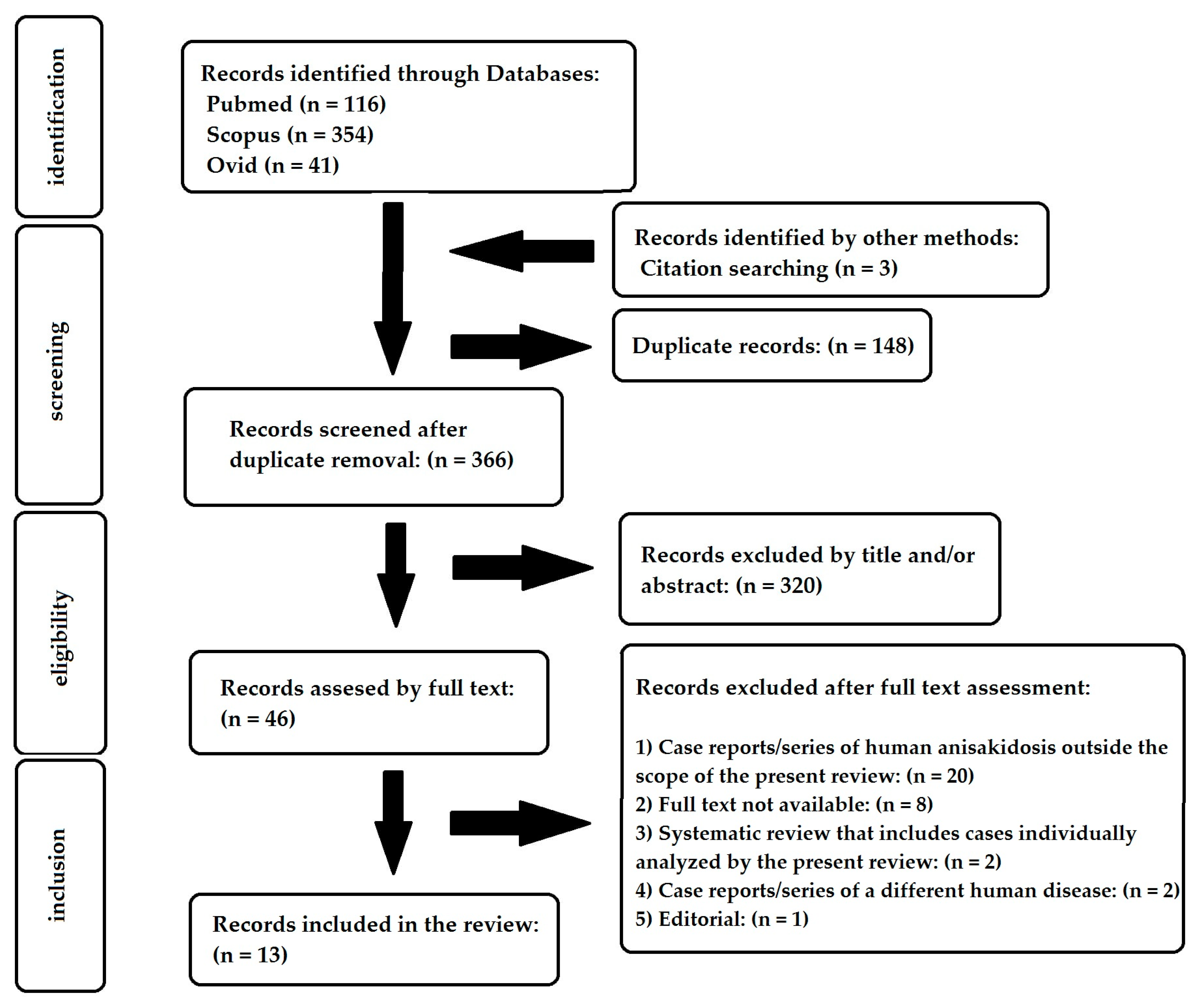
2. Materials and Methods

2.1. Study Eligibility

The literature review included publications that described cases of human anisakidosis with intraoral localization. In that sense, other types of articles such as scoping/narrative reviews, experimental studies, clinical trials, studies concerning other species, etc., were excluded, as were publications in which human anisakidosis was not localized inside the mouth. Cases of allergic anisakidosis/allergy to Anisakis, or cases in which the coughing, vomiting, or passive elimination of larvae through the patients’ mouth was the sole reported manifestation of the infection’s oral cavity involvement, were not within the scope of the present review. No limitation was placed on the included studies’ year of publication or language.

2.2. Search Strategy

A search was performed in PubMed, Scopus, and Ovid databases, with them being accessed for the last time on 29 March 2025.
In PubMed, the following algorithm, which utilized relevant keywords, MeSH terms, and the Boolean operators “OR” and “AND”, was used, and it retrieved 116 records:
((anisakis[MeSH Terms]) OR (anisakiasis[MeSH Terms]) OR (anisakidae) OR (anisakis) OR (anisakiasis) OR (anisakiosis) OR (anisakidosis) OR (pseudoterranova) OR (contracaecum) OR (hysterothylacium) OR (herringworm) OR (codworm) OR (sealworm)) AND ((mouth[MeSH Terms]) OR (mouth mucosa[MeSH Terms]) OR (cheek[MeSH Terms]) OR (tongue[MeSH Terms]) OR (palate[MeSH Terms]) OR (palatine tonsil[MeSH Terms]) OR (lip[MeSH Terms]) OR (mouth floor[MeSH Terms]) OR (oropharynx[MeSH Terms]) OR (gums[MeSH Terms]) OR (oral) OR (intraoral) OR (perioral) OR (oral cavity) OR (oral mucosa) OR (mouth) OR (bucc*) OR (cheek) OR (tongue) OR (gloss*) OR (ling*) OR (palate*) OR (tonsil*) OR (lip*) OR (labia*) OR (mouth floor) OR (sublingual*) OR (oropharynx*) OR (gingiva*) OR (gum*))
Initially, Scopus was accessed using the algorithm below:
((anisakidae) OR (anisakis) OR (anisakiasis) OR (anisakiosis) OR (anisakidosis) OR (pseudoterranova) OR (contracaecum) OR (hysterothylacium) OR (herringworm) OR (codworm) OR (sealworm)) AND ((oral) OR (intraoral) OR (perioral) OR (oral cavity) OR (oral mucosa) OR (mouth) OR (bucc*) OR (cheek) OR (tongue) OR (gloss*) OR (ling*) OR (palate*) OR (tonsil*) OR (lip*) OR (labia*) OR (mouth floor) OR (sublingual*) OR (oropharynx*) OR (gingiva*) OR (gum*))
However, since an enormous number of records (3754) were retrieved, it was decided to limit the search only to records in which the algorithm keywords were present in their title, abstract, or keywords. The same issue appeared when accessing the Ovid database, so the same limitation was placed. Finally, 345 and 41 publications were retrieved from Scopus and Ovid, respectively.
After duplicate removal, title and abstract screening, and assessment of full text for eligibility (Illustration 1), 10 studies were included in the present review. Additionally, diligent forward and backward citation search of the literature identified 3 more relevant records, which raised the total number of studies included in the present review to 13 (Figure 1).

2.3. Data Extraction

Data from the aforementioned selected publications was extracted, entered in a Microsoft Excel file, and double-checked. The collected data concerned the following parameters: study publication year, country the case was observed in, patient age, patient sex, history of raw or undercooked seafood consumption, anatomical localization of the infection, patient’s main complaint and clinical presentation, time between seafood consumption and symptom onset, time between symptom onset and medical consultation, imaging tests, laboratory tests, larval species, number of larvae, larvae degree of maturation, larvae state of preservation, method of larvae identification, the involvement of other organs, treatment, recovery, follow-up, and possible complications.

3. Results

3.1. Included Studies and Epidemiological Data

The present review identified 13 individual case reports of oral mucosa anisakidosis, which were published in the period 1971–2022 (Table 1). The majority (6/13) of cases were reported in Asia, with 5 cases in Japan, 5 in Korea, and 1 in Oman. One case was reported in Europe, specifically in Italy, and one in South America, specifically in Chile. The sex and age of patients arereported in all 13 cases. Females accounted for 61.5% (8/13) of the patients. The age of patients ranged from 6 to 69 years, with a median age of 39 (Table 1).
In 12/13 cases, fish or cephalopods were prepared and eaten either at home or in an establishment near the patient’s place of residence (Table 1). In only one case, diagnosed in Italy, the patient reported that she had consumed raw fish during a trip toJapan [20].
The patients’ medical history and general condition were mentioned in 5/13 cases. In three cases, patients reported no past medical history [20,32,34]. In one out of five cases, the patient was an underweight 6-year-old child [29], and in one out of five cases, it was mentioned that the 69-year-old patient had hypertension and hyperlipidemia, but it was not mentioned whether or not she was taking medication [36].

3.2. Anatomical Localization and Clinical Presentation

The palatine tonsil was the most common site of infection, with 5/13 cases being localized there. In 3/13 cases the buccal mucosa was involved, either alone or in addition to the labial or the lingual mucosa. The soft palate and the tongue were affected each in 2/13 cases, while in 1/13 cases, the precise anatomical localization of infection within the oral cavity was not specified (Table 1).
In 12/13 cases, patients associated the onset of their symptoms with the consumption of raw or undercooked seafood (Table 1). In one case, although the 6-year-old patient regularly consumed fish and prawns as part of her diet, neither had she ever consumed raw or undercooked seafood, nor was seafood consumption temporally related to the appearance of her symptoms, so her infection was instead attributed to the inadequate cooking of seafood [29].
The time between the consumption of raw seafood and the onset of symptoms was mentioned in 10/13 cases (Table 2). In 9/10 cases symptoms appeared immediately, or within minutes of eating infected fish or cephalopods. In 1/10 cases their onset was delayed and started 4 days later. Furthermore, it wasmentioned that in 7/10 cases, patients sought medical consultation within hours or a few days from symptom onset, with only one patient waiting for 2 months (Table 2).
The patient’s main complaint or reason for consultation was mentioned in all 13 cases (Table 2). The most common symptom, reported in8/13 cases, was oral or pharyngeal pain, characterized by acute onset and intense severity. Other reported symptoms were foreign body sensation and throat irritation, each present in 3/13 cases, as well as sticking sensation in the mouth, throat blockage sensation, swallowing difficulty, skin rash, and foreign body observation, each reported in 1/13 cases. In one case, the patient complained of symptoms suggestive of chronic recurrent tonsillitis and adenoiditis, but those symptoms were not specified.

3.3. Diagnosis

The findings of the clinical or endoscopic visual examination of the affected tissues were discussed in 10/13 cases (Table 2). In 9/10 cases, one or more larvae were observed, and in 4 of those cases, larval penetration of the mucosa was observed. The presence of the worm or worms was accompanied by edema in 2/10 cases, by erythematous and edematous mucosal lesions in 2/10 cases, and by bilateral tonsil enlargement in 1/10 cases. In the remaining 1/13 case, no worms were observed, but a submucosal nodule could be palpated in the buccal mucosa, beneath an inflammatory edematous lesion.
The utilization of laboratory tests, such as complete blood count (CBC), inflammatory markers such as C-reactive protein (CRP),erythrocyte sedimentation rate (ESR), and antibody serology, was reported in 8/13 cases. In three out of eight cases, all laboratory investigations were reported to be normal, with no further details on the tests performed [20,29,31]. Laboratory test results of the remaining fivecases are shown in Table 3.
Imaging was used in only 1/13 case, which presented with a painful submucosal nodule in the buccal mucosa. In that case, Ultrasonography (USG) and Magnetic Resonance Imaging (MRI) without contrast were both utilized for the investigation of the lesion [20]. Ultrasonography revealed a hypoechoic cystic lesion with peripheral vascularization, within which linear hyperechoic structures were observed. MRI, on the other hand, showed a round cystic nodular lesion with homogenous walls and liquid content [20].

3.4. Parasitological Findings

The etiological agent responsible for the infection was reported to belong to genus Anisakis in 10/13 cases, with 2 cases specifying it as Anisakis simplex, without further specifying whether it belonged to the Anisakis simplex complex, or to Anisakis simplex sensu stricto. In the remaining 3/13 cases, the infection was caused by a Pseudoterranova spp. larva, which in one case was specified as belonging to the species Pseudoterranova azarasi (Table 4). The number of larvae responsible for the infection was mentioned in 12/13 cases (Table 4). In the majority (10/12) of cases, a single larva was observed in the oral cavity, and in the remaining two cases, 8 and 20 larvae were removed from the affected tissues, respectively. In 8/13 cases, the larva or larvae were alive and moving at the time of observation and removal (Table 4). Additionally, all three larvae belonging to genus Pseudoterranova were characterized as fourth-stage (L4) larvae.
The method of parasite identification was mentioned in 9/13 cases (Table 4). In 8/13 cases, it consistedinmicroscopic identification of the nematode genus and/or species, while in 1/13 cases, identification was performed by polymerase chain reaction (PCR).

3.5. Involvement of Other Organs

Investigation of concurrent anisakidosis of other organs wasdiscussed in 5/13 cases. In two patients, endoscopy of the entirety, or part of the gastrointestinal tract was performed [27,34]; in one patient, a full-body Computed Tomography (CT) scan was performed [20], and in one patient, only stool analysis was performed [29]. In one patient, the investigation included chest and abdomen CT scan, endoscopy, and stool analysis [32]. In four out of the five/5 aforementioned patients, no signs of gastrointestinal or ectopic anisakidosis were detected [20,29,32,34], while in one case, additional larvae were found in the central portion of the patient’s esophagus [27].

3.6. Treatment and Prognosis

Details regarding treatment are mentioned in all 13 cases (Table 5). Larva or larvae removal by forceps, either directly, or endoscopically, was performed in 10/13 cases. In addition, due to significant laryngeal edema, one patient received intravenous antibiotics and corticosteroids after the removal of the nematode from his tongue. The remaining 3/13 cases were treated surgically, as the direct removal of the larva or larvae was not possible, either due to encapsulation of the larvae, or due to their deep localization in the tissues.
Patient recovery was discussed in 11/13 cases and wasconsidered good in all of them (Table 5). In 10/11 cases, patients were relieved of symptoms either immediately, or a few days after larval removal. Long term (>1 month) follow-up was mentioned in four cases. Follow-up time ranged from 1 to 12 months after larval removal or surgery, with the median time being 4 months. All four patients were completely asymptomatic at follow-up.

4. Discussion

The oral cavity represents an anatomical site where helminthic infections caused by species of roundworms or tapeworms may occasionally develop. These infections occur predominantly in endemic countries and regions and typically present as lesions resembling a benign tumor, a mucocele, or a traumatic lesion [37].
Anisakidosis affecting the oral mucosa appears to be exceptionally rare, although it is likely an underdiagnosed and underreported condition, as appears to be the case with anisakidosis in general [1,2], although the oral localization itself could offer the possibility of earlier diagnosis compared to other forms of anisakidosis. This becomes evident by the fact that although oropharyngeal anisakidosis manifesting as “tingling throat syndrome” has been known for some time [24,25], the present review identified only 13 records, all of which were case reports of individual cases of oral anisakidosis. In 2021, Suzuki et al., in addition to presenting a case of Pseudoterranovosis of the base of the tongue that they observed, provided a review of eight cases of oral anisakidosis from the world literature. Yet, the present review is, to the best of our knowledge, the first review of anisakidosis with oral mucosa localization, and it attempted to exhaustively collect and analyze the complete epidemiological, clinical, diagnostic, parasitological, therapeutic, and prognostic data concerning the aforementioned disease.
In the majority (8/13) of cases included in the present review, patients presented with acute throat or mouth pain, which appeared shortly after eating raw or undercooked seafood. In 9/13 cases the diagnosis of helminthic infection could be reached immediately, as one or more larvae were observed, either directly or endoscopically. Two cases in particular require further comment. In the first case, reported by Bhargava et al. in 1996 [29], a six-year-old girl presented with symptoms suggestive of chronic recurrent tonsillitis. Histopathology after tonsillectomy revealed crosssections of a dead and degenerating anisakid nematode in the tonsillar parenchyma. There was no eosinophilic infiltration or granuloma formation, and the authors argue that the presence of the larva might have been incidental and completely unrelated to the clinical symptoms. The second case, reported by Iacomino et al. in 2020 [20], is of great interest, as on the one hand, pathologically, it resembled chronic gastrointestinal anisakidosis [1,12], and on the other hand, clinically and ultrasonographically, it resembled more common parasitic diseases of the oral cavity such as filariasis and cysticercosis, which are characterized by worm encapsulation and granuloma formation [37]. The evolution from an acute to a chronic inflammatory process couldbe attributed to the long period of time (2 months) between the onset of the symptoms and medical consultation. This wasnot the case for the other cases included in the present review, since the acute pain experienced by patients most likely prompted them to immediately seek medical attention.
Anisakid larvae appear to reach the oral mucosa, either directly after ingestion, or less often, through upward migration from the stomach [19,32]. In the present review, 10/13 cases were attributed to Anisakis spp. and 3/13 to Pseudoterranova spp. In the international literature, Anisakis spp. nematodes appear to be responsible for 97% of cases of anisakidosis [1], which has, to some extent, led to conflation of the terms “anisakidosis” and “anisakiasis” [2]. Pseudoterranova spp. larvae are darker in color and larger compared to those of Anisakis spp. and are thought to cause milder symptoms, because they succeed less often at penetrating the gastric or the intestinal mucosa [19,31]. This phenomenon was also observed in the present review, where mucosal penetration was observed in 0/3 cases caused by Pseudoterranova spp. and in 6/10 cases caused by Anisakis spp. larvae. In the majority (10/13) of cases, only a single larva was responsible for the infection. In two cases, at least 8 and 20 larvae, respectively, were removed from the oral mucosa. The authors of the aforementioned cases attribute the infection by multiple worms to the consumption of raw cephalopods (cuttlefish and squid), from which the intestines, which contained multiple parasites, had not been removed before consumption [26,27]. A single larva, or a small number of larvae, is responsible for the vast majority of cases of human anisakidosis [1,5], although cases in which up to 150–200 nematodes were observed in the gastric or intestinal mucosa of patients have been reported [38]. Additionally, in all three cases caused by Pseudoterranova spp. larvae, the larvae were morphologically characterized as L4-stage larvae. Several cases of human infection by L4-stage adult anisakid larvae have been reported [2]; however, according to the best of the authors’ knowledge, the literature does not provide an answer on whether this maturation happens inside the human body or at some point before human ingestion.
Two different mechanisms are involved in the pathogenesis of human anisakidosis: direct tissue damage by invading larvae and IgE-mediated allergic reactions [39]. In the present review, 5/13 cases presented with edema, or edematous and erythematous mucosal lesions, which the authors attributed to a localized allergic reaction [36]. Nevertheless, cases of allergic anisakidosis per se were excluded from the present review, by study design. The main reason for this exclusion is the fact that definitive diagnosis of allergic anisakidosis, which is attributed mostly to Anisakis spp., is complex, and the elevation of Anisakis-specific IgE antibodies is not considered diagnostic by itself [15]. This is partly due to the fact that two of the most common Anisakis allergens, tropomyosin and paramyosin, exhibit strong cross-reactivity with allergens from other invertebrates, including crustaceans, mites, and other nematode genera [5,40].
Anisakis sensitization rates vary amongst populations and range from 0.4 to 27.4% in the general population [41]. True Anisakis allergy, on the other hand, is considerably rarer [5], and it might affect up to 7% of the general population in endemic countries [40]. Heffler et al. reported that in Sicily, Italy, 527 out of the 3419 subjects were sensitized to Anisakis simplex, and based on their anamnesis, 29 of them were diagnosed as allergic to Anisakis [42]. Two out of the twenty-ninepatients recalled at least one episode of oral allergy syndrome, which is defined as itching and edema localized inside the tissues of the oral cavity and the perioral area, as their only symptom after eating raw or undercooked seafood. Oral allergy syndrome, either as the sole manifestation of allergic anisakidosis, or in addition to generalized allergic or gastrointestinal symptoms, has been reported by the large observational study of Sánchez-López et al. in Catalonia, Spain [43], and by some case reports [44,45,46]. A case reported by Eguia et al. in 2003 [15] is of exceptional interest. Researchers present the case of a 59-year-old Spanish woman, whose only complaint was the appearance of ulcerative and erosive inflammatory lesions in her gingival and labial mucosa after fish consumption. After detailed history and serological investigation, the diagnosis of allergic gingivostomatitis due to Anisakis sp. was made.
Lastly, the main limitation associated with the present review should be stated. The present review includes, analyzes, and summarizes only cases extracted from individual case reports. Data from case reports might not reflect the true epidemiological, clinical, diagnostic, and therapeutic data regarding a clinical entity, such as anisakidosis of the oral mucosa in this case. Furthermore, as explained in the Methodology section, in the Scopus and Ovid databases, the search was limited only to records in which the algorithm keywords were present in their title, abstract, or keywords. This may have led to the omission of some relevant cases in the present review. Another significant limitation of the present study is the fact that the detailed taxonomic profile of the etiological agents responsible for oral anisakidosis could only partially be elucidated, since the parasite species was identifiedin only three of the cases. In two of these cases, the method by which the identification was reached was not mentioned, while the third case is the only case in which PCR, the gold standard for species identification, was utilized. This lack of provided information could be attributed to either the high cost for the utilization of molecular methods or to the possible indifference of some clinicians. It is of utmost importance for the advancement of clinical, pathological, epidemiological, and ecological knowledge regarding oral anisakidosis that in the future, clinicians provide detailed information regarding the species responsible for the parasitic infections they diagnose or manage. Lastly, one more limitation of the present study that is known to the authors, is the fact that Shamsi et al., in their recent review [2], state that 2 of the 13 case reports included in the present review were actually incorrectly diagnosed as anisakidosis. The authors of the present study find the argumentation provided by Shamsi et al. to be weak and by no means adequate enough to warrant removal of those case reports from the present review. However, this last fact has to be mentioned to the reader in the context of the clinical heterogeneity of human anisakidosis and the difficulties present in correctly diagnosing the latter.
Anisakidosis is considered an emerging disease, since the number of cases of human anisakidosis is steadily increasing worldwide [2,20]. That could be attributed to a range of factors, such as higher rates of intermediate host infection due to climate change, higher degree of suspicion among clinicians, and the adoption of foreign eating habits, such as Japanese sushi and sashimi or Latin American ceviche, by populations that do not consume raw or uncooked seafood as part of their traditional diet [2,20]. In this sense, it might be necessary that greater attention is given to this parasitic disease, as well as the possibility of its localization in the oral cavity in the future.

5. Conclusions

Anisakidosis is an emerging, cosmopolitan, and underdiagnosed parasitic diseasecaused by the accidental ingestion of third-stage anisakid larvae belonging to the genera Anisakis, Pseudoterranova, and Contracaecum. Although it has a worldwide distribution, this disease is endemic in countries and areas of the world where the consumption of raw or undercooked seafood is the dietary norm. In most cases, human anisakidosis causes gastrointestinal symptoms of varying severity; however, other forms of anisakidosis, such as ectopic and allergic anisakidosis have been described. In this sense, the aim of the present review is to explore the possibility of anisakidosis with localization in the oral mucosa, the uppermost part of the gastrointestinal tract. A search of the literature indicates that cases of infections caused by Anisakis spp. and Pseudoterranova spp. have been reported in the oral cavity. Oral anisakidosis appears to be exceptionally rare and in most cases occurs with a characteristic clinical picture, resembling acute gastrointestinal anisakidosis, which is defined by the onset of acute mouth or throat pain immediately after the consumption of raw seafood and by the observation of one or more larvae, either lying on or penetrating the oral mucosa. Despite its rarity, dental health professionals and other clinicians should be aware of this disease and the possibility of its intraoral localization, since environmental factors on the one hand, and the adoption of foreign dietary habits on the other, will likely make anisakidosis a much more common disease worldwide in the near future.

Funding

This research received no external funding.

Institutional Review Board Statement

Not applicable.

Informed Consent Statement

Not applicable.

Data Availability Statement

The original data presented in the study are included in the article; further inquiries can be directed to the corresponding author.

Conflicts of Interest

The authors declare no conflicts of interest.

References

  1. Adroher-Auroux, F.J.; Benítez-Rodríguez, R. Anisakiasis and Anisakis: An underdiagnosed emerging disease and its main etiological agents. Res. Vet. Sci. 2020, 132, 535–545. [Google Scholar] [CrossRef]
  2. Shamsi, S.; Barton, D.P. A critical review of anisakidosis cases occurring globally. Parasitol. Res. 2023, 122, 1733–1745. [Google Scholar] [CrossRef]
  3. Nadler, A.S.; Hudspeth, S.S.D. Ribosomal DNA and Phylogeny of the Ascaridoidea (Nemata: Secernentea): Implications for morphological Evolution and Classification. Mol. Phylogenetics Evol. 1998, 10, 221–236. [Google Scholar] [CrossRef] [PubMed]
  4. Shamsi, S. Recent advances in our knowledge of Australian anisakid nematodes. Int. J. Parasitol. Parasites Wildl. 2014, 3, 178–187. [Google Scholar] [CrossRef]
  5. Nieuwenhuizen, N.E.; Lopata, A.L. Anisakis—A food-borne parasite that triggers allergic host defences. Int. J. Parasitol. 2013, 43, 1047–1057. [Google Scholar] [CrossRef]
  6. Anderson, R.C. Order Rhabditida. In Nematode Parasites of Vertebrates: Their Development and Transmission, 2nd ed.; CABI Publishing: Wallingford, UK, 2000; pp. 19–39. [Google Scholar] [CrossRef]
  7. Hochberg, N.S.; Hamer, D.H. Anisakidosis: Perils of the deep. Clin. Infect. Dis. 2010, 51, 806–812. [Google Scholar] [CrossRef]
  8. Anisakiasis; U.S Centers for Disease Control and Prevention. Available online: https://www.cdc.gov/dpdx/anisakiasis/index.html (accessed on 29 March 2025).
  9. Shamsi, S. Parasite loss or parasite gain? Story of Contracaecum nematodes in antipodean waters. Parasite Epidemiol. Control 2019, 4, e00087. [Google Scholar] [CrossRef] [PubMed]
  10. Shamsi, S.; Briand, J.M.; Justine, J. Occurrence of Anisakis (Nematoda: Anisakidae) larvae in unusual hosts in Southern hemisphere. Parasitol. Int. 2017, 66, 837–840. [Google Scholar] [CrossRef]
  11. Shamsi, S.; Sheorey, H. Seafood-borne parasitic diseases in Australia: Are they rare or underdiagnosed? Intern. Med. J. 2018, 48, 591–596. [Google Scholar] [CrossRef]
  12. Mathison, B.A.; Pritt, B.S. Parasites of the Gastrointestinal Tract. In Encyclopedia of Infection and Immunity; Elsevier: Amsterdam, The Netherlands, 2022; Volume 3, pp. 136–203. [Google Scholar] [CrossRef]
  13. Fiorenza, E.A.; Wendt, C.A.; Dobkowski, K.A.; King, T.L.; Pappaionou, M.; Rabinowitz, P.; Samhouri, J.F.; Wood, C.L. It’s a wormy world: Meta-analysis reveals several decades of change in the global abundance of the parasitic nematodes Anisakis spp. and Pseudoterranova spp. in marine fishes and invertebrates. Glob. Change Biol. 2020, 26, 2854–2866. [Google Scholar] [CrossRef]
  14. Yang, S.; Pei, X.; Li, Y.; Zhan, L.; Tang, Z.; Chen, W.; Song, X.; Yang, D. Epidemical study of third stage larvae of Anisakis spp. infections in marine fishes in China from 2016 to 2017. Food Control 2020, 107, 106769. [Google Scholar] [CrossRef]
  15. Eguia, A.; Aguirre, J.M.; Echevarria, M.A.; Martinez-Conde, R.; Pontón, J. Gingivostomatitis after eating fish parasitized by Anisakis simplex: A case report. Oral. Surg. Oral. Med. Oral. Pathol. Oral. Radiol. 2003, 96, 437–440. [Google Scholar] [CrossRef] [PubMed]
  16. Daschner, A. Chapter 31—Risks and Possible Health Effects of Raw Fish Intake. In Fish and Fish Oil in Health and Disease Prevention; Raatz, K.S., Bibus, M.D., Eds.; Academic Press: Cambridge, MA, USA, 2016; pp. 341–353. [Google Scholar] [CrossRef]
  17. Jackson, J.G. The “New Disease” Status of Human Anisakiasis and North American Cases: A Review. J. Milk. Food Technol. 1975, 38, 769–773. [Google Scholar] [CrossRef]
  18. Dilauro, S.; Crum-Cianflone, N.F. Ileitis: When it is not Crohn’s disease. Curr. Gastroenterol. Rep. 2010, 12, 249–258. [Google Scholar] [CrossRef]
  19. Torres, P.; Jercic, M.I.; Weitz, J.C.; Dobrew, E.K.; Mercado, R.A. Human pseudoterranovosis, an emerging infection in Chile. J. Parasitol. 2007, 93, 440–443. [Google Scholar] [CrossRef]
  20. Iacomino, E.; Sinatti, G.; Pasqua, M.; Tucci, C.; Picchi, G.; Cipolloni, G.; Marco, G. Anisakis in oral cavity: A rare case of an emerging disease. Oral Maxillofac. Surg. Cases 2020, 6, 100136. [Google Scholar] [CrossRef]
  21. Cardona, V.; Guilarte, M.; Labrador-Horrillo, M. Molecular diagnosis usefulness for idiopathic anaphylaxis. Curr. Opin. Allergy Clin. Immunol. 2020, 20, 248–252. [Google Scholar] [CrossRef] [PubMed]
  22. Kawanishi, K.; Ikeda, Y.; Furotani, M.; Tsuboi, S.; Kanno, T.; Niwa, T.; Nagaoka, T.; Tabata, Y.; Kitano, M. Fifty—Illimeter abscess in the ileum caused by perforation from anisakiasis successfully treated with conservative therapy without drainage. Oxf. Med. Case Rep. 2021, 2021, omab033. [Google Scholar] [CrossRef] [PubMed]
  23. Fujikawa, H.; Kuwai, T.; Yamaguchi, T.; Miura, R.; Sumida, Y.; Takasago, T.; Miyasako, Y.; Nishimura, T.; Iio, S.; Imagawa, H.; et al. Gastric and enteric anisakiasis successfully treated with Gastrografin therapy: A case report. World J. Gastrointest. Endosc. 2018, 10, 69–73. [Google Scholar] [CrossRef]
  24. Lymbery, A.J.; Walters, J.A. Helminth-Nematode: Anisakid Nematodes. In Encyclopedia of Food Safety; Motarjemi, Y., Ed.; Academic Press: Cambridge, MA, USA, 2014; pp. 78–82. [Google Scholar] [CrossRef]
  25. Olson, A.C., Jr.; Lewis, M.D.; Hauser, M.L. Proper identification of anisakine worms. Am. J. Med. Technol. 1983, 49, 111–114. [Google Scholar] [PubMed]
  26. Park, J.Y.; Park, Y.J.; Ko, H.S.; Song, Y.J. Anisakiasis in oral cavity: A case report. Korean J. Otolaryngol.-Head. Neck Surgery. 2006, 49, 763–765. [Google Scholar]
  27. Choi, S.K.; Kim, C.K.; Kim, S.H.; Jo, D.I. Anisakiasis Involving the Oral Mucosa. Arch. Craniofacial Surg. 2017, 18, 261–263. [Google Scholar] [CrossRef]
  28. Kim, C.H.; Chung, B.S.; Moon, Y.I.; Chun, S.H. A case report on human infection with Anisakis sp. in Korea. Kisaengchunghak Chapchi. 1971, 9, 39–43. [Google Scholar] [CrossRef]
  29. Bhargava, D.; Raman, R.; El Azzouni, M.Z.; Bhargava, K.; Bhusnurmath, B. Anisakiasis of the tonsils. J. Laryngol. Otol. 1996, 110, 387–388. [Google Scholar] [CrossRef] [PubMed]
  30. Takano, K.; Okuni, T.; Murayama, K.; Himi, T. A Case Study of Anisakiasis in the Palatine Tonsils. Adv. Otorhinolaryngol. 2016, 77, 125–127. [Google Scholar] [CrossRef] [PubMed]
  31. Fukui, S.; Matsuo, T.; Mori, N. Palatine Tonsillar Infection by Pseudoterranovaazarasi. Am. J. Trop. Med. Hyg. 2020, 103, 8. [Google Scholar] [CrossRef]
  32. Cha, E.J.; Kim, J.S.; Heo, S.J. Anisakiasis in Palatine Tonsil. J. Craniofacial Surg. 2022, 33, 692–694. [Google Scholar] [CrossRef]
  33. Kumagai, M. An unusual body in the soft palate. Nihon Univ. J. Med. 2006, 48, 79–81. [Google Scholar]
  34. Choi, J.H.; Kim, E.J.; Park, S.H.; Kim, S.W. A Case of Anisakiasis Invading the Oropharynx. Korean J. Otorhinolaryngol.-Head. Neck Surg. 2015, 58, 284–286. [Google Scholar] [CrossRef]
  35. Kumagai, K.; Hirose, Y.; Yoshida, S. Woman with Foreign Body on Her Tongue. Ann. Emerg. Med. 2018, 72, 121–122. [Google Scholar] [CrossRef]
  36. Suzuki, S.; Bandoh, N.; Goto, T.; Uemura, A.; Sasaki, M.; Harabuchi, Y. Severe laryngeal edema caused by Pseudoterranova species: A case report. Medicine 2021, 100, 24456. [Google Scholar] [CrossRef]
  37. Hassona, Y.; Scully, C.; Delgado-Azanero, W.; de Almeida, O.P. Oral helminthic infestations. J. Investig. Clin. Dent. 2015, 6, 99–107. [Google Scholar] [CrossRef] [PubMed]
  38. Baptista-Fernandes, T.; Rodrigues, M.; Castro, I.; Paixão, P.; Pinto-Marques, P.; Roque, L.; Belo, S.; Ferreira, P.M.; Mansinho, K.; Toscano, C. Human gastric hyperinfection by Anisakis simplex: A severe and unusual presentation and a brief review. Int. J. Infect. Dis. 2017, 64, 38–41. [Google Scholar] [CrossRef]
  39. Aibinu, I.E.; Smooker, P.M.; Lopata, A.L. Anisakis Nematodes in Fish and Shellfish—From infection to allergies. Int. J. Parasitol. Parasites Wildl. 2019, 9, 384–393. [Google Scholar] [CrossRef]
  40. Moneo, I.; Carballeda-Sangiao, N.; González-Muñoz, M. New Perspectives on the Diagnosis of Allergy to Anisakis spp. Curr. Allergy Asthma Rep. 2017, 17, 27. [Google Scholar] [CrossRef]
  41. Mazzucco, W.; Raia, D.D.; Marotta, C.; Costa, A.; Ferrantelli, V.; Vitale, F.; Casuccio, A. Anisakis sensitization in different population groups and public health impact: A systematic review. PLoS ONE 2018, 13, 0203671. [Google Scholar] [CrossRef]
  42. Heffler, E.; Sberna, M.E.; Sichili, S.; Intravaia, R.; Nicolosi, G.; Porto, M.; Liuzzo, M.T.; Picardi, G.; Fichera, S.; Crimi, N. High prevalence of Anisakis simplex hypersensitivity and allergy in Sicily, Italy. Ann. Allergy Asthma Immunol. 2016, 116, 146–150. [Google Scholar] [CrossRef]
  43. Sánchez-López, J.; Gázquez, V.; Rubira, N.; Valdesoiro, L.; Guilarte, M.; Garcia-Moral, A.; Depreux, N.; Soto-Retes, L.; De Molina, M.; Luengo, O.; et al. Food allergy in Catalonia: Clinical manifestations and its association with airborne allergens. Allergol. Immunopathol. 2017, 45, 48–54. [Google Scholar] [CrossRef] [PubMed]
  44. Mattiucci, S.; Fazii, P.; De Rosa, A.; Paoletti, M.; Megna, A.S.; Glielmo, A.; De Angelis, M.; Costa, A.; Meucci, C.; Calvaruso, V.; et al. Anisakiasis and gastroallergic reactions associated with Anisakis pegreffii infection, Italy. Emerg. Infect. Dis. 2013, 19, 496–499. [Google Scholar] [CrossRef]
  45. Shigehira, Y.; Inomata, N.; Nakagawara, R.; Okawa, T.; Sawaki, H.; Nakamura, K.; Kobayashi, Y.; Shiomi, K.; Ikezawa, Z. A case of an allergic reaction due to Anisakis simplex after the ingestion of salted fish guts made of Sagittated calamari: Allergen analysis with recombinant and purified Anisakis simplex allergens. Arerugi 2010, 59, 55–60. [Google Scholar] [PubMed]
  46. Barbarroja-Escudero, J.; Rodriguez-Rodriguez, M.; Sanchez-Gonzalez, M.J.; Antolin-Amerigo, D.; Alvarez-Mon, M. Anisakis simplex: A new etiological agent of Kounis syndrome. Int. J. Cardiol. 2013, 167, 187–189. [Google Scholar] [CrossRef] [PubMed]
Figure 1. Flowchart of the study screening and selection process.
Parasitologia 05 00041 g001
Table 1. Main epidemiological characteristics of the included studies.
Ref.Publication YearSexAgeCountryAnatomical LocalizationConsumed Food
[19]2007M40ChileOral cavityPomfret (raw, ceviche)
[26]2006M31KoreaOral mucosa
(buccal mucosa and tongue)
Squid (raw)
[27]2017M39KoreaOral mucosa
(buccal and labial mucosa)
Cuttlefish (raw)
[20]2019F59ItalyOral mucosa (buccal mucosa)Fish (raw)
[28]1971F27KoreaPalatine tonsilSquid (raw)
[29]1996F6OmanPalatine tonsilMackerel, sardines, prawns
(likely improperly cooked)
[30]2016M68JapanPalatine tonsilTuna (raw, sashimi)
[31]2020F25JapanPalatine tonsil(raw, sashimi)
[32]2022F54KoreaPalatine tonsilFish (raw)
[33]2006M35JapanSoft PalateSquid
[34]2015F46KoreaSoft PalateHalibut, tuna (raw, sashimi)
[35]2018F38JapanTongueSquid (raw, sushi)
[36]2021F69JapanTongueJacopever (raw, sashimi)
Table 2. Clinical findings of the included studies. Empty cells in this table indicate that that piece of information wasnot provided by the authors of the respective case.
Ref.Time from Food Consumption to Symptom OnsetTime from Symptom Onset to ConsultationPatient’s Main ComplaintClinical/Endoscopic Findings
[19]--Foreign body sensation between the teeth-
[26]Immediately-Oral painMoving whitish worms measuring 1 × 10 cm penetrating the oral mucosa.
[27]30 min1 dayOral and sub-sternal painErythematous and edematous lesions. In the center of each lesion, a white worm was observed penetrating the oral mucosa.
[20]Immediately2 monthsNeck rash, right cheek painAn erythematous and edematous lesion in the oral mucosa, under which a nodule with a smooth surface and of fibrous texture was palpated.
[28]--Foreign body sensation and swallowing difficultyEnlargement of both palatine tonsils. A white worm found in the left tonsillar crypt.
[29]--Symptoms suggestive of chronic recurrent tonsillitis and adenoiditis-
[30]Immediately3 daysThroat painRight submandibular edematous swelling. Intraorally, a mucosal swelling involving the right tonsil and the soft palate, with a white worm observed over the tonsil.
[31]Immediately5 daysThroat pain and irritationA moving black worm in the left tonsil.
[32]Immediately10 daysThroat pain and irritation, foreign body sensationA moving white worm, which was seen penetrating the mucosa of the right tonsillar crypt.
[33]Immediately2 hThroat pain-
[34]30 min-Pain around the uvulaA moving brown worm measuring 3 × 0.5 cm, which was seen penetrating the mucosa of the left pharyngopalatine arch.
[35]Immediately10 hSticking sensation, observation of a foreign body on the tongueA white worm, surrounded by an area of erythema and swelling, on the left half of the dorsum of the tongue.
[36]4 days8 hThroat blockage feeling and irritationEdema of the left arytenoid and the epiglottis. A moving white-yellowish worm observed on the base of the tongue.
Table 3. Laboratory findings of the included studies. Empty cells in this table indicate that that piece of information wasnot provided by the authors of the respective case.
Ref.CBC with Leucocyte DifferentiationInflammatory MarkersAntibody Serology
[27]NormalNormal CRP -
[30]Eosinophilia with normal leucocyte countMinor CRP elevationElevated Total Serum IgE,
Anisakis-specific IgE highly positive,
Anisakis-specific IgG positive,
Anisakis-specific IgA positive
[32]--Normal Total Serum IgE,
Anisakis-specific IgE negative
[34]NormalNormal ESR-
[36]NormalMinor CRP elevationElevated Total Serum IgE,
Anisakis-specific IgE highly positive,
IgE specific for fish and squid negative
Table 4. Parasitological findings of the included studies. Empty cells in this table indicate that that piece of information is not provided by the authors of the respective case.
Ref.Number of LarvaeLarvae SpeciesLarvae State of PreservationMethod of Parasite Identification
[19]1Pseudoterranova sp.-Microscopic
[26]20Anisakis simplexAlive-
[27]8Anisakis simplexAlive-
[20]1Anisakis sp.-Microscopic
[28]1Anisakis sp.AliveMicroscopic
[29]1Anisakis sp.Dead (degenerating)Microscopic
[30]1Anisakis sp.AliveMicroscopic
[31]1Pseudoterranova azarasiAliveMolecular (PCR)
[32]1Anisakis sp.AliveMicroscopic
[33]-Anisakis sp.--
[34]1Anisakis sp.AliveMicroscopic
[35]1Anisakis sp.--
[36]1Pseudoterranova sp.AliveMicroscopic
Table 5. Information regarding treatment and recovery of the patients. Empty cells in this table indicate that that piece of information wasnot provided by the authors of the respective case.
Ref.TreatmentRecovery and Follow-Up
[19]Larvae removal-
[26]Surgery (mucosal resection)Patient discharged free of symptoms 1 day after larvae removal, free of disease (1 month follow-up)
[27]Larvae removalPatient discharged free of symptoms 1 day after larvae removal
[20]Surgery (nodule excision)Patient discharged free of symptoms post-operation
[28]Larvae removalImmediate symptom improvement after larvae removal
[29]Surgery (tonsillectomy)Free of disease (2-month follow-up)
[30]Larvae removalImmediate symptom improvement after larvae removal, patient discharged free of symptoms 10 days afterwards
[31]Larvae removalImmediate symptom improvement after larvae removal
[32]Larvae removalImmediate symptom improvement after larvae removal, free of disease (12-month follow-up)
[33]Larvae removalImmediate symptom improvement after larvae removal
[34]Larvae removalImmediate symptom improvement after larvae removal, free of disease (1-month follow-up)
[35]Larvae removal-
[36]Larvae removal, intravenous antibiotics, and corticosteroidsSymptom improvement 1 day after larvae removal, patient discharged free of symptoms 3 days afterwards
Disclaimer/Publisher’s Note: The statements, opinions and data contained in all publications are solely those of the individual author(s) and contributor(s) and not of MDPI and/or the editor(s). MDPI and/or the editor(s) disclaim responsibility for any injury to people or property resulting from any ideas, methods, instructions or products referred to in the content.

