Abstract
The finding also represents the first detection of this trematode on the Balkan Peninsula. The study was conducted between 2014 and 2023. Nineteen road-killed polecats, mainly from southern Bulgaria, were examined. A four-year-old male polecat road-killed near Chepelare, a town in the Somlyan Province in the Rhodope Mountains, was infested with six adult Troglotrema acutum within its frontal sinus. The skull exhibited only weakly developed lesions. The prevalence of 5.26% observed in this sample was lower than values reported from other European countries. This finding extends the known southeastern distribution range of T. acutum by over 700 km.
Keywords:
Troglotrema acutum; Trematoda; European polecat; Mustela putorius; first record; Rhodopes; Bulgaria 1. Introduction
The trematode Troglotrema acutum and various nematode species from the genus Skrjabingylus can parasitize the nasal and frontal sinus cavities of various European carnivores. The species T. acutum is the only representative from the class of trematodes that parasitizes the nasal and frontal sinuses of various carnivorous mammals. The presence of adult trematodes can lead to sometimes severe inflammatory reactions with skull injuries [1,2,3,4,5]. The European polecat (Mustela putorius) is considered the most critical definitive host of T. acutum, but other mustelids and canids have also been described as accidental hosts [6]. The eggs of T. acutum are excreted via the feces of the final host. The miracidia hatch in fresh water and infect first intermediate hosts from the subclass Prosobranchia, specifically species from the genus Bythinella. After a period of up to nine months, cercariae are released. It is assumed that this delay is due to the low water temperature in the snails’ habitats. The cercariae then infect various species of anurans, where metacercariae form in the muscle tissue [7]. The final host then eats these second intermediate hosts. Experimental studies on ferrets (Mustela putorius furo) have estimated the prepatent period at 35 to 42 days. Adult trematodes have a lifespan of several years [7]. The parasites can damage the host’s bone structure and cause severe pathologies. Severe infestation with T. acutum can have a detrimental effect on the body condition of polecat hosts [5].
According to current knowledge, the distribution of T. acutum covers the low mountain ranges in western, central, and parts of eastern Europe [8,9]. T. acutum was first described as Distoma acutum, based on specimens from two polecats collected in southwestern Germany near Freiburg im Breisgau. The polecat has been confirmed as a host throughout the known distribution range of T. acutum, with records reported from Spain [10], France [11,12,13,14], Luxembourg [4], Germany summary in [15,16], Switzerland [17,18,19], Austria [20,21,22,23], Italy [24], Slovakia [25,26], the Czech Republic [6], Czechoslovakia [27,28,29,30,31], and Hungary [32]. This report, which provides the first evidence of T. acutum infection in a polecat from Bulgaria, is a significant contribution to the field. It extends the known southeastern distribution range of T. acutum by over 700 km, highlighting our research’s importance in understanding its parasitic behavior and distribution of T. acutum.
2. Material and Methods
Between 2014 and 2023, 19 road-killed polecats were collected mainly from southern Bulgaria (Figure 1; Table S1). After recording the geographic origin and the sex of each animal, the fresh heads of the polecats were frozen and later made available to the author. After thawing, the polecat heads were prepared according to the method of Müller and Heddergott [33]. After removing all tissue and blood in a cold water bath, the skulls were illuminated through the foramen magnum using a strong light source. The frontal sinus was examined for the dark area characteristic of a T. acutum infection. To check for a possible co-infection with nematodes of the genus Skrjabingylus spp., a 2 mm hole was also drilled in the right and left postorbital processes, and these were then rinsed several times with water [34]. In the case of infection, the trematodes were removed using a dissecting needle and by rinsing the frontal sinus several times with water using a syringe. The rinsing water was collected and filtered three times through a sieve with a mesh size of 36 μm (Linker Industrie-Technik, Kassel, Germany) to collect T. acutum eggs. After removal of the trematodes, they were analyzed with a Stemi 2000C stereomicroscope (Carl Zeiss Microscopy GmbH, Jena, Germany). Measurements were taken using a digital image analysis system (Microimage 4, Microsoft Corp.). The trematodes were identified on the basis of the first description by Leuckart [35].
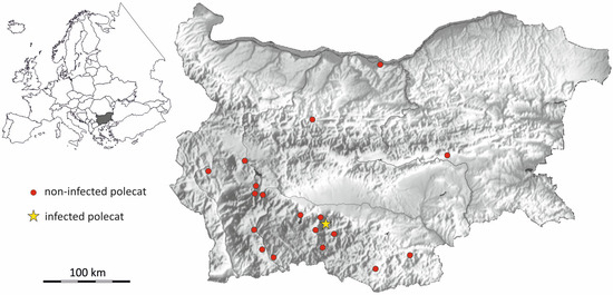
Figure 1.
Sampling locations of the European polecats (Mustela putorius) from Bulgaria analyzed in this study.
The age of the polecats was determined by counting the number of incremental growth lines in the cementum of a canine of the upper jaw [36] using a B1-220A light microscope (Motic, Wetzlar, Germany) at × 40–100 magnification. Animals without growth lines were classified as juveniles, while those with one or more growth lines were classified as adults. The diameters of the skull lesions were measured to an accuracy of 0.01 mm using a digital caliper.
3. Results
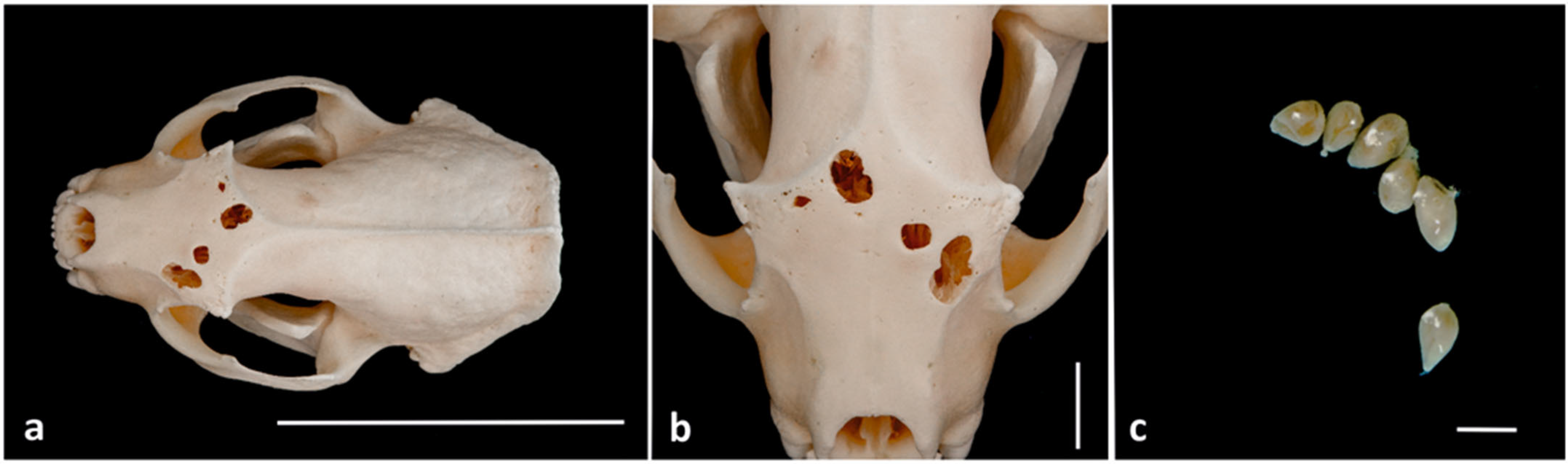
Out of the 19 fresh polecat skulls examined, one (5.26%) was infested with T. acutum (Figure 1). The infested animal was a four-year-old road-killed male (Figure 2a) that was collected on the 15th of November 2023 near the town of Chepelare (41°46′10.6″ N/24°42′24.6″ E) in southern Bulgaria. The site is located in the Somlyan Province, at the northern foothills of the Rhodope Mountains, at an altitude of 780 m above sea level. Six adult T. acutum (Figure 2c) were found firmly attached to the bone structure of the frontal sinus and the ethmoid bone. The examined trematodes had the characteristic pear-shaped form of T. acutum, with a pointed posterior end (Figure 2c). As is typical for digenetic trematodes, an oral sucker was located at the anterior end, and a ventral sucker was positioned in the center of the body. The adult trematodes had an average body length of 2.74 ± 0.31 mm, an average width of 2.05 ± 0.12 mm, and an average height of 1.24 ± 0.14 mm. The infected polecat had four lesions in the frontal bone that were round (0.4 mm) and oval (1.02 to 6.8 mm) in shape (Figure 2b). The skull of the infected polecat and the trematodes are in the author’s collection (Mp_B_144). The infested polecat did not show any co-infection with the nematode S. nasicola.

Figure 2.
The dissected skull of the infected European polecat (Mustela putorius) and the recovered trematodes from Chepelare in Bulgaria. (a) The skull of the infected polecat. Scale: 5 cm. (b) Detailed view of the frontal bone of the infected polecat showing the lesions. Scale: 1 cm. (c) Adult Troglotrema acutum from the frontal sinus of the infected polecat. Scale: 2 mm.
4. Discussion
Here we report the first detection of the trematode T. acutum from Bulgaria and the Balkan Peninsula. This finding is significant, as it suggests that the previously known distribution range of this parasite in south-eastern Europe is much larger than previously assumed. Thus far, there have been no reported cases of T. acutum infestations from this part of Europe. The nearest confirmed record of T. acutum comes from the Bükk Mountains in northern Hungary [32]. The present finding thus extends the known distribution range of the parasite by over 700 km to the southeast. It is possible that the presence of the trematode has been overlooked in the past, especially since the skull lesions that are indicative of infection are not always present [1,4,15].
While it has been suggested that various species of snails can serve as intermediate hosts for T. acutum [6], a recent study on polecats from Germany [5] supported the hypothesis that infections with T. acutum are strongly correlated with the presence of Bythinella snails [15]. Only snails of the genus Bythinella have been confirmed as intermediate hosts by experimental studies [7]. The infected animal was an adult polecat from the Rhodope Mountains in the southern part of the country, a region known as a hotspot for the genus Bythinella [37], which likely acted as a first intermediate host.
The prevalence of 5.26% determined in this study is lower than values reported elsewhere. A prevalence of less than 10% has been reported in polecats from Luxembourg [4], France [14], and the Czech Republic [6]. In contrast, values greater than 10% were found in polecats from Germany [5,15,38]. However, the reported prevalence values can vary substantially within a country. For example, values of 25% [11] and 43% [12] have been reported from France, while values of 18.5% [15], 30.7% [5], and even 100% [39] have been recorded for Germany. Müller and Heddergott [15] suggested that these differences in prevalence may be due to variations in the occurrence of the genus Bythinella.
In rare cases, infestation intensities of more than 100 T. acutum have been reported in polecats [15,18,40]. The infestation intensity (six T. acutum) observed here was low and comparable to findings from Luxembourg [4]. The reason for the great variability in the intensity of infestation is unclear. It can be assumed that the number of matacercariae ingested by eating infected frogs may be one reason for this. The large differences in infestation intensity within small, narrowly defined areas [5,15] could support this hypothesis. It is worth mentioning that, in contrast to other studies [3,4,7,16], no T. acutum eggs were found in the Bulgarian polecat despite multiple flushes of the connective tissue in the paranasal and frontal sinuses. The morphological measurements of the adult T. acutum collected in Bulgaria were consistent with those from other studies [3,4,7,16].
The skull of the infected Bulgarian polecat exhibited lesions that are typical of a T. acutum infection, as described in previous studies [2,3,5,6,15,41]. These lesions were confined to the frontal bone, and the skull did not show the severe spongy swellings observed in infected polecats from Germany [5] and Luxembourg [4]. Previous studies have shown that not all infected animals develop bony skull lesions [4,5,15]. Müller and Heddergott [15] suggest that the presence and severity of the bone lesions may depend on the duration of infection with these trematodes.
The polecat infected with T. acutum from Bulgaria did not show a co-infection with the nematode S. nasicola, as has been reported multiple times in the past from Germany, France, and Austria [1,5,13,15,23]. So far, the author has not encountered any reports of species from the genus Skrjabingylus in Bulgaria.
The present study extends the known southeastern distribution range of T. acutum while emphasizing the variability observed in prevalence across different studies. Further research is necessary to fully understand the factors driving the geographic distribution of the parasite, as well as variation in infestation intensity and lesion presentation.
Supplementary Materials
The following supporting information can be downloaded at: https://www.mdpi.com/article/10.3390/parasitologia4040032/s1, Table S1: Characteristics of the European polecats (Mustela putorius) from Bulgaria analyzed in this study.
Funding
This research received no external funding.
Institutional Review Board Statement
Not applicable.
Informed Consent Statement
Not applicable.
Data Availability Statement
The original contributions presented in this study are included in the paper.
Acknowledgments
The author would like to thank everyone for providing and collecting the road-killed polecats and for their support of the project. Special thanks go to the late Franz Müller (Germany) for his many years of collaboration, particularly for his assistance in planning and executing the project.
Conflicts of Interest
The author declares that he has no conflicts of interest.
References
- Kierdorf, U.; Kierdorf, H.; Konjevie, D.; Lanzar, P. Remarks on cranial lesions in the European polecat (Mustela putorius) caused by helminth parasites. Vet. Arch. 2006, 76, S101–S109. [Google Scholar]
- Ribas, A.; Molina-Vacas, G.; Boadella, M.; Rodriguez-Teijeiro, J.D.; Fernandez-Cardo, R.; Arrizabalaga, A. First report of Troglotrema acutum (Digenea, Troglotrematidae) in the European badger Meles meles in the Iberian Peninsula and presumptive lesions caused in the host. J. Helminthol. 2012, 86, 222–227. [Google Scholar] [CrossRef] [PubMed]
- Heddergott, M.; Frantz, A.C.; Jenrich, J.; Müller, F. Dissections of fresh skulls confirm low prevalence of Troglotrema acutum (Trematoda: Troglotrematidae) in German badgers (Meles meles). Parasitol. Res. 2015, 114, 789–793. [Google Scholar] [CrossRef] [PubMed]
- Heddergott, M.; Steffen, C.; Steinbach, P.; Frantz, A.C. First record of Troglotrema acutum (Trematoda, Troglotrematidae) in European polecats Mustela putorius from Luxembourg. Parasitol. Res. 2021, 120, 2659–2663. [Google Scholar] [CrossRef]
- Frantz, A.C.; Cantú Salazar, L.; Müller, F.; Steinbach, P.; Wittische, J.; Heddergott, M. Interactions of cranial helminths in the European polecat (Mustela putorius): Implications for host body condition. Int. J. Parasitol. Parasites Wildl. 2022, 18, 273–282. [Google Scholar] [CrossRef]
- Koubek, P.; Baruš, V.; Koubkova, B. Troglotrema acutum (Digenea) from carnivores in the Czech Republic. Helminthologia 2004, 41, 25–31. [Google Scholar]
- Vogel, H.; Voelker, J. Über den Lebenszyklus von Troglotrema acutum. Tropenmed. Parasitol. 1978, 29, 385–405. [Google Scholar]
- Skrjabin, K.I. Trematodes of Animals and Humans; Principles of Trematology; Izdatelstvo Akademii Nauk: Moscow, Rusia, 1949; Volume 3, p. 600. (In Russian) [Google Scholar]
- Yamaguti, S. Synopsis of Digenetic Trematodes of Vertebrates; Keigaku Publishing: Tokyo, Japan, 1971; Volumes I and II. [Google Scholar]
- Torres, J.; Feliu, C.; Miquel, J.; Casanova, J.C.; Garcia-Perea, R.; Gisbert, J. Helmitofauna de Mustela putorius Linnaeus, 1758 (Carnivora: Mustelidae) en la peninsula Iberica. Bol. Soc. Hist. Nat. Balear. 1996, 39, 155–165. [Google Scholar]
- Moniez, R. Sur un parasite (Distoma acutum F. S. Leuckart) qui vit dans l’os ethmoide et dans les sinus frontaux du putois. In Revue Biologique du Nord de la France; Le Bigot: Lille, France, 1890; Volume 2, p. 242. [Google Scholar]
- Artois, M.; Blancou, J.; Gerad, Y. Parasitisme du putois (Mustela putorius) par Troglotrema acutum. Rev. Med. Vet. Toulouse 1982, 133, 771–777. [Google Scholar]
- Muller, A. Les Infestations a Skrjabingylus spp. Chez les Mustelides de l’Est de la France. Ph.D. Thesis, Ecole Normale Superieure de Lyon (ENV), Lyon, France, 1989. [Google Scholar]
- Torres, J.; Miquel, J.; Founier, P.; Founier-Chambrillon, C.; Liberge, M.; Fons, R.; Feliu, C. Helminth communities of the autochthonous mustelids Mustela lutreola and M. putorius and the introduced Mustela vison in south-western France. J. Helminthol. 2008, 82, 349–355. [Google Scholar] [CrossRef]
- Müller, F.; Heddergott, M. Befall des Europäischen Iltis Mustela putorius (Mustelidae) mit Troglotrema acutum (Trematoda) und Skrjabingylus nasicola (Nematoda) in der Region Osthessen und der angrenzenden Rhön (Bayern, Hessen und Thüringen). Beitr. Jagd Wildforsch. 2009, 34, 367–383. [Google Scholar]
- Heddergott, M.; Müller, F. First record of Troglotrema acutum (Trematoda: Troglotrematidae) from a pine marten Martes martes in Germany. J. Parasit. Dis. 2020, 44, 105–109. [Google Scholar] [CrossRef] [PubMed]
- Wegelin, H. Merkwürdige Nasenparasiten des Iltis, Putorius foetorius Cuv. Mitt. Thurgau. Nat. Forsch. Ges. 1930, 28, 159–166. [Google Scholar]
- Baer, J.G. Quelques helminthes rares ou peu connus du putois. Rev. Suisse Zool. 1931, 38, 313–334. [Google Scholar]
- Mermod, C.; Debrot, S.; Marchesi, P.; Weber, J.M. Le putois (Mustela putorius L.) en Suisse romande. Rev. Suisse Zool. 1983, 90, 847–856. [Google Scholar] [CrossRef]
- Schumacher, S. Distoma acutum, ein Parasit in den Nebenhöhlen der Nase des Iltis. Osterreich. Waidw. 1929, 2, 362–364. [Google Scholar]
- Kerschagl, W. Jahresbericht über die Untersuchungen kranken Wildes im Jahre 1930. St. Hubertus 1931, 17, 54–56. [Google Scholar]
- Kerschagl, W. Jahresbericht über die Untersuchungen kranken Wildes im Jahre 1932. St. Hubertus 1933, 19, 51–53. [Google Scholar]
- Duscher, G.G.; Harl, J.; Fuehrer, H.P. Evidence of Troglotrema acutum and Skrjabingylus sp. coinfection in a polecat from Lower Austria. Helminthologia 2015, 52, 63–66. [Google Scholar] [CrossRef]
- Marconcini, A.; Tasselli, E. Trematodi e nematodi reperiti nella puzzola (Putorius putorius) in Toscana. Ann. Fac. Med. Vet. Pisa Univ. 1969, 22, 203–216. [Google Scholar]
- Mituch, J.; Hovorka, J.; Hovorka, I.; Vilagiova, I. Helminty masožrave zveri (Carnivora) v modelovom uzemi Tatranskeho Narodneho parku. Folia Venatoria 1992, 22, 191–200. [Google Scholar]
- Demuth, J.; Hromada, M.; Kraeczyk, A.J.; Malecha, A.W.; Tobolka, M.; Tryjanowski, P. Cranial lesions caused by helminth parasites and morphological traits in the European polecat Mustela putorius. Helminthologia 2009, 46, 85–89. [Google Scholar] [CrossRef]
- Ullrich, K. Über das Vorkommen von seltenen oder wenig bekannten Parasiten der Säugetiere und Vögel in Böhmen und Mähren. Prag. Arch. Tiermed. 1930, 10, 19–43. [Google Scholar]
- Stanĕk, M. Dalši nalezy cizopashnych červů u šelem na uzemi ČSSR. Zool. Listy 1963, 12, 359–362. [Google Scholar]
- Svatoš, I. Doplňky k helmintofaunĕ nĕkterych volnĕ žijicich šelem. Zool. Listy 1993, 12, 173–175. [Google Scholar]
- Mituch, J. Helmintofauna masozravcov na Slovensku a v CSSR. Folia Venatoria 1972, 2, 161–171. [Google Scholar]
- Rajsky, D.; Porubčansky, S. Rozširenie cicavice nosnej Troglotrema acutum u lasicovitych šeliem na Slovensku. Veterinařstvi 1989, 39, 78–79. [Google Scholar]
- Vasarhelyi, S. Distoma acutum Leuckart von Ungarn. Zool 1941, 135, 265–270. [Google Scholar]
- Müller, F.; Heddergott, M. Nachweismethodik für einen Befall von Musteliden mit Troglotrema acutum (Trematoda) und Skrjabingylus spp. (Nematoda). Beitr. Jagd Wildforsch. 2009, 34, 385–390. [Google Scholar]
- Heddergott, M. First record of Skrjabingylus petrowi (Nematoda: Metastrongyloidea) in a pine marten (Martes martes) in Germany. Eur. J. Wildl. Res. 2009, 55, 543–546. [Google Scholar] [CrossRef]
- Leuckart, F.S. Zoologische Bruckstücke. III. Helminthologische Beiträge; Forgotten Books: Freiburg, Germany, 1842; p. 60. [Google Scholar]
- Heddergott, M.; Pohl, D.; Steinbach, P.; Cantú Salazar, L.; Müller, F.; Frantz, A.C. Determinants and effects of sinus worm Skrjabingylus nasicola (Nematoda: Metastrongyloidae) infestation in invasive American mink Neovison vison in Germany. Parasitol. Res. 2016, 115, 3449–3457. [Google Scholar] [CrossRef] [PubMed]
- Glöer, P.; Georgiev, D. Bulgaria, a hot spot of biodiversity (Gastropoda: Rissooidea)? J. Conchol. 2011, 40, 489. [Google Scholar]
- Klupiec, P. Die Helminthenfauna des Iltis (Mustela putorius L.) in seinem nordwestdeutschen Verbreitungsgebiet. Ph.D. Thesis, Veterinarmedizinische Hochschule, Hannover, Germany, 2001. [Google Scholar]
- Pohl, L. Über das Vorkommen von Distoma acutum Leuck. bei Putorius putorius L. Jena Z. Naturw. 1912, 48, 563–568. [Google Scholar]
- Olt, A. Untersuchungen von Iltisköpfen auf Parasiten in der Nasen-und Stirnhöhle. Dtsch. Jagerztg. 1929, 93, 83–84. [Google Scholar]
- Lehmensick, R. Über die Veränderungen am Iltis Schädel durch den Befall mit Troglotrema acutum. Z. Parasitenkd. 1942, 12, 659–664. [Google Scholar] [CrossRef]
Disclaimer/Publisher’s Note: The statements, opinions and data contained in all publications are solely those of the individual author(s) and contributor(s) and not of MDPI and/or the editor(s). MDPI and/or the editor(s) disclaim responsibility for any injury to people or property resulting from any ideas, methods, instructions or products referred to in the content. |
© 2024 by the author. Licensee MDPI, Basel, Switzerland. This article is an open access article distributed under the terms and conditions of the Creative Commons Attribution (CC BY) license (https://creativecommons.org/licenses/by/4.0/).