

Unsupervised Metal Artifact Reduction in Dental CBCT Using Fine-Tuned Cycle-Consistent Adversarial Networks

 ,
,  , ,
, ,  and
and
Abstract
1. Introduction
1.1. Research Gap and Motivation
- 1.
- Dependence on Paired Training Data: Supervised deep learning approaches require voxel-aligned artifact-free and artifact-affected image pairs. However, such datasets are practically unattainable in clinical settings due to ethical constraints related to repeated radiation exposure and challenges in achieving perfect anatomical registration.
- 2.
- Sensitivity of Classical MAR Methods: Traditional projection-domain and reconstruction-based techniques rely heavily on accurate metal segmentation and simplified physical models. These approaches often fail under severe artifact conditions and may introduce secondary distortions or excessive smoothing of anatomical structures.
- 3.
- Risk of Structural Hallucinations in GAN-based Methods: While unsupervised GAN frameworks eliminate the need for paired data, they may generate unrealistic textures or distort fine anatomical details if not properly constrained, limiting their reliability for clinical use.
- 4.
- Limited Practical Integration into Clinical Workflows: Many existing MAR solutions require hardware modifications, computationally intensive reconstruction, or manual preprocessing steps, reducing their feasibility for routine deployment in digital dentistry environments.
1.2. Study Overview and Contributions
- 1.
- Addressing the Lack of Paired Data: We implement an unsupervised CycleGAN-based MAR pipeline that eliminates the need for voxel-perfect paired datasets, directly addressing the primary limitation of supervised approaches.
- 2.
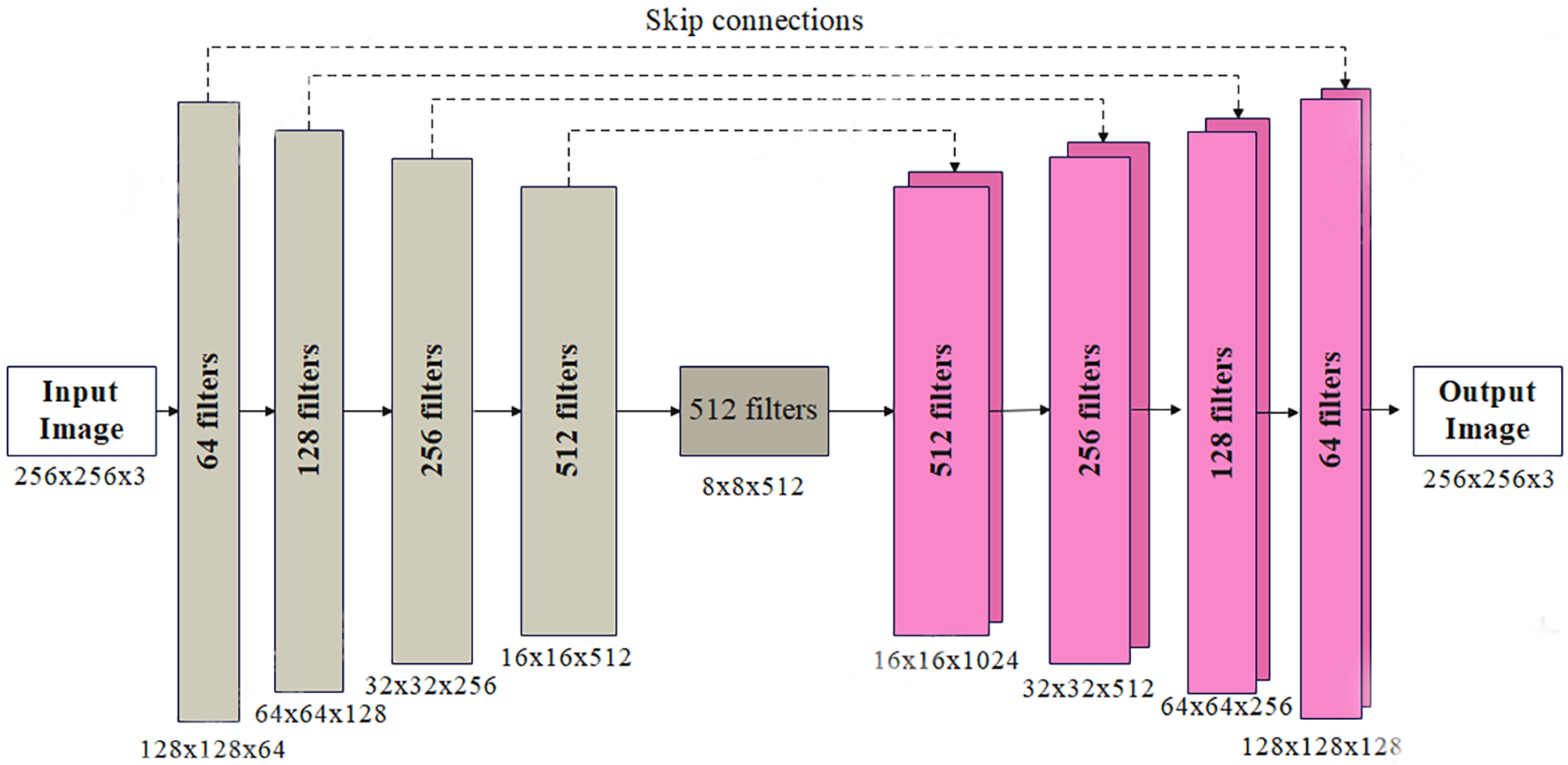
- Improved Anatomical Preservation in GAN Frameworks: By integrating U-Net-based generators with PatchGAN discriminators and carefully tuned loss functions, the proposed model mitigates structural hallucinations while preserving fine anatomical details.
- 3.
- Robustness Beyond Classical MAR Limitations: Unlike traditional methods that depend on metal segmentation or physical modeling, the proposed approach learns artifact distributions directly from data, improving performance under complex artifact conditions.
- 4.
- Practical Workflow Integration: The proposed solution does not require hardware modifications or manual preprocessing, making it suitable for scalable deployment in existing digital dentistry pipelines.
2. Background
2.1. Research Problem
2.2. Traditional and Classical MAR Methods
2.3. Deep Learning Approaches for MAR
2.4. Challenges and Research Gaps
2.5. Contribution
3. Materials and Methods
3.1. Dataset Creation
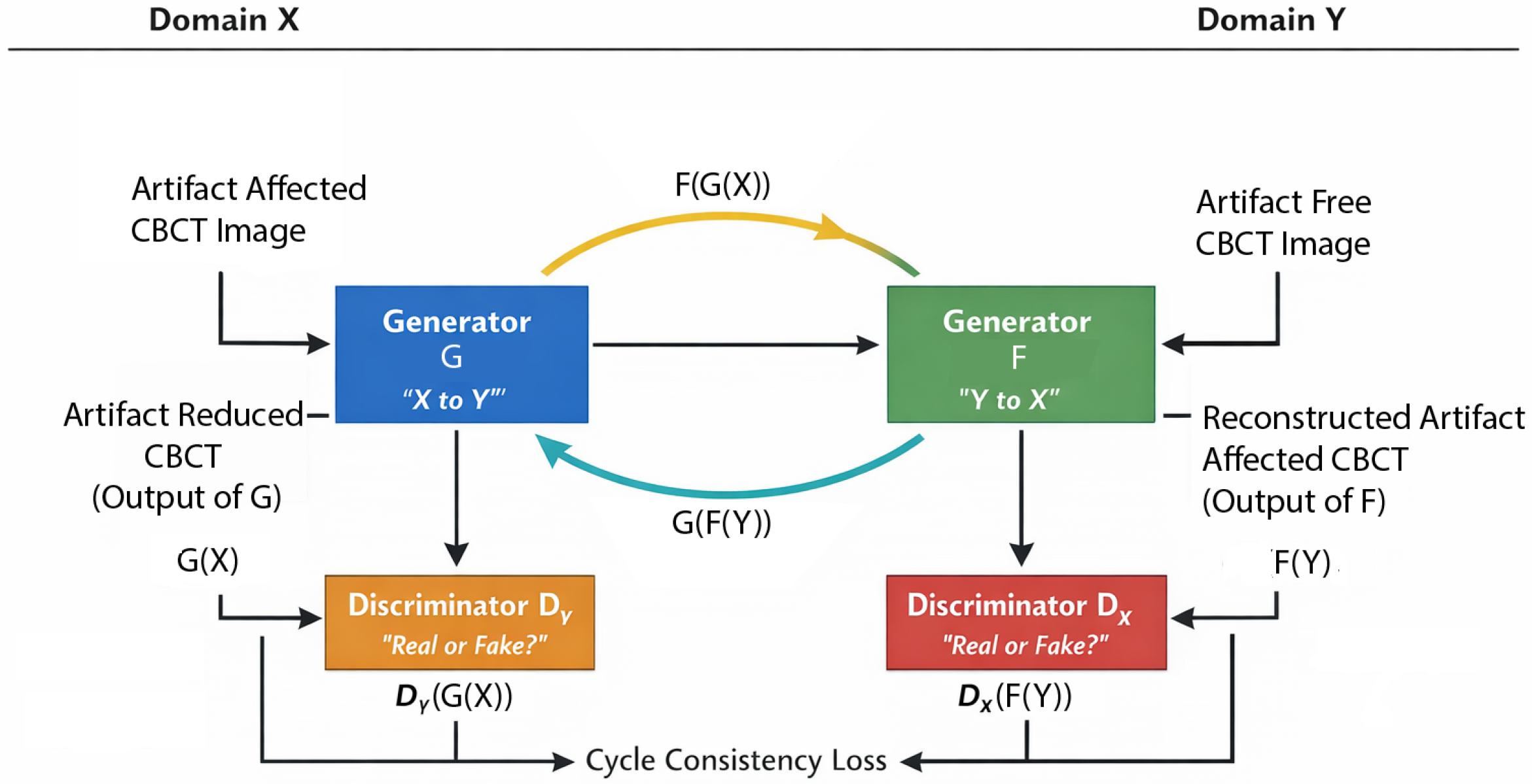
3.2. Proposed Algorithm
3.2.1. Generator: U-Net
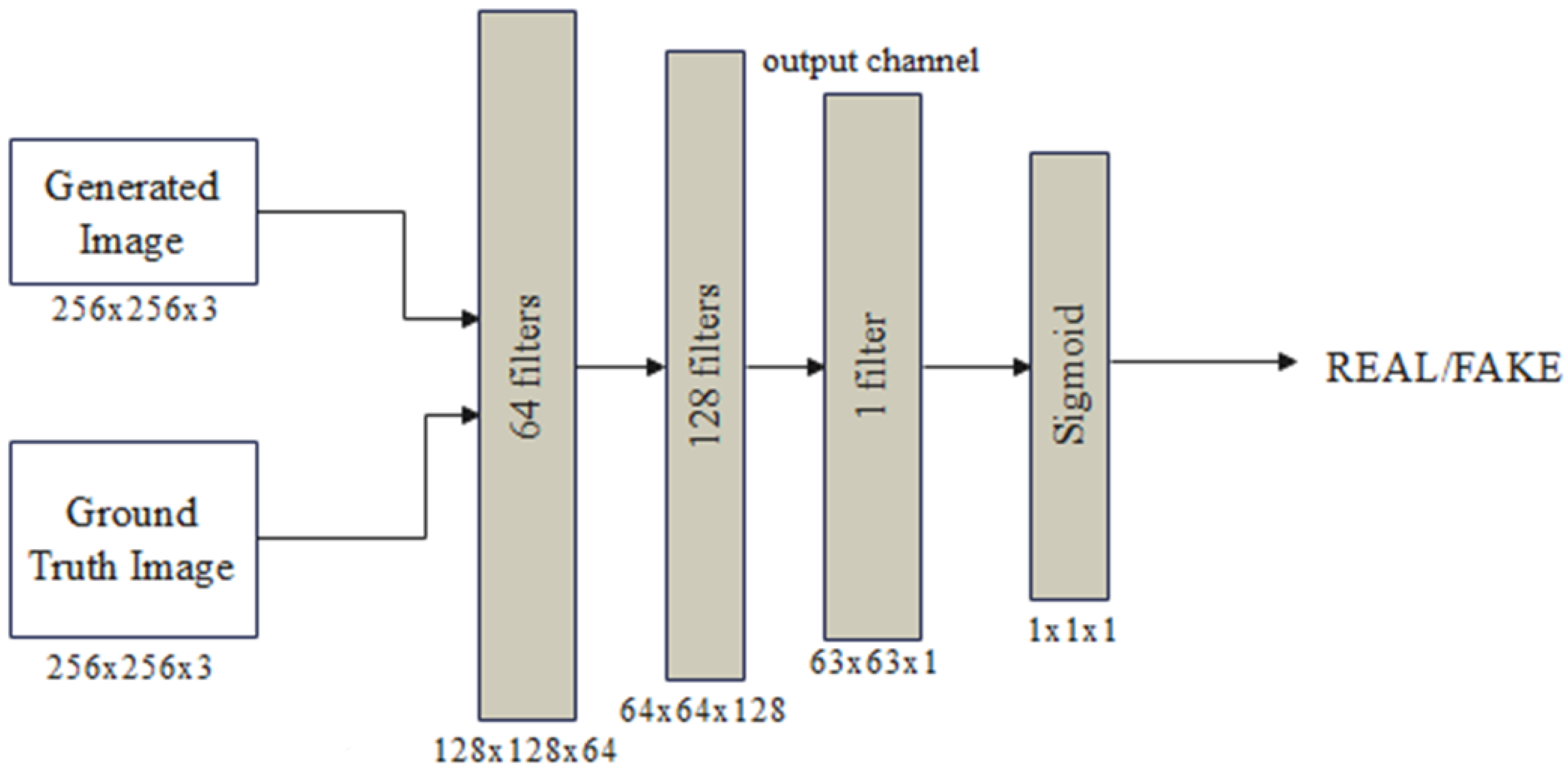
3.2.2. Discriminator: PatchGAN
3.3. Training, Validation, and Testing
4. Results
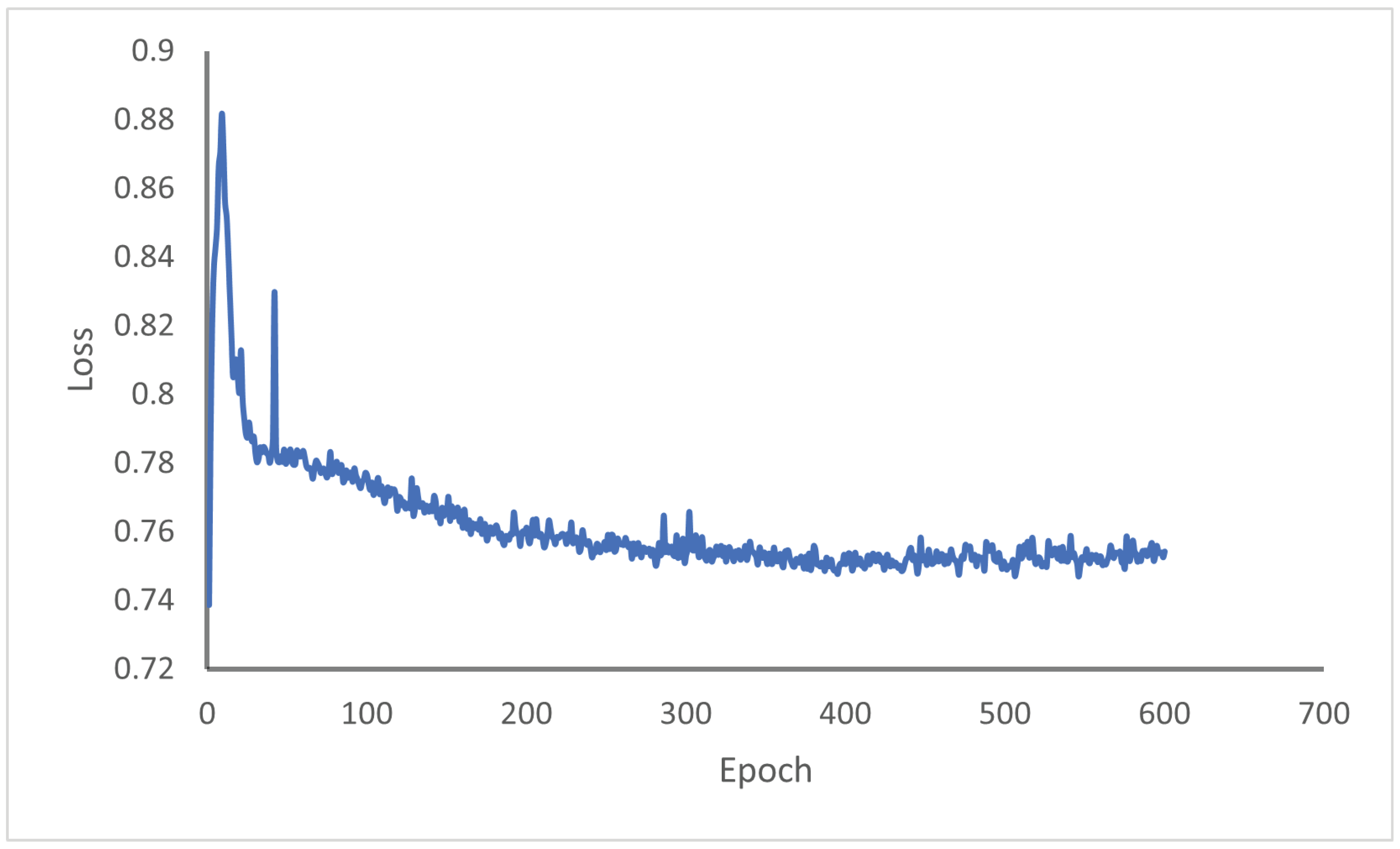
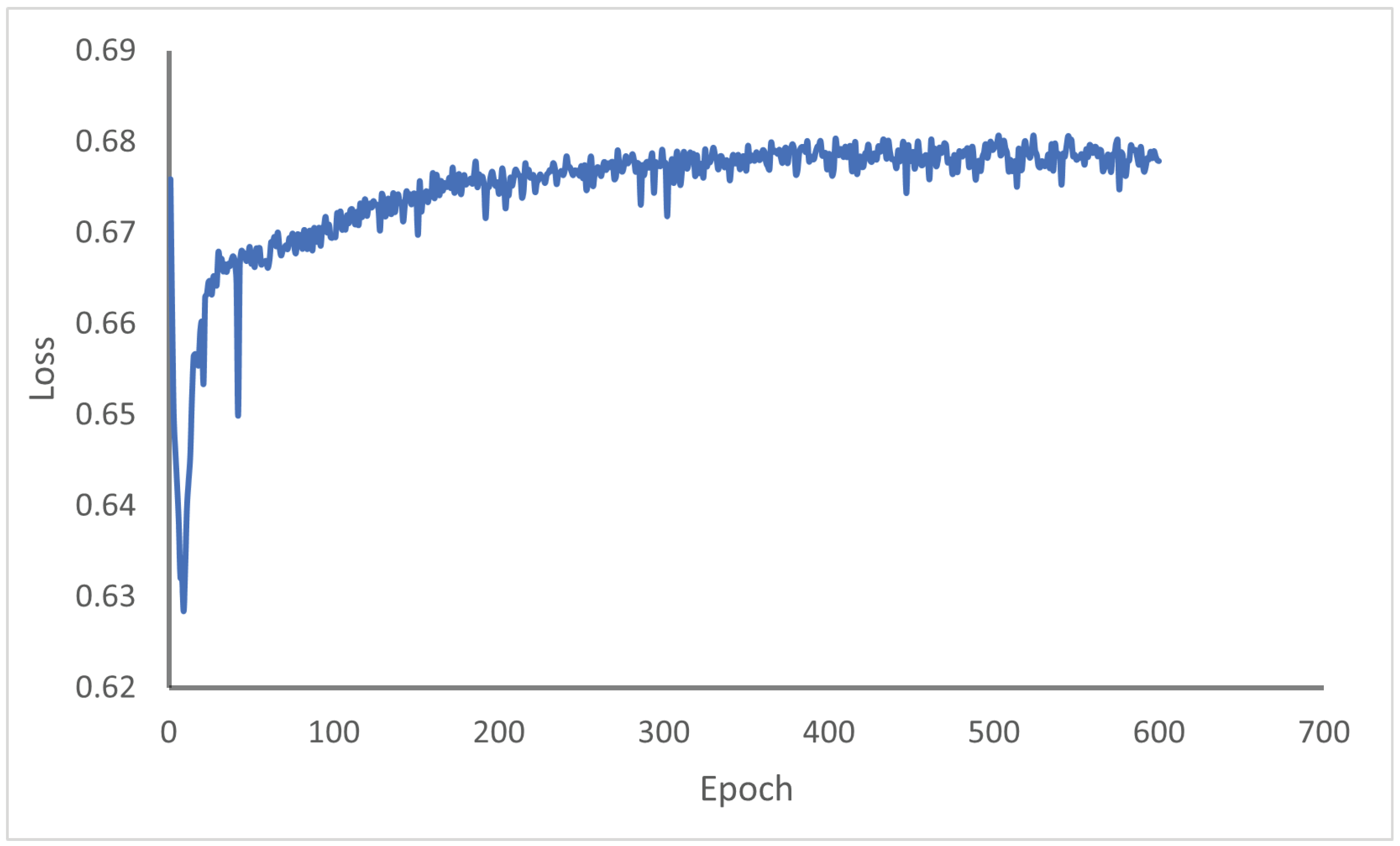
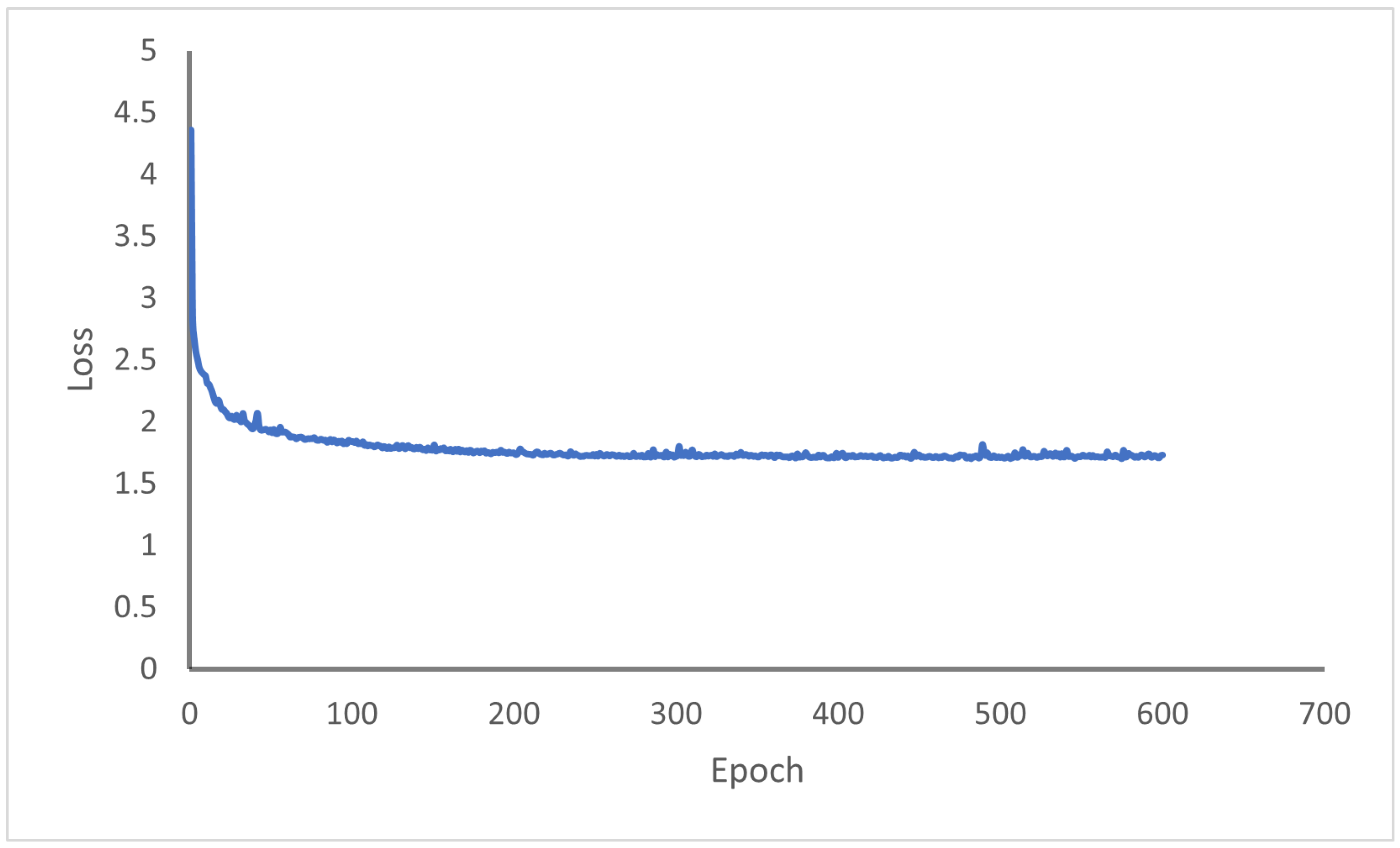
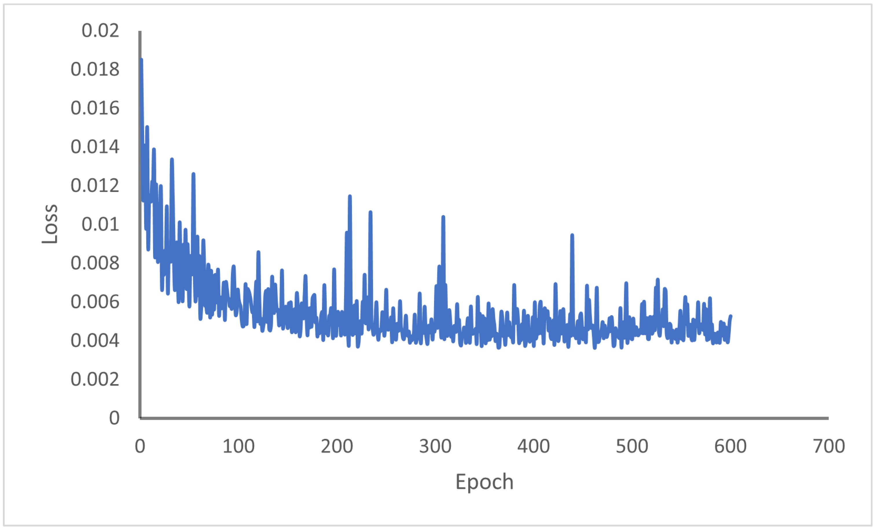
4.1. Training Dynamics and Model Convergence
4.2. Baseline Selection and Evaluation Framework
- Classical Deterministic Baseline: The Normalized Metal Artifact Reduction (NMAR) algorithm [7] was selected as the representative clinical standard. NMAR remains a primary benchmark due to its stability in suppressing streak artifacts through sinogram interpolation, though it often lacks the structural preservation capabilities of generative models.
- Supervised Deep Learning Benchmarks: Two supervised variants—UNet2D1D and UNet2D2D [28]—were integrated to serve as performance upper bounds. These architectures represent the modern standard for encoder–decoder-based image restoration. By comparing our unsupervised framework against these supervised models, we evaluate the trade-offs between synthetic-pair training (which can introduce domain gaps) and our direct clinical-distribution learning strategy.
- Identical Testing Dataset: All models were evaluated on the same held-out test set consisting of axial slices. As detailed in Section 3.3, this partition was performed at the patient level to strictly prevent data leakage and ensure the assessment of model generalizability on unseen anatomy.
- Standardized Preprocessing: Every input image underwent identical Min-Max intensity normalization and was rescaled to a uniform resolution of pixels to maintain architectural consistency across all benchmarks.
Evaluation Metrics and Rationale
- 1.
- Structural Fidelity (PSNR and SSIM): While the Signal-to-Noise Ratio () and Structural Similarity Index Measure are traditionally full-reference metrics, they were strategically utilized here to quantify the anatomical consistency of the model’s output relative to the original artifact-affected inputs. This approach ensures that the generative process preserves the patient’s underlying dental morphology (e.g., pulp chambers and root structures) without introducing structural hallucinations or deformations. In this context, a higher indicates superior preservation of the anatomical “identity” during artifact suppression.
- 2.
- Perceptual Quality (BRISQUE): To assess the naturalness of the reconstructed images without requiring a reference, the Blind/Referenceless Image Spatial Quality Evaluator (BRISQUE) was employed. This no-reference metric provides a standardized measure of perceptual quality, where lower scores indicate a reduction in artificial distortions and “checkerboard” artifacts.
- 3.
- Distributional Consistency (FID): The Fréchet Inception Distance (FID) was used to evaluate the similarity between the feature-space distributions of the generated results and a validated cohort of artifact-free clinical scans. A lower score validates the model’s ability to shift the artifact-affected images into the domain of high-quality, “clean” dental CBCT imaging.
4.3. Ablation Study
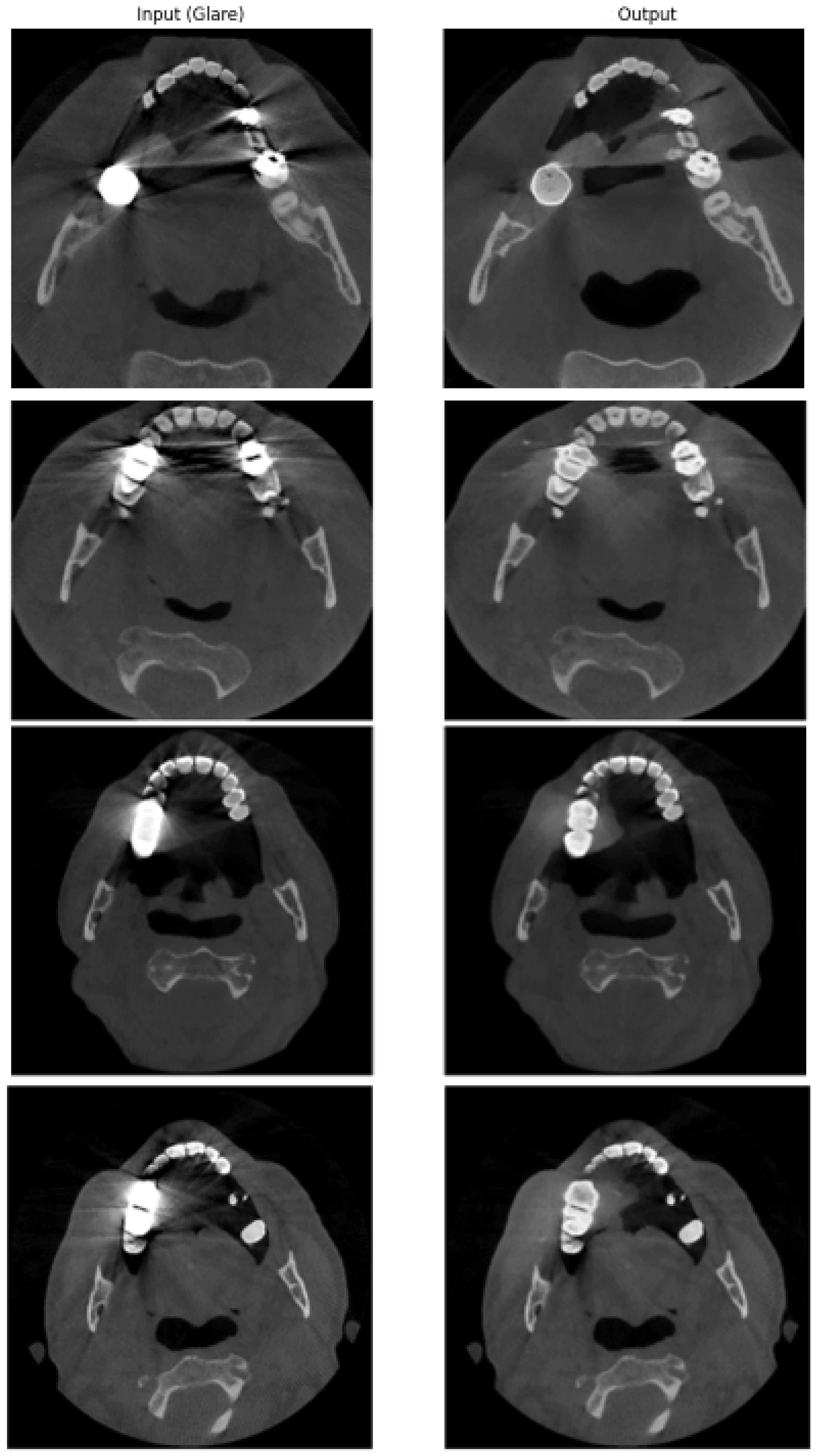
4.4. Quantitative and Qualitative Evaluation of Reconstruction Accuracy
4.5. Computational Efficiency and Inference Time
5. Discussion
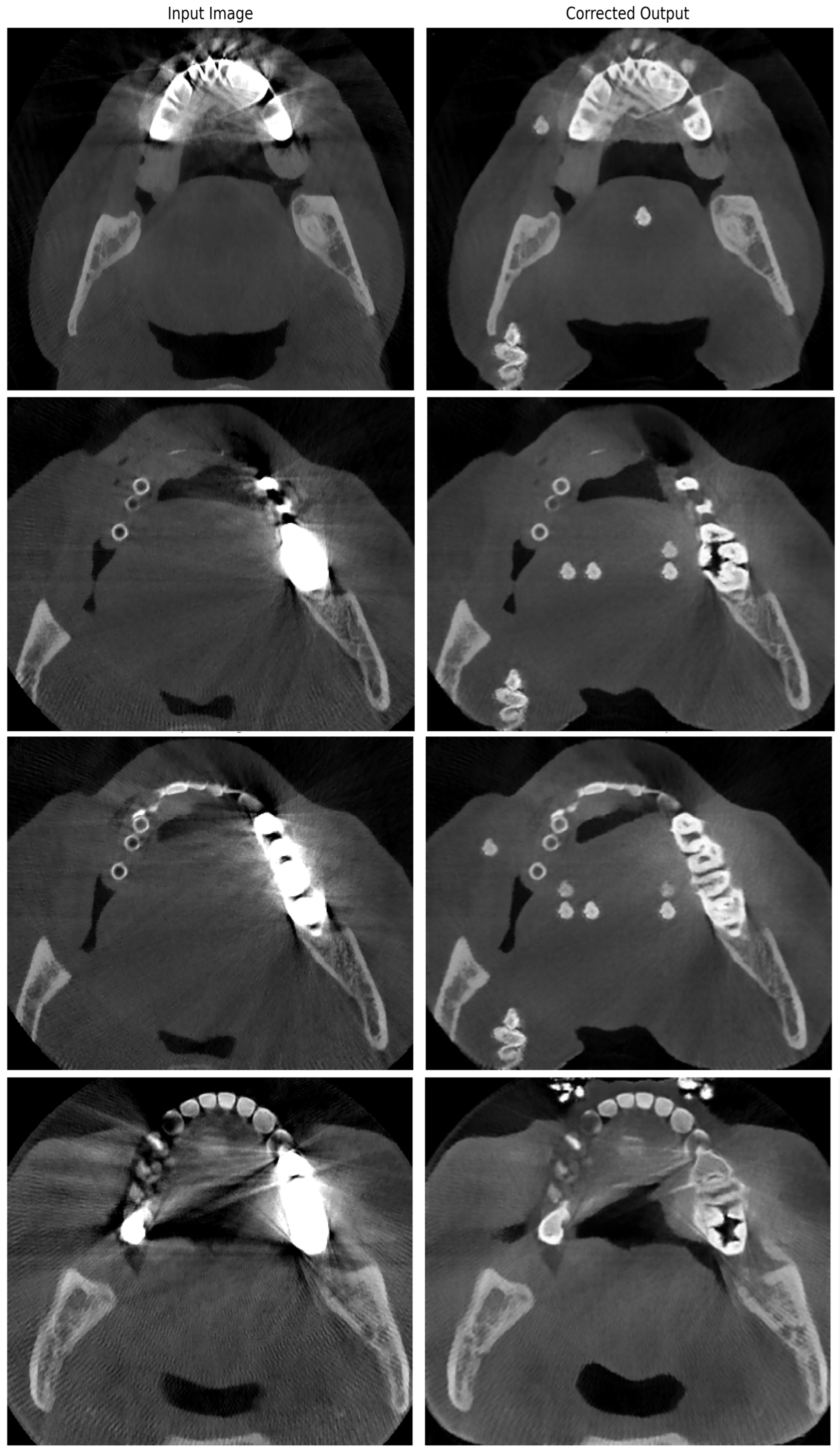
5.1. Qualitative Analysis of Failure Cases and Hallucinations
5.2. Limitations of the Study and Future Work
- 1.
- Lack of Clinical Ground Truth: The most significant challenge in clinical MAR research is the absence of voxel-aligned, artifact-free references for real patients. Although we addressed this by using no-reference metrics ( and ) and structural fidelity indices ( relative to the input), these are proxy measures. Future studies should incorporate physical dental phantoms with known geometries to establish absolute error margins.
- 2.
- Generative Hallucinations and Clinical Oversight: As discussed in Section 5.1, the generative nature of CycleGAN can occasionally introduce anatomical hallucinations in regions of extreme photon starvation. Therefore, the current model must be viewed as a supportive diagnostic tool. Clinical expert validation remains mandatory to distinguish between authentic anatomical restoration and potential AI-induced artifacts.
- 3.
- Two-Dimensional Spatial Context vs. 3D Volumetric Continuity: Our current approach operates on 2D axial slices. While computationally efficient, this ignores the inter-slice dependency along the vertical (z) axis. Future work will focus on extending the unsupervised adversarial learning to 3D volumetric data or 2.5D architectures to enhance vertical structural consistency.
- 4.
- Model Generalizability Across CBCT Vendors: The model was trained and validated using a specific dataset (ToothFairy) which reflects the reconstruction kernels and noise profiles of a particular subset of CBCT scanners. The robustness of the framework across different hardware vendors and scanning protocols (e.g., varying peak voltages or filtration) has not been fully evaluated. Future work will investigate domain adaptation techniques to ensure the model remains scanner-agnostic.
- 5.
- Diverse Metallic Objects and Prosthetics: While the framework effectively reduces artifacts from common dental restorations, its performance on specialized high-density materials—such as titanium implants or orthodontic appliances—has not been fully evaluated. These objects create distinct “photon starvation” patterns that differ from standard fillings. To address this, future work will involve collecting and labeling local clinical data to expand the training distribution, ensuring the model generalizes across a broader range of metallic prosthetics and diverse patient populations.
6. Conclusions
Author Contributions
Funding
Institutional Review Board Statement
Informed Consent Statement
Data Availability Statement
Acknowledgments
Conflicts of Interest
References
- Ibraheem, I. Reduction of artifacts in dental cone beam CT images to improve the three-dimensional image reconstruction. J. Biomed. Sci. Eng. 2012, 5, 409–415. [Google Scholar] [CrossRef]
- Johari, M.; Abdollahzadeh, M.; Esmaeili, F.; Sakhamanesh, V. Metal artifact suppression in dental cone beam computed tomography images using image processing techniques. J. Med. Signals Sens. 2018, 8, 12–24. [Google Scholar] [PubMed] [PubMed Central]
- Barrett, J.F.; Keat, N. Artifacts in CT: Recognition and avoidance. Radiographics 2004, 24, 1679–1691. [Google Scholar] [CrossRef] [Scilit]
- Diehn, F.E.; Michalak, G.J.; DeLone, D.R.; Kotsenas, A.L.; Lindell, E.P.; Campeau, N.G.; Halaweish, A.F.; McCollough, C.H.; Fletcher, J.G. CT dental artifact: Comparison of an iterative metal artifact reduction technique with weighted filtered back-projection. Acta Radiol. Open 2017, 6, 2058460117743279. [Google Scholar] [CrossRef] [Scilit]
- Wajer, R.; Wajer, A.; Kazimierczak, N.; Wilamowska, J.; Serafin, Z. The impact of AI on metal artifacts in CBCT oral cavity imaging. Diagnostics 2024, 14, 1280. [Google Scholar] [CrossRef] [Scilit]
- Shavakhi, M.; Soltani, P.; Aghababaee, G.; Patini, R.; Armogida, N.G.; Spagnuolo, G.; Valletta, A. A quantitative evaluation of the effectiveness of the metal artifact reduction algorithm in cone beam computed tomographic images with stainless steel orthodontic brackets and arch wires: An ex vivo study. Diagnostics 2024, 14, 159. [Google Scholar] [CrossRef] [Scilit] [PubMed]
- Meyer, E.; Raupach, R.; Lell, M.; Schmidt, B.; Kachelrieß, M. Normalized metal artifact reduction (NMAR) in computed tomography. Med. Phys. 2010, 37, 5482–5493. [Google Scholar] [CrossRef] [Scilit] [PubMed]
- Kalender, W.A.; Hebel, R.; Ebersberger, J. Reduction of CT artifacts caused by metallic implants. Radiology 1987, 164, 576–577. [Google Scholar] [CrossRef] [Scilit] [PubMed]
- Lemmens, C.; Faul, D.; Nuyts, J. Suppression of metal artifacts in CT using a reconstruction procedure that combines MAP and projection completion. IEEE Trans. Med. Imaging 2009, 28, 250–260. [Google Scholar] [CrossRef] [Scilit]
- Ho, J.; Jain, A.; Abbeel, P. Denoising Diffusion Probabilistic Models. Adv. Neural Inf. Process. Syst. 2020, 33, 6840–6851. [Google Scholar] [CrossRef] [Scilit]
- Ghani, M.U.; Karl, W.C. Fast Enhanced CT Metal Artifact Reduction Using Data Domain Deep Learning. IEEE Trans. Comput. Imaging 2020, 6, 181–193. [Google Scholar] [CrossRef] [Scilit]
- Gjesteby, L.; Yang, Q.; Xi, Y.; Zhou, L.; Wang, G.; Chen, J. Metal Artifact Reduction in CT: Where Are We After Four Decades? IEEE Access 2016, 4, 5826–5849. [Google Scholar] [CrossRef] [Scilit]
- Amirian, M.; Barco, D.; Herzig, I.; Schilling, F.-P. Artifact Reduction in 3D and 4D Cone-Beam Computed Tomography Images With Deep Learning: A Review. IEEE Access 2024, 12, 10281–10295. [Google Scholar] [CrossRef] [Scilit]
- Lee, J.; Gu, J.; Ye, J.C. Unsupervised CT metal artifact learning using attention-guided β-CycleGAN. IEEE Trans. Med. Imaging 2021, 40, 3932–3944. [Google Scholar] [CrossRef] [Scilit]
- Klages, P.; Benslimane, I.; Riyahi, S.; Jiang, J.; Hunt, M.; Deasy, J.O.; Veeraraghavan, H.; Tyagi, N. Patch-based generative adversarial neural network models for head and neck MR-only planning. Med. Phys. 2020, 47, 626–642. [Google Scholar] [CrossRef] [Scilit]
- Zhang, Y.; Yu, H. Convolutional Neural Network Based Metal Artifact Reduction in X-Ray Computed Tomography. IEEE Trans. Med. Imaging 2018, 37, 1370–1381. [Google Scholar] [CrossRef] [Scilit]
- Du, M.; Liang, K.; Xing, Y. Reduction of metal artefacts in CT with Cycle-GAN. In Proceedings of the 2018 IEEE Nuclear Science Symposium and Medical Imaging Conference Proceedings (NSS/MIC), Sydney, NSW, Australia, 10–17 November 2018; IEEE: Piscataway, NJ, USA, 2018; pp. 1–3. [Google Scholar] [CrossRef] [Scilit]
- Yi, X.; Walia, E.; Babyn, P. Generative adversarial network in medical imaging: A review. Med. Image Anal. 2019, 58, 101552. [Google Scholar] [CrossRef] [Scilit] [PubMed]
- University of Modena and Reggio Emilia and Radboud University, ToothFairy Dataset for MICCAI 2023 ToothFairy Challenge. Available online: https://ditto.ing.unimore.it/toothfairy/ (accessed on 15 January 2026).
- Hur, J.; Shin, Y.-G.; Lee, H. Hybrid model-based and deep learning-based metal artifact reduction method in dental cone-beam computed tomography. Nucl. Eng. Technol. 2023, 55, 2854–2864. [Google Scholar] [CrossRef] [Scilit]
- Soltani, P.; Spagnuolo, G.; Angelone, F.; Rezaeiyazdi, A.; Mohammadzadeh, M.; Maisto, G.; Moaddabi, A.; Cernera, M.; Armogida, N.G.; Amato, F.; et al. Artificial Intelligence for Artifact Reduction in Cone Beam Computed Tomographic Images: A Systematic Review. Appl. Sci. 2026, 16, 396. [Google Scholar] [CrossRef] [Scilit]
- Choi, Y.; Kim, H.; Baek, J.; Lee, H. Dual Domain Diffusion Guidance for 3D CBCT Metal Artifact Reduction. In Proceedings of the IEEE/CVF Winter Conference on Applications of Computer Vision, Waikoloa, HI, USA, 3–8 January 2024; IEEE: Piscataway, NJ, USA, 2024; pp. 1–10. [Google Scholar] [CrossRef] [Scilit]
- Shi, Y.; Xu, J.; Shen, D. MARformer: An Efficient Metal Artifact Reduction Transformer for Dental CBCT Images. arXiv 2023, arXiv:2311.09590. [Google Scholar] [CrossRef] [Scilit]
- Wang, H.; Zhang, W.; Xu, Y.; Li, H.; Ren, P. WaterCycleDiffusion: Visual-textual fusion empowered underwater image enhancement. Inf. Fusion 2026, 127, 103693. [Google Scholar] [CrossRef] [Scilit]
- Liu, H.; Chen, L.; Lu, X.; Wang, H.; Bai, L.; Wang, M.; Ren, P. A visual-textual mutual guidance fusion network for remote sensing visual question answering. Pattern Recognit. 2026, 176, 113258. [Google Scholar] [CrossRef] [Scilit]
- Boktor, M.; Ecclestone, B.R.; Ye, J.A.; Reza, P.H. Multi-channel feature extraction for virtual histological staining of photon absorption remote sensing images. Sci. Rep. 2024, 14, 2009. [Google Scholar] [CrossRef] [Scilit] [PubMed]
- Hindarto, D.; Handayani, E.T.E. Revolution in Image Data Collection: CycleGAN as a Dataset Generator. Sinkron J. Penelit. Tek. Inform. 2024, 8, 444–454. [Google Scholar] [CrossRef] [Scilit]
- Hauptmann, A.; Arridge, S.; Lucka, F.; Muthurangu, V.; Steeden, J.A. Real-time cardiovascular MR with spatio-temporal artifact suppression using deep learning—Proof of concept in congenital heart disease. Magn. Reson. Med. 2019, 81, 1143–1156. [Google Scholar] [CrossRef] [Scilit] [PubMed]






| Hyperparameter | Value |
|---|---|
| Learning rate | |
| Optimizer | Adam |
| 0.5 | |
| 0.999 | |
| Batch size | 1 |
| Number of epochs | 600 |
| 18 | |
| 15 | |
| Image resolution | |
| Normalization range |
| Configuration | BRISQUE (↓) | FID (↓) | PSNR (↑) * | SSIM (↑) * |
|---|---|---|---|---|
| Full Model (Proposed) | 10.82 | 157.04 | 25.96 | 0.9105 |
| Suboptimal Weights () | 12.95 | 168.43 | 23.18 | 0.8714 |
| Without Identity Loss () | 13.67 | 174.21 | 22.45 | 0.8520 |
| Without U-Net Skip Connections | 14.32 | 181.57 | 21.05 | 0.8211 |
| Method | BRISQUE (↓) | FID (↓) | PSNR (↑) * | SSIM (↑) * |
|---|---|---|---|---|
| Baseline (Artifact-Affected) | 207.03 | - | - | |
| NMAR (Meyer et al.) [7] | 202.68 | 29.96 | 0.8802 | |
| UNet2D1D (Supervised) [28] | 263.28 | 16.64 | 0.5611 | |
| UNet2D2D (Supervised) [28] | 248.77 | 16.32 | 0.5480 | |
| Proposed CycleGAN | 25.96 | 0.9105 |
| Method | Inference Time (ms) |
|---|---|
| UNet2D1D | |
| UNet2D2D | |
| Proposed CycleGAN (Generator) |
Disclaimer/Publisher’s Note: The statements, opinions and data contained in all publications are solely those of the individual author(s) and contributor(s) and not of MDPI and/or the editor(s). MDPI and/or the editor(s) disclaim responsibility for any injury to people or property resulting from any ideas, methods, instructions or products referred to in the content. |
© 2026 by the authors. Licensee MDPI, Basel, Switzerland. This article is an open access article distributed under the terms and conditions of the Creative Commons Attribution (CC BY) license.
Share and Cite
Chamika, T.; Dhanapala, S.N.A.; Nimalaweera, S.; Dissanayake, M.B.; Jayasinghe, R.D. Unsupervised Metal Artifact Reduction in Dental CBCT Using Fine-Tuned Cycle-Consistent Adversarial Networks. Digital 2026, 6, 31. https://doi.org/10.3390/digital6020031
Chamika T, Dhanapala SNA, Nimalaweera S, Dissanayake MB, Jayasinghe RD. Unsupervised Metal Artifact Reduction in Dental CBCT Using Fine-Tuned Cycle-Consistent Adversarial Networks. Digital. 2026; 6(2):31. https://doi.org/10.3390/digital6020031
Chicago/Turabian StyleChamika, Thamindu, Sithum N. A. Dhanapala, Sasindu Nimalaweera, Maheshi B. Dissanayake, and Ruwan D. Jayasinghe. 2026. "Unsupervised Metal Artifact Reduction in Dental CBCT Using Fine-Tuned Cycle-Consistent Adversarial Networks" Digital 6, no. 2: 31. https://doi.org/10.3390/digital6020031
APA StyleChamika, T., Dhanapala, S. N. A., Nimalaweera, S., Dissanayake, M. B., & Jayasinghe, R. D. (2026). Unsupervised Metal Artifact Reduction in Dental CBCT Using Fine-Tuned Cycle-Consistent Adversarial Networks. Digital, 6(2), 31. https://doi.org/10.3390/digital6020031