Review

Factors Influencing Late Breast Toxicity After Radiotherapy: A Scoping Review

by
Riccardo Ray Colciago
1,
Chiara Chissotti
2,
Federica Ferrario
2,*,
Ilenia Manno
1,2,
Matteo Mombelli
1,2,
Giulia Rossano
1,2,
Lorenzo De Sanctis
1,2 and
Stefano Arcangeli
1,2
1
Medicine and Surgery Department, University of Milan Bicocca, 20126 Milano, Italy
2
Radiation Oncology Unit, Fondazione IRCCS San Gerardo dei Tintori, 20900 Monza, Italy
*
Author to whom correspondence should be addressed.
BioChem 2025, 5(2), 13; https://doi.org/10.3390/biochem5020013
Submission received: 14 March 2025 / Revised: 20 May 2025 / Accepted: 27 May 2025 / Published: 30 May 2025

Abstract

:
Radiation therapy offers well-established benefits in enhancing loco-regional control, distant disease control, and breast-cancer-specific survival. However, it is not without its challenges, particularly in breast cancer patients, where advances in systemic therapies and other treatment modalities have significantly improved survival outcomes. As radiation oncologists, our responsibility is to deliver the most effective treatments while minimizing toxicity for each patient. This scoping review aims to retrieve and assess the literature on factors associated with increased radiation-induced late breast toxicity. Specifically, we seek to identify both non-modifiable variables and those that can be influenced by the choices made by radiation oncologists. This review highlights which clinical decisions could directly impact late breast toxicity following adjuvant radiation therapy after breast-conserving surgery.

1. Introduction

Together with surgery and systemic therapies, radiation therapy (RT) is a cornerstone in the treatment of breast cancer (BC) patients, as depicted in Figure 1.
Veronesi et al. (2002) demonstrated that breast-conserving surgery combined with RT offered comparable disease control to radical mastectomy among 701 women randomized between 1973 and 1980 [1]. Since then, significant advancements in medicine have emerged, including the development of novel drugs that have revolutionized medical oncology [2] and technological innovations that have transformed radiation oncology in terms of techniques, dose prescription, and irradiation volumes [3].
Radiation therapy has well-established benefits in improving loco-regional control, distant disease control, and BC-specific survival [4,5,6]. In their most recent meta-analysis, the Early Breast Cancer Trialists’ Collaborative Group demonstrated that modern RT could also enhance overall survival [6]. This analysis included data on nodal irradiation from 16 trials, encompassing a total of 14,324 patients. While earlier studies showed that RT improved cancer control and disease-specific mortality without impacting overall survival—due to increased non-BC mortality—the eight more recent trials revealed that regional nodal irradiation significantly reduced recurrence, BC mortality, and overall mortality. The absolute 15-year gains were 2.6%, 3.0%, and 3.0%, respectively [6].
The benefits of RT appear to be independent of advancements in systemic therapy. In a recent secondary analysis of the ALTTO trial [7], RT was associated with improvements in both loco-regional control and overall survival, despite patients receiving modern systemic treatments, including targeted therapies. Although the total number of events remained low, these findings highlight the sustained relevance of RT as a key component of multidisciplinary breast cancer management, even amidst the evolving development of novel systemic treatments.
However, RT is not without its challenges. This is especially relevant for BC patients, where advances in systemic therapies and other treatment modalities have significantly improved survival outcomes [2]. In long-surviving breast cancer patients, it is of paramount importance to optimize treatment strategies. As radiation oncologists, we have both the responsibility and the opportunity to deliver the most effective therapies while minimizing toxicity, tailoring each treatment to the individual patient’s needs.
The aim of this scoping review is to compile and discuss current evidence on factors associated with increased breast toxicity following irradiation, with an emphasis on aspects where radiation oncologists can actively influence patient outcomes.

2. Materials and Methods

Between September 2024 and January 2025, we conducted a systematic search of the NCBI PubMed, Scopus, and Embase databases. We used specific search terms related to breast late toxicity, including “breast cancer”, “long-term results”, and “factors associated”, combined with terms related to radiotherapy and radiation oncology. Our goal was to identify studies reporting data on factors associated with late toxicities ≥ 6 months after the end of adjuvant RT for BC. Additionally, we examined reference lists for relevant studies to ensure comprehensive coverage.

2.1. Selection Criteria

Studies were included if they met the following criteria:
-
Primarily investigated factors associated with long-term effects on the breast in the context of radiotherapy or radiation oncology.
-
Provided either an abstract or full-text availability.
-
Were written in English.

2.2. Ethical Considerations

This review adhered to ethical guidelines and did not involve human subjects or personal data.

3. Results

3.1. Quality Assessment

Our research identified 66 published studies, all supporting the evidence that multiple factors contribute to long-term breast toxicity following RT. While some variables are non-modifiable, serving only as risk factors for increased toxicity, others can be actively adjusted by the radiation oncologist. The selected studies in our review emphasized the potential benefits of optimizing radiation treatment, particularly in terms of prescribed dose, target volume, and irradiation technique.
Breast radiation toxicity can be influenced by various factors, which can be broadly categorized as modifiable and non-modifiable. Modifiable factors, often linked to physician decisions, may lead to either increased or reduced toxicity levels, while non-modifiable factors are inherent to the patient. Understanding these determinants is crucial for predicting toxicity, allowing for personalized treatment strategies that minimize risk and optimize patient outcomes.
The included papers provide a broad range of evidence, as outlined in Table 1. Specifically, insights into non-modifiable factors are primarily drawn from retrospective analyses, whereas modifiable factors are more commonly assessed through randomized phase III trials. Additionally, these trials adopt different follow-up durations for the evaluation of late outcomes, introducing potential bias due to inconsistent reporting timeframes.

3.2. Non-Modifiable Factors

3.2.1. Bioinformatic Tools

Radiation-induced toxicity is influenced by individual radiosensitivity, which may be intrinsically determined. A flowchart illustrating how patients’ omics features could aid in predicting treatment outcomes is presented in Figure 2. While evidence on genetic susceptibility to radiation toxicity remains limited, research suggests that radiosensitivity is a heritable trait [8]. Several studies have explored the association between single-nucleotide polymorphisms (SNPs) and RT toxicity across various treatment sites [9,10].
In the context of BC, Jandu et al. [11] conducted a study involving 1640 patients to investigate the relationship between specific SNPs and breast toxicity two years post-RT. The researchers examined over 7 million SNPs, employing adjusted regression models that accounted for clinical and treatment-related covariates, as well as population substructure. Their findings revealed associations between eight distinct SNPs and grade ≥ 2 nipple retraction, breast edema, induration, and grade ≥ 1 lymphedema.
Despite these insights, the current evidence remains insufficient to establish robust predictive risk models for radiation therapy. In contrast, for systemic treatments, some predictive tools are emerging. For instance, Magnuson et al. [12] developed a model to estimate the risk of severe toxicity in older breast cancer patients undergoing chemotherapy. This tool, based on clinical and sociodemographic factors, demonstrated adequate accuracy in predicting hospitalization risk. However, in the context of radiation therapy, the integration of genetic and molecular data into clinical decision-making is still limited. This highlights a critical need for further research aimed at advancing precision oncology and improving individualized radiotherapy strategies for breast cancer patients.

3.2.2. Modern Detection Methods

Radiomics has been widely applied to predict disease control, treatment response, and survival outcomes in BC patients [13]. However, its application in evaluating normal tissue late responses remains limited, with most studies focusing on acute side effects like radiodermatitis and pneumonitis [14,15].
For late breast toxicity, Avanzo et al. [16] conducted an analysis involving 165 patients treated with partial breast irradiation. Among these patients, 41 (24.8%) developed radiation-induced fibrosis after an average follow-up period of five years. Utilizing simulation computed tomography (CT) scans, they derived relative electron density distributions and dose maps, based on Hounsfield Units and Biologically Effective Dose, respectively. Alongside these imaging features, clinical and demographic data were included in the analysis. From this comprehensive dataset, the authors developed a predictive algorithm incorporating seven variables. This model achieved a sensitivity of 0.83 (95% CI 0.80–0.86), a specificity of 0.75 (95% CI 0.71–0.77), and an area under the curve of 0.86 (95% CI 0.85–0.88). Their findings suggest that textural features extracted from simulation CT scans have significant predictive value for late radiation-induced fibrosis.
Soltani et al. [17] developed a dosiomic clinical model to predict the onset of skin toxicity in 32 breast cancer patients treated with adjuvant RT. Their analysis identified several dosiomic features significantly associated with radiation-induced skin toxicity, highlighting the potential of dosiomics in toxicity prediction. Similarly, Zhou et al. [18] explored the use of ultrasound histogram analysis as a non-invasive imaging biomarker to predict and monitor acute radiation-induced toxicity. Their findings suggest that variations in ultrasound histograms may offer a valuable tool for longitudinal assessment of treatment-related skin changes in breast cancer patients undergoing radiotherapy. These insights underscore the potential of modern detection methods in refining patient selection for RT, aiming to minimize the risk of long-term adverse effects.

3.2.3. Age

Batenburg et al. [19] conducted a systematic review of the literature to identify factors associated with increased late toxicity following RT. Most studies evaluating the role of age did not find a significant association between age and toxicity. However, Lilla et al. [20] reported a significant (p < 0.01) increase in fibrosis and telangiectasia in older patients. Their study included 416 individuals who underwent RT after breast-conserving surgery. Similar findings were observed by Barnett et al. [21], who analyzed multivariate regressions for factors associated with late toxicity in a cohort of 1014 patients treated with intensity-modulated radiation therapy (IMRT). Their results indicate that older age was significantly associated with increased breast edema (p < 0.0005) and breast pain (p = 0.007).

3.2.4. Breast Volume

Breast volume has long been considered a factor associated with higher toxicity rates in BC patients, affecting both acute and late toxicities [22]. Barnett et al. [23] showed that larger breast volume was significantly associated with breast shrinkage (p < 0.0005), telangiectasia (p < 0.0005), breast edema (p < 0.0005), and pigmentation changes (p = 0.003).
Similar findings were reported in the DBCG HYPO trial [23], which analyzed 1333 patients, stratifying breast size with a cut-off of 600 mL for small versus large breasts. In this cohort, breast induration was correlated with irradiated breast volume, but only in the oldest age group (≥65 years) (p = 0.005), with no significant correlation observed in younger patients (p = 0.82).
Conversely, De Rose et al. [24] reported data on 352 patients with a 5-year follow-up. Patients received 40.05 Gy to the whole breast with a simultaneous integrated boost of 48 Gy in 15 fractions. None of the analyzed variables, including boost volume > 70 cm3, TSA > 400 cm2, and breast size > 1500 cm3, were significantly associated with late toxicity. Only cosmetic outcomes were significantly affected by a boost size > 70 cm3.
While these studies suggest a potential correlation between breast volume and increased late toxicities, there are concerns about the role of dose inhomogeneity, which is strongly linked to breast size [25]. It remains unclear whether breast volume or dose inhomogeneity plays a more critical role in late toxicity outcomes. In a retrospective analysis by Colciago et al. [26], data from 425 BC patients with a minimum follow-up of 5 years were examined. Fibrosis was associated with a breast volume ≥ 1000 cm3 (p = 0.04), while the impact of breast volume on edema was less certain (p = 0.055). However, the volume of the breast receiving 105% of the prescribed dose was correlated with both breast size and late toxicity. This suggests that employing more homogeneous radiation techniques may help mitigate late toxicities, a topic that will be further discussed in a subsequent section.

3.3. Modifiable Factors

3.3.1. Comorbidities

Several patient-related factors have been associated with a higher risk of increased late toxicity following breast irradiation [21]. Conditions such as diabetes, hypertension, and hypercholesterolemia are known to promote endothelial dysfunction, potentially impairing the ability to repair damage in irradiated tissues [27,28]. However, evidence linking these conditions to breast toxicity remains limited, with most data focusing on acute toxicity, particularly for hypertension and hypercholesterolemia [29]. Diabetes, in contrast, has demonstrated a clearer correlation with increased rates of fat necrosis. In a series of 343 patients who underwent partial breast irradiation, multivariate regression analysis showed that diabetes doubled the incidence of fat necrosis (p = 0.02) [30].
Smoking, on the other hand, has consistently shown a strong correlation with multiple toxicities and is perhaps the only truly modifiable risk factor. Notably, early smoking cessation (within six months of cancer diagnosis) has been shown to improve survival by 2.1 to 3.9 years [31]. Regarding late breast toxicity, Digesù et al. [32] evaluated 447 patients treated with conventional fractionation (CF) (130/447) or hypofractionated (HF) (317/447) RT. With a median follow-up of 52 months, the study reported low toxicity rates overall, but multivariate analysis identified smoking as a significant risk factor for late grade 1 skin toxicity (HR: 2.15). Similarly, the Danish Breast Cancer Group analyzed data from 1333 patients enrolled in the HYPO trial to investigate factors associated with increased late toxicity. Among these, 178 patients developed grade 2–3 induration. Smoking was found to significantly double the risk of induration in patients aged 50–64 and those aged ≥ 65 years [23].

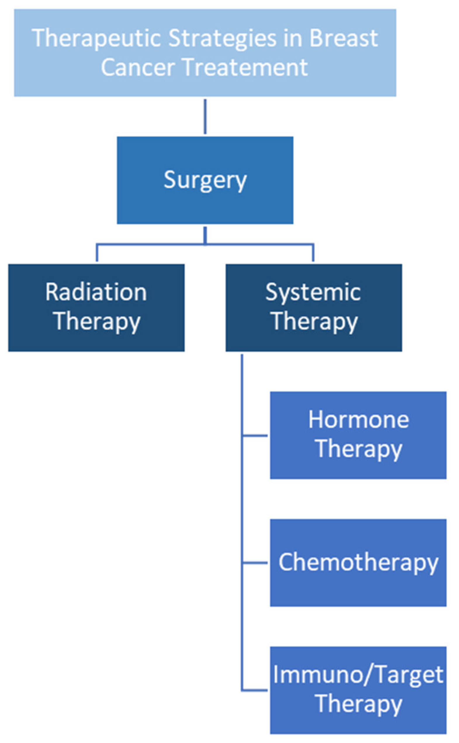
3.3.2. Systemic Therapy

The role of systemic therapy in BC has been a subject of ongoing debate, with several studies suggesting no clear correlation between chemo-hormonal therapy and breast toxicity [19]. In a cross-sectional survey conducted by Ishiyama et al. [33] involving 247 patients, cases of grade 2 or higher toxicity were relatively rare (0.52–7.8%). However, multivariate analysis indicated that chemotherapy was associated with increased edema (p = 0.04). Additionally, Keller et al. [34] analyzed outcomes in 946 patients treated with HF adjuvant RT, finding that chemotherapy was linked to a higher incidence of side effects at the 5-year follow-up.
Within the context of hormonal therapy, numerous studies have compared tamoxifen and aromatase inhibitors, though few have specifically addressed late breast toxicity, often focusing more on systemic side effects [35]. Notably, Meattini et al. [36] investigated treatment de-escalation strategies in patients with favorable prognoses. Their phase 3 randomized multicenter trial included women aged 70 years or older with histologically confirmed stage I, luminal A-like BC. The study compared single-modality endocrine therapy (control arm) to RT (experimental arm), with co-primary endpoints being quality of life and local recurrence. Interim analysis at 24 months, involving 207 patients, showed that RT had a lesser impact on quality of life (p = 0.045) and resulted in fewer treatment-related adverse events compared with the control group.

3.3.3. Dose

As radiation oncologists, treatment-related factors are central to our ability to influence patients’ toxicity outcomes following irradiation. The variables directly affected by the choices of the radiation oncologist include prescription dose, volumes of interest, and irradiation techniques.
In the context of BC, adjuvant RT is typically prescribed under three main regimens: CF (50 Gy in 25 fractions), HF (40–42 Gy in 15–16 fractions), and ultra-HF (26 Gy in 5 fractions). Dose schedules in terms of dose per fraction and Biologically Effective Dose (BED) are shown in Table 2. The BED [37] for disease control, assuming an α/β ratio of 3.7 [38] Gy, is 77.0 Gy, 68.9 Gy, and 62.5 Gy, respectively. Conversely, in terms of late toxicity, with an α/β ratio of 3 Gy, the BED values are 83.3 Gy for CF, 75.7 Gy for HF, and 71.0 Gy for ultra-HF. Importantly, the overall treatment time does not have a statistically significant impact on photographic changes in breast appearance (p = 0.29) [39]. Nevertheless, the adoption of HF in daily clinical practice has been slow [40], likely due to concerns regarding late toxicities. However, evidence indicates that HF RT is as safe as, or less toxic than, CF regimens.
Whelan et al. [41] randomized 1234 women to receive whole-breast irradiation in either a 3- or 5-week schedule. The control group received 50 Gy in 25 fractions, while the experimental arm received 42.4 Gy in 16 fractions. After a 10-year follow-up, there were no differences in disease recurrence. Skin and cosmetic toxic effects were slightly lower with the CF schedule compared with HF (29.5% and 28.7% versus 33.2% and 30.2%, respectively). However, subcutaneous toxicity was less frequent in the HF arm (51.9% versus 54.7%), though these differences were not statistically significant.
Haviland et al. [42] analyzed 4451 patients enrolled in the START-A and B trials, comparing CF and HF RT schedules. Disease control was comparable between the regimens, but moderate or severe breast induration, telangiectasia, and breast edema were significantly less common with the HF schedule.
Reddy et al. [43] conducted a study involving 287 patients randomized from 2011 to 2014 to receive either 42.4 Gy in 16 fractions or 50 Gy in 25 fractions to the whole breast. Digital photographs taken one year post-treatment were analyzed for six quantitative measures of breast symmetry. Hypofractionated treatment yielded better results in terms of nipple positioning and vertical symmetry, with no significant differences in other cosmetic outcomes.
The DBCG HYPO trial [44] randomized 1854 patients to receive either 50 Gy in 25 fractions or 40 Gy in 15 fractions, with a primary endpoint of 3-year grade 2–3 breast induration. At three years, induration rates were 11.8% in the control arm versus 9.0% in the experimental arm, yielding a risk difference of −2.7% (p = 0.07). At a median follow-up of over seven years, HF was associated with significantly lower rates of induration (p = 0.029), cosmetic complications (p = 0.032), dyspigmentation (p < 0.001), and edema (p = 0.044).
Finally, Lu et al. [45] conducted a meta-analysis comparing the safety and efficacy of HF and CF fractionated RT for BC. Of 288 identified studies, 35 were selected, comprising data from 18,246 patients. While no differences were observed in disease control, HF was non-inferior in terms of breast pain, breast atrophy, and lymphedema. Additionally, HF regimens were associated with significantly lower rates of skin toxicity (p < 0.01) and patient fatigue (p < 0.01).
Evidence on ultra-HF irradiation remains limited. The FAST trial [46] reported data on 915 women randomized to receive either 50 Gy in 25 fractions or 30 Gy or 28.5 Gy in 5 once-weekly fractions, with cosmetic outcomes as the primary endpoint. At five years, changes in photographic breast appearance were significantly higher in the 30 Gy group (p = 0.019) but not in the 28.5 Gy group (p = 0.686) compared with the control. Physician-assessed normal tissue effects were more frequent in the 30 Gy group (p < 0.001) but not in the 28.5 Gy arm (p = 0.248). At a median follow-up of 9.9 years, disease control was comparable across all three groups.
The FAST-Forward trial [47], a phase III randomized trial, compared three dose schedules for adjuvant RT in BC patients: 40 Gy in 15 fractions, 27 Gy in 5 daily fractions, and 26 Gy in 5 daily fractions. At five years, disease control was similar across the groups. However, substantial clinician-assessed toxicities were observed in 9.9% of patients in the 40 Gy group, 15.4% in the 27 Gy group, and 11.9% in the 26 Gy group. The 27 Gy arm exhibited significantly higher side effects compared with the control group (p < 0.0001), whereas the 26 Gy arm showed comparable outcomes to the 40 Gy group (p = 0.20).
Sigaudi et al. [48] proposed a comparison between 28.5 Gy in 5 once-weekly fractions and 26 Gy in 5 daily fractions in their prospective study. An interim analysis of 70 patients revealed that at six months, the 26 Gy group experienced 16.7% grade 2 skin induration, 8.3% grade 2 breast pain, and 8.3% fair cosmetic outcomes, with no grade ≥ 3 toxicities reported. In the 28.5 Gy group, no grade ≥ 3 side effects or fair/worse cosmetic outcomes were documented. However, these findings are preliminary.

3.3.4. Volumes

Optimizing irradiation volumes is essential for a radiation oncologist. The objective is to minimize these volumes as much as possible in order to reduce toxicity while maintaining optimal disease control. Several studies suggest that most local recurrences occur at the site of the primary tumor [49,50]. Veronesi et al. [49] randomly assigned 579 women with breast carcinoma to undergo quadrantectomy and axillary dissection, with (299) or without (280) RT. Among 69 ipsilateral breast tumor recurrences (IBTR), 59 (85.5%) occurred within the so-called “scar area”. Gage et al. [50] analyzed 1628 patients who underwent tumor excision and received a dose > 60 Gy to the tumor bed. The crude recurrence rates at 5 and 10 years were 5.7% and 9.3% for recurrences in the tumor bed, and 0.9% and 2.8% for recurrences at other sites, respectively. Vicini et al. [51] conducted a pathological analysis to precisely define areas with a higher likelihood of recurrence. Of the 333 cases studied, 303 (90.9%) had residual microscopic disease within 15 mm of the initial tumor.
In this context, as outlined in several randomized phase III trials (Table 3), external beam partial breast irradiation (PBI) has been shown to be comparable to whole-breast irradiation (WBI) in terms of oncological outcomes in highly selected patients. However, concerns remain regarding the optimal dose schedule to be used [52]. The UK IMPORT LOW trial evaluated PBI in a three-arm non-inferiority study [53]. A total of 2018 women with tumors ≤ 3 cm and 0–3 involved axillary nodes were randomized to receive external beam PBI of 40 Gy in 15 fractions (experimental arm), a combination of 36 Gy WBI + 40 Gy to the tumor bed (reduced dose arm), or 40 Gy in 15 fractions of WBI alone (control arm). Local control, regional recurrence, distant relapse, and overall survival were similar between the groups. At the 5-year follow-up, changes in breast appearance were significantly better in the PBI group (p < 0.0001). Harder or firmer breasts were significantly less frequent in the PBI arm (p = 0.002 and p < 0.0001, respectively) compared with the WBI group. Patient-reported changes in breast appearance were lower after PBI compared with WBI alone (15% vs. 27%, respectively, p < 0.001).
Whelan et al. [54], in the OCOG-RAPID trial, randomized 2135 BC patients (N0) with tumors ≤ 3 cm to receive either external beam PBI or WBI. In the experimental arm, 38.5 Gy in 10 fractions was delivered twice daily (at least six hours apart), while the control group received 42.56 Gy in 16 fractions or 50 Gy in 25 fractions, with or without a 10 Gy boost in 4–5 daily fractions. Disease control was comparable between PBI and WBI. However, late grade ≥ 2 and grade 3 toxicities were 32% vs. 13% (p < 0.0001) and 4.5% vs. 1.0% (p < 0.0001), respectively. Specifically, PBI led to a higher incidence of breast induration and skin telangiectasia. Cosmetic outcomes at 7 years were also worse with PBI compared with WBI. Fair or poor results, as assessed by nurses, were seen in 36% of the PBI group and 19% of the WBI group.
The NSABP B-39/RTOG 0413 [55] trial design was similar to the OCOG-RAPID trial. Vicini et al. adopted the same therapeutic approach but modified some selection criteria: eligible women were 18 years or older with tumors ≤ 3 cm and 0–3 involved axillary nodes. The PBI group reported more grade ≥ 3 side effects (10% vs. 7%). Additionally, physician-assessed cosmesis at 36 months, as reported by White et al. [56], was worse for the experimental arm.
These findings are further supported by the IRMA trial [57], which compared 38.5 Gy in 10 fractions of twice-daily PBI to WBI in 3309 patients. At a median follow-up of five years, adverse cosmetic outcomes were significantly higher in the PBI arm (p = 0.012). Additionally, late soft tissue toxicity (grade ≥ 3) was notably more frequent in the PBI group (p < 0.0001).
In the FLORENCE trial [58], eligible patients were women over 40 years with early-stage BC ≤ 25 mm. Randomization compared 30 Gy in 5 fractions of PBI over two weeks vs. 50 Gy in 25 daily fractions of WBI (+/− a 10 Gy boost to the tumor bed in 5 daily fractions). The 10-year cumulative incidence of IBTR was 2.5% (n° of patients = 6) in the WBI group and 3.7% (n = 9) in the PBI group, respectively (p = 0.40) [59]. Ten-year overall survival was similar between the groups (p = 0.86). PBI was associated with 5 (2%) cases of grade 2 late toxicities, while WBI reported 7 (2.7%) cases of grade 2 late effects. Furthermore, fair or poor cosmetic outcomes were more frequent in the WBI group compared with the PBI group: 1.9% vs. 0% (p = 0.0001) and 14.6% vs. 0.8% (p = 0.0001) for patient and physician-assessed outcomes, respectively.
Nevertheless, Colciago et al. [60] conducted a case–control study using real-world data, comparing 536 patients evenly divided between PBI (30 Gy in 5 daily fractions) and WBI (42.40 Gy in 16 fractions) cohorts. Multivariate Cox proportional hazard regression revealed a significantly higher incidence of fat necrosis in the PBI group (HR = 2.2, 95% CI: 1.2–4.0; p = 0.01).
Therefore, as shown in Table 4, selected patients with early-stage BC should be considered for PBI over WBI due to reduced late toxicities and improved cosmetic outcomes. The recommended dose schedules are 40.05 Gy in 15 fractions or 30 Gy in 5 fractions. However, the RAPID-like regimen is strongly discouraged.

3.3.5. Technique

The choice of irradiation technique often lies between two options: 3D conformal radiation therapy (3DCRT) or IMRT/volumetric arc-modulated radiation therapy (VMAT).
Donovan et al. [61] investigated the effects of WBI in a cohort of 360 patients, who were randomized to receive either conformal RT or IMRT. The study revealed that changes in breast appearance were 1.7 times more likely to be reported in the 3DCRT arm compared with IMRT (p = 0.008). Furthermore, IMRT significantly reduced the incidence of palpable induration, indicating its potential advantage in preserving cosmetic outcomes. Hörner-Rieber et al. [62] conducted a prospective, multicenter, randomized phase III trial comparing 3DCRT and IMRT for BC irradiation. A total of 502 patients were randomized, and after a median follow-up of 5.1 years, no significant differences were observed in disease control, late toxicities, or cosmetic outcomes. Mukesh et al. [63] randomized 815 BC patients to receive 40 Gy in 15 fractions delivered via 3DCRT versus IMRT. The primary endpoints were late cosmetic and breast toxicity outcomes. IMRT demonstrated fewer instances of suboptimal cosmesis (p = 0.027) and skin telangiectasia (p = 0.021) in univariate analysis. These findings were consistent in multivariate analysis for both outcomes (p = 0.038 and p = 0.031, respectively).
As shown in Figure 3, achieving optimal treatment target conformity and uniform dose distribution with 3DCRT can be challenging, often resulting in higher maximum doses to small sections of organs at risk (OARs) such as the heart and lungs [64]. IMRT and VMAT offer improved conformity and dose distribution, but they can increase low-dose radiation exposure to OARs, including the heart, lungs, and contralateral breast. Additionally, IMRT and VMAT require greater expertise and increased monitor unit usage from the linear accelerator, which may lead to higher resource consumption [65].
Deep inspiration breath hold (DIBH) has shown dosimetric advantages with both 3DCRT and IMRT/VMAT techniques. Berg et al. [66] analyzed 1327 treatment plans comparing free-breathing to respiratory gating techniques. Median target coverage was comparable between right-sided free-breathing and left-sided respiratory gating patients, while free-breathing left-sided patients showed less target coverage. Mean heart dose and mean lung dose were significantly lower in the left-sided respiratory gating group compared with the free-breathing left-sided group.
Despite these dosimetric advantages, clinical evidence on the impact of these techniques on late heart toxicities remains limited. Given the widespread availability of these advanced techniques for whole-breast irradiation (WBI), it is crucial to select the most suitable method to achieve optimal treatment outcomes. This includes ensuring proper target coverage, dose homogeneity, and sparing of OARs, leveraging all available technological advancements.

3.3.6. Boost Administration

Boost administration has proven beneficial for disease control in selected patients, as demonstrated by several randomized trials [67,68,69,70,71,72]. However, boost therapy is associated with worse outcomes. Colette et al. [73] explored predictors of fibrosis 10 years post-treatment in their “boost versus no boost” trial. Their analysis of 5178 patients revealed that moderate to severe late toxicities occurred in 485 patients (26.9%) in the boost group compared with 230 patients (12.6%) in the no-boost group (p < 0.0001).
To mitigate this toxicity, radiation oncologists have two strategies: improved patient selection and enhanced target delineation. The omission of boost may be considered for patients over 70 years of age with hormone receptor-positive tumors of low or intermediate grade resected with widely negative (≥2 mm) margins, following ASTRO guidelines [74]. As depicted in Table 4, strong indications for boost include patients with invasive cancer who are ≤50 years of age, patients aged 51 to 70 years with high-grade tumors, or those with positive margins [70]. Conversely, the St. Gallen expert consensus conference suggested omitting boosts in patients younger than 60 years with low-grade tumors or a favorable biological profile [75].
Target delineation of the tumor bed presents significant interobserver variability. It is critical to understand that higher boost doses correlate with increased toxicities. Addressing this challenge requires a multidisciplinary approach involving radiation oncologists, surgeons, and radiologists. The use of clips in the lumpectomy cavity is essential for accurate target delineation. Additionally, integrating preoperative imaging such as CT and MRI can further enhance precision [76].

4. Discussion

This review provides an extensive and in-depth analysis of the complex interplay between non-modifiable and modifiable factors influencing late breast toxicity after radiotherapy, unveiling its multifaceted nature. As shown in Table 1, if, on one hand, inherent patient characteristics such as genetic predisposition, age, and breast volume may serve as substantial predictors of toxicity [8,21,25], a number of adjustable treatment parameters under the control of radiation oncologists can significantly reduce adverse effects [77].
Genetic radiosensitivity emerges as a pivotal factor in oncology, offering novel pathways for personalized treatment regimens [12,78]. Although its full integration into clinical practice remains challenging, advancements in genetic profiling hold tremendous promise for the future of oncologic strategies [79]. As these genetic models become more sophisticated, they will enable tailored radiotherapy approaches that can minimize toxicity without losing clinical effectiveness. Additionally, radiomics—a rapidly evolving field that leverages advanced imaging techniques to extract and analyze complex textural features—presents a promising tool for enhancing risk stratification and predictive accuracy, empowering clinicians to proactively adjust treatment plans [18]. To foster a more personalized approach to treatment, it is crucial to embrace a model-based probability as a guiding tool for selecting treatments. In this context, personalized radiotherapy means tailoring radiation doses according to each patient’s individual risk (e.g., escalating or de-escalating doses for high-risk and low-risk patients, respectively). To aim at this purpose, it becomes paramount to integrate clinical data with insights into biological mechanisms, thereby enhancing the predictive value of these models [80].
Non-modifiable risk factors such as age and breast volume consistently emerge as significant contributors to late breast toxicity. Older patients frequently develop higher rates of fibrosis and breast edema, compromising both aesthetic and functional outcomes [20,21], while larger breast volumes introduce challenges due to dose inhomogeneity, leading to increased toxicity risks [25,26]. These findings emphasize the need for individualized radiation plans that consider unique anatomical and physiological characteristics, ensuring optimal and patient-specific care. A key focus of this review is on modifiable factors that can mitigate late breast toxicity. Among these, the selection of radiation dose and fractionation schedules is particularly influential. Hypofractionation, which administers higher doses over fewer sessions, has demonstrated efficacy in reducing toxicity without compromising therapeutic outcomes, establishing its value in modern radiotherapy [42,43,44,45]. Advanced irradiation techniques such as IMRT and VMAT further enhance therapeutic outcomes by improving dose homogeneity and minimizing radiation exposure to surrounding healthy tissues [61,62,63]. Despite the resource demands associated with these technologies, their benefits in reducing late toxicities and improving cosmetic results are well documented [64].
Furthermore, patient positioning during radiotherapy, adaptive planning based on anatomical changes, and the use of bolus materials are critical modifiable elements that contribute to optimal dose delivery and reduced toxicity [81]. Comprehensive patient management also plays a crucial role in mitigating modifiable risk factors, including lifestyle interventions such as smoking cessation, weight management, and diabetes control [82]. Pharmacological interventions, including the use of radioprotective agents, are also being explored as adjuncts to minimize toxicity [83]. Personalized patient education and counseling enhance adherence to these lifestyle modifications, ultimately contributing to better long-term outcomes [84]. Additionally, careful selection and administration of systemic therapies, including chemotherapy and hormonal treatments, help balance oncological efficacy with potential side effects, ensuring an individualized approach to treatment [85].
Our findings highlight the critical importance of meticulous treatment planning and technological integration in minimizing late breast toxicity. Optimizing target volumes, employing advanced imaging for precise delineation, and adhering to evidence-based dose constraints are essential strategies for effective radiotherapy. As the field continues to evolve, integrating genetic and radiomic data into clinical workflows holds vast potential for enhancing personalized care, improving patient outcomes, and ensuring a higher quality of life post radiotherapy. In conclusion, late breast toxicity post radiotherapy is influenced by a complex interplay of inherent and modifiable factors. A specific emphasis on modifiable factors reveals that radiation oncologists can significantly reduce toxicity through informed clinical decisions regarding dose selection, advanced irradiation techniques, adaptive planning, and comprehensive patient management. Future research should continue to refine these modifiable parameters, integrate emerging technologies, and validate innovative approaches to achieve the dual goal of effective cancer control and minimal toxicity, ultimately enhancing the quality of life for BC survivors.

5. Conclusions

This review unveils that late breast toxicity post radiotherapy is influenced by a complex spectrum of factors, both inherent and adjustable. Radiation therapy has well-established benefits in improving loco-regional control, distant disease control, breast cancer-specific survival, and overall survival in patients with breast cancer. These advantages are particularly clear with the adoption of modern RT techniques, which have demonstrated efficacy irrespective of contemporary systemic therapies. However, in the setting of long-term survivorship, RT poses specific challenges, with late-onset toxicities representing a critical factor affecting quality of life outcomes.
Radiation oncologists play a pivotal role in mitigating these effects through informed clinical decisions, technological advancements, and comprehensive patient management. Future research should focus on validating genetic predictors, refining radiomic models, and optimizing treatment protocols to achieve the dual objectives of maximizing tumor control with minimal toxicity. Personalized treatment approaches will massively contribute to improved patient outcomes and quality of life.

Author Contributions

Conceptualization, R.R.C. and S.A.; methodology, R.R.C. and S.A.; investigation, R.R.C.; writing—original draft preparation, R.R.C. and S.A.; writing—review and editing, C.C., F.F., I.M., M.M., G.R. and L.D.S.; supervision, S.A. All authors have read and agreed to the published version of the manuscript.

Funding

This research received no external funding.

Acknowledgments

Pietro Invernizzi, I.R.C.C.S. San Gerardo dei Tintori.

Conflicts of Interest

The authors declare no conflicts of interest.

Abbreviations

The following abbreviations are used in this manuscript:
RTRadiation therapy
BCBreast cancer
SNPsSingle-nucleotide polymorphism
CTComputed tomography
IMRTIntensity-modulated radiation therapy
CFConventional Fractionation
HFHypofractionated
AUCArea under the curve
BEDBiologically effective dose
IBTRIpsilateral breast tumor recurrences
PBIPartial breast irradiation
WBIWhole-breast irradiation
3DCRT3D conformal radiation therapy
VMATVolumetric arc modulated radiation therapy
OAROrgans at risk
DIBHDeep inspiration breath hold

References

  1. Veronesi, U.; Cascinelli, N.; Mariani, L.; Greco, M.; Saccozzi, R.; Luini, A.; Aguilar, M.; Marubini, E. Twenty-year follow-up of a randomized study comparing breast-conserving surgery with radical mastectomy for early breast cancer. N. Engl. J. Med. 2002, 347, 1227–1232. [Google Scholar] [CrossRef] [PubMed]
  2. Tong, C.W.S.; Wu, M.; Cho, W.C.S.; To, K.K.W. Recent Advances in the Treatment of Breast Cancer. Front. Oncol. 2018, 8, 227. [Google Scholar] [CrossRef] [PubMed]
  3. Garibaldi, C.; Jereczek-Fossa, B.A.; Marvaso, G.; Dicuonzo, S.; Rojas, D.P.; Cattani, F.; Starzyńska, A.; Ciardo, D.; Surgo, A.; Leonardi, M.C.; et al. Recent advances in radiation oncology. ecancer Med. Sci. 2017, 11, 785. [Google Scholar] [CrossRef]
  4. EBCTCG (Early Breast Cancer Trialists’ Collaborative Group); McGale, P.; Taylor, C.; Correa, C.; Cutter, D.; Duane, F.; Ewertz, M.; Wang, Y. Effect of radiotherapy after mastectomy and axillary surgery on 10-year recurrence and 20-year breast cancer mortality: Meta-analysis of individual patient data for 8135 women in 22 randomised trials. Lancet 2014, 383, 2127–2135. [Google Scholar] [CrossRef] [PubMed]
  5. Early Breast Cancer Trialists’ Collaborative Group (EBCTCG); Darby, S.; McGale, P.; Correa, C.; Taylor, C.; Arriagada, R.; Clarke, M.; Cutter, D.; Davies, C.; Ewertz, M.; et al. Effect of radiotherapy after breast-conserving surgery on 10-year recurrence and 15-year breast cancer death: Meta-analysis of individual patient data for 10 801 women in 17 randomised trials. Lancet 2011, 378, 1707–1716. [Google Scholar] [CrossRef]
  6. Taylor, C.; Dodwell, D.; McGale, P.; Hills, R.K.; Berry, R.; Bradley, R.; Braybrooke, J.; Clarke, M.; Gray, R.; Holt, F.; et al. Radiotherapy to regional nodes in early breast cancer: An individual patient data meta-analysis of 14,324 women in 16 trials. Lancet 2023, 402, 1991–2003. [Google Scholar] [CrossRef]
  7. De Santis, M.C.; Samy, F.; De Rose, F.; Meduri, B.; Lambertini, M.; Sotiriou, C.; Carnevale, M.G.; Verderio, P.; Aspitia, M.; Piccart, M.; et al. 152MO Post-mastectomy radiation therapy in patients with human epidermal growth factor receptor 2 (HER2)-positive breast cancer: Analysis of the adjuvant lapatinib and/or trastuzumab treatment optimization (ALTTO) trial [BIG 2-06/NCCTG N063D (Alliance)]. ESMO Open 2025, 10, 104706. [Google Scholar] [CrossRef]
  8. Roberts, S.; Spreadborough, A.; Bulman, B.; Barber, J.; Evans, D.; Scott, D. Heritability of cellular radiosensitivity: A marker of low-penetrance predisposition genes in breast cancer? Am. J. Hum. Genet. 1999, 65, 784–794. [Google Scholar] [CrossRef]
  9. Kerns, S.L.; Dorling, L.; Fachal, L.; Bentzen, S.; Pharoah, P.D.; Barnes, D.R.; Gómez-Caamaño, A.; Carballo, A.M.; Dearnaley, D.P.; Peleteiro, P.; et al. Meta-analysis of Genome Wide Association Studies Identifies Genetic Markers of Late Toxicity Following Radiotherapy for Prostate Cancer. eBioMedicine 2016, 10, 150–163. [Google Scholar] [CrossRef]
  10. Barnett, G.C.; Elliott, R.M.; Alsner, J.; Andreassen, C.N.; Abdelhay, O.; Burnet, N.G.; Chang-Claude, J.; Coles, C.E.; Gutiérrez-Enríquez, S.; Fuentes-Raspall, M.J.; et al. Individual patient data meta-analysis shows no association between the SNP rs1800469 in TGFB and late radiotherapy toxicity. Radiother. Oncol. 2012, 105, 289–295. [Google Scholar] [CrossRef]
  11. Jandu, H.K.; Veal, C.D.; Fachal, L.; Luccarini, C.; Aguado-Barrera, M.E.; Altabas, M.; Azria, D.; Baten, A.; Bourgier, C.; Bultijnck, R.; et al. Genome-wide association study of treatment-related toxicity two years following radiotherapy for breast cancer. Radiother. Oncol. 2023, 187, 109806. [Google Scholar] [CrossRef] [PubMed]
  12. Magnuson, A.; Sedrak, M.S.; Gross, C.P.; Tew, W.P.; Klepin, H.D.; Wildes, T.M.; Muss, H.B.; Dotan, E.; Freedman, R.A.; O’Connor, T.; et al. Development and Validation of a Risk Tool for Predicting Severe Toxicity in Older Adults Receiving Chemotherapy for Early-Stage Breast Cancer. J. Clin. Oncol. 2021, 39, 608–618. [Google Scholar] [CrossRef]
  13. Llorián-Salvador, Ó.; Windeler, N.; Martin, N.; Etzel, L.; Andrade-Navarro, M.A.; Bernhardt, D.; Rost, B.; Borm, K.J.; Combs, S.E.; Duma, M.N.; et al. CT-based radiomics for predicting breast cancer radiotherapy side effects. Sci Rep. 2024, 14, 20051. [Google Scholar] [CrossRef]
  14. Feng, H.; Wang, H.; Xu, L.; Ren, Y.; Ni, Q.; Yang, Z.; Ma, S.; Deng, Q.; Chen, X.; Xia, B.; et al. Prediction of radiation-induced acute skin toxicity in breast cancer patients using data encapsulation screening and dose-gradient-based multi-region radiomics technique: A multicenter study. Front. Oncol. 2022, 12, 1017435. [Google Scholar] [CrossRef] [PubMed]
  15. Kraus, K.M.; Oreshko, M.; Bernhardt, D.; Combs, S.E.; Peeken, J.C. Dosiomics and radiomics to predict pneumonitis after thoracic stereotactic body radiotherapy and immune checkpoint inhibition. Front. Oncol. 2023, 13, 1124592. [Google Scholar] [CrossRef] [PubMed]
  16. Avanzo, M.; Pirrone, G.; Vinante, L.; Caroli, A.; Stancanello, J.; Drigo, A.; Massarut, S.; Mileto, M.; Urbani, M.; Trovo, M.; et al. Electron Density and Biologically Effective Dose (BED) Radiomics-Based Machine Learning Models to Predict Late Radiation-Induced Subcutaneous Fibrosis. Front. Oncol. 2020, 10, 490. [Google Scholar] [CrossRef]
  17. Soltani, S.; Aliasgharzadeh, A.A.; Fadavi, P.; Bagherpour, Z.; Moradi, H.; Safari, M.; Beigi, M. Enhanced prediction of radiation-induced skin toxicity in breast cancer patients using a hybrid dosiomics-clinical model. J. Radiat. Res. Appl. Sci. 2025, 18, 101382. [Google Scholar] [CrossRef]
  18. Zhou, B.; Wang, J.; Yang, X.; Henry, S.; Lin, J.Y.; Torres, M.A.; Liu, T. Ultrasound Histogram Assessment of Acute Breast Toxicity After Breast Cancer Radiation Therapy: A Prospective Longitudinal Study. Ultrasound Med. Biol. 2022, 49, 309–317. [Google Scholar] [CrossRef]
  19. Batenburg, M.; Bartels, M.; Maarse, W.; Witkamp, A.; Verkooijen, H.; Bongard, H.v.D. Factors Associated with Late Local Radiation Toxicity after Post-Operative Breast Irradiation. Breast J. 2022, 2022, 6745954. [Google Scholar] [CrossRef]
  20. Lilla, C.; Ambrosone, C.B.; Kropp, S.; Helmbold, I.; Schmezer, P.; von Fournier, D.; Haase, W.; Sautter-Bihl, M.-L.; Wenz, F.; Chang-Claude, J. Predictive factors for late normal tissue complications following radiotherapy for breast cancer. Breast Cancer Res. Treat. 2007, 106, 143–150. [Google Scholar] [CrossRef] [PubMed]
  21. Barnett, G.C.; Wilkinson, J.S.; Moody, A.M.; Wilson, C.B.; Twyman, N.; Wishart, G.C.; Burnet, N.G.; Coles, C.E. The Cambridge Breast Intensity-modulated Radiotherapy Trial: Patient- and treatment-related factors that influence late toxicity. Clin. Oncol. 2011, 23, 662–673. [Google Scholar] [CrossRef] [PubMed]
  22. Ratosa, I.; Jenko, A.; Oblak, I. Breast size impact on adjuvant radiotherapy adverse effects and dose parameters in treatment planning. Radiol. Oncol. 2018, 52, 233–244. [Google Scholar] [CrossRef]
  23. Thomsen, M.S.; Alsner, J.; Lutz, C.M.; Berg, M.; Jensen, I.; Lorenzen, E.L.; Nielsen, H.M.; Jakobsen, E.H.; Stenbygaard, L.; Nielsen, M.H.; et al. Breast induration and irradiated volume in the DBCG HYPO trial: The impact of age, smoking, and boost. Radiother. Oncol. 2024, 201, 110574. [Google Scholar] [CrossRef] [PubMed]
  24. De Rose, F.; Fogliata, A.; Franceschini, D.; Iftode, C.; D’agostino, G.R.; Comito, T.; Franzese, C.; Di Brina, L.; Clerici, E.; Loi, M.; et al. Hypofractionated Whole Breast Irradiation and Simultaneous Integrated Boost in Large-breasted Patients: Long-term Toxicity and Cosmesis. Clin. Breast Cancer 2020, 20, 527–533. [Google Scholar] [CrossRef]
  25. Neal, A.; Torr, M.; Helyer, S.; Yarnold, J. Correlation of breast dose heterogeneity with breast size using 3D CT planning and dose-volume histograms. Radiother. Oncol. 1995, 34, 210–218. [Google Scholar] [CrossRef]
  26. Colciago, R.R.; Cavallo, A.; Magri, M.C.; Vitullo, A.; La Rocca, E.; Giandini, C.; Bonfantini, F.; Di Cosimo, S.; Baili, P.; Sant, M.; et al. Hypofractionated whole-breast radiotherapy in large breast size patients: Is it really a resolved issue? Med. Oncol. 2021, 38, 107. [Google Scholar] [CrossRef]
  27. Gimbrone, M.A., Jr.; García-Cardeña, G. Endothelial Cell Dysfunction and the Pathobiology of Atherosclerosis. Circ. Res. 2016, 118, 620–636. [Google Scholar] [CrossRef] [PubMed]
  28. Wang, J.; Boerma, M.; Fu, Q.; HauerJensen, M. Significance of endothelial dysfunction in the pathogenesis of early and delayed radiation enteropathy. World J. Gastroenterol. 2007, 13, 3047–3055. [Google Scholar] [CrossRef]
  29. Rattay, T.; Seibold, P.; Aguado-Barrera, M.E.; Altabas, M.; Azria, D.; Barnett, G.C.; Bultijnck, R.; Chang-Claude, J.; Choudhury, A.; Coles, C.E.; et al. External Validation of a Predictive Model for Acute Skin Radiation Toxicity in the REQUITE Breast Cohort. Front. Oncol. 2020, 10, 575909. [Google Scholar] [CrossRef]
  30. Colciago, R.R.; La Rocca, E.; Giandini, C.; Mateo, A.R.; Bedini, N.; Capri, G.; Folli, S.; Lozza, L.; Meroni, S.; Emanuele, P.; et al. One-week external beam partial breast irradiation: Survival and toxicity outcomes. J. Cancer Res. Clin. Oncol. 2023, 149, 10965–10974. [Google Scholar] [CrossRef]
  31. Cinciripini, P.M.; Kypriotakis, G.; Blalock, J.A.; Karam-Hage, M.; Beneventi, D.M.; Robinson, J.D.; Minnix, J.A.; Warren, G.W. Survival Outcomes of an Early Intervention Smoking Cessation Treatment After a Cancer Diagnosis. JAMA Oncol. 2024, 31, e244890. [Google Scholar] [CrossRef]
  32. Digesù, C.; Deodato, F.; Macchia, G.; Cilla, S.; Pieri, M.; Zamagni, A.; Farioli, A.; Buwenge, M.; Ferrandina, G.; Morganti, A.G. Hypofractionated radiotherapy after conservative surgery may increase low–intermediate grade late fibrosis in breast cancer patients. Breast Cancer Targets Ther. 2018, 10, 143–151. [Google Scholar] [CrossRef]
  33. Ishiyama, H.; Niino, K.; Hosoya, T.; Hayakawa, K. Results of a questionnaire survey for symptom of late complications caused by radiotherapy in breast conserving therapy. Breast Cancer 2006, 13, 197–201. [Google Scholar] [CrossRef]
  34. Keller, L.M.; Sopka, D.M.; Li, T.; Klayton, T.; Li, J.; Anderson, P.R.; Bleicher, R.J.; Sigurdson, E.R.; Freedman, G.M. Five-year results of whole breast intensity modulated radiation therapy for the treatment of early stage breast cancer: The fox chase cancer center experience. Int. J. Radiat. Oncol. 2012, 84, 881–887. [Google Scholar] [CrossRef] [PubMed]
  35. Early Breast Cancer Trialists’ Collaborative Group (EBCTCG); Bradley, R.; Braybrooke, J.; Gray, R.; Hills, R.K.; Liu, Z.; Pan, H.; Peto, R.; Dodwell, D.; McGale, P.; et al. Aromatase inhibitors versus tamoxifen in premenopausal women with oestrogen receptor-positive early-stage breast cancer treated with ovarian suppression: A patient-level meta-analysis of 7030 women from four randomised trials. Lancet Oncol. 2022, 23, 382–392. [Google Scholar] [CrossRef] [PubMed]
  36. Meattini, I.; Poortmans, P.M.; Marrazzo, L.; Desideri, I.; Brain, E.; Hamaker, M.; Lambertini, M.; Miccinesi, G.; Russell, N.; Saieva, C.; et al. Exclusive endocrine therapy or partial breast irradiation for women aged ≥70 years with luminal A-like early stage breast cancer (NCT04134598—EUROPA): Proof of concept of a randomized controlled trial comparing health related quality of life by patient reported outcome measures. J. Geriatr. Oncol. 2021, 12, 182–189. [Google Scholar] [CrossRef] [PubMed]
  37. Fowler, J.F. 21 years of Biologically Effective Dose. Br. J. Radiol. 2010, 83, 554–568. [Google Scholar] [CrossRef]
  38. Brunt, A.; Haviland, J.; Kirby, A.; Somaiah, N.; Wheatley, D.; Bliss, J.; Yarnold, J. Five-fraction Radiotherapy for Breast Cancer: FAST-Forward to Implementation. Clin. Oncol. 2021, 33, 430–439. [Google Scholar] [CrossRef]
  39. Haviland, J.S.; Bentzen, S.M.; Bliss, J.M.; Yarnold, J.R.; START Trial Management Group. Prolongation of overall treatment time as a cause of treatment failure in early breast cancer: An analysis of the UK START (Standardisation of Breast Radiotherapy) trials of radiotherapy fractionation. Radiother. Oncol. 2016, 121, 420–423. [Google Scholar] [CrossRef]
  40. Meattini, I.; Palumbo, I.; Becherini, C.; Borghesi, S.; Cucciarelli, F.; Dicuonzo, S.; Fiorentino, A.; Spoto, R.; Poortmans, P.; Aristei, C.; et al. The Italian Association for Radiotherapy and Clinical Oncology (AIRO) position statements for postoperative breast cancer radiation therapy volume, dose, and fractionation. Radiol. Med. 2022, 127, 1407–1411. [Google Scholar] [CrossRef]
  41. Whelan, T.J.; Pignol, J.-P.; Levine, M.N.; Julian, J.A.; MacKenzie, R.; Parpia, S.; Shelley, W.; Grimard, L.; Bowen, J.; Lukka, H.; et al. Long-term results of hypofractionated radiation therapy for breast cancer. N. Engl. J. Med. 2010, 362, 513–520. [Google Scholar] [CrossRef] [PubMed]
  42. Haviland, J.S.; Owen, J.R.; Dewar, J.A.; Agrawal, R.K.; Barrett, J.; Barrett-Lee, P.J.; Dobbs, H.J.; Hopwood, P.; Lawton, P.A.; Magee, B.J.; et al. The UK Standardisation of Breast Radiotherapy (START) trials of radiotherapy hypofractionation for treatment of early breast cancer: 10-year follow-up results of two randomised controlled trials. Lancet Oncol. 2013, 14, 1086–1094. [Google Scholar] [CrossRef] [PubMed]
  43. Reddy, J.P.; Lei, X.; Huang, S.-C.; Nicklaus, K.M.; Fingeret, M.C.; Shaitelman, S.F.; Hunt, K.K.; Buchholz, T.A.; Merchant, F.; Markey, M.K.; et al. Quantitative Assessment of Breast Cosmetic Outcome After Whole-Breast Irradiation. Int. J. Radiat. Oncol. 2016, 97, 894–902. [Google Scholar] [CrossRef]
  44. Offersen, B.V.; Alsner, J.; Nielsen, H.M.; Jakobsen, E.H.; Nielsen, M.H.; Krause, M.; Stenbygaard, L.; Mjaaland, I.; Schreiber, A.; Kasti, U.-M.; et al. Hypofractionated Versus Standard Fractionated Radiotherapy in Patients With Early Breast Cancer or Ductal Carcinoma In Situ in a Randomized Phase III Trial: The DBCG HYPO Trial. J. Clin. Oncol. 2020, 38, 3615–3625. [Google Scholar] [CrossRef]
  45. Lu, Y.; Hui, B.; Yang, D.; Li, Y.; Li, B.; Zhou, L.; Xu, L.; Tang, F.; Wang, W.; Chen, R.; et al. Efficacy and safety analysis of hypofractionated and conventional fractionated radiotherapy in postoperative breast cancer patients. BMC Cancer 2024, 24, 181. [Google Scholar] [CrossRef]
  46. Brunt, A.M.; Haviland, J.S.; Sydenham, M.; Agrawal, R.K.; Algurafi, H.; Alhasso, A.; Barrett-Lee, P.; Bliss, P.; Bloomfield, D.; Bowen, J.; et al. Ten-Year Results of FAST: A Randomized Controlled Trial of 5-Fraction Whole-Breast Radiotherapy for Early Breast Cancer. J. Clin. Oncol. 2020, 38, 3261–3272. [Google Scholar] [CrossRef]
  47. Murray Brunt, A.; Haviland, J.S.; Wheatley, D.A.; Sydenham, M.A.; Alhasso, A.; Bloomfield, D.J.; Chan, C.; Churn, M.; Cleator, S.; Coles, C.E.; et al. Hypofractionated breast radiotherapy for 1 week versus 3 weeks (FAST-Forward): 5-year efficacy and late normal tissue effects results from a multicentre, non-inferiority, randomised, phase 3 trial. Lancet 2020, 395, 1613–1626. [Google Scholar] [CrossRef]
  48. Sigaudi, V.; Zannetti, M.; Ferrara, E.; Manfredda, I.; Mones, E.; Loi, G.; Krengli, M.; Franco, P. Ultra-Hypofractionation for Whole-Breast Irradiation in Early Breast Cancer: Interim Analysis of a Prospective Study. Biomedicines 2022, 10, 2568. [Google Scholar] [CrossRef] [PubMed]
  49. Veronesi, U.; Marubini, E.; Mariani, L.; Galimberti, V.; Luini, A.; Veronesi, P.; Salvadori, B.; Zucali, R. Radiotherapy after breast-conserving surgery in small breast carcinoma: Long-term results of a randomized trial. Ann. Oncol. 2001, 12, 997–1003. [Google Scholar] [CrossRef]
  50. Gage, I.; Recht, A.; Gelman, R.; Nixon, A.J.; Silver, B.; Bornstein, B.A.; Harris, J.R. Long-term outcome following breast-conserving surgery and radiation therapy. Int. J. Radiat. Oncol. 1995, 33, 245–251. [Google Scholar] [CrossRef]
  51. Vicini, F.A.; Kestin, L.L.; Goldstein, N.S. Defining the clinical target volume for patients with early-stage breast cancer treated with lumpectomy and accelerated partial breast irradiation: A pathologic analysis. Int. J. Radiat. Oncol. 2004, 60, 722–730. [Google Scholar] [CrossRef] [PubMed]
  52. Goldberg, M.; Whelan, T.J. Accelerated Partial Breast Irradiation (APBI): Where Are We Now? Curr. Breast Cancer Rep. 2020, 12, 275–284. [Google Scholar] [CrossRef]
  53. Coles, C.E.; Griffin, C.L.; Kirby, A.M.; Titley, J.; Agrawal, R.K.; Alhasso, A.; Bhattacharya, I.S.; Brunt, A.M.; Ciurlionis, L.; Chan, C.; et al. Partial-breast radiotherapy after breast conservation surgery for patients with early breast cancer (UK IMPORT LOW trial): 5-year results from a multicentre, randomised, controlled, phase 3, non-inferiority trial. Lancet 2017, 390, 1048–1060. [Google Scholar] [CrossRef]
  54. Whelan, T.J.; Julian, J.A.; Berrang, T.S.; Kim, D.-H.; Germain, I.; Nichol, A.M.; Akra, M.; Lavertu, S.; Germain, F.; Fyles, A.; et al. External beam accelerated partial breast irradiation versus whole breast irradiation after breast conserving surgery in women with ductal carcinoma in situ and node-negative breast cancer (RAPID): A randomised controlled trial. Lancet 2019, 394, 2165–2172. [Google Scholar] [CrossRef] [PubMed]
  55. Vicini, F.A.; Cecchini, R.S.; White, J.R.; Arthur, D.W.; Julian, T.B.; Rabinovitch, R.A.; Kuske, R.R.; Ganz, P.A.; Parda, D.S.; Scheier, M.F.; et al. Long-term primary results of accelerated partial breast irradiation after breast-conserving surgery for early-stage breast cancer: A randomised, phase 3, equivalence trial. Lancet 2019, 394, 2155–2164. [Google Scholar] [CrossRef]
  56. White, J.; Winter, K.; Cecchini, R.; Vicini, F.; Arthur, D.; Kuske, R.; Rabinovitch, R.; Sehkon, A.; Khan, A.; Chmura, S.; et al. Cosmetic outcome from post lumpectomy whole breast irradiation (WBI) versus partial breast irradiation (PBI) on the NRG oncology/NSABP B39-RTOG 0413 phase III clinical trial. Int. J. Radiat. Oncol. 2019, 105, S3–S4. [Google Scholar] [CrossRef]
  57. Meduri, B.; Baldissera, A.; Iotti, C.; Scheijmans, L.J.; Stam, M.R.; Parisi, S.; Boersma, L.J.; Ammendolia, I.; Koiter, E.; Valli, M.; et al. Cosmetic Results and Side Effects of Accelerated Partial-Breast Irradiation Versus Whole-Breast Irradiation for Low-Risk Invasive Carcinoma of the Breast: The Randomized Phase III IRMA Trial. J. Clin. Oncol. 2023, 41, 2201–2210. [Google Scholar] [CrossRef]
  58. Livi, L.; Meattini, I.; Marrazzo, L.; Simontacchi, G.; Pallotta, S.; Saieva, C.; Paiar, F.; Scotti, V.; Cardillo, C.D.L.; Bastiani, P.; et al. Accelerated partial breast irradiation using intensity-modulated radiotherapy versus whole breast irradiation: 5-year survival analysis of a phase 3 randomised controlled trial. Eur. J. Cancer 2015, 51, 451–463. [Google Scholar] [CrossRef]
  59. Meattini, I.; Marrazzo, L.; Saieva, C.; Desideri, I.; Scotti, V.; Simontacchi, G.; Bonomo, P.; Greto, D.; Mangoni, M.; Scoccianti, S.; et al. Accelerated Partial-Breast Irradiation Compared With Whole-Breast Irradiation for Early Breast Cancer: Long-Term Results of the Randomized Phase III APBI-IMRT-Florence Trial. J. Clin. Oncol. 2020, 38, 4175–4183. [Google Scholar] [CrossRef]
  60. Colciago, R.R.; La Rocca, E.; Giandini, C.; Carnevale, M.G.; Bianchi, G.V.; Maugeri, I.; Depretto, C.; Meroni, S.; Cavallo, A.; Pignoli, E.; et al. Fat necrosis after accelerated partial breast irradiation or hypofractionated whole breast irradiation: A case-control study. Tumori J. 2024, 110, 451–461. [Google Scholar] [CrossRef]
  61. Donovan, E.; Bleakley, N.; Denholm, E.; Evans, P.; Gothard, L.; Hanson, J.; Peckitt, C.; Reise, S.; Ross, G.; Sharp, G.; et al. Randomised trial of standard 2D radiotherapy (RT) versus intensity modulated radiotherapy (IMRT) in patients prescribed breast radiotherapy. Radiother. Oncol. 2007, 82, 254–264. [Google Scholar] [CrossRef] [PubMed]
  62. Hörner-Rieber, J.; Forster, T.; Hommertgen, A.; Haefner, M.F.; Arians, N.; König, L.; Harrabi, S.B.; Schlampp, I.; Weykamp, F.; Lischalk, J.W.; et al. Intensity Modulated Radiation Therapy (IMRT) With Simultaneously Integrated Boost Shortens Treatment Time and Is Noninferior to Conventional Radiation Therapy Followed by Sequential Boost in Adjuvant Breast Cancer Treatment: Results of a Large Randomized Phase III Trial (IMRT-MC2 Trial). Int. J. Radiat. Oncol. 2021, 109, 1311–1324. [Google Scholar] [CrossRef]
  63. Mukesh, M.B.; Barnett, G.C.; Wilkinson, J.S.; Moody, A.M.; Wilson, C.; Dorling, L.; Hak, C.C.W.; Qian, W.; Twyman, N.; Burnet, N.G.; et al. Randomized Controlled Trial of Intensity-Modulated Radiotherapy for Early Breast Cancer: 5-Year Results Confirm Superior Overall Cosmesis. J. Clin. Oncol. 2013, 31, 4488–4495. [Google Scholar] [CrossRef]
  64. De Rose, F.; De Santis, M.C.; Lucidi, S.; Colciago, R.R.; Marino, L.; Cucciarelli, F.; La Rocca, E.; Di Pressa, F.; Lohr, F.; Vanoni, V.; et al. Dose constraints in breast cancer radiotherapy. A critical review. Radiother. Oncol. 2024, 202, 110591. [Google Scholar] [CrossRef]
  65. Fogliata, A.; Burger, H.; Groenewald, A.; Punt, L.; Parkes, J.; Cozzi, L. Intensity Modulated Therapy for Patients With Breast Cancer. Practical Guidelines and Tips for an Effective Treatment Planning Strategy. Adv. Radiat. Oncol. 2024, 9, 101535. [Google Scholar] [CrossRef]
  66. Berg, M.; Lorenzen, E.L.; Jensen, I.; Thomsen, M.S.; Lutz, C.M.; Refsgaard, L.; Nissen, H.D.; Offersen, B.V. The potential benefits from respiratory gating for breast cancer patients regarding target coverage and dose to organs at risk when applying strict dose limits to the heart: Results from the DBCG HYPO trial. Acta Oncol. 2017, 57, 113–119. [Google Scholar] [CrossRef]
  67. Romestaing, P.; Lehingue, Y.; Carrie, C.; Coquard, R.; Montbarbon, X.; Ardiet, J.M.; Mamelle, N.; Gérard, J.P. Role of a 10-Gy boost in the conservative treatment of early breast cancer: Results of a randomized clinical trial in Lyon, France. J. Clin. Oncol. 1997, 15, 963–968. [Google Scholar] [CrossRef] [PubMed]
  68. Graham, P.; Browne, L.; Capp, A.; Fox, C.; Delaney, G.; Kearsley, J. The St George, Wollongong and Liverpool breast boost trial: 1st planned analysis at 6-year mean follow-up. Australas Radiol. 2007, 51 (Suppl. S3), A85. [Google Scholar]
  69. Teissier, E.; Henry, M.; Ramaioli, A.; Lagrange, J.L.; Courdi, A.; Bensadoun, R.J. Boost in conservative treatment: 6 years results of randomized trial. Breast Cancer Res. Treat. 1998, 50, 287. [Google Scholar]
  70. Bartelink, H.; Maingon, P.; Poortmans, P.; Weltens, C.; Fourquet, A.; Jager, J.; Schinagl, D.; Oei, B.; Rodenhuis, C.; Horiot, J.-C.; et al. Whole-breast irradiation with or without a boost for patients treated with breast-conserving surgery for early breast cancer: 20-year follow-up of a randomised phase 3 trial. Lancet Oncol. 2015, 16, 47–56. [Google Scholar] [CrossRef]
  71. Coles, C.; Haviland, J.S.; Kirby, A.M.; Griffin, C.L.; Sydenham, M.A.; Titley, J.C.; Bhattacharya, I.; Brunt, A.M.; Chan, H.Y.C.; Donovan, E.M.; et al. Dose-escalated simultaneous integrated boost radiotherapy in early breast cancer. Radiother. Oncol. 2021, 161, S197–S199. [Google Scholar] [CrossRef]
  72. Chua, B.H.; Link, E.K.; Kunkler, I.H.; Whelan, T.J.; Westenberg, A.H.; Gruber, G.; Bryant, G.; Ahern, V.; Purohit, K.; Graham, P.H.; et al. Radiation doses and fractionation schedules in non-low-risk ductal carcinoma in situ in the breast (BIG 3–07/TROG 07.01): A randomised, factorial, multicentre, open-label, phase 3 study. Lancet 2022, 400, 431–440. [Google Scholar] [CrossRef] [PubMed]
  73. Collette, S.; Collette, L.; Budiharto, T.; Horiot, J.-C.; Poortmans, P.M.; Struikmans, H.; Bogaert, W.V.D.; Fourquet, A.; Jager, J.J.; Hoogenraad, W.; et al. Predictors of the risk of fibrosis at 10 years after breast conserving therapy for early breast cancer—A study based on the EORTC trial 22881–10882 ‘boost versus no boost’. Eur. J. Cancer 2008, 44, 2587–2599. [Google Scholar] [CrossRef] [PubMed]
  74. Smith, B.D.; Bellon, J.R.; Blitzblau, R.; Freedman, G.; Haffty, B.; Hahn, C.; Halberg, F.; Hoffman, K.; Horst, K.; Moran, J.; et al. Radiation therapy for the whole breast: Executive summary of an American Society for Radiation Oncology (ASTRO) evidence-based guideline. Pract. Radiat. Oncol. 2018, 8, 145–152. [Google Scholar] [CrossRef]
  75. Curigliano, G.; Burstein, H.J.; Winer, E.P.; Gnant, M.; Dubsky, P.; Loibl, S.; Colleoni, M.; Regan, M.M.; Piccart-Gebhart, M.; Senn, H.-J.; et al. De-escalating and escalating treatments for early-stage breast cancer: The St. Gallen International Expert Consensus Conference on the Primary Therapy of Early Breast Cancer. Ann. Oncol. 2017, 28, 1700–1712. [Google Scholar] [CrossRef]
  76. Beddok, A.; Kirova, Y.; Laki, F.; Reyal, F.; Salomon, A.V.; Servois, V.; Fourquet, A. The place of the boost in the breast cancer treatment: State of art. Radiother. Oncol. 2022, 170, 55–63. [Google Scholar] [CrossRef]
  77. Pignol, J.-P.; Olivotto, I.; Rakovitch, E.; Gardner, S.; Sixel, K.; Beckham, W.; Vu, T.T.T.; Truong, P.; Ackerman, I.; Paszat, L. A Multicenter randomized trial of breast intensity-modulated radiation therapy to reduce acute radiation dermatitis. J. Clin. Oncol. 2008, 26, 2085–2092. [Google Scholar] [CrossRef]
  78. Andreassen, C.N.; Alsner, J. Genetic variants and normal tissue toxicity after radiotherapy: A systematic review. Radiother. Oncol. 2009, 92, 299–309. [Google Scholar] [CrossRef]
  79. Rosenstein, B.S.; West, C.M.; Bentzen, S.M.; Alsner, J.; Andreassen, C.N.; Azria, D.; Barnett, G.C.; Baumann, M.; Burnet, N.; Chang-Claude, J.; et al. Radiogenomics: Radiobiology enters the era of big data and team science. Int. J. Radiat. Oncol. 2014, 89, 709–713. [Google Scholar] [CrossRef]
  80. Coppes, R.; van Dijk, L. Future of Team-based Basic and Translational Science in Radiation Oncology. Semin. Radiat. Oncol. 2024, 34, 370–378. [Google Scholar] [CrossRef]
  81. Beaton, L.; Bandula, S.; Gaze, M.N.; Sharma, R.A. How rapid advances in imaging are defining the future of precision radiation oncology. Br. J. Cancer 2019, 120, 779–790. [Google Scholar] [CrossRef] [PubMed]
  82. Blaes, A.; Nohria, A.; Armenian, S.; Bergom, C.; Thavendiranathan, P.; Barac, A.; Sanchez-Petitto, G.; Desai, S.; Zullig, L.L.; Morgans, A.K.; et al. Cardiovascular Considerations After Cancer Therapy: Gaps in Evidence and JACC: CardioOncology Expert Panel Recommendations. JACC CardioOncol. 2025, 7, 1–19. [Google Scholar] [CrossRef] [PubMed]
  83. Ramachandran, N.; Ayoub, N.; Agrawal, D.K. Integrating Radioprotective Agents into Post-Mastectomy Radiotherapy: Optimization of Reconstructive Outcomes in Breast Cancer. J. Surg. Res. 2024, 7, 454–465. [Google Scholar] [CrossRef] [PubMed]
  84. Religioni, U.; Barrios-Rodríguez, R.; Requena, P.; Borowska, M.; Ostrowski, J. Enhancing Therapy Adherence: Impact on Clinical Outcomes, Healthcare Costs, and Patient Quality of Life. Medicina 2025, 61, 153. [Google Scholar] [CrossRef] [PubMed]
  85. Loibl, S.; André, F.; Bachelot, T.; Barrios, C.; Bergh, J.; Burstein, H.; Cardoso, M.; Carey, L.; Dawood, S.; Del Mastro, L.; et al. Early breast cancer: ESMO Clinical Practice Guideline for diagnosis, treatment and follow-up. Ann. Oncol. 2023, 35, 159–182. [Google Scholar] [CrossRef]
Figure 1. Treatment options for breast cancer patients.
Biochem 05 00013 g001
Figure 2. Omics diagram.
Biochem 05 00013 g002
Figure 3. Dose distribution: 3D versus VMAT.
Biochem 05 00013 g003
Table 1. Variables and their effect on toxicity.
FactorVariableCommentType of Publication
Genomicsrs643644; rs11345494; rs77311050; rs34063419; rs188287402; rs12657177; rs145328458; rs12443861These SNPs were associated with worse grade ≥ 2 nipple retraction, breast edema, induration, and grade ≥ 1 lymphedemaProspective
RadiomicsElectron distribution and dose mapsThe model achieved good predictive power for breast fibrosisRetrospective
AgeOlder ageMore fibrosis, telangiectasia and edemaRetrospective
Breast VolumeLarge breast volumeMore fibrosis and edemaRetrospective
ComorbiditiesDiabetesMore fat necrosisRetrospective
SmokingWorse skin toxicity and fibrosisRetrospective
Systemic TherapyChemotherapyWorse edemaRetrospective
TamoxifenVery few data supporting worse toxicity
Dose ScheduleHypofractionatedBetter fibrosis, edema, telangiectasia and cosmesis for the whole cohort of patients Randomized Phase III
Volumes of InterestPBIBetter fibrosis and cosmesis but worse fat necrosis compared with WBIRandomized Phase III and Retrospective
Technique of IrradiationIMRT/VMATBetter cosmesis and fibrosis compared with 3DRTRandomized Phase III
Boost AdministrationBoostWorse FibrosisRandomized Phase III
SNP: single-nucleotide polymorphism; PBI: partial breast irradiation; WBI: whole-breast irradiation; IMRT: intensity-modulated radiation therapy; VMAT: volumetric modulated arc therapy; 3DRT: three-dimensional radiation therapy.
Table 2. Dose schedules for Whole-Breast Irradiation.
Dose ScheduleBED for Efficacy *taBED for Efficacy **BED for Toxicity ***
Conventional50 Gy in 25 fractions77.0 Gy57.2 Gy83.3 Gy
Hypofractionated40.05–42.4 Gy in
15–16 fractions
68.9–72.7 Gy57.5–59.5 Gy75.7–79.8 Gy
Ultra-hypo-fractionated26 Gy in 5 fractions62.5 Gy59.5 Gy71
* For the calculation of the BED for efficacy, we used the Fowler formula [30], considering an α/β of 3.7 Gy. ** For the calculation of the taBED for efficacy, we used the time-adjusted Fowler formula [30], considering an α/β of 3.7 Gy and a K of 0.6. *** For the calculation of the BED for toxicity, we used the Fowler formula [30], considering an α/β of 3 Gy BED: biologically effective dose; taBED: time-adjusted BED.
Table 3. Randomized Clinical Trials for External Beam PBI.
TitleInterventionComparisonIBTRToxicity
UK IMPORT LOW [49]40 Gy/15 FxWBI: 40 Gy/15Fx5-y: 0.5% vs. 1.1%Worse WBI (p = 0.002)
OCOG-RAPID [50]38.5 Gy/10 Fx twice dailyWBI: 42.56 Gy/16 Fx +/− Boost5-y: 2.3% vs. 1.7%
(HR 1.27; 0.84–1.91)
Late G ≥ 2:
32% vs. 13% (p < 0.0001)
NSABP B-39/RTOG 0413 [51]38.5 Gy/10 Fx twice dailyWBI: 42.56 Gy/16 Fx +/− Boost4% vs. 3%
(HR 1.22 0.94–1.58)
G ≥ 3:
10% vs. 7%
IRMA [53]38.5 Gy/10 Fx twice dailyWBI: 50–40.05 Gy/25–15 Fx +/− BoostN.D.Worse PBI
(p < 0.0001)
FLORENCE [54]30 Gy/5 Fx in two weeksWBI: 50 Gy/25 Fx +/− Boost5-y: 1.5% vs. 1.4%
(p = 0.86)
10-y: 2.5% vs. 3.7%
(p = 0.40)
G = 2 Late: 2% vs. 2.7%
IBTR: ipsilateral breast tumor recurrence; E/G: excellent/good; HDR: high dose rate; EBI: electron beam irradiation; WBI: whole-breast irradiation; PDR: pulsed dose rate; HR: hazard ratio; F/P: fair/poor.
Table 4. Indication Criteria for PBI and Boost Administration.
PBIBoost Administration
Patients
-
Age ≥ 50
-
Age < 50 (mandatory)
-
Age < 60 *
Surgery
-
Mandatory surgical clips
-
DCIS at ≤2 mm from the surgical margin
-
Infiltrant carcinoma on the surgical margin
Tumor
-
Unifocal lesion
-
Non-lobular invasive histology
-
Limited intraductal carcinoma (<5 mm)
-
Grading < 3
-
Luminal-like type
-
Tumor diameter ≤ 30 mm
-
pN stage = 0 or micrometastases (<2 mm)
-
Grading = 3
-
≥3 positive lymph nodes
-
HER2 + or TNBC types *
Other
-
No chemotherapy
-
Signed informed consent
* limited evidence. PBI: partial breast irradiation; DCIS: ductal carcinoma in situ; pN: pathological nodal; HER2: human epidermal growth factor receptor 2; TNBC: triple negative breast cancer.
Disclaimer/Publisher’s Note: The statements, opinions and data contained in all publications are solely those of the individual author(s) and contributor(s) and not of MDPI and/or the editor(s). MDPI and/or the editor(s) disclaim responsibility for any injury to people or property resulting from any ideas, methods, instructions or products referred to in the content.

