Review

Flavonoids and Flavonoid-Based Nanoparticles for Osteoarthritis and Rheumatoid Arthritis Management

by
Hicham Wahnou
1,*,
Youness Limami
1,2,* and
Mounia Oudghiri
1,*
1
Laboratory of Immunology and Biodiversity, Faculty of Sciences Ain Chock, Hassan II University, B.P 2693, Maarif, Casablanca 20100, Morocco
2
Laboratory of Health Sciences and Technologies, Higher Institute of Health Sciences, Hassan First University of Settat, Settat 26000, Morocco
*
Authors to whom correspondence should be addressed.
BioChem 2024, 4(1), 38-61; https://doi.org/10.3390/biochem4010003
Submission received: 14 January 2024 / Revised: 15 February 2024 / Accepted: 6 March 2024 / Published: 13 March 2024

Abstract

:
Arthritis, a global health burden comprising osteoarthritis and rheumatoid arthritis, demands advanced therapeutic approaches. In this context, flavonoids, a diverse group of naturally occurring compounds abundant in fruits, vegetables, and medicinal plants, have emerged as promising candidates for mitigating the inflammatory processes associated with arthritic conditions. This review aims, first, to provide a comprehensive exploration of the potential of flavonoids, focusing on specific compounds such as quercetin, epigallocatechin-3-gallate (EGCG), apigenin, luteolin, fisetin, silibinin, kaempferol, naringenin, and myricetin. The second section of this review delves into the anti-arthritic activities of these flavonoids, drawing insights from clinical trials and scientific studies. Each flavonoid is scrutinized individually to elucidate its mechanisms of action and therapeutic efficacy in the context of both osteoarthritis and rheumatoid arthritis. The third section of this review highlights the challenges associated with harnessing flavonoids for anti-inflammatory purposes. Bioavailability limitations pose a significant hurdle, prompting the exploration of innovative strategies such as the use of nanoparticles as delivery vehicles. In response to these challenges, the fourth section focuses on the emerging field of flavonoid-based nanoparticles. This includes detailed discussions on quercetin, EGCG, fisetin, and naringenin-based nanoparticles, highlighting formulation strategies and preclinical evidence supporting their potential in arthritis management. The targeted delivery to inflammatory sites and the exploration of synergistic combinations with other compounds are also discussed as promising avenues to enhance the therapeutic impact of flavonoids. This review consolidates current knowledge on flavonoids and their nanoformulations as potential therapeutic interventions for osteoarthritis and rheumatoid arthritis. By addressing challenges and presenting future research directions, this review aims to contribute to the advancement of innovative and effective strategies for alleviating the global burden of arthritis.

Graphical Abstract

1. Introduction

Osteoarthritis (OA) and rheumatoid arthritis (RA) stand as global health challenges, affecting millions of individuals and posing a significant burden on healthcare systems worldwide [1]. In 2019, the prevalence of OA reached approximately 528 million individuals globally, marking a substantial 113% increase since 1990 [1]. Notably, about 73% of those affected were older than 55 years, with females constituting 60% of the population with OA [1]. The knee emerged as the most frequently affected joint, with a prevalence of 365 million cases, followed by the hip and the hand [1]. Furthermore, 344 million people living with OA experience severity levels (moderate or severe) that could benefit from rehabilitation [2]. Also in 2019, approximately 18 million individuals globally had RA, with about 70% of them being women and 55% aged over 55 [1]. Around 13 million people with RA face moderate or severe levels of severity that could benefit from rehabilitation [2]. Although it is a systemic autoimmune disease impacting various body systems, RA predominantly affects the joints of the hands, wrists, feet, ankles, knees, shoulders, and elbows [3].
Despite decades of research and advancements in medical science, the current treatments for these conditions still grapple with limitations and undesirable side effects [4]. The quest for innovative and effective therapeutic approaches has led researchers to explore the vast potential of natural products, marking a paradigm shift in arthritis management. The limitations inherent in conventional treatments, such as non-steroidal anti-inflammatory drugs (NSAIDs) and disease-modifying anti-rheumatic drugs (DMARDs), have underscored the need for alternative strategies [3]. The interest in natural products, particularly phytochemicals, has gained prominence due to their perceived safety and potential efficacy in addressing the complexities of OA and RA [5]. Among these, flavonoids, with their diverse anti-inflammatory and anti-arthritic activities, have emerged as compelling candidates for further investigation [5,6,7,8,9]. Nevertheless, there are challenges in terms of their bioavailability and targeted delivery. Flavonoids, when administered conventionally, may have limited absorption and distribution in the body, thereby reducing their therapeutic effectiveness [10]. Furthermore, the complexity of arthritis, whether OA or RA, presents a multifaceted challenge. These conditions involve not only inflammation but also structural damage to the joints, pain management, and a struggle to maintain overall joint function and mobility [11]. Addressing these aspects comprehensively requires innovative approaches that can target multiple facets of the disease simultaneously.
This review focuses on the fascinating intersection of natural products and advanced delivery systems, specifically examining the potential of flavonoid-based nanoparticles in revolutionizing arthritis management. By delving into the intricacies of various flavonoids, such as quercetin, EGCG, and apigenin, known for their anti-arthritic properties, we aim to provide a comprehensive understanding of their mechanisms of action.
Moreover, the interest of scientists in harnessing the advantages of nanoparticle technology to enhance the innate benefits of phytochemicals takes the center stage [10,12,13]. The unique properties of nanoparticles, including their improved bioavailability and targeted delivery, offer a promising avenue for overcoming the challenges associated with traditional treatments [10,14,15]. As we navigate through the review, we will explore how these nanoscale formulations may amplify the therapeutic impact of flavonoids, potentially mitigating the limitations associated with current arthritis interventions. In essence, this review synthesizes the urgency of addressing OA and RA as global health challenges, the limitations of current treatments, and the burgeoning interest in natural products. By focusing on flavonoid-based nanoparticles, we aim to shed light on the potential breakthroughs that could redefine the landscape of arthritis therapy, offering not only enhanced efficacy but also a safer and more targeted approach to alleviate the burden faced by individuals worldwide.
To guarantee the precision and comprehensiveness of our review, our research team conducted a meticulous data collection and search process across diverse databases, such as Pubmed, Google Scholar, Springer, Elsevier Science Direct, and Web of Science. The focus was on scrutinizing studies spanning the period from 2001 to 2024, with the inclusion of two earlier studies for contextualization. To maintain consistency and conduct a thorough analysis, only articles with English texts were considered, and keywords and heading searches were performed using terms such as flavonoids, flavonoid-based nanoparticles, arthritis, osteoarthritis, rheumatoid arthritis, anti-inflammatory, antioxidant, and challenges. Subsequently, a rigorous selection process was implemented. Prior to screening full-text documents, duplicate papers and irrelevant works were meticulously eliminated. The inclusion criteria were stringent, encompassing original articles and review papers which met specific factors, thereby ensuring the accuracy and quality of the information presented in this paper. In total, 136 references were chosen for our study, including articles dedicated to providing context, epidemiological data, and reports of clinical studies.

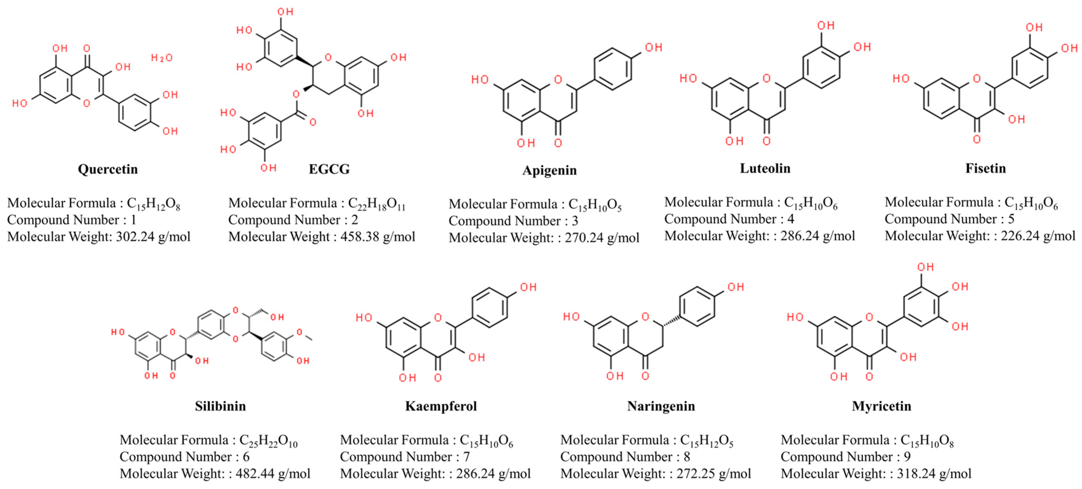
2. Flavonoids’ Anti-Arthritic Activities and Clinical Trial Potential

In the realm of natural compounds, various flavonoids have gained significant attention for their potential therapeutic benefits, particularly in the context of inflammatory and autoimmune diseases. This section focuses on nine well-studied compounds: quercetin, epigallocatechin-3-gallate (EGCG), apigenin, luteolin, fisetin, silibinin, kaempferol, naringenin, and myricetin (Figure 1). Each subsection explores the molecular structures, natural sources, and diverse health-promoting properties of these compounds, shedding light on their antioxidant and anti-inflammatory activities.

2.1. Quercetin

Quercetin’s name is derived from the Latin term “quercetum”, signifying oak forest and named after Quercus [16]. It is abundantly present in various plants in the natural environment, with its primary sources being onions, apples, and tea. It is represented by the molecular formula C15H10O7 [16] (Figure 1). As a naturally occurring polar auxin transport inhibitor, quercetin features a ketocarbonyl group in its molecule, and the oxygen atom on the first carbon is basic, allowing the formation of salts with strong acids. The molecular structure of quercetin encompasses four active groups: a dihydroxy group between the A ring, o-dihydroxy group B, C ring C2, C3 double bond, and 4-carbonyl [17]. The inclusion of a phenolic hydroxyl group and double bonds imparts potent antioxidant and anti-inflammatory activities to quercetin [18]. Firstly, the phenolic hydroxyl group serves as a hydrogen atom donor, allowing quercetin to effectively scavenge and neutralize reactive oxygen species (ROS) and other free radicals [19]. This antioxidant activity helps to prevent oxidative damage to cellular components, including lipids, proteins, and DNA, thereby mitigating inflammation and reducing the risk of chronic diseases associated with oxidative stress [20]. Furthermore, the presence of double bonds in the flavonoid’s backbone contributes to its anti-inflammatory properties by modulating key signaling pathways involved in the inflammatory response. Specifically, quercetin and other flavonoids can inhibit the activity of enzymes such as cyclooxygenase (COX) and lipoxygenase (LOX), which are responsible for the production of pro-inflammatory mediators like prostaglandins and leukotrienes [21]. Additionally, flavonoids can suppress the expression of inflammatory genes by interfering with transcription factors such as nuclear factor-kappa B (NF-κB) and activator protein-1 (AP-1) [22]. This dual mechanism of action, involving both antioxidant and anti-inflammatory effects, makes quercetin a potent therapeutic agent for combating inflammation and oxidative stress-related conditions [23]. In fact, in the context of osteoarthritis and RA, several studies have shown the effectiveness of quercetin in preventing and treating these pathologies [24].
By examining its impact on immune function, studies have revealed that quercetin, at a concentration of 50 µM, significantly (p < 0.0001) curbs the elevation of tumor necrosis factor-alpha (TNF-α), interleukins (IL-18, IL-6), and the nitric oxide (NO) induced by lipopolysaccharides (LPSs) in the Raw264.7 cell line, serving as a monocyte/macrophage-like cell line, indicating a robust anti-inflammatory effect in vitro [25] (Figure 2). These findings align with previously reported research, affirming quercetin’s efficacy as an inhibitor of TNF-α and IL-6 [26,27]. The ability of quercetin to regulate the Toll-like receptor 4/nuclear factor kappa-light-chain-enhancer of activated B (TLR4/NF-κB) signaling pathway has also been evaluated [28], showing the ability to inhibit reactive oxygen species (ROS) production and pro-inflammatory cytokine expression. Quercetin also inhibits neutrophil extracellular traps (NETs)’ release as well the release and enzymatic activity of elastase and myeloperoxidase [29], a common phenomenon shown in arthritis [30]. Furthermore, quercetin also protects A549 cells from the cytotoxicity induced by NETs at concentrations not exceeding 18 μg/mL [29].
In the context of chronic inflammation, which is often linked to increased oxidative stress and the development of diseases, Yeh et al. demonstrated that quercetin partially mitigated the pro-inflammatory effects, synergistically enhancing the inhibitory effects of β-carotene on pro-inflammatory mediator secretion and the DNA-damaging ability of phorbol 12-myristate 13-acetate (PMA)-stimulated HL-60 cells. This action was associated with the antioxidant activity of quercetin and its inhibition of pro-inflammatory cytokine production [31]. Further studies summarized that quercetin exhibits a mixed inhibition mechanism towards adenosine triphosphate (ATP), with its binding site overlapping both ATP’s and inhibitor of nuclear factor kappa B (IκBα)’s binding sites [32]. Adding to its repertoire, Musumeci et al. underscored quercetin’s role as an inhibitor of late-stage inflammatory transmitters like High-Mobility Group Box-1 (HMGB-1), effectively constraining the activation of mitogen-activated protein kinase (MAPK) [33]. Crucially, when delving into the modulation of HMGB-1, which plays a pivotal role in chronic inflammatory and autoimmune diseases, including RA [34], quercetin emerges as a promising agent for intervention and control (Figure 2).
The anti-rheumatoid effects of quercetin were also evaluated using animal models such as adjuvant-induced arthritis (AIA) and collagen-induced arthritis (CIA). In the AIA model, studies demonstrated that quercetin reduced acute-phase edema in rats but showed no significant effects during the chronic phase of the disease [35]. Oral administration of quercetin in chronic rat AIA models resulted in decreased clinical signs and lowered levels of inflammatory markers such as IL-1β (p < 0.001) and MCP-1 (p < 0.05) as well as lipoxygenase levels in the liver (p < 0.001) and lungs (p < 0.001) [36]. Furthermore, the NF-kB and ERK signaling pathways were also reduced [36]. In the CIA model, quercetin exhibited anti-inflammatory and joint-protective properties, surpassing methotrexate in efficacy [37]. It modulated T follicular helper 17/regulatory T (Th17/Treg) balance, inhibited NOD-like receptor protein 3 (NLRP3) inflammasome activation, and activated heme oxygenase-1 (HO-1)-mediated anti-inflammatory responses [38]. Additionally, quercetin protected cartilage by inhibiting NF-κB activation, reducing matrix metalloproteinase 13 (MMP-13) production, and alleviating arthritis symptoms in CIA mice [39]. Furthermore, in zymosan-induced RA models, the dose-dependent intra-articular administration of quercetin reduced hyperalgesia, articular edema, and neutrophil recruitment to the knee joint cavity [40,41] (Figure 3).

2.2. Epigallocatechin-3-Gallate (EGCG)

Epigallocatechin gallate (EGCG), derived from the Latin term “epigallocatechin” and gallate, has a history dating back to ancient times. Abundantly present in various plants, EGCG is notably found in tea leaves, particularly in green tea [42]. Represented by the molecular formula C22H18O11, EGCG is a polyphenolic compound with distinctive chemical characteristics [42] (Figure 1). As a naturally occurring compound, EGCG has been recognized for its potential health benefits. Functioning as a powerful antioxidant, EGCG’s molecular structure includes three phenol rings, giving it unique properties [43].
In fact, the presence of phenol rings in EGCG contributes to its antioxidant properties. The phenolic hydroxyl groups (-OH) attached to the rings enable EGCG to donate hydrogen atoms, thereby neutralizing free radicals and preventing oxidative damage to cells and tissues. Furthermore, the arrangement of the phenol rings in EGCG allows for specific interactions with biological molecules, such as proteins and enzymes [42].
Additionally, the presence of phenol rings in EGCG affects its absorption, metabolism, and bioavailability [44]. The hydroxyl groups on the phenol rings can undergo conjugation reactions in the liver, where they are modified to enhance solubility and facilitate excretion. EGCG has been extensively studied for its potential health-promoting effects, including its role in supporting cardiovascular health and its anti-inflammatory properties [42,43]. Furthermore, EGCG has undergone scrutiny for its potential as an anti-arthritic agent, delving into its capacity to modulate various cellular signaling pathways [45].
Attention has been directed toward exploring its influence on inflammation over the past decade, demonstrating remarkable cartilage and chondrocyte protection. In OA models, it curtails the impact of pro-inflammatory cytokines like IL-1β, TNF-α, and IL-6, effectively mitigating extracellular matrix degradation and apoptosis in chondrocytes [46]. EGCG’s modulation of signaling pathways, particularly its inhibition of NF-κB nuclear translocation, showcases its potential in alleviating inflammatory responses in OA chondrocytes [47]. Additionally, EGCG safeguards human cartilage from IL-1β-induced degradation, exhibiting a dose-dependent reduction in the expression of MMP-1 and MMP-13 [48,49] (Figure 2). Furthermore, EGCG’s selectively inhibits members of the ADAMTS (disintegrin and metalloproteinase with thrombospondin motifs) family of enzymes, such as ADAMTS-1, ADAMTS-4, and ADAMTS-5, leading to the preservation of the collagen structure and the regulation of the cell cycle, suggesting promising avenues for mitigating tissue destruction in joint disorders [50,51].
Beyond cartilage protection, EGCG displays bone-preserving activity by inducing apoptosis in osteoclasts [52], regulating NF-κB p65 [52,53], and inhibiting osteoblast differentiation [53,54]. Its inhibitory effects on IL-6 synthesis in osteoblasts contribute to a reduction in bone resorption and osteoclast formation [55]. Moreover, EGCG exhibits a protective role in preventing DNA fragmentation and apoptosis in osteoblastic cells, reinforcing its potential in maintaining a balanced bone environment [52].
In the realm of synovial fibroblast activity regulation, EGCG emerges as a potent modulator in RA. It selectively inhibits IL-1β-induced chemokine production and MMP-2 activity in RA synovial fibroblasts, underscoring its anti-arthritic properties [56]. EGCG’s capacity to suppress IL-6 and vascular endothelial growth factor (VEGF) synthesis in RA synovial fibroblasts further reinforces its potential therapeutic relevance [57,58]. Notably, EGCG’s downregulation of the anti-apoptotic protein Mcl-1 in RA synovial fibroblasts suggests a selective induction of apoptosis, sensitizing cells to TNFα-induced apoptosis [59,60]. This multifaceted approach positions EGCG as a promising candidate for regulating invasive growth in RA.
The anti-arthritic activity of EGCG has been comprehensively evaluated in vivo using two distinct animal models. In a CIA mouse model, the administration of EGCG-containing green tea extract (GTE) in drinking water showcased remarkable disease-modifying effects [61]. The incidence and severity of CIA were significantly reduced in mice exposed to EGCG, aligning with a substantial inhibition of key inflammatory mediators such as cyclooxygenase-2 (COX-2), interferon-gamma (IFNγ), and TNF-α in arthritic joints [61]. Furthermore, lower levels of total immunoglobulins G (IgG) and type II collagen-specific IgG in the serum and arthritic joints indicated a potential immune-suppressive influence of EGCG on arthritis in this model [61] (Figure 3).
Another animal model, adjuvant-induced arthritis in rats, provided additional insights into EGCG’s therapeutic impact. EGCG selectively inhibited IL-6 synthesis during the onset of arthritis, presenting a crucial mechanism for the observed reduction in inflammation [58]. In EGCG-treated rats, there was a specific decrease in the IL-6 levels in both serum and joints, coupled with an enhancement in the synthesis of soluble gp130 protein—an endogenous inhibitor of IL-6 signaling [58]. Notably, the correlation between reduced arthritis severity in EGCG-treated rats and decreased MMP-2 activity in the joints suggested a potential link between EGCG’s anti-inflammatory effects and the modulation of the matrix’s metalloproteinase activity [58]. Additionally, studies with GTE in adjuvant-induced arthritis demonstrated its immunomodulatory benefits, as GTE administration ameliorated arthritis by inhibiting serum IL-17 levels while concurrently upregulating IL-10 levels [62]. Daily oral administration of GTE in rats modestly improved adjuvant-induced arthritis, marked by decreased levels of chemokines such as MCP-1/CCL2 and GROα/CXCL1, along with the enhanced expression of chemokine receptors in the joints [63]. These findings collectively underscore the potential of EGCG as a promising therapeutic agent in the management of arthritis, with distinct impacts on immune responses, inflammation, and joint integrity in diverse animal models (Figure 3).

2.3. Apigenin

Apigenin, a flavonoid derived from the Latin term “apium” (parsley) and named after its abundance in parsley, has a history rooted in traditional medicine [64,65]. Widely distributed in various plants, apigenin is found in significant amounts in celery, chamomile, onions, and certain fruits, such as oranges [66]. Its molecular formula is C15H10O5, and its unique chemical structure contributes to its distinct properties (Figure 1). As a naturally occurring compound, apigenin is recognized for its antioxidant and anti-inflammatory characteristics [66]. Its molecular structure consists of two benzene rings connected by a pyrone ring, forming a flavone subclass [67]. This structure plays a crucial role in its ability to scavenge free radicals and modulate inflammatory pathways [66].
Studies have explored apigenin’s potential health benefits, including its anti-cancer properties, neuroprotective effects, and its ability to support cardiovascular health [67]. Additionally, apigenin has been investigated for its anxiolytic and sedative properties, suggesting potential applications in the field of mental health [66,67].
In vitro studies consistently demonstrate that inflammatory responses involve MAPKs and NF-κB signaling pathways, highlighting the potential for inflammatory therapy in the targeting of LPS-stimulated signal transduction cascades [68,69]. Apigenin, particularly observed in human THP-1 macrophages, significantly reduces NF-κB activation and LPS-induced ERK1/2 [70]. Apigenin also showcases anti-inflammatory activity in human periodontal ligament (hPDL) cells stimulated by LPS and nicotine [71]. Apigenin, within concentrations of 10 to 40 µM, impedes the production and activity of the HO-1 proteins induced by LPS and nicotine [71]. Furthermore, it significantly reduces the production of various inflammatory mediators, including IL-1β, IL-6, IL-12, TNF-α, prostaglandin (PG) E2, and NO, as well as COX-2 and inducible nitric oxide synthase (iNOS) in hPDL cells exposed to nicotine and LPS [71] (Figure 2). Additional experiments with mouse J774A.1 macrophage cells and human THP-1-induced macrophages underscore the pivotal role of apigenin in inhibiting LPS-induced pro-inflammatory cytokine production (TNF-α, IL-1β, and IL-6) by regulating multiple intracellular signaling pathways [72]. Furthermore, apigenin, akin to other flavonoids, hampers TNF-α synthesis or impairs its activity, contributing to its overall anti-inflammatory effects [72]. The downregulation of NF-κB and TNF-α emerges as a critical mechanism in apigenin’s capacity to inhibit inflammatory processes, suggesting promising therapeutic implications [72] (Figure 2 and Figure 3). Transitioning to in vivo studies, apigenin effectively inhibits LPS-induced inflammation by deactivating NF-κB and dephosphorylating Ser536 in the p65 subunit [70].
Building upon the promising anti-inflammatory properties highlighted in both in vitro and in vivo studies, the exploration of apigenin has extended to clinical trials. In a randomized controlled clinical trial, the effectiveness and safety of chamomile oil enriched with apigenin were investigated for knee OA. Following a regimen of topical chamomile oil application three times a day over a three-week period, patients with knee OA experienced a significant reduction in the need for analgesics (specifically acetaminophen (p = 0.001)). This intervention not only alleviated pain (p < 0.001) but also contributed to an enhancement in the patients’ physical function and stiffness (p < 0.001) [73]. However, the study had limitations, including a short follow-up duration, a relatively small sample size, and an imbalance in gender representation. These factors should be considered when interpreting the results.

2.4. Luteolin

Luteolin, a flavonoid whose name is derived from the botanical term “luteum” (yellow), has a rich history in traditional medicine [74]. Widely distributed in the plant kingdom, luteolin is found in various sources, such as parsley, celery, thyme, peppers, and certain fruits, like oranges [75]. Its molecular formula is C15H10O6, and its unique chemical structure contributes to its distinctive properties [74] (Figure 1). As a naturally occurring compound, luteolin is known for its antioxidant and anti-inflammatory activities [76]. The molecular structure of luteolin includes two benzene rings connected by a pyrone ring, making it a flavone subclass. This structural arrangement is associated with its ability to scavenge free radicals and modulate inflammatory pathways [74].
Research on luteolin has explored its potential health benefits, including its anti-cancer properties, neuroprotective effects, and role in supporting cardiovascular health [77]. Additionally, luteolin has been studied for its anti-allergenic and anti-viral properties, highlighting its diverse range of potential applications in health and wellness [78,79].
In various in vitro studies, luteolin has demonstrated significant anti-inflammatory activity by inhibiting NF-κB activation. For instance, it suppressed the increase in p50 nuclear localization in LPS-stimulated RAW264.7 cells and decreased NO production and iNOS expression [80,81]. Interestingly, luteolin-mediated inhibition of NF-κB signaling was observed in various cell types, including periodontal ligament cells, macrophages, adipocytes, and microglial cells [76]. Luteolin’s mechanism of action extends beyond NF-κB inhibition, as it also influences upstream signaling molecules. In studies focusing on myeloid differentiation primary response 88 (MyD88)-dependent signaling, luteolin inhibited NF-κB-mediated luciferase activity in a dose-dependent manner after the stimulation of the adapter protein MyD88 [81]. It also suppressed the kinase activities of Src and Syk [82]. Additionally, luteolin demonstrated an ability to inhibit TIR-domain-containing adapter-inducing interferon-β (TRIF)-dependent signaling, reducing the expression of various inflammatory molecules [81] (Figure 2).
Moreover, luteolin modulated Akt phosphorylation, a crucial regulator of the NF-κB and AP-1 pathways [81]. By inhibiting Akt phosphorylation, luteolin decreased downstream inflammatory mediator expression in various cellular contexts. Notably, luteolin has also been reported to have effects on various targets within the MAPK signaling pathway. In different cell lines and experimental models, luteolin has been shown to influence MAPK activation in response to different stimuli [76]. Examples of this include the suppression of IL-1β-induced cJun N-terminal kinase (JNK) and p38 kinase activation, the inhibition of the nuclear translocation of AP-1, and the modulation of pro-inflammatory cytokine production. However, Aziz et al. reported conflicting results across studies and cell types [76]. While some research indicates that luteolin decreases the phosphorylation of p38 MAPK, others show an increase in ERK1/2 phosphorylation. Luteolin has also been reported to possess ROS-scavenging activity, potentially linked to its inhibition of the MAPK pathway activated by ROS (Figure 2). In some instances, luteolin has been observed to activate rather than inhibit the MAPK pathway.
In vivo studies have explored the effects of luteolin on inflammatory pathways and their related disorders in animal models of arthritis. Kang et al. demonstrated a comprehensive investigation of the impact of luteolin on MMP-3 protein production, providing insightful findings on its regulatory effects [83]. Their study delved into both in vitro and in vivo aspects, yielding notable results. Firstly, luteolin exhibited a multifaceted influence on the gene expression levels of various key players in extracellular matrix modulation, including MMP-3, MMP-1, MMP-13, ADAMTS-4, and ADAMTS-5, within rabbit articular chondrocytes. Intriguingly, it was found to enhance the gene expression level of collagen, suggesting a potential positive regulatory role in collagen production [83]. Furthermore, in a more recent study, Fei et al. demonstrated a comprehensive investigation into the protective effects of luteolin on IL-1β-stimulated rat chondrocytes and its potential in a monosodium iodoacetate (MIA)-induced model of OA [84]. In vitro, rat chondrocytes were pre-treated with varying concentrations of luteolin before IL-1β stimulation, revealing significant reductions in the production of inflammatory mediators. Luteolin effectively suppressed NO, PGE2, TNF-α, and matrix metalloproteinases (MMP-2, MMP-8, MMP-9) [84] (Figure 2). Additionally, luteolin downregulated the expression of key inflammatory proteins, including iNOS, COX-2, MMP-1, MMP-3, and MMP-13, indicating its potential in mitigating inflammatory cascades initiated by IL-1β [84].
The study extended luteolin’s impact to an in vivo setting, where OA rats received a luteolin treatment. Morphological and ultrastructural scanning electron microscopy (SEM) assessments 45 days post MIA injection revealed that luteolin administration significantly prevented cartilage destruction. Immunohistochemistry demonstrated that luteolin restored collagen II expression, emphasizing its chondroprotective potential. Importantly, in the IL-1β-stimulated chondrocytes, luteolin demonstrated a notable inhibition of NF-κB phosphorylation, suggesting a mechanism through which it modulates the inflammatory response [84] (Figure 3).

2.5. Fisetin

Fisetin, a flavonoid whose name is derived from the botanical source “Fiscus” (related to the fig tree), has a history rooted in traditional medicine [85]. Widely distributed in the plant kingdom, fisetin is found in various sources such as strawberries, apples, persimmons, onions, and cucumbers [86]. Its molecular formula is C15H10O6, and its unique chemical structure contributes to its distinctive properties [85] (Figure 1). As a naturally occurring compound, fisetin is recognized for its antioxidant and anti-inflammatory activities. The molecular structure of fisetin includes two benzene rings connected by a pyrone ring, classifying it as a flavonol subclass. This structural arrangement is associated with its ability to scavenge free radicals and modulate inflammatory pathways [87].
Research on fisetin has explored its potential health benefits, including its anti-cancer properties, neuroprotective effects, and its role in supporting cardiovascular health [85,87]. Fisetin has also been studied for its potential anti-aging properties, with some research suggesting its ability to activate sirtuins, the proteins associated with longevity [88].
Transitioning from the broad exploration of fisetin’s health benefits, it becomes imperative to delve into specific research findings, especially in the context of chronic inflammatory diseases such as arthritis. Notably, a comprehensive study utilized high-throughput technology to identify 1071 genes regulated by fisetin in LPS-treated RAW264.7 cells [89] (Figure 2). The microarray analysis substantiated that fisetin not only inhibited the expression and secretion of inflammatory cytokines but also played a role in facilitating autophagosome–lysosome fusion and degradation within the cells treated with LPS. These effects were achieved through the inhibition of the PI3K/AKT/mTOR signaling pathway [89]. Building upon the preceding research findings, it is essential to transition to a more recent study that provides further insights into the anti-inflammatory mechanisms of fisetin. In this study, fisetin’s impact on pro-inflammatory mediators and cytokines, particularly in LPS-stimulated RAW 264.7 macrophages, was investigated [90]. Notably, fisetin demonstrated the significant inhibition of NO, PGE2, IL-6, and TNF-α expression (Figure 2). Moreover, the study explored the in vivo effects of fisetin using zebrafish larvae as a model. Fisetin not only attenuated LPS-induced mortality and abnormalities in zebrafish larvae but also normalized heart rate [90]. This protective effect was associated with a decrease in macrophage and neutrophil recruitment to the LPS-microinjected inflammatory site, accompanied by the downregulation of pro-inflammatory genes. The molecular mechanisms behind fisetin’s anti-inflammatory activity were elucidated, revealing its inhibition of NF-κB and inactivation of glycogen synthase kinase 3β (GSK-3β) [90]. Fisetin promoted the nuclear localization of β-catenin, further inhibiting NF-κB activity. Pharmacological inhibition experiments supported the crosstalk between GSK-3β/β-catenin and NF-κB signaling pathways, affirming fisetin’s potential as a dietary flavonoid to modulate inflammation and mitigate endotoxic shock.
The focus also shifted to its potential therapeutic role in OA. Zheng et al. study delves into both in vitro and in vivo investigations, aiming to elucidate the impact of fisetin on OA progression [91]. In the in vitro experiments, chondrocytes were pre-treated with fisetin alone or in combination with sirtinol, an inhibitor of SIRT1, before stimulation with IL-1β. The study assessed various markers, including NO, PGE2, TNF-α, and IL-6, and the expression of key genes (COX-2, iNOS, MMP-3, MMP-13, ADAMTS-5, Sox-9, aggrecan, and collagen-II) (Figure 2). The results demonstrated fisetin’s inhibitory effects on inflammatory and catabolic factors as well as its role in preserving key cartilage components [91].
Moving to in vivo experiments using a mouse OA model induced by the destabilization of the medial meniscus (DMM), fisetin’s impact was further investigated through gavage. Fisetin-treated mice exhibited less cartilage destruction, lower Osteoarthritis Research Society International (OARSI) scores, reduced subchondral bone plate thickness, and alleviated synovitis [91] (Figure 3).

2.6. Silibinin

Silibinin, a flavonolignan whose name is derived from the plant Silybum marianum, commonly known as milk thistle, has a history rooted in traditional medicine [92]. Primarily found in milk thistle seeds, silibinin is recognized for its potential health benefits. Its molecular formula is C25H22O10, and its unique chemical structure contributes to its distinctive properties [92] (Figure 1). As a naturally occurring compound, silibinin is renowned for its hepatoprotective effects. The molecular structure of silibinin includes a flavonoid component and a lignan component, giving it a unique dual structure [92]. This structural arrangement is associated with its ability to support liver function and protect against various liver conditions, making it a subject of interest in liver health research [93].
Research on silibinin has explored its potential in various health applications, including arthritis. In the study conducted by Tong et al., it was demonstrated that silibinin exerts significant anti-inflammatory effects in the context of RA [94]. The researchers observed a suppression of cell viability and an increase in the percentage of apoptotic RA-fibroblast-like synoviocytes (FLS) upon silibinin treatment. Notably, the production of inflammatory cytokines in both RA-FLS and a CIA rat model was effectively inhibited by silibinin. Furthermore, Tong et al. investigated the impact of silibinin on macrophage polarization, revealing that it induced M2 polarization in RAW264.7 cells [94]. Importantly, the study demonstrated that silibinin inhibits the differentiation of Th17 cells in vitro. Mechanistically, silibinin was found to suppress the NF-κB pathway in RA-FLS. Additionally, Sirtuin1 (SIRT1) was identified as a key player, with its levels decreasing after silibinin treatment. To corroborate the involvement of SIRT1, RA-FLS transfected with a short hairpin RNA (shRNA) of SIRT1 showed enhanced silibinin-induced apoptosis [94]. Autophagy, another critical cellular process, was markedly decreased in a dose-dependent manner following silibinin treatment. These findings collectively suggest that silibinin inhibits inflammation by targeting the NF-κB pathway, and the involvement of SIRT1 may contribute to silibinin-induced apoptosis. Moreover, silibinin’s inhibition of autophagy in RA-FLS further underscores its potential as a comprehensive therapeutic agent for the treatment of RA [94] (Figure 2). In a recent study, researchers have delved into the therapeutic potential of silybin, a compound derived from milk thistle, in the context of dyslipidemia and arthritis using an AIA rat model. This investigation built upon prior research on compounds related to silibinin and aimed to provide novel insights into the multifaceted effects of silybin [95]. The study uncovered the overexpression of Liver X Receptor alpha (LXRα) and key lipogenic enzymes regulated by LXRα in AIA rats, including lipoprotein lipase (LPL), cholesterol 7α and 27α hydroxylase (CYP7A, CYP27A), adipocyte fatty acid-binding protein (aP2/FABP4), and fatty acid translocase (CD36/FAT). These molecular changes were associated with dyslipidemia during arthritis development. By employing metabolomics, docking technology, and biochemical analyses, the research demonstrated that silybin effectively mitigates dyslipidemia and arthritis symptoms by suppressing the upregulated LXRα and restoring normal lipid metabolism [95]. Crucially, the study explored the interplay between LXRα and the NF-κB pathway. The activation of LXRα was found to exacerbate the inflammatory process induced by LPS. Conversely, the inhibition of LXRα agonism, achieved through siRNA or silybin treatment, resulted in reduced nuclear translocation of NF-κB and decreased induction of downstream cytokines. This insight suggests that LXRα agonism is a pivotal factor in the development of arthritis and represents a potential therapeutic target [95].

2.7. Kaempferol

Kaempferol, a flavonol whose name is derived from the botanical “Kaempferia,” has a rich history in traditional medicine [96]. Widely distributed in the plant kingdom, kaempferol is found in various sources such as tea, broccoli, kale, beans, and strawberries [96]. Its molecular formula is C15H10O6, and its unique chemical structure contributes to its distinct properties [97] (Figure 1). As a naturally occurring compound, kaempferol is recognized for its antioxidant and anti-inflammatory activities [97]. The molecular structure of kaempferol includes two benzene rings connected by a pyrone ring, classifying it as a flavonol subclass. This structural arrangement is associated with its ability to scavenge free radicals and modulate inflammatory pathways [98].
Research on kaempferol has explored its potential health benefits, including its anti-cancer properties, cardiovascular benefits, and role in supporting overall immune health [97,98]. Moreover, investigations have been conducted to delve into the anti-arthritic activity of kaempferol. Recent studies have delved into its specific impact on key inflammatory markers in the context of OA. In a concentration-dependent manner, kaempferol treatment, reaching up to 100 μM, demonstrated a notable reduction in IL-1β-stimulated formations of PGE2 and NO [99]. Interestingly, kaempferol exhibited a dual effect by upregulating the expression of iNOS and Cox-2 in IL-1β-stimulated rat OA chondrocytes [99]. Furthermore, the study shed light on the mechanistic aspects, revealing that kaempferol effectively inhibited the IL-1β-induced degradation of IκBα and the subsequent activation of NF-κB in rat chondrocytes [99]. An in-depth examination of its efficacy and underlying mechanisms was conducted using a CIA mouse model. In this study, kaempferol was administered both intragastrically (200 mg/kg) and intraperitoneally (20 mg /kg) to assess its anti-arthritic effects (Figure 3). The investigation encompassed pharmacodynamic and pharmacokinetic studies, revealing distinct outcomes in terms of the spleen index, the arthritis index, paw thickness, and inflammatory factors in the arthritis-afflicted mice [100]. Notably, the oral administration of kaempferol demonstrated marked anti-arthritis effects despite a relatively low bioavailability and circulatory exposure. Conversely, the intraperitoneal injection of kaempferol, despite achieving higher in vivo exposure, yielded marginal anti-arthritis effects. This discrepancy prompted a closer examination, implicating a potential involvement of the gut in the observed outcomes. A further analysis, including 16S ribosomal RNA profiling, unveiled an imbalance in the intestinal microbiota induced by arthritis, a phenomenon mitigated by kaempferol treatment [100]. A metabolomics study provided additional insights, indicating that kaempferol treatment significantly reversed the perturbation of the metabolites related to energy production, tryptophan, fatty acid, and secondary bile acid metabolisms in the gut contents of CIA mice [100] (Figure 3). Transitioning from the preceding investigations into kaempferol’s anti-arthritic effects, a novel mouse model combining hyperuricemia and gouty arthritis was employed in another study. Kaempferol, recognized for its anti-inflammatory and urate-lowering properties, was administered in various dosages (25, 50, and 100 mg/kg) to assess its impact on uric acid levels, renal function, and joint pathology. Through detailed examinations of kidney and joint tissues, the study delved into kaempferol’s efficacy in mitigating hyperuricemia-induced damage [101]. Histological analyses confirmed that kaempferol effectively ameliorated glomerular and renal tubular lesions as well as inflammatory infiltration in the ankle joints induced by hyperuricemia combined with gouty arthritis. The study further explored the mechanisms underlying kaempferol’s effects, focusing on its modulation of the NOD-like receptor protein 3 (NLRP3) inflammasome and the NF-κB pathway [101] (Figure 2 and Figure 3).

2.8. Naringenin

Naringenin, a flavanone whose name is derived from the botanical source “Citrus” (genus of flowering plants which includes oranges and grapefruits), has a history rooted in traditional medicine [102]. Widely distributed in the plant kingdom, naringenin is found in significant amounts in citrus fruits such as oranges, grapefruits, and tomatoes [103]. Its molecular formula is C15H12O5, and its unique chemical structure contributes to its distinctive properties [104] (Figure 1). As a naturally occurring compound, naringenin is recognized for its antioxidant and anti-inflammatory activities. The molecular structure of naringenin includes two benzene rings connected by a pyrone ring, classifying it as a flavanone subclass [103]. Additionally, naringenin has been studied for its anti-inflammatory effects, suggesting its potential in addressing conditions related to inflammation, such as arthritis [104,105]. Transitioning to the exploration of naringenin’s impact on OA, a study conducted by Wang et al. delved into its effects on the transcriptional expression, secretion, and enzymatic activity of MMP-3 using the murine monosodium iodoacetate (MIA) OA model [106]. Pain behavior assessments, microscopic analyses of knee-joint tissues, and in vitro studies on IL-1β-activated articular chondrocytes provided comprehensive insights [106] (Figure 2). In the MIA rats, naringenin exhibited a substantial reduction in pain behavior and a marked improvement in tissue morphology. Additionally, a significant inhibition of MMP-3 expression was observed, indicating the potential therapeutic impact of naringenin in vivo. Further investigations in primary cultured chondrocytes of rats revealed that naringenin not only reduced the transcriptional expression, secretion, and enzymatic activity of degradative enzymes (MMP-1, MMP-13, ADAMTS-4, and ADAMTS-5) but also exhibited inhibitory effects on the NF-κB pathway in vitro [106] (Figure 2).
Advancing our understanding, a more recent investigation unveiled the noteworthy anti-inflammatory and antioxidant properties of naringenin in a CIA mouse model. The study in question, conducted on DBA/1 CIA mice, demonstrated that naringenin significantly reduced foot inflammation and lowered pro-inflammatory cytokine levels in the serum. Moreover, it bolstered antioxidant capacity within the CIA model [107]. In parallel, in vitro experiments with LPS-induced RAW264.7 cells underscored naringenin’s ability to mitigate pro-inflammatory cytokine and ROS levels (Figure 3). The study delved into the molecular intricacies of this flavonoid’s action, revealing that naringenin activated autophagy and augmented the autophagic flux. Importantly, the disrupting of autophagy, achieved through silencing Atg5 or inhibiting autophagolysosomes using chloroquine, counteracted naringenin’s impact on pro-inflammatory cytokines [107]. Further insights emerged as the study unraveled the involvement of the AMPK/ULK1 signaling pathway. Naringenin’s activation of this pathway was pivotal, as inhibiting AMPK reversed the initiation of autophagy and diminished the secretion of pro-inflammatory cytokines induced by naringenin [107]. Naringin exhibits anti-inflammatory and pro-apoptotic effects in RA fibroblast-like synoviocytes (RA FLSs). These effects are characterized by a reduction in the expression of IL-1, IL-6, and IL-8, an increase in caspase-3 expression, an elevated Bax/Bcl-2 ratio, and a decrease in the expression of MMP-1, MMP-2, and MMP-13. Notably, these actions are attributed to the downregulation of the MAPK/ERK and PI3K/AKT signaling pathways, as reported in a study conducted by Aihaiti et al. [108].

2.9. Myricetin

Myricetin, a flavonol whose name is derived from the botanical “Myrica” (genus of flowering plants which includes the bayberry), has a history deeply rooted in traditional medicine [108]. Widely distributed in the plant kingdom, myricetin is found in various sources such as berries, particularly cranberries, grapes, and a variety of fruits, vegetables, and herbs [109]. Its molecular formula is C15H10O8, and its unique chemical structure contributes to its distinctive properties [108] (Figure 1). As a naturally occurring compound, myricetin is recognized for its antioxidant and anti-inflammatory activities. The molecular structure of myricetin includes three hydroxy groups and a double bond, contributing to its flavonol subclass classification. This structural arrangement is associated with its ability to scavenge free radicals and modulate inflammatory pathways [108,110].
Research on myricetin has explored its potential health benefits, including its anti-arthritic properties. Myricetin has demonstrated its direct inhibition of cathepsin K activity, a potent collagenase expressed in osteoclasts and synovial fibroblasts. The IC50 for recombinant human cathepsin was determined to be 585.3 µmol/L [110]. Furthermore, positive effects of myricetin were observed in murine CIA (Figure 2). Mice with CIA received a daily oral dose of myricetin at 25 mg/kg. Throughout the study, clinical severity assessments of CIA and histopathological assessments were conducted [110]. Biomarkers associated with the histological evaluation of cartilage degradation, including deoxypyridinoline, cartilage oligomeric matrix protein, and the C-terminal telopeptide degradation product of type I collagen (CTX-I), were examined. Treatment with myricetin significantly reduced the levels of biomarkers indicative of cartilage degradation and improved the clinical symptoms of CIA in mice [110] (Figure 3). Furthermore, a recent independent study sought to explore the therapeutic potential of myricetin in countering the IL-21-induced tumor-like characteristics of adjuvant-induced arthritis fibroblast-like synoviocytes (AIA-FLS) [111]. In this investigation, myricetin demonstrated its capacity to suppress IL-21 receptor expression and mitigate the activation of the choline kinase alpha (ChoKα) signaling cascade, including N-Ras, Ral-GDS, and PI3K, in IL-21-induced AIA-FLS [111]. Subsequently, myricetin treatment led to a reduction in ChoKα and phospholipase D2 (PLD2) enzymatic activity, effectively restraining the proliferative, migratory, and invasive properties of AIA-FLSs. These results propose myricetin as a potential anti-arthritic agent, attenuating IL-21-induced hyperproliferation, migration, and invasive behavior of AIA-FLS through the modulation of the ChoKα signaling cascade [111].

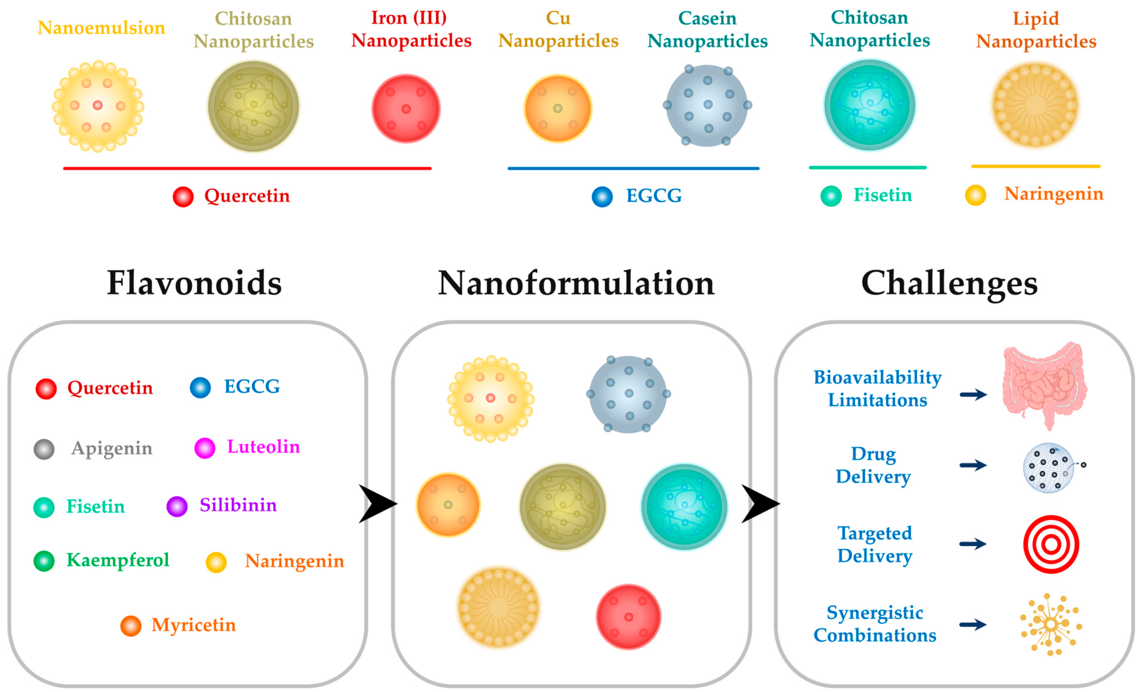
3. Challenges in Harnessing Flavonoids for Anti-Inflammatory Applications and the Potential of Nanoparticles

Flavonoids, despite their promising anti-inflammatory properties, present several challenges that researchers have been actively addressing in order to optimize their therapeutic potential [10]. One significant hurdle is their limited bioavailability, which restricts their efficacy in vivo [10,15]. Flavonoids often exhibit poor solubility, stability, and a rapid metabolism, leading to reduced concentrations at the target site. Additionally, their susceptibility to degradation in the gastrointestinal tract poses a challenge to their absorption [15]. These issues have spurred the exploration of innovative strategies to enhance the delivery and effectiveness of flavonoids in anti-inflammatory applications.

3.1. Bioavailability Limitations

Efforts to overcome the bioavailability challenges of flavonoids have included the exploration of various formulations and delivery systems [10]. Nanoparticles, due to their unique properties, have emerged as a promising avenue to address these limitations. The encapsulation of flavonoids within nanoparticles can enhance their solubility, stability, and protection against premature degradation [10]. This approach aims to improve the absorption of flavonoids, ultimately leading to increased bioavailability and therapeutic efficacy (Figure 4).

3.2. Nanoparticles as Delivery Vehicles

Nanoparticles offer a versatile platform for the controlled and targeted delivery of flavonoids. By encapsulating these compounds within nanoparticles, researchers can achieve sustained-release kinetics, prolonging their presence in the bloodstream and improving their distribution to inflamed tissues [10]. Moreover, nanoparticles can protect flavonoids from enzymatic degradation and facilitate their transport across biological barriers, enhancing their bioavailability [112] (Figure 4).

3.3. Targeted Delivery to Inflammatory Sites

Utilizing nanoparticles for targeted delivery to inflammatory sites represents a significant advancement in drug delivery strategies. This approach capitalizes on the unique properties of nanoparticles, allowing for the precise localization of therapeutic agents in inflamed tissues [113]. Through the engineering of functionalized nanoparticles, it becomes possible to tailor their surface properties to selectively accumulate in areas of inflammation.
Functionalized nanoparticles can be designed to exploit the distinct biological characteristics of inflamed tissues, such as altered vascular permeability and the expression of specific cell surface markers [114]. By incorporating targeting ligands or antibodies onto the nanoparticle surface, they can selectively bind to receptors overexpressed on the inflamed cells or endothelial cells lining inflamed blood vessels [115,116].
Once localized to the inflammatory site, the nanoparticles facilitate a controlled and concentrated release of flavonoids. This localized delivery minimizes systemic exposure of the therapeutic agent, reducing the risk of systemic side effects. Moreover, by concentrating the therapeutic payload at the site of inflammation, targeted delivery maximizes the efficacy of flavonoids in alleviating inflammatory processes [116].
The advantages of targeted delivery to inflammatory sites are twofold [117]. Firstly, it enhances the therapeutic efficacy of flavonoids by ensuring that a higher concentration of the drug reaches the desired site of action. Secondly, it minimizes off-target effects, thereby reducing the likelihood of adverse reactions associated with systemic drug administration.

3.4. Prolonged Circulation Time

A prolonged circulation time refers to the ability of nanoparticles to remain in the bloodstream for an extended period of time, which is a crucial advantage in drug delivery [118]. In the context of arthritis management, a prolonged circulation time is particularly advantageous as it enables the sustained release of flavonoids at the site of inflammation. Chronic inflammatory conditions such as OA and RA require the continuous suppression of inflammation to alleviate symptoms and prevent disease progression [119]. By prolonging the circulation time of flavonoid-loaded nanoparticles, it becomes possible to achieve a sustained and controlled release of these anti-inflammatory compounds, leading to prolonged therapeutic effects and improved patient outcomes.
Moreover, a prolonged circulation time enhances the efficiency of nanoparticle-based drug delivery systems by reducing the frequency of dosing [120]. This not only improves patient compliance but also minimizes the potential side effects associated with high-doses or the frequent administration of therapeutic agents. Additionally, the prolonged presence of nanoparticles in the bloodstream increases the likelihood of their accumulation at the inflamed joint sites, further enhancing the localized delivery of flavonoids and maximizing their therapeutic efficacy [121].

3.5. Enabling Diverse Compounds’ Encapsulation and Synergy

Nanoparticle-based delivery systems offer a versatile platform for encapsulating a diverse array of therapeutic compounds, presenting a significant advancement in drug delivery technology. This capability extends to flavonoids, which can be effectively incorporated into nanoparticles due to their diverse pharmacological properties. One notable advantage of this approach is the potential for synergistic combinations of flavonoids with other anti-inflammatory agents [122,123].
By integrating flavonoids in complementary compounds, nanoparticle formulations can be tailored to achieve enhanced therapeutic effects. This synergy arises from the ability to customize formulations, optimizing the balance between different agents to maximize their collective anti-inflammatory activity [124]. For instance, combining flavonoids with conventional anti-inflammatory drugs or natural extracts rich in bioactive compounds can lead to synergistic effects, surpassing the individual efficacy of each component [125]. Furthermore, the co-delivery of multiple compounds within nanoparticles offers a multifaceted strategy for intervening in inflammatory processes. Flavonoids, known for their antioxidant and anti-inflammatory properties, can be combined with agents targeting specific inflammatory pathways or mediators [126,127]. This comprehensive approach allows nanoparticles to address various aspects of the inflammatory cascade simultaneously, offering a more holistic solution to inflammation-related disorders.
In addition to enhancing therapeutic efficacy, synergistic combinations within nanoparticle-based delivery systems can also mitigate the potential side effects associated with high doses of individual agents [128]. By optimizing the ratio and combination of compounds, nanoparticles can minimize adverse reactions while maximizing therapeutic benefits.

4. Flavonoid-Based Nanoparticles Enhance the Therapeutic Potential

In this section, we delve into the exploration of flavonoid-based nanoparticles and their potential in enhancing therapeutic outcomes, with a specific focus on quercetin, EGCG, fisetin, and naringenin. These nanoparticles aim to overcome the inherent challenges associated with poor absorption and bioavailability, unlocking the full therapeutic potential of these flavonoids in the treatment of inflammatory conditions, particularly RA and OA. Each subsection explores innovative nanoformulations, their impact on inflammatory markers, and their efficacy in preclinical models, shedding light on promising avenues for advancing arthritis treatment strategies.

4.1. Quercetin-Based Nanoparticles

Quercetin, despite its proven effectiveness in treating inflammatory diseases, faces limitations in its application due to poor absorption and bioavailability. Addressing these challenges is crucial for unlocking the full therapeutic potential of quercetin. In light of this, recent research has focused on the development, optimization, and evaluation of a quercetin-loaded nanoemulsion (NE)-based gel specifically designed for managing RA [129]. Cytotoxicity studies on HIG-82 and RAW 264.7 cells revealed no toxic effects on synoviocytes, while also exhibiting a potent inhibitory effect on LPS-induced TNF-α production [129]. Furthermore, the quercetin–NE gel displayed a favorable rheological behavior, a commendable texture profile, and improved drug permeation in comparison to the free quercetin gel (Figure 5). Notably, the gel proved to be non-irritating and effectively inhibited paw edema in rats induced with complete Freund’s adjuvant (CFA) over 24 h, outperforming the free quercetin gel [129].
Alternative strategies have been explored to address the limited use of quercetin due to its poor absorption and bioavailability. One notable approach involves the formulation of quercetin-loaded chitosan nanoparticles (Q-NPs) [130]. The anti-rheumatic efficacy of Q-NPs was evaluated in an FCA-induced arthritic rat model treated with 10 and 20 mg/Kg Q-NPs, respectively. The results demonstrated a significant reduction in ankle diameter after treatment with high-dose Q-NPs (20 mg/Kg) [130]. Moreover, the mice treated with this formulation exhibited a substantial decrease in their TNFα and IL-6 levels. Significant impacts were also observed in the biochemical tests, the hematological parameters, and the oxidative stress parameters [130]. Histopathological examinations further confirmed the anti-rheumatic effect of high-dose Q-NPs on kidney, liver, and ankle tissues (Figure 5).
In an innovative approach to addressing the challenges associated with quercetin, ultrasmall iron–quercetin natural coordination nanoparticles (Fe-Qur NCNs) were also developed [130] (Figure 5). These nanoparticles, created through a simple mixing process, showcased exceptional anti-inflammatory and antioxidant properties. Fe-Qur NCNs retained the innate ability of quercetin to eliminate ROS while also exhibiting improved water solubility and biocompatibility [130]. In vitro experiments revealed that Fe-Qur NCNs effectively mitigated excess ROS, prevented cell apoptosis, and inhibited the polarization of inflammatory macrophages by reducing the activation of NF-κB pathways [130] (Figure 5).

4.2. EGCG-Based Nanoparticles

Expanding on the exploration of nanoparticles derived from epigallocatechin gallate (EGCG), the investigation yielded compelling outcomes. In a study conducted by Wei et al., the biocompatibility of Cu-EGCG nanosheets was demonstrated, accompanied by a sustained release of Cu2+ from the nanoparticles (Figure 5). Noteworthy was the nanosheets’ multi-enzyme-like antioxidative activity, effectively mitigating intracellular ROS [131]. This resulted in a significant reduction in the expression levels of pro-inflammatory cytokines, including TNF-α, iNOS, IL-1β, and IL-6, secreted by RAW264.7 cells, particularly under LPS induction. Moreover, the nanosheets exhibited the capability to inhibit the nuclear translocation of the p65 subunit of NF-kB [131] (Figure 5). In addition to this, they activated the expression of Nrf2, pointing towards a dual mechanism in modulating cellular responses. In the same study, the influence of RAW264.7 macrophages on cartilage inflammation-associated cells, particularly chondrocytes, was explored. Cu-EGCG nanosheets, at a concentration of 80 μg/mL, exhibited a minimal impact on chondrocyte viability. When chondrocytes pre-treated with LPS were co-cultured with different concentrations of Cu-EGCG nanosheets, the expression levels of inflammatory markers and cartilage-related genes (IL-6, IL-1β, TNF-α, MMP-13, MMP-3, ACAN, and Col2a1) were significantly reduced compared to the LPS-treated group. The Cu-EGCG nanosheets effectively mitigated ROS production induced by M1 macrophage-conditioned media (CM), showcasing their antioxidant properties [131] (Figure 5). At the molecular level, the Cu-EGCG nanosheets demonstrated superior anti-inflammatory effects by decreasing the upregulation of inflammatory factors and improving the expression of cartilage-related markers (COL2A1 and ACAN). Furthermore, Cu-EGCG-CM exhibited a better performance than EGCG-CM in reducing the secretion of MMP-13 and IL-6, crucial factors in OA [131].
In another study, researchers explored the enhanced anti-arthritic activity of EGCG through the use of EGCG-loaded nanoparticles (EGC-NPs), particularly in OA and RA cells [132]. EGCG and gallic acid (GA) individually demonstrated inhibitory activity in both OA and RA cells, with a higher sensitivity observed in RA cells. Combining EGCG and GA in a synergistic regimen further increased their inhibitory effects on arthritis cells. The EGC-NPs, with a specific EGCG–GA–casein ratio, exhibited improved cell viability inhibition compared to the EGCG–GA mixture [132].
In vivo experiments using a CIA model in rats demonstrated the potential therapeutic efficacy of EGC-NPs. The EGC-NPs, administered through gavage, significantly alleviated arthritis symptoms, as evidenced by reduced paw swelling, lower arthritis scores, and improved radiographic and histopathological outcomes. The EGC-NPs outperformed the EGCG–GA mixture in terms of anti-arthritic effects. Furthermore, the EGC-NPs effectively suppressed the expression of pro-inflammatory cytokines (TNF-α, IL-1β, IL-6, and IL-8) in arthritic rats, surpassing the effects of the EGCG–GA mixture [132] (Figure 5).

4.3. Fisetin-Based Nanoparticles

To the best of our knowledge, only one recent in vitro study conducted by Nabizadeh et al. investigated the impact of fisetin-loaded nanoparticles (FNPs) on gene expression in chondrocytes pre-treated with IL-1β to simulate OA conditions. IL-1β typically down-regulates cartilage-related genes, such as Sox-9, COL2, and aggrecan. FNPs, administered at 50 μg/mL, effectively prevented the reduction in Sox-9 and COL2 mRNA expression in IL-1β-stimulated chondrocytes. Additionally, FNPs increased the levels of sirtuin 1 (SirT1), a gene influencing chondrocyte differentiation and proliferation, in IL-1β-stimulated chondrocytes. The IL-1β-induced up-regulation of pro-inflammatory cytokines TNF-α and IL-6 was mitigated by FNPs, similar to fisetin. Furthermore, FNPs increased the expression of the anti-inflammatory cytokine IL-10 in IL-1β-stimulated chondrocytes. Overall, the results suggest that FNPs can inhibit inflammatory responses induced by IL-1β and preserve cartilage-related gene expression in OA chondrocyte cells (Figure 5).

4.4. Naringenin-Based Nanoparticles

Similarly, a solitary study investigated the potential augmented anti-arthritic effects of naringenin through a nanoformulation in the form of lipid-based nanoparticles [133]. The study demonstrated the nanoformulation’s effectiveness in significantly reducing arthritis in vivo, using a CFA model. The results exhibited a notable decrease in the RA factor, COX-2, and key inflammatory markers, including IL-6 and TNF-α. Moreover, the histological examination of the ankle joints revealed a reduction in the induced joint damage [133] (Figure 5).

5. Conclusions and Future Direction

In conclusion, this comprehensive review has shed light on the potential of flavonoids and flavonoid-based nanoparticles as promising avenues for the management of OA and RA. The meticulous examination of specific flavonoids, including quercetin, EGCG, apigenin, luteolin, fisetin, silibinin, kaempferol, naringenin, and myricetin, has provided insights into their anti-arthritic activities and clinical implications. However, challenges such as their bioavailability limitations have been recognized, prompting the exploration of innovative solutions like the use of nanoparticles as delivery vehicles. The potential for targeted delivery to inflammatory sites and the exploration of synergistic combinations represent promising strategies to overcome these hurdles and enhance the therapeutic impact of flavonoids. The emerging field of flavonoid-based nanoparticles, exemplified by formulations such as quercetin-, EGCG-, fisetin-, and naringenin-based nanoparticles, holds great promise. These nanoformulations present opportunities to improve drug delivery efficiency and overcome bioavailability challenges. Looking ahead, future research directions should focus on addressing the remaining gaps in our understanding, including further clinical studies, the optimization of delivery strategies, and the exploration of synergistic combinations. In fact, challenges such as ensuring optimal nanoparticle design, scalability, and cost-effectiveness need to be addressed for the successful clinical implementation of flavonoids [134]. Regarding safety profiles, while flavonoids are generally considered safe, concerns may arise with nanoparticle formulations due to potential toxicity or immunogenicity. Therefore, rigorous safety assessments are essential before clinical use [134]. Additionally, patient compliance and bioavailability remain key considerations. Nanoparticles may offer improved compliance through targeted delivery and sustained-release mechanisms. However, issues such as variability in patient response and the potential for adverse reactions must be carefully monitored [134].
The integration of multidisciplinary approaches, encompassing pharmacology, nanotechnology, and clinical medicine, will be pivotal in advancing the translation of these findings into effective therapeutic interventions. In perspective, the culmination of our review underscores the potential of flavonoids and their nanoformulations as valuable assets in the ongoing quest for innovative and effective solutions in arthritis management. By navigating the complexities of their delivery and synergies, these compounds could offer new hope for improved patient outcomes, ushering in a new era of precision medicine in the treatment of OA and RA.

Author Contributions

Conceptualization, H.W. and Y.L.; data curation, H.W., Y.L. and M.O.; figures’ conception, H.W.; writing—original draft preparation, H.W.; writing—review and editing, Y.L. and M.O. All authors have read and agreed to the published version of the manuscript.

Funding

This research received no external funding.

Institutional Review Board Statement

Not applicable.

Informed Consent Statement

Not applicable.

Data Availability Statement

Data (references) are contained within the article.

Conflicts of Interest

The authors declare no conflicts of interest.

References

  1. Long, H.; Liu, Q.; Yin, H.; Wang, K.; Diao, N.; Zhang, Y.; Lin, J.; Guo, A. Prevalence Trends of Site-Specific Osteoarthritis from 1990 to 2019: Findings from the Global Burden of Disease Study 2019. Arthritis Rheumatol. 2022, 74, 1172–1183. [Google Scholar] [CrossRef]
  2. Cieza, A.; Causey, K.; Kamenov, K.; Hanson, S.W.; Chatterji, S.; Vos, T. Global estimates of the need for rehabilitation based on the Global Burden of Disease study 2019: A systematic analysis for the Global Burden of Disease Study 2019. Lancet 2021, 396, 2006–2017. [Google Scholar] [CrossRef]
  3. Stanich, J.A.; Carter, J.D.; Whittum-Hudson, J.; Hudson, A.P. Rheumatoid arthritis: Disease or syndrome? Open Access Rheumatol. Res. Rev. 2009, 1, 179–192. [Google Scholar] [CrossRef]
  4. Lee, Y.C.; Lu, B.; Guan, H.; Greenberg, J.D.; Kremer, J.; Solomon, D.H. Physician Prescribing Patterns and Risk of Future Long-Term Opioid Use Among Patients With Rheumatoid Arthritis: A Prospective Observational Cohort Study. Arthritis Rheumatol. 2020, 72, 1082–1090. [Google Scholar] [CrossRef] [PubMed]
  5. Ma, S.N.; Zaman Huri, H.; Yahya, F. Drug-related problems in patients with rheumatoid arthritis. Ther. Clin. Risk Manag. 2019, 15, 505–524. [Google Scholar] [CrossRef]
  6. Al-Khayri, J.M.; Sahana, G.R.; Nagella, P.; Joseph, B.V.; Alessa, F.M.; Al-Mssallem, M.Q. Flavonoids as Potential Anti-Inflammatory Molecules: A Review. Molecules 2022, 27, 2901. [Google Scholar] [CrossRef] [PubMed]
  7. Ndayambaje, M.; Wahnou, H.; Sow, M.; Chgari, O.; Habyarimana, T.; Karkouri, M.; Limami, Y.; Naya, A.; Oudghiri, M. Exploring the multifaceted effects of Ammi visnaga: Subchronic toxicity, antioxidant capacity, immunomodulatory, and anti-inflammatory activities. J. Toxicol. Environ. Health Part A 2024, 87, 150–165. [Google Scholar] [CrossRef] [PubMed]
  8. Limami, Y.; Pinon, A.; Wahnou, H.; Oudghiri, M.; Liagre, B.; Simon, A.; Duval, R.E. Ursolic Acid’s Alluring Journey: One Triterpenoid vs. Cancer Hallmarks. Molecules 2023, 28, 7897. [Google Scholar] [CrossRef]
  9. Benayad, S.; Wahnou, H.; El Kebbaj, R.; Liagre, B.; Sol, V.; Oudghiri, M.; Saad, E.M.; Duval, R.E.; Limami, Y. The Promise of Piperine in Cancer Chemoprevention. Cancers 2023, 15, 5488. [Google Scholar] [CrossRef] [PubMed]
  10. Wahnou, H.; Liagre, B.; Sol, V.; El Attar, H.; Attar, R.; Oudghiri, M.; Duval, R.E.; Limami, Y. Polyphenol-Based Nanoparticles: A Promising Frontier for Enhanced Colorectal Cancer Treatment. Cancers 2023, 15, 3826. [Google Scholar] [CrossRef]
  11. Hunter, D.J.; Guermazi, A.; Roemer, F.; Zhang, Y.; Neogi, T. Structural correlates of pain in joints with osteoarthritis. Osteoarthr. Cartil. 2013, 21, 1170–1178. [Google Scholar] [CrossRef]
  12. Limami, Y.; Leger, D.Y.; Liagre, B.; Pécout, N.; Viana, M. Ibuprofen-loaded calcium phosphate granules: A new bone substitute for local relieving symptoms of osteoarthritis. Eur. J. Pharm. Sci. Off. J. Eur. Fed. Pharm. Sci. 2021, 158, 105679. [Google Scholar] [CrossRef]
  13. Guo, X.; Lou, J.; Wang, F.; Fan, D.; Qin, Z. Recent Advances in Nano-Therapeutic Strategies for Osteoarthritis. Front. Pharmacol. 2022, 13, 924387. [Google Scholar] [CrossRef]
  14. Wahnou, H.; Youlyouz-Marfak, I.; Liagre, B.; Sol, V.; Oudghiri, M.; Duval, R.E.; Limami, Y. Shining a Light on Prostate Cancer: Photodynamic Therapy and Combination Approaches. Pharmaceutics 2023, 15, 1767. [Google Scholar] [CrossRef] [PubMed]
  15. Hba, S.; Ghaddar, S.; Wahnou, H.; Pinon, A.; El Kebbaj, R.; Pouget, C.; Sol, V.; Liagre, B.; Oudghiri, M.; Limami, Y. Natural Chalcones and Derivatives in Colon Cancer: Pre-Clinical Challenges and the Promise of Chalcone-Based Nanoparticles. Pharmaceutics 2023, 15, 2718. [Google Scholar] [CrossRef]
  16. Lakhanpal, P.; Rai, D.K. Quercetin: A versatile flavonoid. Internet J. Med. Update 2007, 2, 22–37. [Google Scholar] [CrossRef]
  17. Magar, R.T.; Sohng, J.K. A Review on Structure, Modifications and Structure-Activity Relation of Quercetin and Its Derivatives. J. Microbiol. Biotechnol. 2020, 30, 11–20. [Google Scholar] [CrossRef] [PubMed]
  18. Li, Y.; Yao, J.; Han, C.; Yang, J.; Chaudhry, M.T.; Wang, S.; Liu, H.; Yin, Y. Quercetin, Inflammation and Immunity. Nutrients 2016, 8, 167. [Google Scholar] [CrossRef]
  19. Kumar, N.; Goel, N. Phenolic acids: Natural versatile molecules with promising therapeutic applications. Biotechnol. Rep. 2019, 24, e00370. [Google Scholar] [CrossRef] [PubMed]
  20. Tan, B.L.; Norhaizan, M.E.; Liew, W.P.; Sulaiman Rahman, H. Antioxidant and Oxidative Stress: A Mutual Interplay in Age-Related Diseases. Front. Pharmacol. 2018, 9, 1162. [Google Scholar] [CrossRef]
  21. Rudrapal, M.; Eltayeb, W.A.; Rakshit, G.; El-Arabey, A.A.; Khan, J.; Aldosari, S.M.; Alshehri, B.; Abdalla, M. Dual synergistic inhibition of COX and LOX by potential chemicals from Indian daily spices investigated through detailed computational studies. Sci. Rep. 2023, 13, 8656. [Google Scholar] [CrossRef]
  22. Chen, T.; Zhang, X.; Zhu, G.; Liu, H.; Chen, J.; Wang, Y.; He, X. Quercetin inhibits TNF-α induced HUVECs apoptosis and inflammation via downregulating NF-kB and AP-1 signaling pathway in vitro. Medicine 2020, 99, e22241. [Google Scholar] [CrossRef]
  23. Yang, D.; Wang, T.; Long, M.; Li, P. Quercetin: Its Main Pharmacological Activity and Potential Application in Clinical Medicine. Oxidative Med. Cell. Longev. 2020, 2020, 8825387. [Google Scholar] [CrossRef]
  24. Anand David, A.V.; Arulmoli, R.; Parasuraman, S. Overviews of Biological Importance of Quercetin: A Bioactive Flavonoid. Pharmacogn. Rev. 2016, 10, 84–89. [Google Scholar] [CrossRef] [PubMed]
  25. Zhou, X.N.; Han, C.; Song, P.Y.; Zhao, X.H.; Zhong, X.H. Anti-inflammatory effects of luteolin and quercetin in vitro. Prog. Veter. Med. 2017, 38, 56–61. [Google Scholar]
  26. Cessak, G.; Kuzawińska, O.; Burda, A.; Lis, K.; Wojnar, M.; Mirowska-Guzel, D.; Bałkowiec-Iskra, E. TNF inhibitors—Mechanisms of action, approved and off-label indications. Pharmacol. Rep. PR 2014, 66, 836–844. [Google Scholar] [CrossRef] [PubMed]
  27. Paul, A.T.; Gohil, V.M.; Bhutani, K.K. Modulating TNF-alpha signaling with natural products. Drug Discov. Today 2006, 11, 725–732. [Google Scholar] [CrossRef] [PubMed]
  28. Ren, G.Y.; Zhang, B.Y.; Huang, J.L. Protective effects of quercetin on the inflammation of mice RAW264. 7 cells induced by LPS. Chin. Traditional. Patent Med. 2019, 8, 1795–1799. [Google Scholar]
  29. Pereira, G.S.; Percebom, I.; Mendes, S.; Souza, P.S.S.; Diniz, L.F.A.; Costa, M.F.; Lopes, B.R.P.; Toledo, K.A. Quercetin inhibits neutrophil extracellular traps release and their cytotoxic effects on A549 cells, as well the release and enzymatic activity of elastase and myeloperoxidase. Braz. J. Biol. 2022, 84, e252936. [Google Scholar] [CrossRef]
  30. Song, W.; Ye, J.; Pan, N.; Tan, C.; Herrmann, M. Neutrophil Extracellular Traps Tied to Rheumatoid Arthritis: Points to Ponder. Front. Immunol. 2021, 11, 578129. [Google Scholar] [CrossRef]
  31. Yeh, S.-L.; Wang, H.-M.; Chen, P.-Y.; Wu, T.-C. Interactions of β-carotene and flavonoids on the secretion of pro-inflammatory mediators in an in vitro system. Chem.-Biol. Interact. 2009, 179, 386–393. [Google Scholar] [CrossRef]
  32. Avila, C.M.; Romeiro, N.C.; Sant’Anna, C.M.; Barreiro, E.J.; Fraga, C.A. Structural insights into IKKbeta inhibition by natural products staurosporine and quercetin. Bioorganic Med. Chem. Lett. 2009, 19, 6907–6910. [Google Scholar] [CrossRef]
  33. Musumeci, D.; Roviello, G.N.; Montesarchio, D. An overview on HMGB1 inhibitors as potential therapeutic agents in HMGB1-related pathologies. Pharmacol. Ther. 2014, 141, 347–357. [Google Scholar] [CrossRef] [PubMed]
  34. Taniguchi, N.; Kawahara, K.; Yone, K.; Hashiguchi, T.; Yamakuchi, M.; Goto, M.; Inoue, K.; Yamada, S.; Ijiri, K.; Matsunaga, S.; et al. High mobility group box chromosomal protein 1 plays a role in the pathogenesis of rheumatoid arthritis as a novel cytokine. Arthritis Rheum. 2003, 48, 971–981. [Google Scholar] [CrossRef] [PubMed]
  35. Guardia, T.; Rotelli, A.E.; Juarez, A.O.; Pelzer, L.E. Anti-inflammatory properties of plant flavonoids. Effects of rutin, quercetin and hesperidin on adjuvant arthritis in rat. Il Farmaco 2001, 56, 683–687. [Google Scholar] [CrossRef]
  36. Gardi, C.; Bauerova, K.; Stringa, B.; Kuncirova, V.; Slovak, L.; Ponist, S.; Drafi, F.; Bezakova, L.; Tedesco, I.; Acquaviva, A.; et al. Quercetin reduced inflammation and increased antioxidant defense in rat adjuvant arthritis. Arch. Biochem. Biophys. 2015, 583, 150–157. [Google Scholar] [CrossRef]
  37. Haleagrahara, N.; Miranda-Hernandez, S.; Alim, M.A.; Hayes, L.; Bird, G.; Ketheesan, N. Therapeutic effect of quercetin in collagen-induced arthritis. Biomed. Pharmacother. 2017, 90, 38–46. [Google Scholar] [CrossRef] [PubMed]
  38. Yang, Y.; Zhang, X.; Xu, M.; Wu, X.; Zhao, F.; Zhao, C. Quercetin attenuates collagen-induced arthritis by restoration of Th17/Treg balance and activation of Heme Oxygenase 1-mediated anti-inflammatory effect. Int. Immunopharmacol. 2018, 54, 153–162. [Google Scholar] [CrossRef] [PubMed]
  39. Xue, R.; Chen, L.U.; Zhang, C.; Fujita, M.; Li, R.; Yan, S.-M.; Ong, C.K.; Liao, X.; Gao, Q.; Sasagawa, S. Genomic and transcriptomic profiling of combined hepatocellular and intrahepatic cholangiocarcinoma reveals distinct molecular subtypes. Cancer Cell 2019, 35, 932–947. [Google Scholar] [CrossRef]
  40. Guazelli, C.F.S.; Staurengo-Ferrari, L.; Zarpelon, A.C.; Pinho-Ribeiro, F.A.; Ruiz-Miyazawa, K.W.; Vicentini, F.; Vignoli, J.A.; Camilios-Neto, D.; Georgetti, S.R.; Baracat, M.M.; et al. Quercetin attenuates zymosan-induced arthritis in mice. Biomed. Pharmacother. 2018, 102, 175–184. [Google Scholar] [CrossRef]
  41. Guan, F.; Wang, Q.; Bao, Y.; Chao, Y. Anti-rheumatic effect of quercetin and recent developments in nano formulation. RSC Adv. 2021, 11, 7280–7293. [Google Scholar] [CrossRef]
  42. Legeay, S.; Rodier, M.; Fillon, L.; Faure, S.; Clere, N. Epigallocatechin Gallate: A Review of Its Beneficial Properties to Prevent Metabolic Syndrome. Nutrients 2015, 7, 5443–5468. [Google Scholar] [CrossRef]
  43. Zhang, Y.; Lin, H.; Liu, C.; Huang, J.; Liu, Z. A review for physiological activities of EGCG and the role in improving fertility in humans/mammals. Biomed. Pharmacother. 2020, 127, 110186. [Google Scholar] [CrossRef]
  44. Cosme, P.; Rodríguez, A.B.; Espino, J.; Garrido, M. Plant Phenolics: Bioavailability as a Key Determinant of Their Potential Health-Promoting Applications. Antioxidants 2020, 9, 1263. [Google Scholar] [CrossRef]
  45. Ahmed, S. Green tea polyphenol epigallocatechin 3-gallate in arthritis: Progress and promise. Arthritis Res. Ther. 2010, 12, 208. [Google Scholar] [CrossRef]
  46. Ahmed, S.; Rahman, A.; Hasnain, A.; Lalonde, M.; Goldberg, V.M.; Haqqi, T.M. Green tea polyphenol epigallocatechin-3-gallate inhibits the IL-1 beta-induced activity and expression of cyclooxygenase-2 and nitric oxide synthase-2 in human chondrocytes. Free Radic. Biol. Med. 2002, 33, 1097–1105. [Google Scholar] [CrossRef] [PubMed]
  47. Singh, R.; Ahmed, S.; Islam, N.; Goldberg, V.M.; Haqqi, T.M. Epigallocatechin-3-gallate inhibits interleukin-1beta-induced expression of nitric oxide synthase and production of nitric oxide in human chondrocytes: Suppression of nuclear factor kappaB activation by degradation of the inhibitor of nuclear factor kappaB. Arthritis Rheum. 2002, 46, 2079–2086. [Google Scholar] [CrossRef] [PubMed]
  48. Singh, R.; Ahmed, S.; Malemud, C.J.; Goldberg, V.M.; Haqqi, T.M. Epigallocatechin-3-gallate selectively inhibits interleukin-1beta-induced activation of mitogen activated protein kinase subgroup c-Jun N-terminal kinase in human osteoarthritis chondrocytes. J. Orthop. Res. Off. Publ. Orthop. Res. Soc. 2003, 21, 102–109. [Google Scholar] [CrossRef] [PubMed]
  49. Ahmed, S.; Wang, N.; Lalonde, M.; Goldberg, V.M.; Haqqi, T.M. Green tea polyphenol epigallocatechin-3-gallate (EGCG) differentially inhibits interleukin-1 beta-induced expression of matrix metalloproteinase-1 and -13 in human chondrocytes. J. Pharmacol. Exp. Ther. 2004, 308, 767–773. [Google Scholar] [CrossRef] [PubMed]
  50. Adcocks, C.; Collin, P.; Buttle, D.J. Catechins from green tea (Camellia sinensis) inhibit bovine and human cartilage proteoglycan and type II collagen degradation in vitro. J. Nutr. 2002, 132, 341–346. [Google Scholar] [CrossRef]
  51. Vankemmelbeke, M.N.; Jones, G.C.; Fowles, C.; Ilic, M.Z.; Handley, C.J.; Day, A.J.; Knight, C.G.; Mort, J.S.; Buttle, D.J. Selective inhibition of ADAMTS-1, -4 and -5 by catechin gallate esters. Eur. J. Biochem. 2003, 270, 2394–2403. [Google Scholar] [CrossRef] [PubMed]
  52. Hafeez, B.B.; Ahmed, S.; Wang, N.; Gupta, S.; Zhang, A.; Haqqi, T.M. Green tea polyphenols-induced apoptosis in human osteosarcoma SAOS-2 cells involves a caspase-dependent mechanism with downregulation of nuclear factor-kappaB. Toxicol. Appl. Pharmacol. 2006, 216, 11–19. [Google Scholar] [CrossRef]
  53. Lee, J.H.; Jin, H.; Shim, H.E.; Kim, H.N.; Ha, H.; Lee, Z.H. Epigallocatechin-3-gallate inhibits osteoclastogenesis by down-regulating c-Fos expression and suppressing the nuclear factor-kappaB signal. Mol. Pharmacol. 2010, 77, 17–25. [Google Scholar] [CrossRef]
  54. Kamon, M.; Zhao, R.; Sakamoto, K. Green tea polyphenol (-)-epigallocatechin gallate suppressed the differentiation of murine osteoblastic MC3T3-E1 cells. Cell Biol. Int. 2009, 34, 109–116. [Google Scholar] [CrossRef]
  55. Tokuda, H.; Takai, S.; Hanai, Y.; Matsushima-Nishiwaki, R.; Hosoi, T.; Harada, A.; Ohta, T.; Kozawa, O. (-)-Epigallocatechin gallate suppresses endothelin-1-induced interleukin-6 synthesis in osteoblasts: Inhibition of p44/p42 MAP kinase activation. FEBS Lett. 2007, 581, 1311–1316. [Google Scholar] [CrossRef] [PubMed]
  56. Ahmed, S.; Pakozdi, A.; Koch, A.E. Regulation of interleukin-1beta-induced chemokine production and matrix metalloproteinase 2 activation by epigallocatechin-3-gallate in rheumatoid arthritis synovial fibroblasts. Arthritis Rheum. 2006, 54, 2393–2401. [Google Scholar] [CrossRef]
  57. Cronstein, B.N. Interleukin-6--a key mediator of systemic and local symptoms in rheumatoid arthritis. Bull. NYU Hosp. Jt. Dis. 2007, 65 (Suppl. S1), S11–S15. [Google Scholar]
  58. Ahmed, S.; Marotte, H.; Kwan, K.; Ruth, J.H.; Campbell, P.L.; Rabquer, B.J.; Pakozdi, A.; Koch, A.E. Epigallocatechin-3-gallate inhibits IL-6 synthesis and suppresses transsignaling by enhancing soluble gp130 production. Proc. Natl. Acad. Sci. USA 2008, 105, 14692–14697. [Google Scholar] [CrossRef] [PubMed]
  59. Ahmed, S.; Silverman, M.D.; Marotte, H.; Kwan, K.; Matuszczak, N.; Koch, A.E. Down-regulation of myeloid cell leukemia 1 by epigallocatechin-3-gallate sensitizes rheumatoid arthritis synovial fibroblasts to tumor necrosis factor alpha-induced apoptosis. Arthritis Rheum. 2009, 60, 1282–1293. [Google Scholar] [CrossRef]
  60. Liu, H.; Eksarko, P.; Temkin, V.; Haines, G.K., 3rd; Perlman, H.; Koch, A.E.; Thimmapaya, B.; Pope, R.M. Mcl-1 is essential for the survival of synovial fibroblasts in rheumatoid arthritis. J. Immunol. 2005, 175, 8337–8345. [Google Scholar] [CrossRef]
  61. Haqqi, T.M.; Anthony, D.D.; Gupta, S.; Ahmad, N.; Lee, M.S.; Kumar, G.K.; Mukhtar, H. Prevention of collagen-induced arthritis in mice by a polyphenolic fraction from green tea. Proc. Natl. Acad. Sci. USA 1999, 96, 4524–4529. [Google Scholar] [CrossRef]
  62. Kim, H.R.; Rajaiah, R.; Wu, Q.L.; Satpute, S.R.; Tan, M.T.; Simon, J.E.; Berman, B.M.; Moudgil, K.D. Green tea protects rats against autoimmune arthritis by modulating disease-related immune events. J. Nutr. 2008, 138, 2111–2116. [Google Scholar] [CrossRef]
  63. Marotte, H.; Ruth, J.H.; Campbell, P.L.; Koch, A.E.; Ahmed, S. Green tea extract inhibits chemokine production, but up-regulates chemokine receptor expression, in rheumatoid arthritis synovial fibroblasts and rat adjuvant-induced arthritis. Rheumatology 2010, 49, 467–479. [Google Scholar] [CrossRef] [PubMed]
  64. Tomou, E.M.; Papakyriakopoulou, P.; Skaltsa, H.; Valsami, G.; Kadoglou, N.P.E. Bio-Actives from Natural Products with Potential Cardioprotective Properties: Isolation, Identification, and Pharmacological Actions of Apigenin, Quercetin, and Silibinin. Molecules 2023, 28, 2387. [Google Scholar] [CrossRef] [PubMed]
  65. Ko, F.N.; Huang, T.F.; Teng, C.M. Vasodilatory action mechanisms of apigenin isolated from Apium graveolens in rat thoracic aorta. Biochim. Biophys. Acta 1991, 1115, 69–74. [Google Scholar] [CrossRef] [PubMed]
  66. Mushtaq, Z.; Sadeer, N.B.; Hussain, M.; Mahwish; Alsagaby, S.A.; Imran, M.; Mumtaz, T.; Umar, M.; Tauseef, A.; Al Abdulmonem, W.; et al. Therapeutical properties of apigenin: A review on the experimental evidence and basic mechanisms. Int. J. Food Prop. 2023, 26, 1914–1939. [Google Scholar] [CrossRef]
  67. Salehi, B.; Venditti, A.; Sharifi-Rad, M.; Kręgiel, D.; Sharifi-Rad, J.; Durazzo, A.; Lucarini, M.; Santini, A.; Souto, E.B.; Novellino, E.; et al. The Therapeutic Potential of Apigenin. Int. J. Mol. Sci. 2019, 20, 1305. [Google Scholar] [CrossRef]
  68. Izzi, V.; Masuelli, L.; Tresoldi, I.; Sacchetti, P.; Modesti, A.; Galvano, F.; Bei, R. The effects of dietary flavonoids on the regulation of redox inflammatory networks. Front. Biosci.-Landmark 2012, 17, 2396–2418. [Google Scholar] [CrossRef]
  69. Kim, S.; Joo, Y.E. Theaflavin Inhibits LPS-Induced IL-6, MCP-1, and ICAM-1 Expression in Bone Marrow-Derived Macrophages Through the Blockade of NF-κB and MAPK Signaling Pathways. Chonnam Med. J. 2011, 47, 104–110. [Google Scholar] [CrossRef]
  70. Nicholas, C.; Batra, S.; Vargo, M.A.; Voss, O.H.; Gavrilin, M.A.; Wewers, M.D.; Guttridge, D.C.; Grotewold, E.; Doseff, A.I. Apigenin blocks lipopolysaccharide-induced lethality in vivo and proinflammatory cytokines expression by inactivating NF-kappaB through the suppression of p65 phosphorylation. J. Immunol. 2007, 179, 7121–7127. [Google Scholar] [CrossRef]
  71. Jeong, G.S.; Lee, S.H.; Jeong, S.N.; Kim, Y.C.; Kim, E.C. Anti-inflammatory effects of apigenin on nicotine- and lipopolysaccharide-stimulated human periodontal ligament cells via heme oxygenase-1. Int. Immunopharmacol. 2009, 9, 1374–1380. [Google Scholar] [CrossRef] [PubMed]
  72. Zhang, X.; Wang, G.; Gurley, E.C.; Zhou, H. Flavonoid apigenin inhibits lipopolysaccharide-induced inflammatory response through multiple mechanisms in macrophages. PLoS ONE 2014, 9, e107072. [Google Scholar] [CrossRef] [PubMed]
  73. Shoara, R.; Hashempur, M.H.; Ashraf, A.; Salehi, A.; Dehshahri, S.; Habibagahi, Z. Efficacy and safety of topical Matricaria chamomilla L. (chamomile) oil for knee osteoarthritis: A randomized controlled clinical trial. Complement. Ther. Clin. Pract. 2015, 21, 181–187. [Google Scholar] [CrossRef] [PubMed]
  74. Sulaiman, G.M. In vitro study of molecular structure and cytotoxicity effect of luteolin in the human colon carcinoma cells. Eur. Food Res. Technol. 2015, 241, 83–90. [Google Scholar] [CrossRef]
  75. Birt, D.F.; Hendrich, S.; Wang, W. Dietary agents in cancer prevention: Flavonoids and isoflavonoids. Pharmacol. Ther. 2001, 90, 157–177. [Google Scholar] [CrossRef]
  76. Aziz, N.; Kim, M.Y.; Cho, J.Y. Anti-inflammatory effects of luteolin: A review of in vitro, in vivo, and in silico studies. J. Ethnopharmacol. 2018, 225, 342–358. [Google Scholar] [CrossRef]
  77. Lin, Y.; Shi, R.; Wang, X.; Shen, H.M. Luteolin, a flavonoid with potential for cancer prevention and therapy. Curr. Cancer Drug Targets 2008, 8, 634–646. [Google Scholar] [CrossRef]
  78. Harborne, J.B.; Williams, C.A. Advances in flavonoid research since 1992. Phytochemistry 2000, 55, 481–504. [Google Scholar] [CrossRef]
  79. Lu, P.; Zhang, T.; Ren, Y.; Rao, H.; Lei, J.; Zhao, G.; Wang, M.; Gong, D.; Cao, Z. A Literature Review on the Antiviral Mechanism of Luteolin. Nat. Prod. Commun. 2023, 18, 1934578X231171521. [Google Scholar] [CrossRef]
  80. Lee, J.-P.; Li, Y.-C.; Chen, H.-Y.; Lin, R.-H.; Huang, S.-S.; Chen, H.-L.; Kuan, P.-C.; Liao, M.-F.; Chen, C.-J.; Kuan, Y.-H. Protective effects of luteolin against lipopolysaccharide-induced acute lung injury involves inhibition of MEK/ERK and PI3K/Akt pathways in neutrophils. Acta Pharmacol. Sin. 2010, 31, 831–838. [Google Scholar] [CrossRef] [PubMed]
  81. Lee, J.K.; Kim, S.Y.; Kim, Y.S.; Lee, W.-H.; Hwang, D.H.; Lee, J.Y. Suppression of the TRIF-dependent signaling pathway of Toll-like receptors by luteolin. Biochem. Pharmacol. 2009, 77, 1391–1400. [Google Scholar] [CrossRef]
  82. Lee, Y.S.; Kim, M.S.; Lee, D.H.; Kwon, T.H.; Song, H.-H.; Oh, S.-R.; Yoon, D.Y. Luteolin 8-C-β-fucopyranoside downregulates IL-6 expression by inhibiting MAPKs and the NF-κB signaling pathway in human monocytic cells. Pharmacol. Rep. 2015, 67, 581–587. [Google Scholar] [CrossRef]
  83. Kang, B.J.; Ryu, J.; Lee, C.J.; Hwang, S.C. Luteolin Inhibits the Activity, Secretion and Gene Expression of MMP-3 in Cultured Articular Chondrocytes and Production of MMP-3 in the Rat Knee. Biomol. Ther. 2014, 22, 239–245. [Google Scholar] [CrossRef]
  84. Fei, J.; Liang, B.; Jiang, C.; Ni, H.; Wang, L. Luteolin inhibits IL-1β-induced inflammation in rat chondrocytes and attenuates osteoarthritis progression in a rat model. Biomed. Pharmacother. 2019, 109, 1586–1592. [Google Scholar] [CrossRef] [PubMed]
  85. Khan, N.; Syed, D.N.; Ahmad, N.; Mukhtar, H. Fisetin: A dietary antioxidant for health promotion. Antioxid. Redox Signal. 2013, 19, 151–162. [Google Scholar] [CrossRef] [PubMed]
  86. Pal, H.C.; Pearlman, R.L.; Afaq, F. Fisetin and Its Role in Chronic Diseases. Adv. Exp. Med. Biol. 2016, 928, 213–244. [Google Scholar] [CrossRef] [PubMed]
  87. Grynkiewicz, G.; Demchuk, O.M. New Perspectives for Fisetin. Front. Chem. 2019, 7, 697. [Google Scholar] [CrossRef] [PubMed]
  88. Yousefzadeh, M.J.; Zhu, Y.; McGowan, S.J.; Angelini, L.; Fuhrmann-Stroissnigg, H.; Xu, M.; Ling, Y.Y.; Melos, K.I.; Pirtskhalava, T.; Inman, C.L.; et al. Fisetin is a senotherapeutic that extends health and lifespan. EBioMedicine 2018, 36, 18–28. [Google Scholar] [CrossRef] [PubMed]
  89. Sun, Y.; Qin, H.; Zhang, H.; Feng, X.; Yang, L.; Hou, D.X.; Chen, J. Fisetin inhibits inflammation and induces autophagy by mediating PI3K/AKT/mTOR signaling in LPS-induced RAW264.7 cells. Food Nutr. Res. 2021, 65. [Google Scholar] [CrossRef] [PubMed]
  90. Molagoda, I.M.N.; Jayasingha, J.A.C.C.; Choi, Y.H.; Jayasooriya, R.G.P.T.; Kang, C.-H.; Kim, G.-Y. Fisetin inhibits lipopolysaccharide-induced inflammatory response by activating β-catenin, leading to a decrease in endotoxic shock. Sci. Rep. 2021, 11, 8377. [Google Scholar] [CrossRef] [PubMed]
  91. Zheng, W.; Feng, Z.; You, S.; Zhang, H.; Tao, Z.; Wang, Q.; Chen, H.; Wu, Y. Fisetin inhibits IL-1β-induced inflammatory response in human osteoarthritis chondrocytes through activating SIRT1 and attenuates the progression of osteoarthritis in mice. Int. Immunopharmacol. 2017, 45, 135–147. [Google Scholar] [CrossRef]
  92. Bijak, M. Silybin, a Major Bioactive Component of Milk Thistle (Silybum marianum L. Gaernt.)-Chemistry, Bioavailability, and Metabolism. Molecules 2017, 22, 1942. [Google Scholar] [CrossRef]
  93. Kostek, H.; Szponar, J.; Tchórz, M.; Majewska, M.; Lewandowska-Stanek, H. Silibinin and its hepatoprotective action from the perspective of a toxicologist. Prz. Lek. 2012, 69, 541–543. [Google Scholar]
  94. Tong, W.W.; Zhang, C.; Hong, T.; Liu, D.H.; Wang, C.; Li, J.; He, X.K.; Xu, W.D. Silibinin alleviates inflammation and induces apoptosis in human rheumatoid arthritis fibroblast-like synoviocytes and has a therapeutic effect on arthritis in rats. Sci. Rep. 2018, 8, 3241. [Google Scholar] [CrossRef]
  95. Xie, Y.; Feng, S.-L.; Mai, C.-T.; Zheng, Y.-F.; Wang, H.; Liu, Z.-Q.; Zhou, H.; Liu, L. Suppression of up-regulated LXRα by silybin ameliorates experimental rheumatoid arthritis and abnormal lipid metabolism. Phytomedicine Int. J. Phytother. Phytopharm. 2021, 80, 153339. [Google Scholar] [CrossRef]
  96. Wang, S.Y.; Zhao, H.; Xu, H.T.; Han, X.D.; Wu, Y.S.; Xu, F.F.; Yang, X.B.; Göransson, U.; Liu, B. Kaempferia galanga L.: Progresses in Phytochemistry, Pharmacology, Toxicology and Ethnomedicinal Uses. Front. Pharmacol. 2021, 12, 675350. [Google Scholar] [CrossRef]
  97. Chen, A.Y.; Chen, Y.C. A review of the dietary flavonoid, kaempferol on human health and cancer chemoprevention. Food Chem. 2013, 138, 2099–2107. [Google Scholar] [CrossRef]
  98. Devi, K.P.; Malar, D.S.; Nabavi, S.F.; Sureda, A.; Xiao, J.; Nabavi, S.M.; Daglia, M. Kaempferol and inflammation: From chemistry to medicine. Pharmacol. Res. 2015, 99, 1–10. [Google Scholar] [CrossRef] [PubMed]
  99. Zhuang, Z.; Ye, G.; Huang, B. Kaempferol Alleviates the Interleukin-1β-Induced Inflammation in Rat Osteoarthritis Chondrocytes via Suppression of NF-κB. Med. Sci. Monit. Int. Med. J. Exp. Clin. Res. 2017, 23, 3925–3931. [Google Scholar] [CrossRef] [PubMed]
  100. Aa, L.-X.; Fei, F.; Qi, Q.; Sun, R.-B.; Gu, S.-H.; Di, Z.-Z.; Aa, J.-Y.; Wang, G.-J.; Liu, C.-X. Rebalancing of the gut flora and microbial metabolism is responsible for the anti-arthritis effect of kaempferol. Acta Pharmacol. Sin. 2020, 41, 73–81. [Google Scholar] [CrossRef] [PubMed]
  101. Huang, Y.; Li, C.; Xu, W.; Li, F.; Xu, C.; Wu, C.; Wang, Y.; Zhang, X.; Xia, D. Kaempferol suppresses inflammation in mice suffering from both hyperuricemia and gouty arthritis through inhibiting NLRP3 inflammasome and NF-κB pathway. Res. Square 2023. [Google Scholar] [CrossRef]
  102. Salehi, B.; Fokou, P.V.T.; Sharifi-Rad, M.; Zucca, P.; Pezzani, R.; Martins, N.; Sharifi-Rad, J. The Therapeutic Potential of Naringenin: A Review of Clinical Trials. Pharmaceuticals 2019, 12, 11. [Google Scholar] [CrossRef] [PubMed]
  103. Singh, S.; Sharma, A.; Monga, V.; Bhatia, R. Compendium of naringenin: Potential sources, analytical aspects, chemistry, nutraceutical potentials and pharmacological profile. Crit. Rev. Food Sci. Nutr. 2023, 63, 8868–8899. [Google Scholar] [CrossRef] [PubMed]
  104. Uçar, K.; Göktaş, Z. Biological activities of naringenin: A narrative review based on in vitro and in vivo studies. Nutr. Res. 2023, 119, 43–55. [Google Scholar] [CrossRef] [PubMed]
  105. Manchope, M.F.; Casagrande, R.; Verri, W.A., Jr. Naringenin: An analgesic and anti-inflammatory citrus flavanone. Oncotarget 2017, 8, 3766–3767. [Google Scholar] [CrossRef]
  106. Wang, C.C.; Guo, L.; Tian, F.D.; An, N.; Luo, L.; Hao, R.H.; Wang, B.; Zhou, Z.H. Naringenin regulates production of matrix metalloproteinases in the knee-joint and primary cultured articular chondrocytes and alleviates pain in rat osteoarthritis model. Braz. J. Med. Biol. Res. 2017, 50, e5714. [Google Scholar] [CrossRef] [PubMed]
  107. Zhang, W.; Zhang, Y.; Zhang, J.; Deng, C.; Zhang, C. Naringenin ameliorates collagen-induced arthritis through activating AMPK-mediated autophagy in macrophages. Immun. Inflamm. Dis. 2023, 11, e983. [Google Scholar] [CrossRef] [PubMed]
  108. Aihaiti, Y.; Cai, Y.S.; Tuerhong, X.; Yang, Y.N.; Ma, Y.; Zheng, H.S.; Xu, K.; Xu, P. Therapeutic Effects of Naringin in Rheumatoid Arthritis: Network Pharmacology and Experimental Validation. Front. Pharmacol. 2021, 12, 672054. [Google Scholar] [CrossRef]
  109. Ozcan, C.; Yaman, M. Determination of Myricetin in medicinal plants by high-performance liquid chromatography. Instrum. Sci. Technol. 2015, 43, 44–52. [Google Scholar] [CrossRef]
  110. Yuan, X.; Liu, Y.; Hua, X.; Deng, X.; Sun, P.; Yu, C.; Chen, L.; Yu, S.; Liu, S.; Pang, H. Myricetin ameliorates the symptoms of collagen-induced arthritis in mice by inhibiting cathepsin K activity. Immunopharmacol. Immunotoxicol. 2015, 37, 513–519. [Google Scholar] [CrossRef]
  111. Jose, A.M.; Rasool, M. Myricetin ameliorates the IL-21-induced tumorigenic phenotype of adjuvant-induced arthritis FLS by modulating the choline kinase signaling cascade. In Vitro Cell. Dev. Biol.—Anim. 2023, 59, 811–820. [Google Scholar] [CrossRef]
  112. Maus, A.; Strait, L.; Zhu, D. Nanoparticles as delivery vehicles for antiviral therapeutic drugs. Eng. Regen. 2021, 2, 31–46. [Google Scholar] [CrossRef]
  113. Rizvi, S.A.A.; Saleh, A.M. Applications of nanoparticle systems in drug delivery technology. Saudi Pharm. J. SPJ Off. Publ. Saudi Pharm. Soc. 2018, 26, 64–70. [Google Scholar] [CrossRef]
  114. Cerqueira, S.R.; Ayad, N.G.; Lee, J.K. Neuroinflammation Treatment via Targeted Delivery of Nanoparticles. Front. Cell. Neurosci. 2020, 14, 576037. [Google Scholar] [CrossRef]
  115. Liu, J.; Liu, Z.; Pang, Y.; Zhou, H. The interaction between nanoparticles and immune system: Application in the treatment of inflammatory diseases. J. Nanobiotechnol. 2022, 20, 127. [Google Scholar] [CrossRef]
  116. Brusini, R.; Varna, M.; Couvreur, P. Advanced nanomedicines for the treatment of inflammatory diseases. Adv. Drug Deliv. Rev. 2020, 157, 161–178. [Google Scholar] [CrossRef] [PubMed]
  117. Wang, H.; Zhou, Y.; Sun, Q.; Zhou, C.; Hu, S.; Lenahan, C.; Xu, W.; Deng, Y.; Li, G.; Tao, S. Update on Nanoparticle-Based Drug Delivery System for Anti-inflammatory Treatment. Front. Bioeng. Biotechnol. 2021, 9, 630352. [Google Scholar] [CrossRef]
  118. Yoo, J.W.; Chambers, E.; Mitragotri, S. Factors that control the circulation time of nanoparticles in blood: Challenges, solutions and future prospects. Curr. Pharm. Des. 2010, 16, 2298–2307. [Google Scholar] [CrossRef]
  119. Chen, L.; Deng, H.; Cui, H.; Fang, J.; Zuo, Z.; Deng, J.; Li, Y.; Wang, X.; Zhao, L. Inflammatory responses and inflammation-associated diseases in organs. Oncotarget 2018, 9, 7204–7218. [Google Scholar] [CrossRef] [PubMed]
  120. Fan, W.; Peng, H.; Yu, Z.; Wang, L.; He, H.; Ma, Y.; Qi, J.; Lu, Y.; Wu, W. The long-circulating effect of pegylated nanoparticles revisited via simultaneous monitoring of both the drug payloads and nanocarriers. Acta Pharm. Sin. B 2022, 12, 2479–2493. [Google Scholar] [CrossRef]
  121. Chenthamara, D.; Subramaniam, S.; Ramakrishnan, S.G.; Krishnaswamy, S.; Essa, M.M.; Lin, F.-H.; Qoronfleh, M.W. Therapeutic efficacy of nanoparticles and routes of administration. Biomater. Res. 2019, 23, 20. [Google Scholar] [CrossRef]
  122. Shen, W.; Wang, R.; Fan, Q.; Li, Y.; Cheng, Y. Natural polyphenol assisted delivery of single-strand oligonucleotides by cationic polymers. Gene Ther. 2020, 27, 383–391. [Google Scholar] [CrossRef]
  123. Vaz, G.R.; Carrasco, M.C.F.; Batista, M.M.; Barros, P.A.B.; Oliveira, M.D.C.; Muccillo-Baisch, A.L.; Yurgel, V.C.; Buttini, F.; Soares, F.A.A.; Cordeiro, L.M.; et al. Curcumin and Quercetin-Loaded Lipid Nanocarriers: Development of Omega-3 Mucoadhesive Nanoemulsions for Intranasal Administration. Nanomaterials 2022, 12, 1073. [Google Scholar] [CrossRef]
  124. Wu, Y.; Zhang, Y.; Tang, X.; Ye, S.; Shao, J.; Tu, L.; Pan, J.; Chen, L.; Liang, G.; Yin, L. Synergistic anti-oxidant and anti-inflammatory effects of ceria/resatorvid co-decorated nanoparticles for acute lung injury therapy. J. Nanobiotechnol. 2023, 21, 502. [Google Scholar] [CrossRef]
  125. Chen, D.; Liu, X.; Lu, X.; Tian, J. Nanoparticle drug delivery systems for synergistic delivery of tumor therapy. Front. Pharmacol. 2023, 14, 1111991. [Google Scholar] [CrossRef]
  126. Heeba, G.H.; Mahmoud, M.E.; Hanafy, A.A.E. Anti-inflammatory potential of curcumin and quercetin in rats: Role of oxidative stress, heme oxygenase-1 and TNF-α. Toxicol. Ind. Health 2012, 30, 551–560. [Google Scholar] [CrossRef]
  127. Liu, H.; Wang, L.; Li, F.; Jiang, Y.; Guan, H.; Wang, D.; Sun-Waterhouse, D.; Wu, M.; Li, D. The synergistic protection of EGCG and quercetin against streptozotocin (STZ)-induced NIT-1 pancreatic β cell damage via upregulation of BCL-2 expression by miR-16-5p. J. Nutr. Biochem. 2021, 96, 108748. [Google Scholar] [CrossRef] [PubMed]
  128. Yetisgin, A.A.; Cetinel, S.; Zuvin, M.; Kosar, A.; Kutlu, O. Therapeutic Nanoparticles and Their Targeted Delivery Applications. Molecules 2020, 25, 2193. [Google Scholar] [CrossRef] [PubMed]
  129. Gokhale, J.P.; Mahajan, H.S.; Surana, S.J. Quercetin loaded nanoemulsion-based gel for rheumatoid arthritis: In vivo and in vitro studies. Biomed. Pharmacother. 2019, 112, 108622. [Google Scholar] [CrossRef] [PubMed]
  130. Hannan, A.; Akhtar, B.; Sharif, A.; Anjum, F.; Pasha, I.; Khan, A.; Akhtar, M.F.; Saleem, A. Quercetin-loaded chitosan nanoparticles ameliorate adjuvant-induced arthritis in rats by regulating anti-oxidant enzymes and downregulating pro- and inflammatory cytokines. Inflammopharmacology 2023, 31, 287–300. [Google Scholar] [CrossRef] [PubMed]
  131. Wei, H.; Qin, J.; Huang, Q.; Jin, Z.; Zheng, L.; Zhao, J.; Qin, Z. Epigallocatechin-3-gallate (EGCG) based metal-polyphenol nanoformulations alleviates chondrocytes inflammation by modulating synovial macrophages polarization. Biomed. Pharmacother. 2023, 161, 114366. [Google Scholar] [CrossRef] [PubMed]
  132. Zheng, Y.; Xiao, L.; Yu, C.; Jin, P.; Qin, D.; Xu, Y.; Yin, J.; Liu, Z.; Du, Q. Enhanced Antiarthritic Efficacy by Nanoparticles of (−)-Epigallocatechin Gallate–Glucosamine–Casein. J. Agric. Food Chem. 2019, 67, 6476–6486. [Google Scholar] [CrossRef] [PubMed]
  133. Munir, A.; Muhammad, F.; Zaheer, Y.; Ali, M.A.; Iqbal, M.; Rehman, M.; Munir, M.U.; Akhtar, B.; Webster, T.J.; Sharif, A.; et al. Synthesis of naringenin loaded lipid based nanocarriers and their in-vivo therapeutic potential in a rheumatoid arthritis model. J. Drug Deliv. Sci. Technol. 2021, 66, 102854. [Google Scholar] [CrossRef]
  134. Hua, S.; de Matos, M.B.C.; Metselaar, J.M.; Storm, G. Current Trends and Challenges in the Clinical Translation of Nanoparticulate Nanomedicines: Pathways for Translational Development and Commercialization. Front. Pharmacol. 2018, 9, 790. [Google Scholar] [CrossRef]
Figure 1. Chemical structure, molecular formula, and molecular weight of the flavonoids included in our review: quercetin, epigallocatechin-3-gallate (EGCG), apigenin, luteolin, fisetin, silibinin, kaempferol, naringenin, and myricetin.
Biochem 04 00003 g001
Figure 2. Molecular and cellular targets of flavonoids in reducing arthritis, explored trough in vitro studies.
Biochem 04 00003 g002
Figure 3. Molecular and cellular targets of flavonoids in reducing arthritis, explored trough in vivo studies.
Biochem 04 00003 g003
Figure 4. Challenges in optimizing the therapeutic potential of flavonoids by utilizing nanoparticle-based delivery systems, which enhance flavonoids’ bioavailability, stability, and targeted delivery to inflamed tissues.
Biochem 04 00003 g004
Figure 5. Overview of nanoformulations enhancing the therapeutic potential of quercetin, epigallocatechin gallate (EGCG), fisetin, and naringenin for managing osteoarthritis and rheumatoid arthritis.
Biochem 04 00003 g005
Disclaimer/Publisher’s Note: The statements, opinions and data contained in all publications are solely those of the individual author(s) and contributor(s) and not of MDPI and/or the editor(s). MDPI and/or the editor(s) disclaim responsibility for any injury to people or property resulting from any ideas, methods, instructions or products referred to in the content.

