Next Issue
Volume 3, June
Previous Issue
Volume 2, December


BioChem, Volume 3, Issue 1 (March 2023) – 4 articles

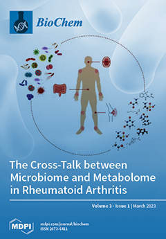
Cover Story (view full-size image): The early stages of the pathological immune response underlying rheumatoid arthritis (RA) occur in mucosal tissues, such as intestinal or oral mucosa, and are strongly associated with the abundance of specific bacterial species. Several studies have proposed microbiome dysbiosis as a trigger of local/systemic immunity activation and joint inflammation. This interplay could be intimately linked to the activation of specific metabolic pathways, resulting in changes in the metabolic profile of RA patients. In the RA scenario, the development of multi “-omics” methodologies may allow a better characterization of RA to improve risk prediction, early diagnosis, prognosis, and monitoring. View this paper
  • Issues are regarded as officially published after their release is announced to the table of contents alert mailing list.
  • You may sign up for e-mail alerts to receive table of contents of newly released issues.
  • PDF is the official format for papers published in both, html and pdf forms. To view the papers in pdf format, click on the "PDF Full-text" link, and use the free Adobe Reader to open them.
Order results
Result details
Select all
Export citation of selected articles as:
14 pages, 644 KiB  
Review
The Cross-Talk between Microbiome and Metabolome in Rheumatoid Arthritis
by Lidia La Barbera, Chiara Rizzo, Giulia Grasso, Federica Macaluso, Federica Camarda, Francesco Ciccia and Giuliana Guggino
BioChem 2023, 3(1), 47-60; https://doi.org/10.3390/biochem3010004 - 13 Mar 2023
Cited by 1 | Viewed by 2894
Abstract
Modern “omics” sciences, including metabolomics and microbiomics, are currently being applied to inflammatory autoimmune diseases, such as rheumatoid arthritis (RA), to investigate the interplay between microbiota, metabolic function, and the immune system. In recent decades, robust evidence has suggested that disruption of the [...] Read more.
Modern “omics” sciences, including metabolomics and microbiomics, are currently being applied to inflammatory autoimmune diseases, such as rheumatoid arthritis (RA), to investigate the interplay between microbiota, metabolic function, and the immune system. In recent decades, robust evidence has suggested that disruption of the normal composition of the microbiome, known as dysbiosis, in the gut and mouth of RA patients contributes to immune dysregulation and alterations in the metabolic pathways, shaping the pathogenesis of the disease and playing a central role in the risk and progression of RA. Metabolic pathways can be influenced by various agents such as the surrounding environment, lifestyle, and exposure to microbiota imbalance. In turn, the body’s metabolic homeostasis influences the immune response, making metabolomics helpful not only to understand pathogenesis pathways, but also to improve early disease detection and therapeutic chances. Combined gut microbiome and metabolome studies set out to unravel the interactions between these two entities, providing insights to discover new treatment targets and potential biomarkers to prevent joint damage. The purpose of this review is to summarize the main recent findings that suggest promising new research directions for the pathogenesis of RA. Full article
Show Figures

Figure 1

16 pages, 684 KiB  
Review
Intracellular Organization of Proteins and Nucleic Acids via Biomolecular Condensates in Human Health and Diseases
by Raffaella Gallo
BioChem 2023, 3(1), 31-46; https://doi.org/10.3390/biochem3010003 - 1 Feb 2023
Viewed by 3283
Abstract
Eukaryotic cells are intracellularly divided into several compartments that provide spatiotemporal control over biochemical reactions. Phase separation of proteins and RNA is emerging as an important mechanism underlying the formation of intracellular compartments that are not delimited by membranes. These structures are also [...] Read more.
Eukaryotic cells are intracellularly divided into several compartments that provide spatiotemporal control over biochemical reactions. Phase separation of proteins and RNA is emerging as an important mechanism underlying the formation of intracellular compartments that are not delimited by membranes. These structures are also known as biomolecular condensates and have been shown to serve a myriad of cellular functions, such as organization of cytoplasm and nucleoplasm, stress response, signal transduction, gene regulation, and immune response. Here, the author will summarize our current understanding of intracellular phase separation, its biological functions, and how this phenomenon is regulated in eukaryotic cells. Additionally, the author will review recent evidence of the role of biomolecular condensates in the development of pathophysiological conditions, with special emphasis on cancer and immune signaling. Full article
(This article belongs to the Special Issue RNA and Protein Dynamics: Latest Advances and Prospects)
Show Figures

Figure 1

16 pages, 3959 KiB  
Article
pH-Selective Reactions to Selectively Reduce Cancer Cell Proliferation: Effect of CaS Nanostructures in Human Skin Melanoma and Benign Fibroblasts
by Olga M. Rodríguez Martínez, Michelle A. Narváez Ramos, Angeliz A. Soto Acevedo, Carolina C. Colón Colón, Darlene Malavé Ramos, Coral Castro Rivera and Miguel E. Castro Rosario
BioChem 2023, 3(1), 15-30; https://doi.org/10.3390/biochem3010002 - 18 Jan 2023
Cited by 3 | Viewed by 2769
Abstract
An acidic extracellular pH value (pHe) is characteristic of many cancers, in contrast to the physiologic pHe found in most benign cells. This difference in pH offers a unique opportunity to design and engineer chemicals that can be employed for [...] Read more.
An acidic extracellular pH value (pHe) is characteristic of many cancers, in contrast to the physiologic pHe found in most benign cells. This difference in pH offers a unique opportunity to design and engineer chemicals that can be employed for pH-selective reactions in the extracellular fluid of cancer cells. The viability of human skin melanoma and corresponding fibroblasts exposed to CaS dispersions is reported. The viability of melanoma cells decreases with CaS dispersion concentration and reaches 57% at 3%, a value easily distinguishable from melanoma control experiments. In contrast, the viability of benign fibroblasts remains nearly constant within experimental error over the range of dispersion concentrations studied. The CaS dispersions facilitate vinculin delocalization in the cytoplasmic fluid, a result consistent with improved focal adhesion kinase (FAK) regulation in melanoma cells. Thermodynamic considerations are consistent with the formation of H2S from CaS in the presence of protons. The thermodynamic prediction is verified in independent experiments with solid CaS and acidic aqueous solutions. The amount of H2S formed decreases with pH. An activation energy for the process of (30 ± 10) kJ/mol in the temperature range of 280 to 330 K is estimated from initial rate measurements as a function of temperature. The total Gibbs energy minimization approach was employed to establish the distribution of sulfides—including H2S in the gas and aqueous phases—from the dissociation of CaS as a function of pH to mimic physiologically relevant pH values. Theoretical calculations suggest that partially protonated CaS in solution can be stable until the sulfur atom bonds to two hydrogen atoms, resulting in the formation of Ca2+ and H2S, which can be solvated and/or released to the gas phase. Our results are consistent with a model in which CaS is dissociated in the extracellular fluid of melanoma cells selectively. The results are discussed in the context of the potential biomedical applications of CaS dispersions in cancer therapies. Full article
Show Figures

Figure 1

14 pages, 1256 KiB  
Review
Current Technical Approaches to Study RNA–Protein Interactions in mRNAs and Long Non-Coding RNAs
by Johanna Mattay
BioChem 2023, 3(1), 1-14; https://doi.org/10.3390/biochem3010001 - 30 Dec 2022
Cited by 1 | Viewed by 4887
Abstract
It is commonly understood that RNA-binding proteins crucially determine the fate of their target RNAs. Vice versa, RNAs are becoming increasingly recognized for their functions in protein regulation and the dynamics of RNA-protein complexes. Long non-coding RNAs are emerging as potent regulators of [...] Read more.
It is commonly understood that RNA-binding proteins crucially determine the fate of their target RNAs. Vice versa, RNAs are becoming increasingly recognized for their functions in protein regulation and the dynamics of RNA-protein complexes. Long non-coding RNAs are emerging as potent regulators of proteins that exert unknown RNA-binding properties and moonlighting functions. A vast array of RNA- and protein-centric techniques have been developed for the identification of protein and RNA targets, respectively, including unbiased protein mass spectrometry and next-generation RNA sequencing as readout. Determining true physiological RNA and protein targets is challenging as RNA–protein interaction is highly dynamic, tissue- and cell-type-specific, and changes with the environment. Here I review current techniques for the analysis of RNA–protein interactions in living cells and in vitro. RNA-centric techniques are presented on the basis of cross-linking or the use of alternative approaches. Protein-centric approaches are discussed in combination with high-throughput sequencing. Finally, the impact of mutations in RNA–protein complexes on human disease is highlighted. Full article
(This article belongs to the Special Issue RNA and Protein Dynamics: Latest Advances and Prospects)
Show Figures

Figure 1

Previous Issue
Next Issue
Back to TopTop