Article

An In Vitro Study on Bond Strength Degradation of Low-Shrinkage Composite Resins to Demineralized Dentin After Thermal Cycling


by
Maria Cristina Borsatto
1,*,
Barbara Jarreta
1,
Jaciara Miranda Gomes-Silva
1,
Patricia Gatón-Hernández
2,
Carolina Paes Torres
1 and
Rodrigo Galo
3
1
Department of Pediatric Clinics, School of Dentistry of Ribeirão Preto, University of São Paulo (USP), Ribeirao Preto 14040-904, SP, Brazil
2
Department of Integrated Pediatric Dentistry, School of Dentistry, University of Barcelona, 08907 Barcelona, Spain
3
Department of Dental Materials and Prosthesis, School of Dentistry of Ribeirão Preto, University of São Paulo (USP), Ribeirao Preto 14040-904, SP, Brazil
*
Author to whom correspondence should be addressed.
Submission received: 10 February 2026 / Revised: 21 March 2026 / Accepted: 2 April 2026 / Published: 7 April 2026

Highlights

What are the main findings?
  • Low-shrinkage composites did not improve bond durability after aging.
  • Filtek Z250 showed the highest bond strength in all conditions.
  • Filtek P90 exhibited complete bond loss after thermocycling.
  • SEM revealed severe interfacial degradation in the P90 group.
What are the implications of the main findings?
Bond durability depends more on interface quality than shrinkage reduction.

Abstract

Objectives: Variations in composite resin composition and aging time remain one of the main reasons for replacing esthetic restorations. This in vitro study aimed to evaluate the microtensile bond strength of a low-shrinkage composite resin on a demineralized dentin surface following adhesive interface degradation. Methods: Seventy-eight extracted human molars were prepared, and artificial caries lesions were induced. For microtensile bond strength (μTBS) testing, 60 teeth were randomly assigned to six experimental subgroups (n = 10 per subgroup) based on restorative system and thermal cycling condition. An additional 18 teeth were randomly assigned to six experimental subgroups (n = 3 each) for SEM analysis. Three restorative systems were evaluated, Z250 (conventional resin), K (Kalore resin), and P90 (Filtek P90 resin), each subjected to two thermal cycling conditions: without thermal cycling (NTC) and 12,000 thermal cycles (TC). Results: In the NTC groups, Z250 exhibited a significantly higher bond strength (25.29 ± 10.91 MPa) compared to K (9.69 ± 11.63 MPa) and P90 (9.81 ± 8.49 MPa) (p < 0.05). Following TC, a numerical decrease in bond strength was observed across all groups. Z250 (13.00 ± 10.76 MPa) maintained a significantly higher bond strength compared to K (4.30 ± 6.40 MPa) and P90 (0 ± 0 MPa) (p = 0.001). Notably, the P90 group showed a near-complete loss of bond strength after TC (0 ± 0 MPa), which was a statistically significant reduction compared to its NTC condition (p = 0.002). SEM analysis revealed a predominance of mixed failures in most experimental groups, while the P90 TC group showed a clear predominance of adhesive failures. Conclusions: This study demonstrates that the conventional Bis-GMA resin (Z250) consistently exhibited superior bond strength to demineralized dentin compared to the low-shrinkage resins (Kalore and Filtek P90) under both non-aged and aged conditions. While all materials experienced a reduction in bond strength after thermal cycling, the Filtek P90 system showed a catastrophic loss of adhesion after aging, indicating its particular susceptibility to degradation. These results emphasize the critical roles of resin chemistry and adhesive system selection in long-term bond durability in compromised dentin.

1. Introduction

Currently, the number of composite resin restorations, particularly in posterior teeth, is increasing, driven by their esthetic advantages and conservative preparation techniques, and they are a popular alternative to dental amalgam [1]. However, the longevity of these restorations remains significantly affected by the consequences of their polymerization shrinkage stress [2,3], which may result in micro-infiltration, marginal discoloration, fissure formation, enamel deflection, and postoperative sensitivity [4,5], thereby affecting their long-term performance [6]. This intrinsic challenge remains a primary focus for materials scientists and clinicians striving to achieve enhanced restorative outcomes [7].
Continuous efforts to improve the physical and mechanical properties of restorative materials have been performed [8]. Manufacturers have sought to strengthen resin composition by developing new monomers [6,9,10] to overcome the limitations of conventional Bis-GMA-based resins. Currently, there is a clear tendency to replace the conventional Bis-GMA (bisphenol glycidyl methacrylate) matrix with a silorane monomer or a combination of UDMA (urethane dimethacrylate) and DX-511 dimethacrylate to reduce polymerization shrinkage [9,10,11].
Siloranes are silicon-based monomers whose molecules have a siloxane core with four attached oxirane rings that are open during polymerization, allowing them to join with another monomer [11,12]. As a result of this mechanism, the initial distance between monomers decreases slightly, resulting in a volumetric shrinkage of less than 1%, whereas methacrylate-based resins contract by more than 2% [2], thereby generating less stress at the adhesive interface [12]. The resin containing the DX-511 monomer has a higher molecular mass, resulting in a compound with lower polymerization shrinkage. This material is easy to handle; the professional has six times the time to perform the carving, and it additionally offers excellent esthetics, color stability, lower insolubility, adequate physical and mechanical properties, and high durability [12,13,14]. These developments represent significant strides in materials science, yet their long-term clinical effectiveness and interactions with dental substrates remain under continuous scrutiny [15,16].
To simulate oral conditions and assess the long-term performance of restorations, in vitro aging techniques have been extensively used to simulate degradation of the adhesive interface [15]. The most commonly used methods are water storage, held for different periods [17], and thermocycling, an aging technique proposed by ISO [18], in which the specimens are subjected to water baths simulating thermal changes in the oral cavity [19]. Other studies [11,20] have combined these two methods to assess adhesive interface degradation, as thermocycling is the most commonly used method to induce interfacial stress [21]. In contrast, water storage has been shown to reduce bond strength after a short period, indicating that bonds degrade over time [22], primarily through hydrolytic effects on the resin–dentin bond [23]. The specific protocol of 12,000 thermal cycles was selected in this study to simulate approximately 6 months of degradation in the oral cavity, based on established correlations in the literature [20,24].
To assess bond strength between the adhesive system and dentin, various study models have been reported. Among these, microtensile bond strength (μTBS) is recognized as a reliable test [25], as it requires fewer teeth and a joining area of about 1 mm2, which makes the resulting values more sensitive and reliable compared to macro-shear or macro-tensile tests [26,27]. This method allows for a more localized assessment of the adhesive interface, reducing the incidence of cohesive failures within the bulk material and providing a clearer indication of the true adhesive strength [28].
Despite the theoretical advantages of reduced polymerization shrinkage, particularly in stress relief at the adhesive interface, it remains unclear whether these benefits translate into improved long-term bonding performance, especially in compromised substrates such as demineralized dentin. Therefore, it is important not only to assess the immediate bonding effectiveness of low-shrinkage composites but also to critically evaluate their durability after aging. In this context, the present study aims to investigate whether the reduction in polymerization shrinkage effectively contributes to enhanced adhesive stability over time or whether other material-related factors may play a more decisive role [6].
Thus, regarding the continuous development of esthetic restorative materials and the persistent challenge of achieving durable adhesion to dentin, particularly under conditions simulating long-term oral exposure, the need for a better understanding of the behavior of these resins in long-term tests, as well as their adhesion to dentin, necessitates further research to expand knowledge in this area [29]. The current literature remains inconsistent regarding the performance of low-shrinkage composites, particularly after aging, underscoring the need for further investigation [30]. The aim of this study is therefore to evaluate these crucial aspects. The null hypothesis is that the bond strength of adhesive systems to composite resin and aging time do not affect μTBS for the dentin substrate.

2. Materials and Methods

2.1. Sample Preparation

Seventy-eight human third molars were extracted for this study. The sample size (60 teeth for the microtensile test and 18 additional teeth for scanning electron microscopy analysis) was determined based on similar previous studies. It was designed to provide adequate statistical power to detect significant differences among groups while maintaining ethical and laboratory feasibility [28]. Teeth from the Human Teeth Biobank of the Ribeirao Preto School of Dentistry were meticulously cleaned with ultrasound. They received standardized prophylaxis to remove any debris and ensure a uniform surface. Dentin was stored in distilled water at 4 °C, according to established protocols, to preserve its integrity before preparation.
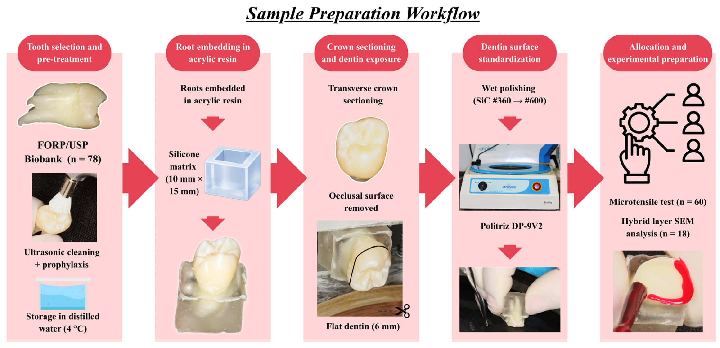
The tooth roots were embedded in acrylic resin using a silicone matrix (1.0 cm in diameter and 1.5 cm in height). The crowns were sectioned transversely, removing the occlusal surface to expose the dentin (Minitom; Struers, Copenhagen, Denmark); standardizing a flat dentin surface of approximately 6 mm in diameter. Subsequently, the exposed dentin was polished with wet #360- and #600-grit silicon carbide (SiC) paper in a low-speed polishing machine (Politriz DP-9V2; Struers, Copenhagen, Denmark) (Figure 1).

2.2. Experimental Design and Bonding Procedures

For the artificial induction of caries lesions, the specimens were meticulously coated with two layers of proof cosmetic nail polish, ensuring that only the dentinal surface to be treated was exposed. This approach, based on the Manesh et al. model [31], is recognized for its efficiency and simplicity in creating standardized artificial caries lesions that mimic in vivo conditions in a controlled manner. Individual immersion in 40 mL of demineralizing solution at 37 °C for 12 consecutive days, without renewal, was established to achieve a consistent degree of dentin demineralization, essential for studying bonding to compromised substrates [32].
Afterwards, specimens were prepared using a spherical drill steel No. 4 in a low-speed handpiece (KaVo in Brazil Ind. Com., Joinville, SC, Brazil), which was replaced every five preparations. Preparation procedures were simulated in the clinic to preserve as much tissue as possible. On the prepared dentin, restorations were performed based on the restorative system to be assessed.
The dentin specimens were randomly assigned to 3 groups for the microtensile test using computer-generated random numbers, based on the restorative systems used: group Z250—Clearfill SE Bond adhesive system + Filtek Z250 resin (conventional matrix of Bis-GMA—conventional composite resin); group K—Clearfill SE Bond adhesive system + Kalore resin (combination of UDMA monomer with DX-511/low-shrinkage composite resin of polymerization); group P90—P90 adhesive system + Filtek P90 resin (matrix of silorane monomer/low-shrinkage composite resin of polymerization).
All materials were used in accordance with the manufacturers’ recommendations. Specifically, for groups Z250 and K, the Clearfil SE Bond primer was actively rubbed into the demineralized dentin for 20 s, followed by gentle air-drying for 5 s until the solvent evaporated and a glossy surface was observed. Subsequently, a thin layer of Clearfil SE Bond bonding agent was applied, left undisturbed for 10 s, gently air-dried for 3 s to ensure even distribution, and light-cured for 10 s. For group P90, the P90 adhesive system was applied to the demineralized dentin with a gentle rubbing motion for 20 s, followed by gentle air-drying for 5 s, and then light-cured for 10 s. Resin composite build-ups (~4 mm) were done in increments (~1 mm) up to 4 mm, with each increment light-cured for 20 s. Photo-activation of all materials was performed using an Ultralux halogen lamp (DabiAtlante SA Ind. Médico Odontológicas, Ribeirão Preto, SP, Brasil), with the output intensity rigorously monitored with a curing radiometer RD-7 (Ecel Ind. e Com. Ltd., Ribeirão Preto, SP, Brazil) every 10 restored specimens. This systematic control maintained a constant minimum light intensity of 600 mW/cm2, minimizing variability in delivered light energy and ensuring uniform polymerization across all experimental groups [33].
After the bonding procedures, the specimens in each group were divided into subgroups (n = 10) to assess bond strength at the adhesive interface using the challenge test: water immersion for 24 h (NTC) and thermocycling (TC). A total of 12,000 thermal cycles was performed in a thermal cycler (MSCT-3; São Carlos, SP, Brazil), with each cycle consisting of water baths at 5 °C to 55 °C, a dwell time of 30 s, and a transfer time of 7 s. This protocol was selected as a well-established, widely used method in the literature for simulating approximately 6 months of intraoral degradation [20,34]. The choice of this period aims to evaluate bonding performance at a clinically relevant stage of aging, before severe degradation fully compromises the interface.

2.3. Microtensile Bond Strength Testing

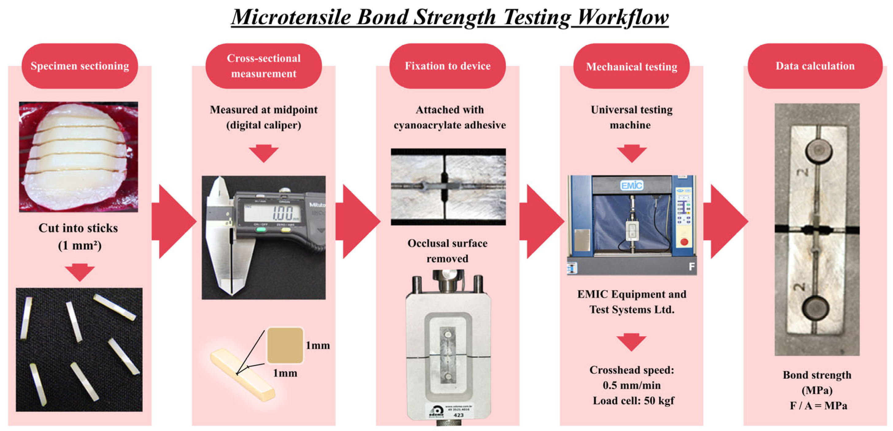
After thermocycling (TC) and non-thermocycling (NTC) protocols, specimens were cut into sticks with a cross-sectional area of approximately 1 mm2. The cross-sectional area of each stick was measured at the midpoint of each section with a digital caliper (Mitutoyo, Tokyo, Japan). Then the specimens were attached to a universal testing machine (EMIC Equipment and Test Systems Ltd., São José dos Pinhais, PR, Brazil) with a cyanoacrylate-based adhesive (Super Bonder Gel, Loctite Corporation, São Paulo, SP, Brazil). The test was conducted at a crosshead speed of 0.5 mm/min, using a 50 kgf load cell. Microtensile bond strength values were calculated in MPa (Figure 2).

2.4. Failure Mode and Scanning Electron Microscopy Analysis

After the µTBS tests, the fracture mode of the specimens was observed using a scanning electron microscope (EVO, Carl Zeiss, Oberkochen, Baden-Württemberg, Germany). The fracture modes were classified as adhesive failure at the interface among the resin composite, adhesive resin, and hybrid layer; cohesive failure in dentine; cohesive failure in resin composite; and mixed failure, with both adhesive and cohesive failures observed in the same specimens.
For hybrid layer analysis, 18 specimens were randomly assigned to 6 experimental groups (n = 3 per group) using a similar computer-generated randomization process. After receiving restorative procedures and thermal cycling, they were sectioned longitudinally, yielding fragments approximately 2 mm thick. Specimens were polished and prepared for SEM. The samples were fixed on aluminum stubs and sputter-coated with gold in a vacuum metallizing equipment (SDC 050, Bal-Tec AG, Balzers, Liechtenstein) and then evaluated by scanning electron microscopy (EVO Carl Zeiss, Oberkochen, Baden-Wurttemberg, Germany) at the Department of Chemistry, Faculty of Philosophy, Sciences and Languages of Ribeirão Preto, University of São Paulo.

2.5. Statistical Analysis

Data were assessed using the Kolmogorov–Smirnov and Shapiro–Wilk normality tests, which indicated that the data were not normally distributed. In consequence, the chosen sample test was the Kruskal–Wallis test at the 5% significance level, and the complementary parametric test was the Mann–Whitney test.
The images of the fractured specimens from the microtensile tests were classified according to fracture type. The analysis of micrography of adhesive interfaces achieved by SEM was descriptive.

3. Results

3.1. Microtensile Bond Strength

The mean microtensile bond strength (µTBS) values and respective standard deviations for the experimental groups are presented in Table 1. A numerical reduction in bond strength was observed for all materials after thermal cycling (TC) compared with non-thermal cycling (NTC), indicating a negative effect of simulated aging on the adhesive interface.
Under non-aged conditions (24 h NTC), the Z250 group showed a mean µTBS of 25.29 (±10.91) MPa, which was statistically higher (p < 0.05) than that of both the K (9.69 ± 11.63 MPa) and P90 (9.81 ± 8.49 MPa) groups, with no significant difference between the latter two. These findings indicate superior initial bonding performance of the conventional Z250 system.
After thermal cycling, although all groups exhibited lower mean bond strength values, statistical significance between NTC and TC conditions was observed only for the P90 group (p = 0.002). Even after aging, Z250 maintained significantly higher bond strength (13.00 ± 10.76 MPa) compared with K (4.30 ± 6.40 MPa) and P90 (0 ± 0 MPa) (p = 0.001). The P90 group exhibited nearly complete loss of bond strength after thermal cycling, indicating severe degradation of the adhesive interface under simulated aging conditions.
Compared with the thermally cycled groups, the tested groups after 24 h of storage showed a significant difference only in the P90 group (p = 0.002), indicating that this material was particularly sensitive to the simulated aging challenge, undergoing marked degradation in bond strength over time.

3.2. Failure Mode and Scanning Electron Analysis

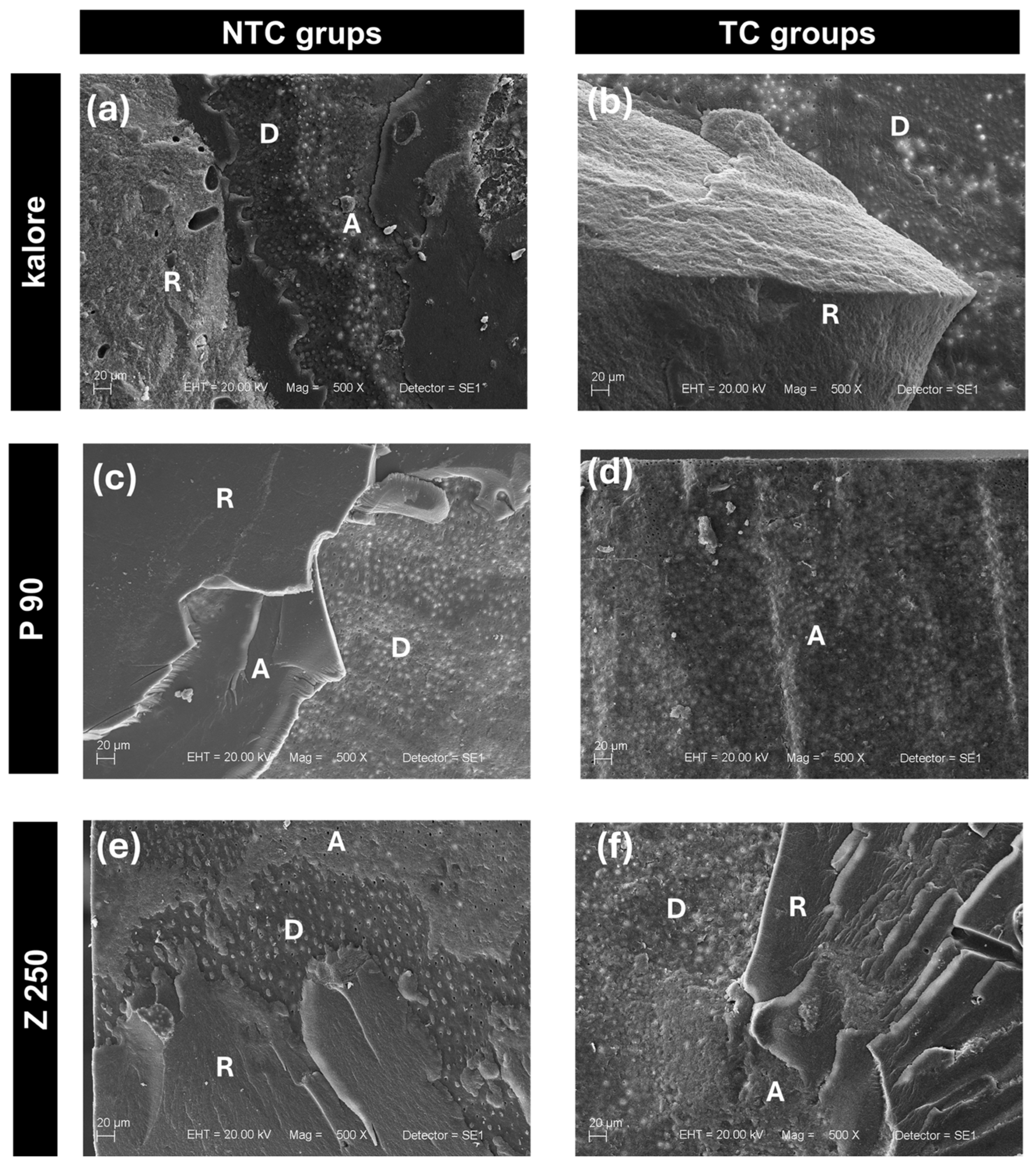
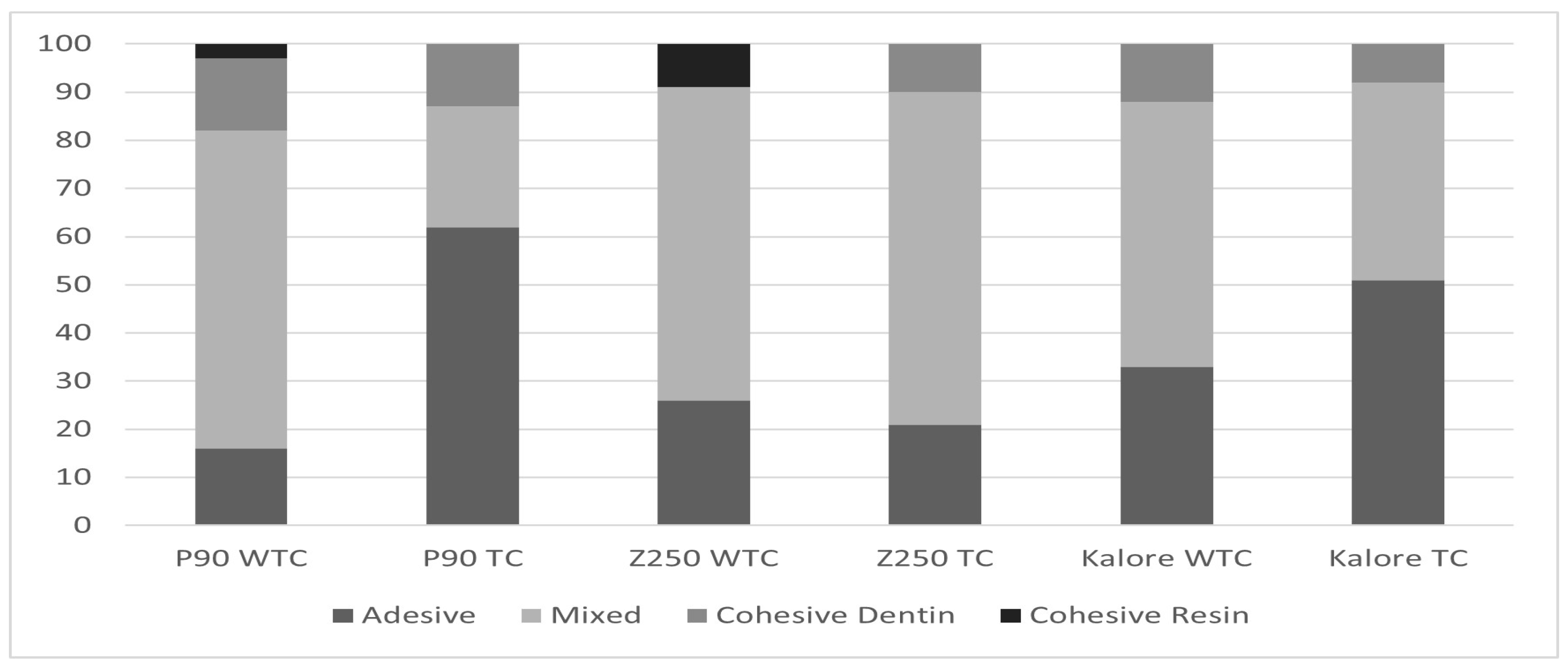
The fracture patterns observed after the µTBS tests were analyzed by scanning electron microscopy (SEM) and are illustrated in Figure 3 and summarized in Figure 4. Overall, mixed failure predominated in most experimental groups, indicating effective stress transfer at the adhesive interface and failure involving both adhesive and cohesive components.
As shown in Figure 4, the Z250 and Kalore groups, under both non-thermal cycling (NTC) and thermal cycling (TC) conditions, exhibited predominantly mixed failures. In contrast, the P90 group after thermal cycling (P90 TC) showed a clear predominance of adhesive failures at the interface between the resin composite, adhesive layer, and hybrid layer (Figure 3d), suggesting severe degradation of the adhesive bond, which is consistent with the near-zero µTBS values observed for this group.
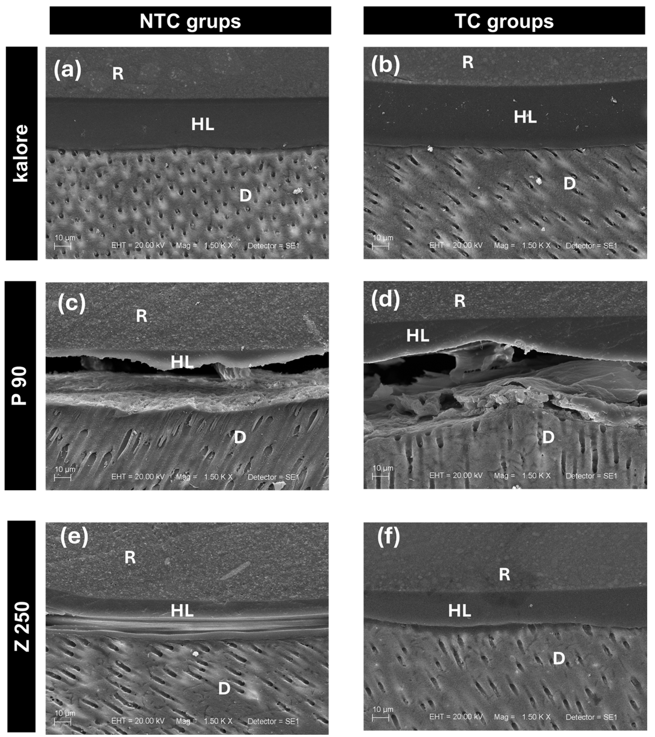
SEM analysis of the adhesive interface (Figure 5) revealed distinct morphological characteristics among the systems. For the Clearfil SE Bond adhesive used with Z250 resin, images obtained under NTC conditions (Figure 5e) showed a thick adhesive layer and pronounced resin tag formation within the dentinal tubules. After thermal cycling (Figure 5f), this general pattern was maintained; however, gaps at the adhesive interface became evident, suggesting micro-separations caused by aging-induced degradation.
Similar morphological features were observed in the Kalore group under NTC conditions, including a relatively thick adhesive layer and resin tags (Figure 5a). After thermal cycling, although tag formation was still detectable, images revealed signs of interfacial disorganization and degradation (Figure 5b).
In contrast, the P90 adhesive system exhibited unfavorable interfacial characteristics even under NTC conditions, with limited tag formation and microcracks at the adhesive interface (Figure 5c). After thermal cycling, the P90 TC group showed extensive interfacial cracking, gaps, an almost complete absence of resin tags, and difficulty in identifying a continuous hybrid layer (Figure 5d). These morphological findings strongly corroborate the adhesive failure pattern and the drastic reduction in bond strength observed for this group after thermal cycling.

4. Discussion

New resins have been developed with novel compositions, particularly those considered to exhibit low polymerization shrinkage. The primary objective of these innovations is to reduce stress at the adhesive interface immediately after photoactivation, thereby theoretically extending the clinical life of dental restorations [30]. However, knowledge regarding the adhesion values of these materials to the dental substrate, especially after aging challenges that simulate the oral environment, remains crucial for their clinical validation. Thus, this study assessed the microtensile bond strength of composites with different chemical compositions, such as Kalore (UDMA/DX-511), Filtek P90 (silorane), and Filtek Z250 (conventional Bis-GMA), to provide insights into their performance under simulated degradation conditions.
In this study, the Filtek P90 resin was used with its corresponding adhesive system, the P90 adhesive system, specifically formulated to interact with silorane chemistry. In contrast, for the Kalore and Filtek Z250 resins, the Clearfil SE Bond adhesive system was selected. This choice was based on the fact that these resins lack proprietary adhesive systems, and Clearfil SE Bond is a widely recognized two-step self-etch adhesive system known for its simplified technique, clinical stability, and good mechanical strength [35]. The Clearfil SE Bond system has a pH of approximately 2, which is considered “mild” in terms of its aggressiveness. This lower pH favors the preservation of hydroxyapatite crystals around collagen fibers, thereby facilitating the formation of a robust hybrid layer and dentinal tags, as observed in our SEM analyses of the Z250 and K groups [36,37]. Similarly, the P90 adhesive system is considered “mild” with a pH of 2.7 [38]. However, SEM observations of this system revealed distinct behavior in the hybrid layer and tag formation, as discussed in the results.
Regarding the initial methodology, artificial caries was induced prior to cavity preparation using the Manesh et al. [28] model, as it is an efficient and straightforward technique for creating in vitro demineralized dentin substrates that mimic clinically relevant lesions. The subsequent removal of demineralized dentin with a low-speed steel drill and the placement of restorations on artificially carious rather than healthy dentin was a crucial methodological choice. This approach enabled the study to evaluate adhesive performance in a more challenging, clinically relevant scenario in which dentin often exhibits some degree of demineralization [39]. After demineralized dentin removal, self-etching adhesive systems were used, classified by their adhesive strategy and number of application steps [29], with mild-pH systems selected for the present study.
The microtensile test results consistently revealed that the Z250 group (conventional resin with Clearfil SE Bond) exhibited higher bond strength values compared to both the K (Kalore with Clearfil SE Bond) and P90 (Filtek P90 with P90 adhesive system) groups, under both non-aged (NTC) and thermocycled (TC) conditions (Table 1). This finding suggests that, in this study, conventional Z250 resin, when combined with a two-step self-etch adhesive system, exhibited greater adhesive stability. This performance may be attributed to the Bis-GMA matrix of Z250, which may possess more favorable wetting and infiltration properties than low-shrinkage monomers, particularly in demineralized dentin [40,41].
Our finding that Clearfil SE Bond yielded higher adhesion values than silorane-based systems or low-shrinkage resins is corroborated by Feitosa et al. [42], who also reported higher bond strength for this adhesive. Similarly, Giacobbi and Vandewalle [43] observed a decrease in microtensile bond strength values for both silorane and Clearfil SE Bond adhesive systems after aging. Still, they found no statistically significant difference between the resins, whereas our results showed that Z250 remained superior. This discrepancy could be explained by variations in the aging protocol (5000 cycles in Feitosa et al.’s study versus 12,000 cycles in ours) or specific differences in caries lesion induction. Other researchers [42] also pointed to the greater bond strength of Z100 resin (conventional) compared to silorane, attributing this to the sensitivity of the single-step adhesive to polymerization shrinkage stress. These results collectively reinforce the idea that, despite advances in low-shrinkage monomers, adhesive interface stability remains a significant challenge for these new formulations, particularly in complex substrates such as demineralized dentin [44].
These findings highlight an important conceptual implication of the present study. Although low-shrinkage composite systems have been developed with the expectation of reducing polymerization stress and, consequently, improving the integrity of the adhesive interface [4,12], our results did not demonstrate superior bonding durability for these materials when compared to the conventional system [15,26]. This suggests that factors such as adhesive chemistry, wettability, viscosity, and the ability to form a stable hybrid layer may have a more critical influence on long-term performance than shrinkage reduction alone [25,29,36,40]. Therefore, the assumption that lower polymerization shrinkage necessarily leads to improved clinical outcomes should be reconsidered, particularly in challenging substrates such as demineralized dentin [23,28].
Although the incremental insertion technique was standardized in our study to minimize polymerization stress, as recommended by Van Ende et al. [45] and Almeida et al. [46], who demonstrated higher bond strength with this technique, the intrinsic viscosity of the materials can significantly affect adhesive performance. Our results, particularly for the P90 group, which exhibited the lowest bond strength values and catastrophic failures after aging, may be partly explained by the high viscosity of the P90 silorane resin.
Authors such as those of [45,47] suggest that low-shrinkage polymerization materials with high viscosity may present adaptation failures to the dentin substrate, resulting in poor wetting and, consequently, a compromised adhesive bond. Our SEM analyses (Figure 5c,d), which revealed the presence of interface cracks and difficulty forming or maintaining tags and a hybrid layer in the P90 group, even under NTC conditions and exacerbated after TC, corroborate this hypothesis. High viscosity may have impeded adequate penetration of the adhesive into the dentinal tubules and the demineralized collagen network, resulting in a weakened adhesive interface susceptible to degradation.
Thermocycling is a standard method for simulating thermal changes in the mouth and evaluating the aging of restorations [11,40,48]. In our study, 12,000 thermal cycles, equivalent to approximately 6 months of aging in the oral cavity, resulted in a generalized decrease in bond strength across all groups (Table 1). This reduction is consistent with the literature, in which studies such as Krajagta and Srisawasdi [49] also reported decreased adhesion after aging, albeit with a shorter aging time (3 months). The decrease in bond strength for the Z250 group (conventional resin) can be attributed to the hydrolytic and thermal degradation of adhesive interface bonds over time.
For group K, the Kalore resin, despite being a low-shrinkage composite, also exhibited a significant reduction, suggesting that the UDMA/DX-511 matrix, although designed for lower shrinkage, remains susceptible to degradation under prolonged thermal stress [50]. Most notably, the P90 group exhibited the largest decrease in bond strength after thermal cycling, with an average value of 0 MPa. This catastrophic failure underscores the sensitivity of the silorane/P90 adhesive system to simulated aging.
The observed low bond strength of the P90 group, culminating in near-zero values after thermal cycling, may be multifactorial. One key factor appears to be bubble formation at the adhesive interface, a phenomenon exacerbated by water accumulation. The P90 adhesive system, despite being “mild”, possesses hydrophobic characteristics and a high concentration of specific monomers, such as HEMA (2-hydroxyethyl methacrylate), which, paradoxically, can induce water movement from dentin to the interface, leading to accumulation and bubble formation [29,44]. Our SEM images (Figure 5c,d) support this hypothesis, revealing significant gaps and cracks and the absence of discernible dentinal tags or an intact hybrid layer, particularly after aging. The ineffectiveness in forming a well-defined hybrid layer for P90 may be directly attributable to the difficulty of wetting and infiltrating the adhesive into the demineralized dentin collagen network, thereby compromising the formation of a mechanically and chemically stable resin–dentin interdiffusion [28]. This failure to form a homogeneous adhesive interface makes the P90 system particularly vulnerable to degradation by thermocycling aging.
SEM images of the adhesive interface provided crucial morphological insight into the functional results. For the groups using the Clearfill SE Bond adhesive system (Z250 and K), the presence of thick and dense adhesive layers, with well-defined tags within the dentinal tubules, was observed (Figure 5a,b,e,f). This morphology is characteristic of effective resin interdiffusion and good hybrid-layer formation, contributing to the relatively higher bond strengths observed for these groups. However, even in these systems, the presence of gaps after aging (Figure 5f) indicates that thermocycling-induced degradation can still compromise the interface’s integrity, albeit to a lesser extent.
In striking contrast, the adhesive interface of the P90 system (Figure 5c,d) exhibited prominent interfacial gaps and cracks in both the initial state and, more acutely, after aging, in addition to the absence of dentinal tags and a detectable hybrid layer. This morphological discrepancy between the adhesive systems is a fundamental finding and largely accounts for the very low microtensile bond strength values observed in the P90 group, particularly after aging. The failure to establish a micro-mechanically retentive and chemically stable adhesive interface, as evidenced by SEM, predisposed the P90 system to accelerated degradation under thermal stress, leading to complete bond loss.

5. Conclusions

These innovative results suggest that, for demineralized dentin, the choice of adhesive system and composite resin has a critical impact on bond durability under aging. The superior performance of the conventional Z250 resin, in conjunction with Clearfil SE Bond, highlights the importance of a robust adhesive system and resin chemistry that promote stable interdiffusion in a compromised dentin substrate. In contrast, systems such as the silorane P90 may present significant limitations in challenging clinical scenarios after aging. Future in vivo studies and long-term clinical trials are necessary to validate these in vitro observations fully and to account for the multifactorial influences of the biological oral environment, which cannot be fully replicated in laboratory settings.

Author Contributions

Conceptualization, M.C.B. and B.J.; methodology, B.J. and J.M.G.-S.; validation, R.G., C.P.T. and P.G.-H.; formal analysis, R.G.; investigation, B.J. and C.P.T.; data curation, R.G.; writing—original draft preparation, M.C.B. and B.J.; writing—review and editing, R.G. and J.M.G.-S.; visualization, M.C.B.; supervision, M.C.B.; project administration, M.C.B.; funding acquisition, P.G.-H. All authors have read and agreed to the published version of the manuscript.

Funding

This study was financed in part by the Coordination for the Improvement of Higher Education Personnel (CAPES), Brazil—Finance Code 001.

Institutional Review Board Statement

The present study was conducted in accordance with the Declaration of Helsinki and was approved by the Research Ethics Committee of the Ribeirão Preto School of Dentistry, University of São Paulo, Brazil (approval number 283.612, issued on 16 May 2013).

Informed Consent Statement

The teeth were obtained from the Human Teeth Biobank of the Ribeirao Preto School of Dentistry.

Data Availability Statement

The raw data supporting the conclusions of this article will be made available by the authors on request.

Conflicts of Interest

The authors declare no conflicts of interest.

Abbreviations

The following abbreviations are used in this manuscript:
NTCWithout thermal cycling
TCThermal cycle
Bis-GMABisphenolglycidyl methacrylate
UDMAUrethane dimethacrylate
μTBSMicrotensile
SEMScanning electron microscope
TBSMicrotensile bond strength
HEMA2-hydroxyethyl methacrylate

References

  1. Sidhu, P.; Sultan, O.S.; Math, S.Y.; Ab Malik, N.; Wilson, N.H.F.; Lynch, C.D.; Blum, I.R.; Daood, U. Current and future trends in the teaching of direct posterior resin composites in Malaysian dental schools: A cross-sectional study. J. Dent. 2021, 110, 103683. [Google Scholar] [CrossRef]
  2. Baracco, B.; Perdigão, J.; Cabrera, E.; Ceballos, L. Two-year clinical performance of a low-shrinkage composite in posterior restorations. Oper. Dent. 2013, 38, 591–600. [Google Scholar] [CrossRef] [PubMed]
  3. Jang, J.H.; Park, S.H.; Hwang, I.N. Polymerization shrinkage and depth of cure of bulk-fill resin composites and highly filled flowable resin. Oper. Dent. 2015, 40, 172–180. [Google Scholar] [CrossRef]
  4. Albeshir, E.G.; Alsahafi, R.; Albluwi, R.; Balhaddad, A.A.; Mitwalli, H.; Oates, T.W.; Hack, G.D.; Sun, J.; Weir, M.D.; Xu, H.H.K. Low-Shrinkage Resin Matrices in Restorative Dentistry—Narrative Review. Materials 2022, 15, 2951. [Google Scholar] [CrossRef]
  5. Gonzalez-Lopez, S.; Vilchez, M.A.; de Haro-Gasquet, F.; Ceballos, L.; de Haro-Munoz, C. Cuspal flexure of teeth with composite restorations subjected to occlusal loading. J. Adhes. Dent. 2007, 9, 11–15. [Google Scholar]
  6. Zheng, S.; Chen, H.; Lin, Q.; Zhu, S. Effect of dentin conditioners on dentin bond strength: A systematic review and meta-analysis. J. Prosthet. Dent. 2024, 132, 509.e1–509.e11. [Google Scholar] [CrossRef] [PubMed]
  7. Topa-Skwarczyńska, M.; Ortyl, J. Photopolymerization shrinkage: Strategies for reduction, measurement methods and future insights. Polym. Chem. 2023, 14, 2145–2158. [Google Scholar] [CrossRef]
  8. Ilie, N.; Hickel, R. Investigations on a methacrylate-based flowable composite based on the SDR™ technology. Dent. Mater. 2011, 27, 348–355. [Google Scholar] [CrossRef]
  9. Soares, C.J.; Bicalho, A.A.; Tantbirojn, D.; Versluis, A. Polymerization shrinkage stresses in a premolar restored with different composite resins and different incremental techniques. J. Adhes. Dent. 2013, 15, 341–350. [Google Scholar] [CrossRef]
  10. Bicalho, A.; Pereira, R.; Zanatta, R.; Franco, S.; Tantbirojn, D.; Versluis, A.; Soares, C.J. Incremental filling technique and composite material—Part I: Cuspal deformation, bond strength, and physical properties. Oper. Dent. 2014, 39, e83–e92. [Google Scholar] [CrossRef]
  11. Amaral, F.L.; Colucci, V.; Souza-Gabriel, A.E.; Chinelatti, M.A.; Palma-Dibb, R.G.; Corona, S.A. Bond durability in erbium: Yttrium–aluminum–garnet laser-irradiated enamel. Lasers Med. Sci. 2010, 25, 155–163. [Google Scholar] [CrossRef] [PubMed]
  12. Maghaireh, G.A.; Taha, N.A.; Alzraikat, H. The Silorane-Based Resin Composites: A Review. Oper. Dent. 2017, 42, E24–E34. [Google Scholar] [CrossRef]
  13. Ilie, N.; Jelen, E.; Clementino-Luedemann, T.; Hickel, R. Low-shrinkage composite for dental application. Dent. Mater. J. 2007, 26, 149–155. [Google Scholar] [CrossRef] [PubMed]
  14. Naoum, S.J.; Ellakwa, A.; Morgan, L.; White, K.; Martin, F.E.; Lee, I.B. Polymerization profile analysis of resin composite dental restorative materials in real time. J. Dent. 2012, 40, 64–70. [Google Scholar] [CrossRef]
  15. Schmidt, M.; Dige, I.; Kirkevang, L.-L.; Vaeth, M.; Hørsted-Bindslev, P. Five-year evaluation of a low-shrinkage silorane resin composite material: A randomized clinical trial. Clin. Oral Investig. 2015, 19, 245–251. [Google Scholar] [CrossRef] [PubMed]
  16. Wu, Z.; Sun, K.; Wang, W.; Xue, Q.; Tonin, B.S.H.; Watts, D.C.; Fu, J.; Wang, H. Characterization of dental light-curing resin composites incorporating multiple modified low-shrink monomers. Dent. Mater. 2024, 40, 1244–1251. [Google Scholar] [CrossRef]
  17. Shono, Y.; Terashita, M.; Shimada, J.; Kozono, Y.; Carvalho, R.M.; Russell, C.M.; Pashley, D.H. Durability of resin–dentin bonds. J. Adhes. Dent. 1999, 1, 211–218. [Google Scholar]
  18. Nakabayashi, N.; Pashley, D.H. Hybridization of Dental Hard Tissues; Quintessence Publishing: Tokyo, Japan, 2000. [Google Scholar]
  19. Malysa, A.; Wezgowiec, J.; Grzebieluch, W.; Danel, D.P.; Wieckiewicz, M. Effect of thermocycling on the bond strength of self-adhesive resin cements used for luting CAD/CAM ceramics to human dentin. Int. J. Mol. Sci. 2022, 23, 745. [Google Scholar] [CrossRef]
  20. Amaral, F.L.; Colucci, V.; Palma-Dibb, R.G.; Corona, S.A. Assessment of in vitro methods used to promote adhesive interface degradation: A critical review. J. Esthet. Restor. Dent. 2007, 19, 340–354. [Google Scholar] [CrossRef]
  21. Bedran-de-Castro, A.K.; Pereira, P.N.; Pimenta, L.A.F. Long-term bond strength of restorations subjected to thermal and mechanical stresses over time. Am. J. Dent. 2004, 17, 337–341. [Google Scholar]
  22. Carrilho, M.R.O.; Carvalho, R.M.; Tay, F.R.; Pashley, D.H. Effects of storage media on mechanical properties of adhesive systems. Am. J. Dent. 2004, 17, 104–108. [Google Scholar] [PubMed]
  23. Hashimoto, M.; Ohno, H.; Sano, H.; Kaga, M.; Oguchi, H. In vitro degradation of resin–dentin bonds analyzed by microtensile bond test, scanning, and transmission electron microscopy. Biomaterials 2003, 24, 3795–3803. [Google Scholar] [CrossRef] [PubMed]
  24. Ghavami-Lahiji, M.; Firouzmanesh, M.; Bagheri, H.; Jafarzadeh Kashi, T.S.; Razazpour, F.; Behroozibakhsh, M. The effect of thermocycling on the degree of conversion and mechanical properties of a microhybrid dental resin composite. Restor. Dent. Endod. 2018, 43, e26. [Google Scholar] [CrossRef] [PubMed]
  25. De Munck, J.; Van Landuyt, K.; Peumans, M.; Poitevin, A.; Lambrechts, P.; Braem, M.; Van Meerbeek, B. A critical review of the durability of adhesion to tooth tissue: Methods and results. J. Dent. Res. 2005, 84, 118–132. [Google Scholar] [CrossRef]
  26. da Silva, G.R.; Araújo, I.S.; Pereira, R.D.; Barreto, B.C.; do Prado, C.J.; Soares, C.J.; Martins, L.R. Microtensile bond strength of methacrylate and silorane resins to enamel and dentin. Braz. Dent. J. 2014, 25, 327–331. [Google Scholar] [CrossRef]
  27. Sano, H.; Shono, T.; Sonoda, H.; Takatsu, T.; Ciucchi, B.; Carvalho, R.; Pashley, D.H. Relationship between surface area for adhesion and tensile bond strength—Evaluation of a microtensile bond test. Dent. Mater. 1994, 10, 236–240. [Google Scholar] [CrossRef]
  28. Pashley, D.H.; Carvalho, R.M. Dentine permeability and dentine adhesion. J. Dent. 1997, 25, 355–372. [Google Scholar] [CrossRef]
  29. Van Meerbeek, B.; Yoshihara, K.; Yoshida, Y.; Mine, A.; De Munck, J.; Van Landuyt, K.L. State of the art of self-etch adhesives. Dent. Mater. 2011, 27, 17–28. [Google Scholar] [CrossRef]
  30. Ilie, N.; Hickel, R. Resin composite restorative materials. Aust. Dent. J. 2011, 56, 59–66. [Google Scholar] [CrossRef]
  31. Manesh, S.K.; Darling, C.L.; Fried, D. Nondestructive assessment of dentin demineralization using polarization-sensitive optical coherence tomography after exposure to fluoride and laser irradiation. J. Biomed. Mater. Res. B Appl. Biomater. 2009, 90, 802–812. [Google Scholar] [CrossRef]
  32. Koibuchi, H.; Yasuda, N.; Nakabayashi, N. Bonding to dentin with a self-etching primer: The effect of smear layers. Dent. Mater. 2001, 17, 122–126. [Google Scholar] [CrossRef]
  33. Rueggeberg, F.A. State-of-the-art: Dental photocuring—A review. Dent. Mater. 2011, 27, 39–52. [Google Scholar] [CrossRef]
  34. Gale, M.S.; Darvell, B.W. Thermal cycling procedures for laboratory testing of dental restorations. J. Dent. 1999, 27, 89–99. [Google Scholar] [CrossRef]
  35. Kameyama, A.; Aizawa, K.; Kato, J.; Hirai, Y. Tensile bond strength of single-step self-etch adhesives to Er: YAG laser-irradiated dentin. Photomed. Laser Surg. 2009, 27, 3–10. [Google Scholar] [CrossRef] [PubMed]
  36. Yoshida, Y.; Nagakane, K.; Fukuda, R.; Nakayama, Y.; Okazaki, M.; Shintani, H.; Inoue, S.; Tagawa, Y.; Suzuki, K.; De Munck, J.; et al. Comparative study on adhesive performance of functional monomers. J. Dent. Res. 2004, 83, 454–458. [Google Scholar] [CrossRef]
  37. Van Meerbeek, B.; De Munck, J.; Yoshida, Y.; Inoue, S.; Vargas, M.; Vijay, P.; Van Landuyt, K.; Lambrechts, P.; Vanherle, G. Adhesion to enamel and dentin: Current status and future challenges. Oper. Dent. 2003, 28, 215–235. [Google Scholar] [PubMed]
  38. Saar, M.; Kane, A.W.; Vreven, J.; Mine, A.; Van Landuyt, K.L.; Peumans, M.; Lambrechts, P.; Van Meerbeek, B.; De Munck, J. Microtensile bond strength and interfacial characterization of 11 contemporary adhesives bonded to bur-cut dentin. Oper. Dent. 2010, 35, 94–104. [Google Scholar] [CrossRef] [PubMed]
  39. Nakajima, M.; Sano, H.; Urabe, I.; Tagami, J.; Pashley, D.H. Bond strengths of resin to caries-affected dentin. J. Dent. Res. 1995, 74, 1581–1588. [Google Scholar] [CrossRef]
  40. Pashley, D.H.; Tay, F.R.; Breschi, L.; Tjäderhane, L.; Carvalho, R.M.; Carrilho, M.; Tezvergil-Mutluay, A. State of the art etch-and-rinse adhesives. Dent. Mater. 2011, 27, 1–16. [Google Scholar] [CrossRef]
  41. Takano, S.; Takahashi, R.; Tabata, T.; Saito, T.; Yamada, M.; Iijima, M. Bonding performance of universal adhesive systems with dual-polymerising resin cements to various dental substrates: In vitro study. BMC Oral Health 2025, 25, 101. [Google Scholar] [CrossRef]
  42. Feitosa, V.P.; Medina, A.D.; Puppin-Rontani, R.M.; Correr-Sobrinho, L.; Sinhoreti, M.A. Effect of resin coat technique on bond strength of indirect restorations after thermal and load cycling. Bull. Tokyo Dent. Coll. 2010, 51, 111–118. [Google Scholar] [CrossRef]
  43. Giacobbi, M.F.; Vandewalle, K.S. Microtensile bond strength of a new silorane-based composite resin adhesive. Gen. Dent. 2012, 60, 148–152. [Google Scholar]
  44. Hatayama, T.; Tabata, T.; Kibe, K.; Ikeda, M.; Sumi, Y.; Shimada, Y. Bonding performance of a new resin core system with a low-polymerization-shrinkage monomer to root canal dentin. Polymers 2024, 16, 3389. [Google Scholar] [CrossRef] [PubMed]
  45. Van Ende, A.; De Munck, J.; Mine, A.; Lambrechts, P.; Van Meerbeek, B. Does a low-shrinking composite induce less stress at the adhesive interface? Dent. Mater. 2010, 26, 215–222. [Google Scholar] [CrossRef] [PubMed]
  46. Almeida e Silva, J.S.; Rolla, J.N.; Baratieri, L.N.; Monteiro, S. The influence of different placement techniques on the microtensile bond strength of low-shrink silorane composite bonded to Class I cavities. Gen. Dent. 2011, 59, 233–237. [Google Scholar]
  47. Baltacıoğlu, İ.H.; Desmerel, G.; Öztürk, B.; Aydin, F.; Orhan, K. Marginal adaptation of bulk-fill resin composites with different viscosities in class II restorations: A micro-CT evaluation. BMC Oral Health 2024, 13, 228. [Google Scholar] [CrossRef]
  48. Eichler, E.; Vach, K.; Schlueter, N.; Jacker-Guhr, S.; Luehrs, A.-K. Dentin adhesion of bulk-fill composites and universal adhesives in class I-cavities with high C-factor. J. Dent. 2024, 142, 104852. [Google Scholar] [CrossRef]
  49. Krajangta, N.; Srisawasdi, S. Microtensile bond strength of silorane-based resin composite and its corresponding adhesive in Class I occlusal restorations. Am. J. Dent. 2011, 24, 346–353. [Google Scholar]
  50. Ferracane, J.L. Hygroscopic and hydrolytic effects in dental polymer networks. Dent. Mater. 2006, 22, 211–222. [Google Scholar] [CrossRef] [PubMed]
Figure 1. Extracted teeth were cleaned, stored, embedded in resin, sectioned to obtain dentin disks, polished, and allocated to experimental tests.
Oral 06 00041 g001
Figure 2. TC/NTC → sectioning into sticks (~1 mm2) → cross-sectional area measurement → fixation with cyanoacrylate → tensile testing (0.5 mm/min; 50 kgf) → bond strength calculation (MPa).
Oral 06 00041 g002
Figure 3. Representative fracture patterns of the experimental groups: (a) mixed fracture for Kalore NTC, (b) mixed fracture for Kalore TC, (c) mixed fracture for P90 NTC, (d) adhesive fracture for P90 TC, (e) mixed fracture for Z250 NTC, and (f) mixed fracture for Z250 TC. Labels are defined as follows: A = adhesive layer/interface; R = resin composite; D = dentin.
Oral 06 00041 g003
Figure 4. Percentage distribution of fracture patterns (adhesive, mixed, cohesive in dentin, and cohesive in resin) observed for the P90, Z250, and Kalore groups under different conditions.
Oral 06 00041 g004
Figure 5. Representative images of the adhesive interface, where R indicates resin, HL the hybrid layer, and D dentin, and the arrow highlights the presence of a gap at the adhesive interface in (a) Kalore NTC, (b) Kalore TC, (c) P90 NTC, (d) P90 TC, (e) Z250 NTC, and (f) Z250 TC.
Oral 06 00041 g005
Table 1. The mean value and standard deviation of microtensile strength of the distinct groups.
Group24 h/Non-Thermocycling (NTC)12.000 Thermocycling (TC)
Z25025.29 (10.91) Aa13.00 (10.76) Aa
K9.69 (11.63) Ba4.30 (6.40) Ba
P909.81 (8.49) Ba0 (0) Bb
Lowercase letters (a, b) indicate statistically significant differences (p < 0.05) within each row (comparison between NTC and TC conditions for the same resin type). Uppercase letters (A, B) indicate statistically significant differences (p < 0.05) within each column (comparison between different resin types under the same aging condition). Groups sharing the same letter are not statistically different.
Disclaimer/Publisher’s Note: The statements, opinions and data contained in all publications are solely those of the individual author(s) and contributor(s) and not of MDPI and/or the editor(s). MDPI and/or the editor(s) disclaim responsibility for any injury to people or property resulting from any ideas, methods, instructions or products referred to in the content.