Article

The Fracture Strength of Acrylic Palatal Prostheses After Microwave and Chlorhexidine Disinfection: A Comparative In Vitro Study

by
Faten Khalid Al-Kadi
1,*,
Jwan Fateh Abdulkareem
1 and
Saja Kareem Esmael
2
1
Department of Prosthodontics, College of Dentistry, University of Sulaimani, Sulaymaniyah 46001, Iraq
2
Dean of the Dentistry College, Ashur University, Baghdad 10047, Iraq
*
Author to whom correspondence should be addressed.
Submission received: 24 August 2025 / Revised: 9 October 2025 / Accepted: 10 October 2025 / Published: 13 October 2025

Abstract

Background/Objectives: Disinfection of removable prostheses is essential for controlling oral infections, yet the methods employed may compromise the mechanical reliability of denture base materials. This study evaluated the effect of microwave irradiation and immersion in 0.2% chlorhexidine solution on the fracture strength of three commonly used acrylic denture bases. Methods: Forty-five standardised maxillary palatal denture bases were fabricated from cross-linked conventional, high-impact, and light-cured acrylic resins. The specimens were divided equally into three treatment groups: water storage (control), immersion in 0.2% chlorhexidine solution for 30 min twice weekly, and microwave disinfection at 650 W for three minutes, each continued for four weeks. Fracture strength was determined by using the Universal testing machine. The data were analysed with one-way ANOVA followed by Bonferroni post hoc testing. Results: Cross-linked and high-impact acrylic resins exhibited significantly greater fracture strength than light-cured acrylics (p < 0.001). The overall ANOVA showed no statistically significant differences among disinfection methods (p = 0.069); however, post hoc comparisons revealed significant reductions in fracture strength within specific material groups following microwave disinfection. This effect was most pronounced in the light-cured group, whereas immersion in chlorhexidine produced no significant changes. Notably, the fracture strength of all groups remained above clinically acceptable thresholds. Conclusions: Microwave disinfection negatively influences the mechanical integrity of acrylic denture bases, particularly those fabricated from light-cured resins. In contrast, immersion in 0.2% chlorhexidine preserves fracture strength, supporting its use as a safe and effective protocol for routine disinfection in dental practice.

Graphical Abstract

1. Introduction

A wide range of polymers has been applied in clinical dentistry [1,2]; however, refs. [1,2] poly(methyl methacrylate) (PMMA) remains the most widely used material because of its favourable properties. It is employed in the fabrication of temporary crowns, dentures, retainers, restorations, and artificial teeth, and is also considered the material of choice for obturators [3]. PMMA’s popularity as a denture base material is primarily attributed to its ease of processing and colouring, satisfactory mechanical behaviour, biocompatibility, cost-effectiveness, and relatively low toxicity. According to the method of polymerisation, PMMA resins may be classified as heat-cured, chemically cured, light-cured, or microwave-cured. Heat-cured PMMA relies on benzoyl peroxide as the initiator, while light-cured PMMA employs photosensitisers such as camphorquinone to initiate polymerisation through visible light exposure [4].
Despite its widespread use, polymerisation shrinkage, limited flexural strength, and susceptibility to fractures are among the limitations of PMMA [5]. Although heat-cured PMMA is generally biocompatible, residual monomers may induce cytotoxicity, tissue inflammation, and mucosal irritation. Denture-based materials must possess adequate mechanical strength to guarantee biofunctionality due to the intricate masticatory stresses present in the oral environment. Prolonged water immersion further compromises PMMA by increasing water sorption and diminishing flexural strength [6].
Denture base materials must possess sufficient mechanical strength to withstand the complex functional and masticatory forces of the oral cavity. They are subjected to tensile, compressive, and shear stresses that may lead to fatigue, crack formation, and eventual fracture. Furthermore, accidental impacts, such as dropping during handling or intraoral trauma, pose a significant risk of breakage. Thus, achieving high impact strength is essential to ensure durability without compromising other critical properties [7,8].
The oral cavity supports a highly diverse microbial population due to its nutrient-rich environment, moisture, favourable temperature, and numerous surfaces for colonisation. While most microorganisms are harmless, particular species can lead to serious infections. Denture use further contributes to plaque accumulation by providing additional surfaces for microbial adherence and biofilm formation [9].
The curing method and type of acrylic resin are key factors influencing surface alterations after chemical disinfection, as the release of residual components can directly affect surface roughness. Light-cured resins generally exhibit higher levels of elution, while heat-polymerised resins achieve greater monomer conversion and consequently lower residual monomer content. From a clinical perspective, a roughness value of 0.2 µm is regarded as the threshold for microbial adhesion, with colonisation increasing beyond this limit [10].
Daily cleaning of dentures is essential for preventing biofilm accumulation and denture stomatitis, regardless of the material used. Chemical cleansers provide a convenient option, particularly for elderly individuals with limited cognitive or physical ability to perform mechanical cleaning, and are available in various forms such as tablets, pastes, gels, and solutions [11]. Among disinfection methods, chemical immersion and microwave irradiation are widely acknowledged as practical and effective approaches. Microwaving offers a rapid, cost-efficient, and accessible solution, though concerns remain regarding its influence on material properties and the absence of standardised protocols. Chlorhexidine gluconate, considered the gold standard of chemical disinfectants due to its broad-spectrum antimicrobial and antiplaque activity, represents a user-friendly option; however, its effects on the mechanical properties of denture base resins require further investigation [9,12,13,14].
The structural durability of acrylic denture resins after microwave oven disinfection is still debated [15]. Moreover, evidence indicates that heat-polymerised dentures may experience dimensional alterations during disinfection [16], underscoring the imperative for a thorough investigation to elucidate these consequences. This study aims to assess the impact of standard cleaning procedures on denture base materials’ structural integrity. This study specifically investigates the impact of microwave oven disinfection and immersion in 0.2% chlorhexidine on the fracture strength of various acrylic palatal denture bases. The null hypothesis posits that neither microwave disinfection nor 0.2% chlorhexidine significantly impacts the fracture strength of these materials.

2. Materials and Methods

A comparative in vitro experimental study was performed to evaluate the fracture strength of acrylic palatal prostheses under different disinfection protocols.

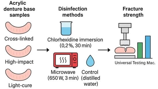
2.1. Fabrication of Test Specimens

In order to closely simulate clinical conditions, this investigation employed a standard model of an edentulous maxillary arch to create test samples. Following the manufacturer’s recommended water/powder ratio of 30 mL to 100 g, a silicone mould that resembled the edentulous maxilla was meticulously filled with Type III dental stone (Zhermack, Badia Polesine, Rovigo, Italy). In total, forty-five casts were effectively prepared for subsequent procedures.
A uniform pattern was established utilising base plate wax (Cavex Modelling Wax, Holland, Haarlem, Netherlands) with a consistent thickness of 2.5 mm applied to each stone cast to guarantee uniform denture base thickness throughout all samples.
Fifteen palatal denture bases were fabricated from each of the following materials:
  • Conventional heat-cure acrylic resin;
  • High-impact acrylic resin;
  • Light-cure acrylic resin.
The types of materials, along with their manufacturers, curing methods, and mixing ratios, are detailed in Table 1.
The wax was removed by rinsing under running hot water, after which the exposed plaster surfaces were sealed with a single coat of sealant. The heat-cured acrylic denture base resin (conventional and high impact) was subsequently packed into the moulds, and polymerisation was conducted in a water bath at 70 °C for one hour, followed by boiling for 30 min to complete the curing process.
In contrast, the light-cured acrylic plate was carefully adapted onto the prepared casts to ensure uniform thickness and precise adaptation. Excess material along the borders was meticulously trimmed, and the polymerisation process was performed in a light-curing unit at 25 °C for three to five minutes.
The thickness of the denture bases (2.5 mm) was verified using a digital calliper (precision ±0.01 mm) (LEZACO, ART.2771, Shanghai, China). Any dimensional discrepancies were carefully corrected by trimming the bases to the predetermined measurements.
After fabrication, all specimens were stored in distilled water at 25 ± 1 °C for 24 h before initiating the disinfection protocols.

2.2. Specimen Grouping and Disinfection Protocols

Forty-five specimens were prepared in Figure 1a,b, with fifteen specimens for each material group. Each material group was subdivided into three disinfection protocol groups (n = 5), as follows:
  • Group I (Control Group): Specimens were stored in distilled water at room temperature (25 ± 0.5 °C) for four weeks before testing.
  • Group II: Specimens were disinfected by immersion in 160 mL of 0.20% chlorhexidine digluconate solution (Kin, Laboratorios Kin, Barcelona, Spain) for 30 min, twice weekly, over four weeks [9].
  • Group III: Specimens underwent disinfection using a conventional household microwave at 650 W for three minutes, twice weekly, for four weeks [9,17,18]. Each specimen was placed in a 250 mL glass flask containing 160 mL of distilled water. The microwave used was a standard unmodified domestic model (Kenwood MW577), equipped with a rotating turntable to ensure even exposure, with the flask consistently positioned at the centre of the turntable. Its specifications included a power consumption of 1450 W, an output power of 900 W, a turntable diameter of 316 mm, and an operating voltage of 230 V at 60 Hz (Kenwood Ltd., Havant, Hampshire, UK).
Figure 1. (a) Acrylic denture base specimen, front view; (b) acrylic denture base specimen, back view; (c) The acrylic denture base specimen was positioned on the testing machine table [19]; (d) Fracture Pattern of Tested Acrylic Denture Base Specimen Across Different Material Types.
Oral 05 00078 g001
Following each disinfection cycle, all specimens were thoroughly rinsed and stored in distilled water at 25 °C.
The sample size consisted of 45 specimens (five per group), determined using (version 3.1.9.7, Heinrich Heine University Düsseldorf, Düsseldorf, Germany) with a significant level of 0.05 and a statistical power of 80% to detect large differences among groups. In contrast to flexural strength tests that generally use small, standardised bar-shaped specimens, the GPower software (version 3.1.9.7, Heinrich Heine University Düsseldorf, Düsseldorf, Germany) present study employed full palatal denture base samples, which are larger and more representative of clinical prostheses; thus, the chosen sample size was considered sufficient for reliable and clinically relevant analysis.
A priori power analysis was conducted using GPower (one-way ANOVA, fixed effects, omnibus; α = 0.05, 1–β = 0.80). In the absence of prior protocol-specific data, a large effect size (Cohen’s f = 0.40) was assumed as a conventional benchmark. This calculation indicated that 64 specimens per group would be required to achieve adequate statistical power. Given the exploratory full-palatal-base design and practical constraints, we used n = 5 specimens per group, which permits detection only of very large effects. Accordingly, the findings should be interpreted with caution regarding smaller differences. This choice was further supported by evidence from our previous full palatal base study on fracture strength of acrylic denture base materials [19] as well as another related research [20], both of which demonstrated large between-group effects under similar testing conditions.

2.3. Fracture Strength Testing

Fracture testing was performed using a universal testing machine (G.U.N.T. Gerätebau GmbH, Hamburg, Germany) equipped with a cylindrical steel loading flat end head of 30 mm diameter, operating at a crosshead speed of 10 mm/min [19]. The acrylic palatal denture bases were placed on the sub-table with the tissue side facing upward, Figure 1c. A compressive force, reaching up to 5000 N, was applied along the midline of each specimen until complete fracture occurred [19,20], Figure 1d. The palatal base experiences bending (flexural) stresses; thus, the test reflects resistance to midline compressive loading that induces flexure rather than a standardised three-point bending test.
Each specimen’s fracture force (KN) was determined from the stress–strain curve generated during the compression test. The maximum load at the point of fracture was recorded, with fracture detected by a sudden decline in the compression force on the curve.

2.4. Statistical Analysis

In this study, statistical analysis was performed using SPSS software (IBM SPSS Statistics, version 31, IBM Corp., Armonk, NY, USA). The mean and standard deviation (SD) were calculated for each group. The Shapiro–Wilk test was applied to assess the normality of data distribution, confirming no significant deviation from normality (p > 0.05); therefore, justifying the use of parametric tests. A one-way analysis of variance (ANOVA) was conducted to compare group means, followed by a Bonferroni post hoc analysis to identify pairwise differences and determine adjusted p-values. The level of statistical significance was set at α = 0.05.

3. Results

The mean, standard deviation, and ANOVA results for the fracture strength (KN) of the denture-based materials tested are summarised in Table 2.
The Shapiro–Wilk test confirmed that the data met the normality assumption (p > 0.05), permitting parametric statistical methods.
A one-way ANOVA was performed to assess the effect of disinfection protocols on the fracture strength of the tested denture base materials. The overall analysis indicated no statistically significant differences among the three disinfection methods (p = 0.069).
Bonferroni post hoc analysis, however, revealed highly significant differences between material types. Both cross-linked conventional acrylic and high-impact acrylic demonstrated significantly greater fracture strength than the light-cured acrylic (p < 0.001), while the difference between cross-linked and high-impact acrylics was not significant (p = 0.316, Table 3).
Among the disinfection protocols, the highest mean fracture strength was observed in specimens stored in distilled water (control group), whereas the lowest values were consistently associated with microwave-disinfected samples.
Although protocol-level differences were not statistically significant, the descriptive data highlighted numerical trends. Cross-linked acrylic in the control group recorded the highest mean value (1.74 ± 0.44 kN), closely followed by high-impact acrylic after chlorhexidine immersion (1.72 ± 0.40 kN). In contrast, light-cured acrylic subjected to microwave disinfection exhibited the lowest mean fracture strength (0.62 ± 0.02 kN).

4. Discussion

Edentulous patients primarily exhibit vertical masticatory movements with limited amplitude, while lateral excursions are minimal and may contribute to occlusal imbalance. Consequently, compressive forces are the dominant stresses acting on complete dentures in clinical settings [5]. Fracture of complete upper dentures arises from a combination of complex mechanical factors, including the viscoelasticity and uneven thickness of the edentulous mucosa, asymmetrical loading, variations in denture base thickness, and functional stresses such as bending and torsion [21]. Given these challenges, this study focused on evaluating the resistance of complete dentures to compressive loading, defined as fracture strength.
Prior research supports this approach by highlighting compressive strength as a clinically relevant indicator of denture base performance under functional loading [5]. To endure such multidirectional stresses and ensure long-term durability, denture base materials must demonstrate reliable mechanical properties, with fracture strength being one of the most essential for ensuring clinical durability [21]. Although not a standardised three-point bending test, the midline compression primarily generates flexural stresses in the denture base; therefore, the observed fracture-load trends can be interpreted in terms of flexural behaviour of the materials [20].
The growing risk of blood-borne pathogen transmission during dental procedures emphasises the need for strict cross-infection control [22]. Dental prostheses have been identified as potential vectors for cross-contamination between patients and dental professionals. Moreover, biofilm formation and microbial proliferation present infectious risks and financial strain on healthcare systems. Bacterial adhesion and plaque accumulation in the oral environment are linked to various common conditions, including denture-induced stomatitis, often accompanied by fungal colonisation [6]. As a result, the application of disinfectant agents with effective antimicrobial properties is highly desirable [23]. Ideally, such disinfectants should effectively inhibit microbial growth without compromising the denture base material’s mechanical integrity, particularly the fracture strength.
The findings of this study partially reject the null hypothesis, which proposed that neither microwave disinfection nor 0.2% chlorhexidine immersion would significantly affect the fracture strength of acrylic denture base materials. The results demonstrated that microwave treatment negatively influenced the fracture strength across all tested materials, indicating a substantial effect of this disinfection method. In contrast, immersion in 0.2% chlorhexidine resulted in no statistically significant reduction in fracture strength, suggesting that this protocol is less detrimental to the structural integrity of the materials. Furthermore, light-cure acrylic consistently exhibited significantly lower fracture resistance compared to cross-linked and high-impact acrylic resins, regardless of the disinfection protocol applied.
The findings showed that microwave and 0.2% chlorhexidine disinfection protocols impacted the fracture strength of the tested denture base materials, with the most affected being light-cure acrylics. However, all measured values remained above levels considered sufficient for clinical function. The lowest average strength recorded was 0.62 kN (620 N), which is still higher than the typical biting forces in edentulous patients, generally ranging from 100 to 300 N, with some reports suggesting a maximum of around 500 N. Although ISO 20795-1:2013 [24] does not specify a minimum required fracture force, the values obtained in this study are consistent with those reported in previous studies as adequate for the safe and effective use of maxillary denture which indicate that thresholds in the range of 100–300 N are adequate for the safe and effective use of maxillary denture bases [25,26,27]. Therefore, even though microwave disinfection led to a statistically significant reduction in strength, especially in light-cure materials, the tested cross-linked conventional and high-impact acrylic types maintained acceptable mechanical performance for clinical applications.
Previous studies indicate that the variability in the effects of microwave disinfection on denture base resins may result from differences in irradiation protocols, such as exposure duration, microwave power, material composition, and evaluation techniques. For example, denture-based materials exhibit different behaviours depending on whether microwave irradiation is conducted under dry or wet conditions. Water immersion is commonly adopted to enhance the disinfection process [22,28]. However, water begins to boil approximately 90 s into irradiation [22,28], potentially raising the temperature beyond the glass transition point of the acrylic resin. This elevated temperature may increase the material’s flexibility, releasing internal stresses formed during processing, resulting in dimensional changes such as warping [22,29,30].
Furthermore, high temperatures may facilitate the migration of residual monomer molecules to active polymer chain sites, leading to additional polymerisation and subsequent shrinkage [31,32,33]. Microwave disinfection has been shown to reduce the flexural strength of denture base acrylics, particularly with longer exposure times. Hamouda and Ahmed [22] reported that both five- and fifteen-minute irradiation cycles, whether in water or dry, caused significant weakening due to monomer boiling and porosity formation.
Basso et al. evaluated the effect of microwave irradiation at 650 W for three minutes on the dimensional stability of maxillary complete dentures. Irradiation once a week for four weeks produced no significant changes, whereas exposure four times weekly led to substantial distortion in the occlusal vertical dimension. The observed alterations were linked to the release of internal stresses in the denture base polymer during repeated heat cycles [34].
In the current study, microwave disinfection resulted in a statistically significant decrease in fracture strength, particularly in the light-cure acrylic group. Despite this reduction, all tested materials maintained fracture resistance above clinically acceptable thresholds. These findings are inconsistent with Consani et al. [18], who reported no detrimental effects on the mechanical properties of acrylic resins following microwave disinfection under different conditions. However, our findings align with other studies that employed longer or more frequent disinfection cycles, which have shown similar reductions in mechanical performance.
In contrast to microwave treatment, immersion in 0.2% chlorhexidine showed no statistically significant impact on the fracture strength of the tested denture base materials, indicating its compatibility with the mechanical integrity of acrylic resins. In line with these findings, a previous study demonstrated that when applied at clinically acceptable concentrations and exposure durations, chlorhexidine exerts minimal influence on denture base polymers’ physical and mechanical properties [35].
The favourable performance may be attributed to the interaction between chlorhexidine and the acrylic material. Acrylic resins, owing to their polar nature, readily absorb water and aqueous disinfectants such as chlorhexidine. This uptake disrupts the intermolecular interactions that stabilise the polymer chains, which leads to the material’s softening. While the initial effects may appear minimal, repeated absorption and desorption cycles can progressively weaken the material’s internal structure, eventually forming microcracks and irreversible degradation. Moreover, elevated disinfection temperatures can accelerate hydrolytic degradation, underscoring the necessity for meticulously controlled disinfection techniques to maintain material integrity [36].
Gaol and Ritonga [23] established that chlorhexidine exerts a negligible influence on the characteristics of acrylic resin, signifying its appropriateness as a disinfectant with minimal risk of undermining the material’s structural integrity. Inconsistent with the findings of Kannaiyan et al. [37], who observed that immersing heat-cured acrylic resin samples in chlorhexidine for ten minutes resulted in significant degradation of their physical qualities, indicating that prolonged exposure may adversely impact material performance.
Light-cured acrylics are known for their excellent flexural, tensile, and compressive strengths, which can be attributed to their high degrees of polymerisation, crystalline compositions, and fewer internal voids. Complete polymerisation plays a key role in enhancing the material’s properties and the overall durability of the prosthesis [38]. The high conversion rate through light activation and the reduced air entrapment during fabrication further support these advantages [39]. Additionally, many researchers have favoured light-cured denture base resins for their dependable strength, minimal residual monomer, user-friendly handling, and the benefit of avoiding complex conventional processing steps [40].
In the present study, distinct variations in fracture strength were observed among the three acrylic materials, with light-cured acrylic consistently exhibiting the lowest values regardless of the disinfection protocol employed. This inferior mechanical performance is likely attributed to its lower degree of polymerisation and reduced cross-linking density, which limits its ability to resist mechanical stress. These findings align with AlQahtani and Haralur’s previous study, highlighting certain drawbacks of light-cured acrylics, including increased water absorption and reduced impact strength. In the same study, light-cured acrylic also exhibited the lowest flexural and fracture strength among the evaluated acrylic resins, further supporting the results observed in the current study [41]. In contrast to previous studies, the lower fracture strength observed in the light-cured acrylic specimens in this study may be attributed to formulation or manufacturer-related variability, as suggested in the literature, since polymerisation rate and cross-linking density remained unassessed. Differences in composition, initiators, and curing protocols can significantly influence the mechanical performance of light-cured resins. By comparison, cross-linked conventional and high-impact acrylics demonstrated superior and comparable fracture strength, indicating greater structural reliability for clinical applications. An unusually low value was observed in the cross-linked control group, which slightly increased variability; however, this outlier did not alter the overall interpretation or conclusions of the study.
Beyond laboratory outcomes, future investigations should also incorporate patient-centred measures, such as comfort, satisfaction, and long-term prosthesis longevity in daily function. These considerations would complement mechanical testing and provide a broader perspective on how disinfection protocols influence both clinical performance and the quality of life for denture wearers.
While the present study offers meaningful insights into the fracture strength of commonly used denture base acrylics, several limitations warrant consideration. The materials tested were restricted to three conventional resins; evaluating newer alternatives, such as polyetheretherketone (PEEK) and 3D-printed resins, would expand the scope, especially given their increasing role in prosthodontics. Incorporating nanoparticles may also enhance mechanical strength while providing antimicrobial benefits. In addition, the loading setup employed in this study differs from a standardised flexural strength test, and future work is recommended to include conventional flexural testing for direct comparison. Finally, although the in vitro design ensured standardised conditions, further studies simulating clinical situations—such as thermal cycling, masticatory forces, and microbial exposure—are needed to enhance the clinical applicability of the findings.

5. Conclusions

  • Cross-linked acrylic demonstrated the highest reliability in fracture strength across all disinfection protocols.
  • High-impact acrylic showed comparable average strength but was more sensitive to the type of disinfection applied.
  • Microwave disinfection led to a statistically significant reduction in fracture strength, particularly in light-cured acrylics, though all materials remained within clinically acceptable limits.
  • Immersion in 0.2% chlorhexidine for 30 min, twice weekly, had no significant impact on fracture strength, indicating its compatibility with the mechanical integrity of acrylic resins.
  • Importantly, repeated chlorhexidine exposure and uncontrolled microwave parameters (power and duration) may adversely affect the structural integrity of denture base materials, underscoring the need for proper disinfection protocols and strict regulation of concentration, immersion time, and irradiation settings.

Author Contributions

Conceptualisation, F.K.A.-K. and J.F.A.; methodology, F.K.A.-K.; software, F.K.A.-K.; validation, F.K.A.-K. and J.F.A.; formal analysis, F.K.A.-K.; investigation, F.K.A.-K.; resources, F.K.A.-K.; data curation, F.K.A.-K. and J.F.A.; writing—original draft preparation, F.K.A.-K.; writing—review and editing, F.K.A.-K.; visualisation, F.K.A.-K.; supervision, J.F.A. and S.K.E.; project administration, F.K.A.-K., J.F.A. and S.K.E. All authors have read and agreed to the published version of the manuscript.

Funding

This research received no external funding.

Data Availability Statement

The data supporting this study’s findings are available upon request from the corresponding author [Al-Kadi, F.K.] to preserve data integrity.

Acknowledgments

The authors acknowledge the University of Sulaimani—Technical Institute for conducting the mechanical testing by providing the necessary facilities and technical support.

Conflicts of Interest

The authors declare no conflicts of interest.

Abbreviations

The following abbreviations are used in this manuscript:
ANOVAA one-way analysis of variance
ChxChlorhexidine
gGram
hHour(s)
HzHertz
ISOInternational Organization for Standardization
KNKilonewton
Mac.Machine
minMinute(s)
mLMillilitre
mmMillimetre
NNewton
nNumber
pp-value (the calculated probability)
PMMAPoly(methyl methacrylate)
PEEKPolyetheretherketone
SDStandard Deviation
SEStandard Error
SPSSStatistical Package for the Social Sciences
Tt-statistic
UTMUniversal Testing Machine
UVUltraviolet curing
VVolt (Voltage)
WWatt
wt%Weight Percentage
°CDegree Celsius
%Percent
=Equals
< Less than
3DThree-Dimensional
α(alpha) is the threshold for significance, commonly set at 0.05.

References

  1. Deb, S. Polymers in dentistry. Proc. Inst. Mech. Eng. Part H J. Eng. Med. 1998, 212, 453–464. [Google Scholar] [CrossRef] [PubMed]
  2. Al-Kadi, F.K.; Adbulkareem, J.F.; Azhdar, B.A. Evaluation of the Mechanical and Physical Properties of Maxillofacial Silicone Type A-2186 Impregnated with a Hybrid Chitosan–TiO2 Nanocomposite Subjected to Different Accelerated Ageing Conditions. Biomimetics 2023, 8, 539. [Google Scholar] [CrossRef]
  3. Xu, X.; He, L.; Zhu, B.; Li, J.; Li, J. Advances in polymeric materials for dental applications. Polym. Chem. 2017, 8, 807–823. [Google Scholar] [CrossRef]
  4. Hassan, M.; Asghar, M.; Din, S.U.; Zafar, M.S. Chapter 8. In Thermoset Polymethacrylate-Based Materials for Dental Applications; Elsevier: Amsterdam, The Netherlands, 2019; pp. 273–308. [Google Scholar]
  5. Modiga, C.; Stoia, A.; Leretter, M.T.; Chiş, A.C.; Ardelean, A.-V.; Azar, E.-R.; Kapor, G.; Pop, D.-M.; Romînu, M.; Sinescu, C.; et al. Mechanical Assessment of Denture Polymers Processing Technologies. J. Funct. Biomater. 2024, 15, 234. [Google Scholar] [CrossRef] [PubMed]
  6. Zafar, M.S. Prosthodontic Applications of Polymethyl Methacrylate (PMMA): An Update. Polymers 2020, 12, 2299. [Google Scholar] [CrossRef] [PubMed]
  7. Alqutaibi, A.Y.; Baik, A.; Almuzaini, S.A.; Farghal, A.E.; Alnazzawi, A.A.; Borzangy, S.; Aboalrejal, A.N.; AbdElaziz, M.H.; Mahmoud, I.I.; Zafar, M.S. Polymeric Denture Base Materials: A Review. Polymers 2023, 15, 3258. [Google Scholar] [CrossRef]
  8. Elshereksi, N.W.; Kundie, F.A.; Muchtar, A.; Azhari, C.H. Protocols of improvements for PMMA denture base resin: An overview. J. Met. Mater. Miner. 2022, 32, 1–11. [Google Scholar] [CrossRef]
  9. Gupta, R.; Aggarwal, R.; Aeran, H. Comparison of Various Methods of Disinfecting Acrylic Dentures: Assessment of Antimicrobial Efficacy and Dimensional Changes. Int. J. Oral Health Dent. 2016, 2, 143. [Google Scholar] [CrossRef]
  10. Elhagali, A.F.; Sharaf, M.Y.; Abd El-Aziz, M.E.-S.A.; Ali Bayiumy, A.S.; Refaei, M.A.A.; Al-Agamy, A.H.; Ali, A.; Elakel, A.; Altayyar, R.; Alzahrani, R.; et al. The Effects of Different Chemical Disinfectants on the Strength, Surface, and Color Properties of Conventional and 3D-Printed Fabricated Denture Base Materials. Prosthesis 2025, 7, 24. [Google Scholar] [CrossRef]
  11. Çakmak, G.; Hess, J.A.; Dönmez, M.B.; Yılmaz, D.; Alhotan, A.; Schimmel, M.; Peutzfeldt, A.; Yilmaz, B. Effect of polishing and denture cleansers on the surface roughness of new-generation denture base materials and their colour change after cleansing. J. Prosthodont. 2024, 33, 783–790. [Google Scholar] [CrossRef]
  12. Webb, B.C.; Thomas, C.J.; Harty, D.W.S.; Willcox, M.D. Effectiveness of two methods of denture sterilisation. J. Oral Rehabil. 1998, 25, 416–423. [Google Scholar] [CrossRef] [PubMed]
  13. Polyzios, G.L.; Zissis, A.J.; Yannikakis, S.A. The effect of glutaraldehyde and microwave disinfection on some properties of acrylic denture resin. Int. J. Prosthodont. 1995, 8, 150–154. [Google Scholar]
  14. Andrucioli, M.C.; deMacedo, L.D.; Panzeri, H.; Lara, E.G.; Paranhos, H.D. Comparison of two cleansing pastes for the removal of biofilm from dentures and palatal lesions in patients with atrophic chronic candidiasis. Braz. Dent. J. 2004, 15, 220–224. [Google Scholar] [CrossRef] [PubMed]
  15. Pavan, S.; Arioli Filho, J.N.; dos Santos, P.H.; de Mollo, F.A., Jr. Effect of microwave treatments on dimensional accuracy of maxillary acrylic resin denture base. Braz. Dent. J. 2005, 16, 119–123. [Google Scholar] [CrossRef]
  16. Basavanna, J.M.; Jujare, R.H.; Varghese, R.K.; Singh, D.; Gaurav, A. Effects of Laboratory Disinfecting Agents on Dimensional Stability of Three Commercially Available Heat-Cured Denture Acrylic Resins in India: An In-Vitro Study. J. Clin. Diagn. Res. 2016, 10, ZC27–ZC31. [Google Scholar] [CrossRef]
  17. Yildirim-Bicer, A.Z.; Peker, I.; Akca, G.; Celik, I. In Vitro Antifungal Evaluation of Seven Different Disinfectants on Acrylic Resins. BioMed Res. Int. 2014, 519098. [Google Scholar] [CrossRef]
  18. Consani, R.L.X.; Vieira, E.H.M.; Mesquita, M.F.; Mendes, W.B.; Arioli-Filho, J.N. Effect of microwave disinfection on physical and mechanical properties of acrylic resins. Braz. Dent. J. 2008, 19, 348–353. [Google Scholar] [CrossRef]
  19. Al-Kadi, F.K.A.; Abdulkareem, J.F.; Al-jmoor, C.A. Fracture Strength of Palatal Denture Base Constructed from Different Acrylic Denture Base Materials. Eur. Sci. J. 2015, 11, 346–354. [Google Scholar]
  20. Prasad, D.K.; Dogra, E.S.; Hegde, C. A Study to Evaluate Midline Fracture Resistance of Maxillary Acrylic Denture Base Reinforced with Metal Pattern in Various Palatal Contours. World J. Dent. 2020, 11, 287–293. [Google Scholar] [CrossRef]
  21. Prombonas, A.; Poulis, N.A.; Yannikakis, S.A. The Impact of Notches on the Fracture Strength of Complete Upper Dentures: A Novel Biomechanical Approach. Eur. Sci. J. 2019, 15, 433. [Google Scholar] [CrossRef]
  22. Hamouda, I.M.; Ahmed, S.A. Effect of Microwave Disinfection on Mechanical Properties of Denture Base Acrylic Resin. J. Mech. Behav. Biomed. Mater. 2010, 3, 480–487. [Google Scholar] [CrossRef]
  23. Gaol, E.L.; Ritonga, P.W.U. Effect of Chlorhexidine and Castor Oil on Surface Roughness of Heat-Cured Acrylic Resin Denture Base. J. Dent. Syiah Kuala 2023, 8, 24–30. [Google Scholar] [CrossRef]
  24. ISO 20795-1:2013; Denture Base Polymers. ISO: Geneva, Switzerland, 2013.
  25. Craig, R.G.; Powers, J.M. Restorative Dental Materials, 11th ed.; Mosby: St. Louis, MO, USA, 2002. [Google Scholar]
  26. Zappini, G.; Kammann, A.; Wachter, W. Influence of acrylic resin polymerisation methods on fracture resistance of denture bases. J. Prosthet. Dent. 2003, 90, 256–260. [Google Scholar] [CrossRef]
  27. Anusavice, K.J.; Shen, C.; Rawls, H.R. Phillips’ Science of Dental Materials, 12th ed.; Elsevier: St. Louis, MO, USA, 2012. [Google Scholar]
  28. De Souza, R.F.; Paranhos, H.F.O.; Lovato da Silva, C.H.; Ito, I.Y. Effects of immersion solution on biofilm formation on acrylic resin denture base surfaces. Braz. Dent. J. 2009, 20, 127–131. [Google Scholar] [CrossRef]
  29. Polyzois, G.L.; Frangou, M.J. Influence of curing method, reinforcement, and fluoride treatment on some properties of denture base materials. J. Oral Rehabil. 2001, 28, 426–432. [Google Scholar] [CrossRef]
  30. Polychronakis, N.; Yannikakis, S.; Zissis, A. The effect of repeated microwaving disinfection on the dimensional stability of acrylic dentures. Acta Stomatol. Croat. 2014, 48, 279–284. [Google Scholar] [CrossRef] [PubMed]
  31. Campanha, N.H.; Pavarina, A.C.; Vergani, C.E.; Machado, A.L.; Giampaolo, E.T. Temperature rise in dentures during microwave disinfection. Braz. Dent. J. 2005, 16, 115–120. [Google Scholar] [CrossRef]
  32. Machado, A.L.; Giampaolo, E.T.; Pavarina, A.C.; Jorge, J.H. Simultaneous microwave disinfection and drying of complete dentures. J. Prosthodont. 2009, 18, 589–593. [Google Scholar] [CrossRef]
  33. Lima, J.F.M.; Orsi, I.A.; Borie, E.; Cândido, L.M.; Compagnoni, M.A. Effect of microwave disinfection on dimensional stability of a denture base acrylic resin. J. Prosthodont. 2009, 18, 123–129. [Google Scholar] [CrossRef]
  34. Basso, M.F.M.; Giampaolo, E.T.; Machado, A.L.; Pavarina, A.C.; Vergani, C.E. Evaluation of the occlusion vertical dimension of complete dentures after microwave disinfection. Gerodontology 2011, 29, e611–e617. [Google Scholar] [CrossRef]
  35. Da Silva, F.C.; Kimpara, E.T.; Mancini, M.N.G.; Balducci, I.; Jorge, A.O.C.; Koga-Ito, C.Y. Effectiveness of six different disinfectants on removing five microbial species and effects on the topographic characteristics of acrylic resin. J. Prosthodont. 2008, 17, 627–633. [Google Scholar] [CrossRef]
  36. Schwindling, F.S.; Rammelsberg, P.; Stober, T. Effect of Chemical Disinfection on the Surface Roughness of Hard Denture Base Materials: A Systematic Literature Review. Int. J. Prosthodont. 2014, 27, 215–225. [Google Scholar] [CrossRef] [PubMed]
  37. Kannaiyan, K.; Rakshit, P.; Bhat, M.P.S.; Sadasiva, S.K.K.; Babu, S.C.; Ummer, H. Effect of Different Disinfecting Agents on Surface Roughness and Colour Stability of Heat-Cured Acrylic Denture Material: An In Vitro Study. J. Contemp. Dent. Pract. 2023, 24, 68–74. [Google Scholar] [CrossRef]
  38. Hashem, M.; Alsaleem, S.O.; Assery, M.K.; Abdeslam, E.B.; Vellappally, S.; Anil, S. A comparative study of the mechanical properties of the light-cure and conventional denture base resins. Oral Health Dent. Manag. 2014, 13, 311–315. [Google Scholar] [PubMed]
  39. Qasim, S.; Al-Khuraif, S.; Kumar, R. An Investigation into the Impact and Flexural Strength of Light-Cure Denture Resin Reinforced with Carbon Nanotubes. World Appl. Sci. J. 2012, 18, 808–812. [Google Scholar] [CrossRef]
  40. Mumcu, E.; Cilingir, A.; Gencel, B.; Sülün, T. Flexural Properties of a Light-Cure and a Self-Cure Denture Base Material Compared to Conventional Alternatives. J. Adv. Prosthodont. 2011, 3, 136–139. [Google Scholar] [CrossRef]
  41. AlQahtani, M.; Haralur, S.B. Influence of Different Repair Acrylic Resin and Thermocycling on the Flexural Strength of Denture Base Resin. Medicina 2020, 56, 50. [Google Scholar] [CrossRef]
Table 1. Summary of acrylic resin materials used in the study.
Material TypeCommercial Brand/ManufacturerMixing Ratio/Polymerisation Method
Conventional Heat-Cure Acrylic ResinVillacryl H Plus, Zhermack, Italy24 g powder: 10.5 mL liquid
High-Impact Acrylic ResinVertex Dental, Netherlands21 g powder: 10 mL liquid
Light-Cure Acrylic ResinCavex, NetherlandsLight polymerisation time: 3–5 min, using 27 W Dental Lab Light Curing Unit (Denshine, Guangzhou, China) with UVA bulbs.
Note: All materials were processed per the respective manufacturers’ guidelines.
Table 2. ANOVA Analysis of Variations in Fracture Strength (KN) of Acrylic Denture Bases among Different Disinfection Methods.
Disinfection ProtocolsDenture Base MaterialsSpecimen Numbers (n)Mean (Std)ANOVA
Water (control) Cross-linked acrylic denture base 5 1.74 (0.44) p-value = 0.069
High-impact acrylic denture base 5 1.36 (0.17)
Light-cure acrylic denture base 5 0.76 (0.08)
Chemical cleaner (0.2% chlorhexidine disinfection) Cross-linked acrylic denture base 5 1.19 (0.19)
High-impact acrylic denture base 5 1.72 (0.4)
Light-cure acrylic denture base 5 0.62 (0.07)
Microwave Cross-linked acrylic denture base 5 1.16 (0.18)
High-impact acrylic denture base 5 1.35 (0.28)
Light-cure acrylic denture base 5 0.62 (0.02)
Table 3. Bonferroni Post Hoc Analysis of Differences among Acrylic Denture Base Types.
Denture Base MaterialsMean DifferenceStd. ErrorTp
Cross-linked acrylic denture baseHigh-impact acrylic denture base−0.110.06−1.750.316
Cross-linked acrylic denture baseLight-cure acrylic denture base0.70.0611.15<0.001
High-impact acrylic denture baseLight-cure acrylic denture base0.810.0612.9<0.001
Disclaimer/Publisher’s Note: The statements, opinions and data contained in all publications are solely those of the individual author(s) and contributor(s) and not of MDPI and/or the editor(s). MDPI and/or the editor(s) disclaim responsibility for any injury to people or property resulting from any ideas, methods, instructions or products referred to in the content.

