Article

Proposal of a Cephalometric Method in Computed Tomography to Mandibular Analysis in Infants with Pierre Robin Sequence Treated by Fast and Early Mandibular Osteo-Distraction: Pilot Study

by
Francesca Imondi
1,
Adriana Assunta De Stefano
1,2,*,
Rachele Podda
1,
Martina Horodynski
1,
Roberto Antonio Vernucci
1,
Valentina Mazzoli
1,
Piero Cascone
3 and
Gabriella Galluccio
1
1
Department of Odontostomatological and Maxillo-Facial Sciences, Sapienza University of Rome, 00161 Rome, Italy
2
Faculty of Dentistry, Central University of Venezuela, Caracas 1070, Venezuela
3
Saint Camillus International University of Health and Medical Sciences, 00131 Rome, Italy
*
Author to whom correspondence should be addressed.
Submission received: 27 March 2025 / Revised: 10 July 2025 / Accepted: 5 August 2025 / Published: 13 August 2025

Abstract

Background: Newborns with Pierre Robin Sequence (PRS) usually show varying degrees of upper airway obstruction and difficulty feeding due to severe micrognatia. Mandibular distraction osteogenesis has become popular as an alternative treatment option when other medical or surgical techniques are unsatisfactory. The aim of this study is to test a three-dimensional (3D) cephalometric method in computed tomography (CT) to measure effective mandibular and midface length, and maxillomandibular ratio (Md/Mx ratio), as a mode of growth normalization expression in PRS patients before and after Fast and Early Mandibular Osteo-distraction (FEMOD), for assessing the diagnostic method and the efficacy of surgical treatment. Methods: In this retrospective pilot study, six PRS patients treated via the FEMOD surgical protocol were included. The measurements of effective maxillary and mandibular length were performed on 3D reconstructions from pre-surgical (T1) and post-surgical CT (T2). The growth disparity between the mandible and the maxilla was verified in T1 and was compared with the measurements obtained from the adaptation of the McNamara Norms; the correction of growth disproportion after FEMOD was assessed. Results: In T1, the PRS patients’ mandibular length and the Md/Mx ratio were smaller than the expected mandibular length (p = 0.029) and the expected Md/Mx ratio (p = 0.028). In T2, the PRS patients’ mandibular length and the Md/Mx ratio did not show significant differences from the expected results (p = 0.461 and p = 0.400). Conclusions: The 3D cephalometric analysis identifies the disproportion in pre-surgical maxillomandibular growth between PRS and reference measurements, and demonstrates that FEMOD allows the achievement of proportionality in the growth of the maxillomandibular complex in PRS patients.

1. Introduction

Pierre Robin Sequence (PRS) is a congenital anomaly characterized by micrognathia, glossoptosis and airway obstruction, often associated with a “U-shaped” cleft palate [1]. The incidence of the disease is considered to be around 1:8500–1:20,000 live births, without distinction between males and females [2]. It is not a syndrome in itself, but rather a sequence of events during intrauterine life that leads to the typical clinical presentation at birth: the hypoplasic or retro-positioned lower jaw may determine the upward displacement of the tongue, which, consequently, prevents the physiological fusion of the palatine processes that will form the hard palate [3].
The diagnosis of PRS is typically made at birth. An early diagnosis can be made in the second trimester of pregnancy through prenatal imaging; high-resolution ultrasonography is routinely used to identify structural malformations that may anticipate other phenotypical anomalies, syndromes, or genetic malformations [3,4].
The diagnostic evaluation of PRS in the post-natal period is based on physical examination, fibro-naso-laryngoscopy, bronchoscopy, and polysomnography, and is intended to detect the site of the airway obstruction and its severity [5].
The treatment of PRS requires a complex and multidisciplinary approach, and may significantly vary for every newborn, as a result of the variability of phenotypical picture, clinical manifestations, the level of stability of the upper airways and the rate of complications, such as associated syndromes. Treatment options may be divided into two groups, namely, non-surgical and surgical [6].
Non-surgical treatments are conducted in cases of infants without or with mild airway obstruction, and my include appropriate instructions regarding feeding techniques given to parents, the lateral or prone positioning of the newborn, nasopharyngeal intubation, and orthodontic devices [7,8].
Surgical treatments include the subperiosteal release of the floor of the mouth, tracheostomy, lip–tongue adhesion and mandibular distraction osteogenesis (MDO) [9].
The clinical–diagnostic protocol applied in our department for the management of PRS includes medical history, physical examination, low-dose Computed Tomography—for the evaluation of mandibular anatomy, mental foramens, airway volume—and polysomnography—to evaluate the severity of apnea and respiratory distress [5,10]. When severe respiratory symptoms are present, mandibular Distraction Osteogenesis is performed a few days after birth (“Fast and early mandibular osteodistraction”, FEMOD), to avoid tracheostomy or prolonged endotracheal intubation [11]. If the patient also presents cleft palate, an orthopedic palatal plate is used to immediately solve the feeding disorder, encouraging the baby to drink through the baby bottle; the intention is to replace the anatomic function of the palate, fill the palatal cleft and promote mandibular growth until the surgical palatoplasty can be performed, usually six months later [7].
The morphology and length of the mandibular body may differ significantly in individuals affected by PRS, compared to healthy individuals. Therefore, the pre-surgical evaluation of mandibular morphology and degree of micrognathia during the planning phase of the FEMOD may help to measure the degree of sagittal discrepancy, as well as the amount of growth disproportionality with the maxilla [5,11,12,13].
The dimension and morphology of the lower jaw can be assessed by various tools, such as low-dose multisection Computed Tomography (CT), Cone Beam Computed Tomography (CBCT), lateral cephalograms, plaster casts, three-dimensional photography and direct clinical measurements with calipers [14].
The measurements in individuals with craniofacial malformations require a comparison with the reference values, which are obtained in healthy children of the same age, to verify if there is the same growth proportionality; however, for children under one year of age, there are few studies on reference values [15,16,17,18].
The cephalometric reference values of the craniofacial growth of healthy individuals at early ages are difficult to obtain, since the use of X-ray images in unaffected healthy controls is considered unethical [16,19,20].
McNamara [21], taking as reference the Boston norms and the samples from the Burlington Orthodontic Research Centre, extrapolated a geometric relationship between the “effective” length of the maxilla and the mandibular length on latero-lateral skull radiographs, which is not related to the sex or age of the subject. These evaluations were performed via a cephalometric method in which, when knowing the midface length, it is possible to estimate the length of the mandible. By subtracting the effective midface length from the mandibular length, the maxillomandibular differential can also be determined. The effective midface length was determined by measuring from the Condylion to point A, after which the effective mandibular length was determined from the Condylion to Gnation.
This cephalometric method has also been applied to CT images [22,23,24] and could offer a useful tool to assess the amount of micrognathia in PRS patients who have undergone a CT before the FEMOD, as well as to measure the response to surgical treatment.
Therefore, the aim of this pilot study was to test a three-dimensional (3D) cephalometric method on the CTs of PRS patients to measure the effective mandibular length, the effective midface length and the maxillomandibular ratio (Md/Mx ratio), according to McNamara, before and after FEMOD, along with the amount of mandibular advancement after surgery. The study hypothesis was that this 3D cephalometric analysis may help to evaluate the pre-surgical disproportionality in PRS patients when compared to reference measurements, as well as the evaluation of the effectiveness of FEMOD in achieving proportionality in the growth of the maxillomandibular complex.

2. Methods

This retrospective pilot study was approved by the medical ethics commission of Policlinico Umberto I Rome, Italy (prot. No.4663). The study was performed in accordance with the 1964 Declaration of Helsinki and its subsequent amendments.

2.1. Patients

Six infants diagnosed with PRS, who presented severe micrognathia and were treated by the FEMOD surgical protocol in our Department of Orthodontics and Maxillofacial Sciences between 2007 and 2017, were included in the study. Among those, we further selected only the subjects who underwent pre- and post-surgical craniofacial computed tomographic scans in multi-planar and three-dimensional reformats, as part of the treatment protocol for mandibular distraction. The patients affected by other malocclusions or syndromes, those who did not undergo FEMOD, and those without the complete CT documentation were excluded.
Patients underwent bilateral mandibular distraction with the FEMOD protocol within 3 months of birth. The protocol followed for the activation of the distraction devices was first proposed by Cicchetti et al. [5]. The activation protocol is divided into three phases, as follows: an initial distraction performed intraoperatively (2.0 mm) after the piezosurgical osteotomies; after two days, an activation of 1.5 mm twice a day for 6–9 days until patient reaches a Class III mandible position to minimize the risk of relapse; the consolidation phase, which lasts 4 weeks.

2.2. Cephalometrics Analysis

Maxillofacial noncontrast CT scans consisting of 0.625 mm axial tomograms, with reconstruction in the coronal and sagittal planes, were used. The CT scans were taken by the same medical personnel using the same equipment. The CT images were processed through the open source software RadiAnt DICOM Viewer (Medixant version 2022.1.1) following the software’s default parameters to obtain the 3D reconstructions of the skull [25]. In this environment, landmarks were manually located and a series of measurements were taken, following McNamara’s analysis, on both pre-surgical (T1) and post-surgical CTs (T2)—the right and left effective mandibular length, effective midface length and maxillomandibular ratio [21]. All measurements were performed by the same observer (Figure 1).
The effective midface length (CoA) (right and left) was measured as the distance in mm between the Condylion (Co), the most posterior superior point of the right and left mandibular profile, and the A point, the most posterior point of the anterior concavity of the maxilla, between the anterior nasal spine and the alveolar process.
The effective mandibular length (CoGn) (right and left) was measured from the Co (right and left) to the Gnation (Gn), the most anterior inferior point of the mandibular symphysis.
The maxillo-mandibular ratio (Md/Mx) was expressed as the ratio between the CoGn and the CoA on the right and left sides.
As the measurements of CoA and CoGn were taken at T1 and T2 on the right and left side of each patient, no significant differences between the sides were observed when the measurements were compared; therefore, the mean value between right and left side was used to present the results.
The evaluation of the growth ratio before and after distraction was performed by both evaluating the maxillomandibular ratio and assessing the proportionality of growth based on the midface length. In fact, according to the McNamara’s analysis, if the measure of the effective midface length is known, it is possible to estimate the effective mandibular length and whether this value is proportionate to the maxillary growth. McNamara reported a table of “Composite Norms”, based on the Burlington Orthodontic Centre’s data, with correspondence between a certain value of effective midface length and the corresponding effective mandibular length [21].
As the lower value of the Co–A measure in the original table was 80 mm, in order to properly apply McNamara’s table to our newborn population, we obtained new Compositive Norms by adding the missing values lower than 80 mm into the Co–A column; then, we reconstructed the expected mandibular length for every midface length.
For every Co–A value, obtained by subtracting 5 mm from the previous value in the table, the corresponding CoGn value was calculated as a 3 mm range, as in the original table, with each minimum obtained by subtracting 8 mm from the previous minimum value. Besides this, when analyzing the Md/Mx ratio, a ratio of 1.22–1.25 was observed when CoA was 80 mm, and the ratio increased approximately by 0.02 every 5 mm of CoA length. Therefore, we have completed the table following this mathematical rule in descending order (Table 1).
The T1 measurements were compared with those from the table obtained from the adaptation of the McNamara Norms to evaluate the growth discrepancy between the mandible and the maxilla. The degree of mandibular advancement was measured (in mm) by comparing the measurements in T2 and T1, and the correction of growth disproportion (Md-Mx Ratio) after the mandibular distraction (T2) was assessed.
In the pre-surgical CT (T1), considering the patient’s CoA measurement as a reference and using Table 1, the T1 expected mandibular length was obtained. The difference between the effective mandibular length measurement and the T1 expected mandibular length was calculated; negative values indicate that the patient’s mandibular length is smaller than the normal value considered in the table.
In the CT taken at surgical follow-up (T2), in the same way as T1, the T2 expected mandibular length was obtained. The effective mandibular length measurement and the T2 expected mandibular length were compared; negative values indicate a mandibular length of the patient lower than the expected normal.

2.3. Statistical Analysis

One operator (AADS) performed all the measurements; the same operator repeated the measurements after 1 month on all patients in a random order to assess the intra-rater repeatability. A second investigator (RP) performed the entire protocol for the assessment of the inter-rater reliability. The Intraclass Correlation Coefficient (ICC) was calculated to evaluate the intra and inter-rater reliability; values below 0.5 indicate poor reliability, moderate is denoted by values between 0.5 and 0.75, good by values between 0.75 and 0.9, and any value above 0.9 indicates excellent reliability. The ICC for intra-rater repeatability ranged between 0.79 and 0.92, with good to excellent reliability for all measurements. The ICC for inter-rater reliability showed moderate to good reliability (0.66–0.87) for all measurements. In cases of large discrepancies, a third operator (RAV) repeated the entire protocol.
The normality of the distributions was verified with the Shapiro–Wilk test; as the data did not show normal distribution, the Wilcoxon signed-ranks test was performed to compare the two matched pairs of samples, which were pre-FEMOD and post-FEMOD effective mandibular lengths, with the expected pre-FEMOD and post-FEMOD mandibular lengths, according to the McNamara table. The null hypothesis was that there were no differences between the measured and expected mandibular lengths, in both T1 and T2.
Data were entered into a statistical database for analysis (IBM SPSS Statistics version 25.0 IBM Corp.). For all analyses, a p value of 0.05 was considered significant.

3. Results

From the initial population of 52 newborns affected by PRS, 6 met the inclusion criteria (3 males, 3 females) and were included in this study (Table 2).
The pre-surgical measurements (T1) are displayed in Table 3. The difference between the measured and the expected CoGn measurements, derived from the Proposted New Compositive Norms derived from McNamara’s original Compositive Norms (Table 1), was negative in all patients; therefore, the pre-surgical mandibular length was smaller than the expected measure. This difference was statistically significant (p = 0.028). A statistically significant difference was also observed between the measured Md/Mx ratio and the expected Md/Mx ratio (p = 0.029).
Table 4 shows the measurements of the surgical follow-up (T2). The values of CoA and CoGn increased with respect to T1 in all patients except patient No. 2, which still showed a negative differential value, despite the CoGn having increased after distraction.
After the mandibular distraction, the measured CoGn length was not statistically different from the expected length (p = 0.400), and the post-surgical measured Md/Mx ratio did not show significant differences from the expected Md/Mx ratio (p = 0.461). Therefore, in T2, we cannot reject the null hypothesis.

4. Discussion

In this study, we tested a cephalometric method of Computed Tomography to measure the proportionality of growth between upper and lower jaws in a population of patients affected by PRS. We also tested if the FEMOD was able to correct the impairment between the skeletal proportionality, using cephalometric values derived from McNamara’s analysis—the effective mandibular length, the effective midface length and the maxillomandibular ratio [21].
The study was possible only in patients who had undergone surgery and had available CT scans. The patients included in the study underwent pre-distraction and post-distraction CT for surgical purposes. Pre-surgical CT is used to define the mandibular anatomy, identify any possible vascular anomalies associated with PRS, dental gems, and the path of the inferior alveolar nerve and the mental foramens, and to evaluate the real volume of the airway [26,27]. The post-surgery CT is mandatory if one is seeking to evaluate post-surgical complications, such as the unexpected development of mandibular asymmetry, the failure of the distraction devices, or the absence or delay of bone fusion, as well as the extent of the distraction and how much the upper airway increases [11,27].
The availability of 3D CT reconstructions of the skull allowed us to perform the cephalometric measurements on both sides, compared to the classic two-dimensional projections made for the age of growth. Sam et al. [28] demonstrated that bilateral points (such as Co and Gn) are associated with a better representation of the anatomic reality in 3D compared to 2D; therefore, each side, left or right, can be assessed independently, without the overlap of other anatomic structures that may interfere in the evaluation. Furthermore, the use of 3D images makes possible to assess whether the mandibular elongation obtained with distraction was similar on both sides, thus assessing the differences in maxillomandibular differential and growth proportionality on both sides [13].
However, as the measurements did not show significant differences between the two sides, in all cases pre- and post-FEMOD, the mean between sides was used to present the results, as also reported by other papers in the literature [13,22,23]. Liu et al. [29] also compared the maxillomandibular growth between healthy and PRS patients, and evaluated the pre- and post-operative condylar position in PRS patients before and after the MDO, using linear and angular measurements on 3D CT. They reported the upward movement of the mandibular condyles with a decrease in the upper articular space and increases in the inter-gonial and inter-condylion distances, confirming the increase in the mandibular dimensions after the MDO.
McNamara’s analysis, originally undertaken via 2D cephalometry, has also been proposed in the context of 3D CT reconstructions by Wong et al. [22]. They proposed to create a database of the normal values of McNamara’s original cephalometric analysis in adult patients not affected by dento-facial deformities. In this study, we have proposed an extension of the table based on the analysis of the maxillo-mandibular differential, as the original Composite Norm proposed by McNamara cannot be used in the evaluation of the mandibular length in infants. We have proposed this extension because, according to the author, the ratio between the middle and lower thirds is independent of sex and age.
The preoperative CT scans of the patients included in the study were used to evaluate the length of the middle third, the effective mandibular length, and the maxillo-mandibular differential. The existence of a disproportion in maxillo-mandibular growth was expressed by the negative maxillo-mandibular differential in T1, as the length of the mandible was shorter than it is normally, as an effect of the micrognathia [12].
The measurements performed on the postoperative CT show that the effective mandibular length obtained after FEMOD reached the expected mandibular value, with the correction of the proportionality of maxillomandibular growth, demonstrating that the FEMOD increases the mandibular length. Only one patient had a negative maxillomandibular differential, suggesting a minor mandibular advancement compared to the other patients. This patient had the lowest follow-up at the time of the post-operative CT, which could have influenced the results.
However, these results should be interpreted with caution, as mandibular lengthening may also determine the posterosuperior movement of the Co point, as demonstrated in the literature, leading to an increase in the effective midface length (defined as the Co–A distance) [28,29,30]. This increase does not seem to occur in patients affected by PRS but not subjected to MDO. Krimmel et al. [13] compared the growth curves between healthy patients and patients of the same age and sex affected by PRS who did not undergo MDO, and observed reduced vertical and sagittal mandibular growth, as well as the fact that the midface was retruded.
Besides the reduced Co–Gn length, the mandibular shape of PRS patients is more elliptical, as the major differences associated with the condition were here observed posteriorly to the gonial angle, namely, a shorter ramus, steeper gonial angle, and reduced oblique length of the mandibular body, while the other measures of body and symphysis were similar to those in the control group, as was the shape of the mandibular body and the submental areas in the axial view [12]. Susarla et al. [31] reported that, after MDO, patients show a consolidation in the morphology of their mandibular body and symphysis that is explicitly similar to that of the mandible of patients not affected by PRS. These results are concordant with those in our study, as we observed that in the post-FEMOD evaluation, the effective and expected mandibular lengths were matched, showing a normal proportion of growth.
This study has many limitations. Firstly, it was a retrospective study, with limitations such that it was only possible in relation to the availability of the pre- and post-distraction CTs. A CT in infants is ethically justified only for pre- and post-surgical evaluations. Heterogeneity in the time elapsed from surgery to post-operative CT is a study limitation too. The lack of a control group without craniofacial malformations prevented us from comparing the proposed values with a control cohort.
Given the small number of patients enrolled, this study can only be considered a pilot study for a multicenter approach. The evaluation of growth proportionality and maxillomandibular differential should be conducted on a larger scale, with pre- and post-surgical CT scans available for a larger number of patients.

5. Conclusions

Patients affected by PRS present severe micrognathia that influences the growth proportionality between the upper and lower jaws.
The proposed New Compositive Norms derived from McNamara’s original Compositive Norms could offer a valuable diagnostic tool to evaluate the proportional growth between maxilla and mandibular jaws in Pierre Robin patients, as well as in the context of other craniofacial disorders.
Through a cephalometric analysis via computed tomography, it is possible to suggest a disproportion in pre-surgical maxillomandibular growth between PRS and reference measurements, and to verify that FEMOD ensures proportionality in the growth of the maxillomandibular complex in PRS patients. The McNamara analysis may help to numerically assess the lack of proportionality, as well as the outcome of the mandibular distraction.

Author Contributions

Conceptualization, F.I., P.C. and G.G.; methodology, A.A.D.S. and M.H.; software, F.I. and R.P.; validation, F.I. and R.P.; formal analysis, R.A.V.; investigation, M.H. and V.M.; resources, P.C.; data curation, R.A.V. and V.M.; writing—original draft preparation, F.I.; writing—review and editing, A.A.D.S.; visualization, G.G.; supervision, G.G. and P.C.; All authors have read and agreed to the published version of the manuscript.

Funding

This research received no specific grant from any funding agency in the public, commercial, or not-for-profit sectors.

Institutional Review Board Statement

This study was approved by the Ethical Board of the University "Sapienza" of Rome, Italy (approval number 4663), date: 29 September 2017.

Informed Consent Statement

Informed consent for participation was obtained from all subjects involved in the study.

Data Availability Statement

The article contains the original contributions made in the study. Any additional questions can be directed to the corresponding author.

Conflicts of Interest

The authors declare that there are no conflicts of interest.

References

  1. Elliott, M.A.; Studen-Pavlovich, D.A.; Ranalli, D.N. Prevalence of selected pediatric conditions in children with Pierre Robin sequence. Pediatr. Dent. 1995, 17, 106–111. [Google Scholar]
  2. Santoro, M.; Coi, A.; Barišić, I.; Pierini, A.; Addor, M.; Baldacci, S.; Ballardini, E.; Boban, L.; Braz, P.; Cavero-Carbonell, C.; et al. Epidemiology of Pierre-Robin sequence in Europe: A population-based EUROCAT study. Paediatr. Perinat. Epidemiol. 2021, 35, 530–539. [Google Scholar] [CrossRef]
  3. Scott, A.R.; Tibesar, R.J.; Sidman, J.D. Pierre Robin Sequence: Evaluation, Management, Indications for Surgery, and Pitfalls. Otolaryngol. Clin. N. Am. 2012, 45, 695–710. [Google Scholar] [CrossRef]
  4. Bronshtein, M.; Blazer, S.; Zalel, Y.; Zimmer, E.Z. Ultrasonographic diagnosis of glossoptosis in fetuses with Pierre Robin sequence in early and mid pregnancy. Am. J. Obstet. Gynecol. 2005, 193, 1561–1564. [Google Scholar] [CrossRef]
  5. Cicchetti, R.; Cascone, P.; Caresta, E.; Papoff, P.; Miano, S.; Cerasaro, C.; Ramieri, V.; Midulla, F.; Moretti, C. Mandibular distraction osteogenesis for neonates with Pierre Robin sequence and airway obstruction. J. Matern. Fetal Neonatal Med. 2012, 25 (Suppl. 4), 133–135. [Google Scholar] [CrossRef] [PubMed]
  6. Hsieh, S.T.; Woo, A.S. Pierre Robin Sequence. Clin. Plast. Surg. 2019, 46, 249–259. [Google Scholar] [CrossRef] [PubMed]
  7. Galluccio, G.; Mazzoli, V.; Vernucci, R.; Silvestri, A.; Barbato, E. Neonatal Functional Treatment for Pierre Robin Sequence. Turk. J. Orthod. 2019, 32, 151–159. [Google Scholar] [CrossRef] [PubMed]
  8. Côté, A.; Fanous, A.; Almajed, A.; Lacroix, Y. Pierre Robin sequence: Review of diagnostic and treatment challenges. Int. J. Pediatr. Otorhinolaryngol. 2015, 79, 451–464. [Google Scholar] [CrossRef]
  9. Almajed, A.; Viezel-Mathieu, A.; Gilardino, M.S.; Flores, R.L.; Tholpady, S.S.; Côté, A. Outcome Following Surgical Interventions for Micrognathia in Infants with Pierre Robin Sequence: A Systematic Review of the Literature. Cleft Palate-Craniofacial J. 2017, 54, 32–42. [Google Scholar] [CrossRef]
  10. Kurian, C.; Ehsan, Z. Sleep and respiratory outcomes in neonates with Pierre Robin sequence: A concise review. Sleep Breath. 2019, 24, 1–5. [Google Scholar] [CrossRef]
  11. Cascone, P.; Papoff, P.; Arangio, P.; Vellone, V.; Calafati, V.; Silvestri, A. Fast and early mandibular osteodistraction (FEMOD) in severe Pierre Robin Sequence. J. Cranio-Maxillofacial Surg. 2014, 42, 1364–1370. [Google Scholar] [CrossRef]
  12. Susarla, S.M.; Vasilakou, N.; Kapadia, H.; Egbert, M.; Hopper, R.A.; Evans, K.N. Defining mandibular morphology in Robin sequence: A matched case-control study. Am. J. Med Genet. Part A 2017, 173, 1831–1838. [Google Scholar] [CrossRef]
  13. Krimmel, M.; Kluba, S.; Breidt, M.; Bacher, M.; Dietz, K.; Buelthoff, H.; Reinert, S. Three-dimensional assessment of facial development in children with pierre robin sequence. J. Craniofacial Surg. 2009, 20, 2055–2060. [Google Scholar] [CrossRef] [PubMed]
  14. Breugem, C.C.; Evans, K.N.; Poets, C.F.; Suri, S.; Picard, A.; Filip, C.; Paes, E.C.; Mehendale, F.V.; Saal, H.M.; Basart, H.; et al. Best Practices for the Diagnosis and Evaluation of Infants with Robin Sequence: A Clinical Consensus Report. JAMA Pediatr. 2016, 170, 894–902. [Google Scholar] [CrossRef] [PubMed]
  15. Brons, S.; Meulstee, J.W.; Loonen, T.G.; Nada, R.M.; Kuijpers, M.A.; Bronkhorst, E.M.; Bergé, S.J.; Maal, T.J.; Kuijpers-Jagtman, A.M. Three-dimensional facial development of children with unilateral cleft lip and palate during the first year of life in comparison with normative average faces. PeerJ 2019, 7, e7302. [Google Scholar] [CrossRef]
  16. Brons, S.; Meulstee, J.W.; Nada, R.M.; Kuijpers, M.A.R.; Bronkhorst, E.M.; Bergé, S.J.; Maal, T.J.J.; Kuijpers-Jagtman, A.M.; Arakeri, G. Uniform 3D meshes to establish normative facial averages of healthy infants during the first year of life. PLoS ONE 2019, 14, e0217267. [Google Scholar] [CrossRef] [PubMed]
  17. Krimmel, M.; Breidt, M.; Bacher, M.; Müller-Hagedorn, S.; Dietz, K.; Bülthoff, H.; Reinert, S.; Kluba, S. Three-dimensional normal facial growth from birth to the age of 7 Years. Plast. Reconstr. Surg. 2015, 136, 490e–501e. [Google Scholar] [CrossRef]
  18. Van Der Haven, I.; Mulder, J.W.; Van Der Wal, K.G.H.; Hage, J.J.; De Lange-De Klerk, E.S.M.; Haumann, T.J. The jaw index: New guide defining micrognathia in newborns. Cleft Palate-Craniofacial J. 1997, 34, 240–241. [Google Scholar] [CrossRef]
  19. Liu, Y.P.; Behrents, R.G.; Buschang, P.H. Mandibular growth, remodeling, and maturation during infancy and early childhood. Angle Orthod. 2010, 80, 97–105. [Google Scholar] [CrossRef]
  20. O’Sullivan, E.; van de Lande, L.S.; El Ghoul, K.; Koudstaal, M.J.; Schievano, S.; Khonsari, R.H.; Dunaway, D.J.; Zafeiriou, S. Growth patterns and shape development of the paediatric mandible—A 3D statistical model. Bone Rep. 2022, 16, 101528. [Google Scholar] [CrossRef]
  21. McNamara, J.A. A method of cephalometric evaluation. Am. J. Orthod. 1984, 86, 449–469. [Google Scholar] [CrossRef]
  22. Wong, R.; Chau, A.; Hägg, U. 3D CBCT McNamara’s cephalometric analysis in an adult southern Chinese population. Int. J. Oral Maxillofac. Surg. 2011, 40, 920–925. [Google Scholar] [CrossRef]
  23. Dos Santos, R.M.G.; De Martino, J.M.; Haiter Neto, F.; Passeri, L.A. Cone-beam computed tomography-based three-dimensional McNamara cephalometric analysis. J. Craniofacial Surg. 2018, 29, 895–899. [Google Scholar] [CrossRef]
  24. Santos, R.M.G.; De Martino, J.M.; Haiter Neto, F.; Passeri, L.A. Cone beam computed tomography-based cephalometric norms for Brazilian adults. Int. J. Oral Maxillofac. Surg. 2018, 47, 64–71. [Google Scholar] [CrossRef] [PubMed]
  25. Barreto, M.S.; da Silva Barbosa, I.; Miranda Leite-Ribeiro, P.; de Araújo, T.M.; Almeida Sarmento, V. Accuracy of the measurements from multiplanar and sagittal reconstructions of CBCT. Orthod. Craniofacial Res. 2019, 23, 223–228. [Google Scholar] [CrossRef] [PubMed]
  26. Mohamed, A.M.; Al Bishri, A.; Haroun Mohamed, A. Distraction osteogenesis as followed by CT scan in Pierre Robin sequence. J. Cranio-Maxillofac. Surg. 2011, 39, 412–419. [Google Scholar] [CrossRef]
  27. Meyers, A.B.; Zei, M.G.; Denny, A.D. Imaging neonates and children with Pierre Robin sequence before and after mandibular distraction osteogenesis: What the craniofacial surgeon wants to know. Pediatr. Radiol. 2015, 45, 1392–1402. [Google Scholar] [CrossRef] [PubMed]
  28. Sam, A.; Currie, K.; Oh, H.; Flores-Mir, C.; Lagravére-Vich, M. Reliability of different three-dimensional cephalometric landmarks in cone-beam computed tomography: A systematic review. Angle Orthod. 2018, 89, 317–332. [Google Scholar] [CrossRef]
  29. Liu, J.; Chen, Y.; Li, F.; Wu, W.; Hao, J.; Luo, D.; Wang, H. Condylar positions before and after bilateral mandibular distraction osteogenesis in children with Pierre Robin sequence. Int. J. Oral Maxillofac. Surg. 2018, 47, 57–63. [Google Scholar] [CrossRef]
  30. Mimouni, G.; Merlob, P.; Mimouni, F.B.; Bin-Nun, A. The goniomaxillar length/goniomandibular length ratio in normal newborn infants: A clinical tool for defining chin position abnormalities. Am. J. Med. Genet. Part A 2020, 185, 46–49. [Google Scholar] [CrossRef]
  31. Susarla, S.M.; Evans, K.N.; Kapadia, H.; Vasilakou, N.; Egbert, M.A.; Hopper, R.A. Distraction Osteogenesis Normalizes Mandibular Body-Symphysis Morphology in Infants with Robin Sequence. J. Oral Maxillofac. Surg. 2018, 76, 169–179. [Google Scholar] [CrossRef] [PubMed]
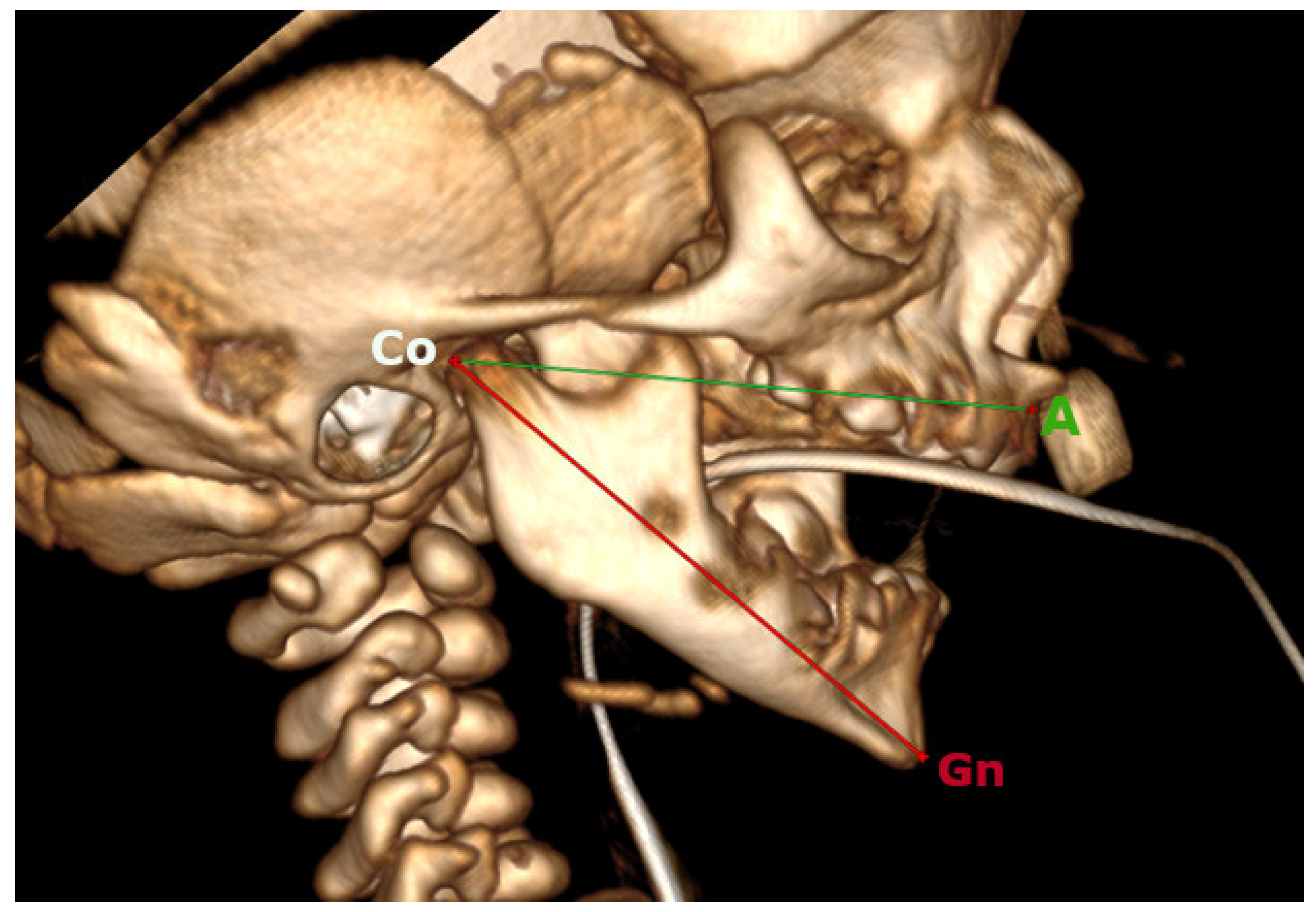
Figure 1. Effective mandibular length (CoGn) and effective midface length (CoA); the CT 3D reconstruction.
Oral 05 00058 g001
Table 1. Proposed new Compositive Norms derived from McNamara’s original Compositive Norms [21].
CoACoGn
Min
CoGn
Max
Ratio Md/Mx MinRatio Md/Mx Max
3536381.031.08
4042441.051.10
4548501.071.12
5054571.091.14
5561641.111.16
6068711.131.18
6575781.151.19
7082851.171.21
7590941.191.23
80971001.211.25
851051081.241.27
901131161.261.29
951221251.281.32
1001301331.301.33
1051381411.311.34
Table 2. Patients included in the study. F: female. M: male. T1 = date (expressed in days from birth) of the pre-surgical CT. T2 = date (expressed in days from birth) of the post-surgical CT.
PatientSexT1
(Days of Born)
FEMOD
(Days of Born)
T2
(Days of Born)
1F2141581
2M131585
3M7189778
4F7797401
5M714668
6F621267
Table 3. Pre-surgical (T1) measurements.
PatientT1. CoA
(mm)
T1. CoGn
(mm)
Expected
T1. CoGn
(mm)
Difference
Measured T1. CoGn–Expected T1. CoGn
(mm)
p Value Expected
T1. CoGn Vs. Measured T1. CoGn
T1 Md/Mx ratioExpected
T1. Md/Mx Ratio
p Value
Expected T1. Md/Mx Ratio Vs.
Measured T1 Md/Mx Ratio
134.932.436−3.60.028 *0.931.030.029 *
242.138.644−5.40.921.06
343.642.646−3.40.981.06
442.041.544−2.50.991.06
542.537.144−6.90.871.06
635.033.036−3.00.941.03
*: p-value significative.
Table 4. Surgical follow-up (T2) measurements. Wilcoxon analysis between measured Md/Mx ratio and expected Md/Mx expected for CoA and CoGn.
PatientT2. CoA
(mm)
T2. CoGn
(mm)
Expected
T2. CoGn
(mm)
Difference
Measured T2. CoGn–Expected T2.CoGn
(mm)
p Value Expected
T2. CoGn Vs. Measured T2. CoGn
T2 Md/Mx RatioExpected
T2. Md/Mx Ratio
p Value
Expected T2. Md/Mx Ratio Vs.
Measured T2 Md/Mx Ratio
161.470.45700.50.4001.151.140.461
247.247.451−3.61.011.08
358.968.2680.21.161.16
452.958.8580.81.111.11
565.476.9760.91.181.17
647.359.5527.51.261.13
Disclaimer/Publisher’s Note: The statements, opinions and data contained in all publications are solely those of the individual author(s) and contributor(s) and not of MDPI and/or the editor(s). MDPI and/or the editor(s) disclaim responsibility for any injury to people or property resulting from any ideas, methods, instructions or products referred to in the content.