Case Report

Persistent Remission of Angioimmunoblastic T-Cell Lymphoma and Associated Immune-Mediated Thrombotic Thrombocytopenic Purpura After Multimodal Therapy: A Case Report


1
Department of Hematology, Oncology, Infectious Diseases and Palliative Care Medicine, Alb-Fils Klinik, 73035 Göppingen, Germany
2
Centre for Cancer Immunology, Faculty of Medicine, University of Southampton, Southampton SO17 1BJ, UK
*
Author to whom correspondence should be addressed.
Submission received: 27 January 2026 / Revised: 21 February 2026 / Accepted: 25 February 2026 / Published: 2 March 2026

Abstract

Angioimmunoblastic T-cell lymphoma (AITL) is a rare subtype of peripheral T-cell lymphoma (PTCL) and is frequently associated with autoimmune phenomena. Clinically, AITL shows an aggressive disease course and poor prognosis with currently available treatment strategies. We here report the case of a 64-year-old female patient who was diagnosed with AITL and showed a complicated clinical course due to concurrent immune-mediated thrombotic thrombocytopenic purpura (iTTP). To our knowledge, the presented case highlights a previously unreported association of both conditions. Treatment, including chemotherapy and iTTP-directed treatments, resulted in rapid clinical improvement and sustained remission of both the AITL and the concurrent iTTP. In AITL, transformed T-follicular helper cells (TFHs) are particularly thought to mediate hypersecretion of cytokines and excessive autoantibody production. Immunological disturbances to large parts mediated through these transformed TFHs are thought to trigger autoimmune conditions, as seen with iTTP in this patient. At 36 months post-treatment, the patient remains in complete remission for both AITL and iTTP. This case highlights the complex immunopathological relationship between AITL and autoimmune disorders possibly impeding diagnosis and treatment in a timely manner.

1. Introduction

Angioimmunoblastic T-cell lymphoma (AITL) is a rare but distinct entity within the peripheral T-cell lymphoma (PTCL) spectrum [1]. Clinically, AITL is characterized by systemic involvement, including generalized lymphadenopathy, hepatosplenomegaly, and extranodal and cutaneous manifestations. The disease is associated with profound immune dysregulation and inflammation, leading to autoimmune phenomena and a high incidence of infections [2,3]. AITL-related autoimmune manifestations comprise a wide range, such as cryoglobulinemia complicated with cryoglobulinemic glomerulonephritis [4], polyneuropathy [4,5] or vasculitis [6], and may as well target different cellular compartments of the hematologic cellular system such as those found in pure red cell aplasia [7], amongst others. The prognosis is poor, reaching a 5-year overall survival of only 44%, and autologous stem cell transplantation (ASCT) can be conducted to improve the outcome [1].
We here report a case of AITL associated with the presentation of severe immune-mediated thrombotic thrombocytopenic purpura (iTTP), a previously unreported association. Immediate treatment addressing both pathogenic processes by using chemotherapy and iTTP-directed treatment approaches led to a sustained remission of both the lymphoma and the iTTP.

2. Case Presentation

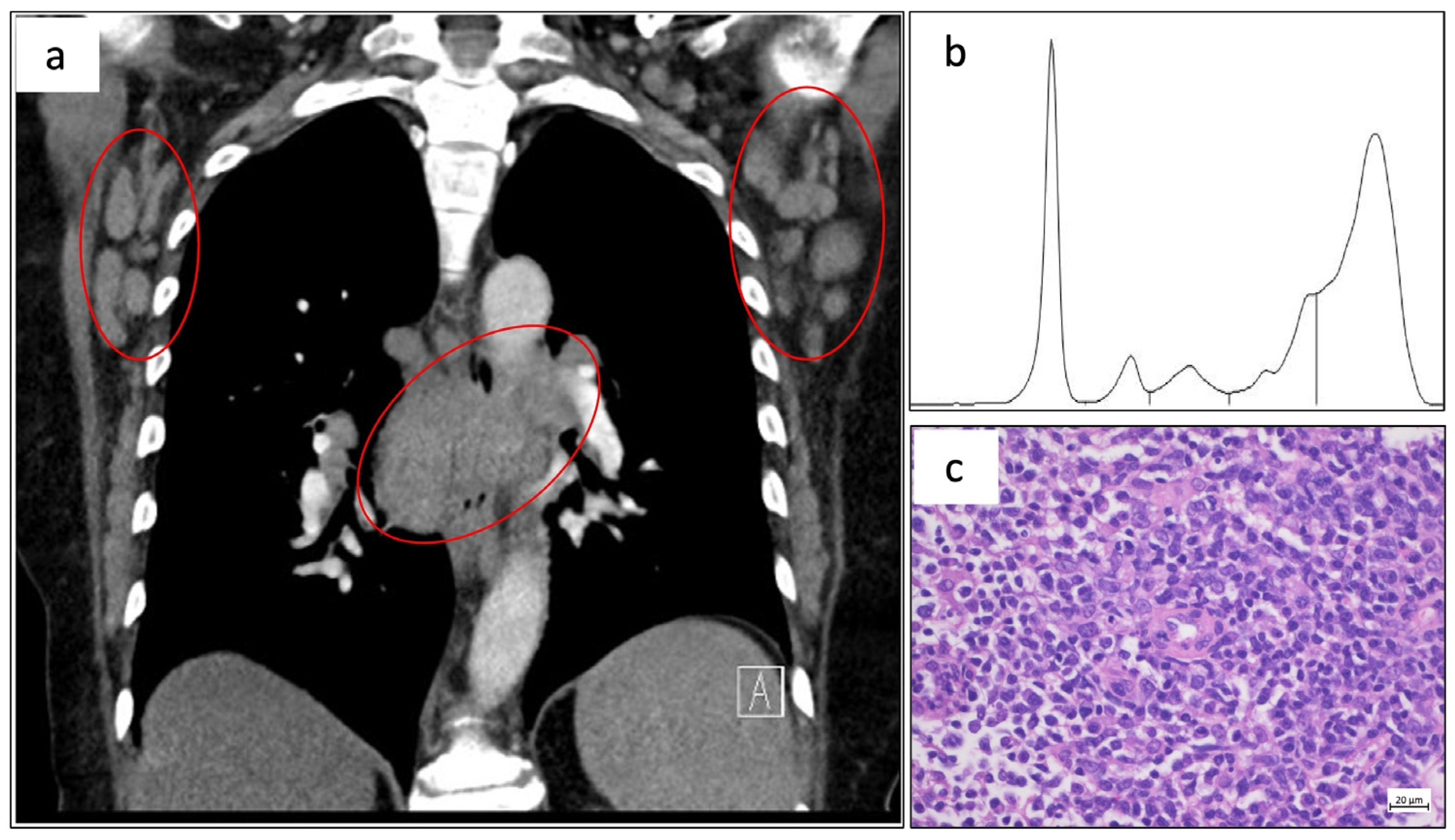
A 64-year-old woman was admitted to our hospital for suspected deep vein thrombosis of the right leg. She had suffered from viral infection, clinically suspected as hand-foot-and-mouth disease, several weeks ago. SARS-CoV-2 vaccination (mRNA-based) was also given weeks before admission. Thrombosis of the right popliteal vein was confirmed. Additionally, generalized lymphadenopathy up to 7.8 cm was found in the physical examination and a consecutive computed tomography (CT) scan (Figure 1a). At admission, peripheral blood tests showed leukocytosis (15.5 × 10³/µL; 11.25 × 10³/µL neutrophils and 0.6 × 10³/µL lymphocytes), normal hemoglobin (13.5 g/dL) and normal thrombocytes (297 × 103/µL). Further laboratory tests revealed increased lactate dehydrogenase, high levels of polyclonal IgG (Figure 1b), positive rheumatoid factor and anti-smooth muscle antibodies (Table 1).
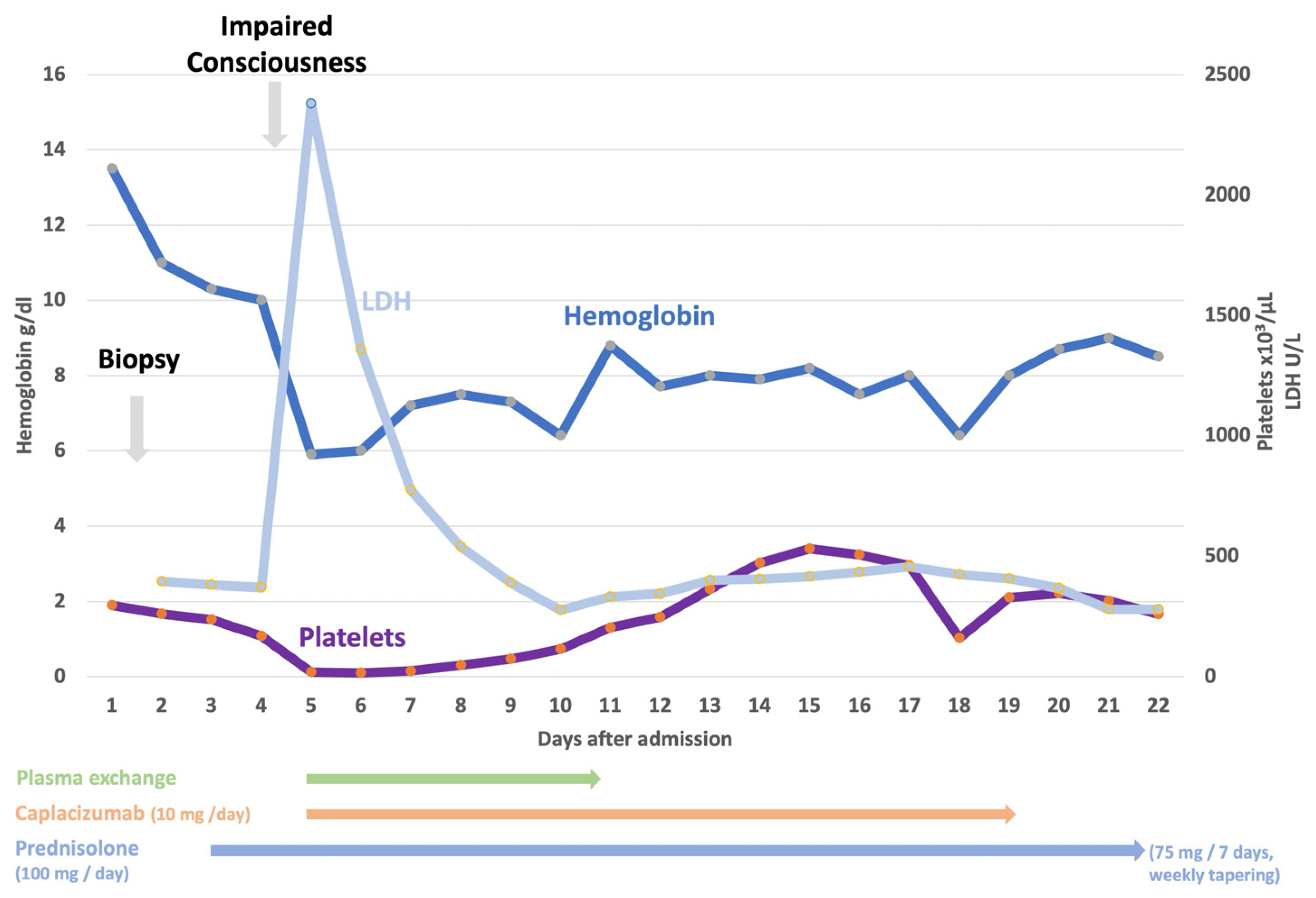
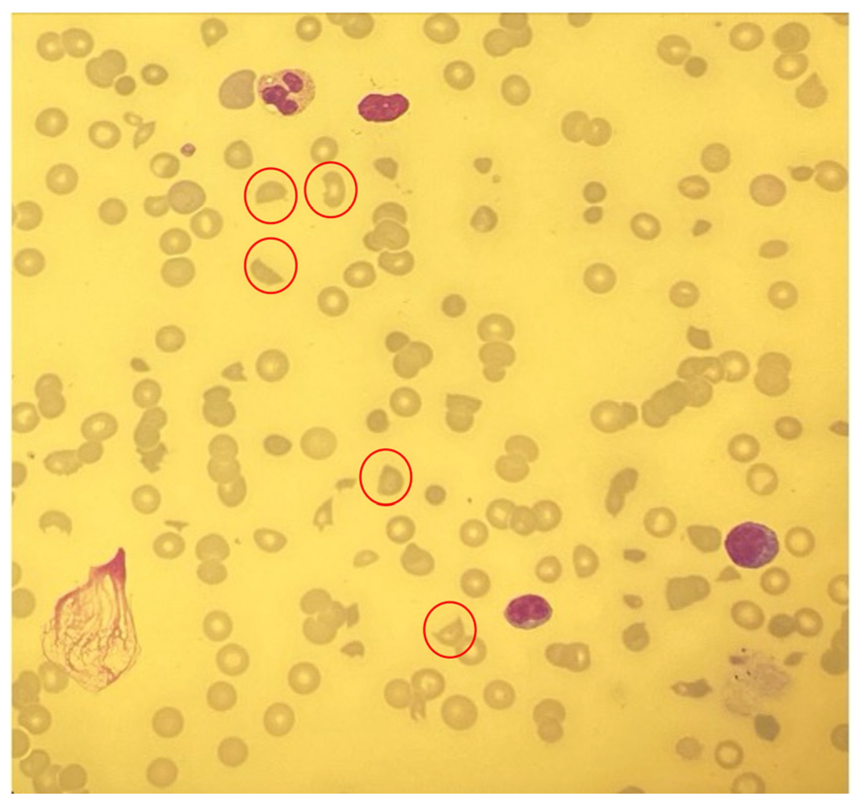
Suspecting a lymphoproliferative disorder, biopsies from a right-side axillary lymph node and the bone marrow were taken, and steroid prephase treatment was initiated. Preliminary pathology results of the bone marrow and lymph node biopsies were consistent with AITL (Figure 1c) and were sent for a second opinion to a reference pathology laboratory. After a few days, the patient’s condition subsequently deteriorated and she developed impaired consciousness with progressive disorientation and bruising. Repeated laboratory tests showed severe hemolytic anemia (hemoglobin 5.9 g/dL) and thrombocytopenia (18 × 103/µL) (Table 1, Figure 2). Numerous schistocytes were detected in the peripheral blood smear, indicating thrombotic microangiopathy (TMA) (Figure 3). Immediate plasma exchange was initiated, and prednisolone 100 mg per day continued. The ADAMTS13 activity was <1%, and further sample workup revealed anti-ADAMTS13 antibodies (>100 U/mL) (Table 1). Thus, the diagnosis of iTTP was established.
Given the suspected association between AITL and the highly active iTTP, the cytoreductive steroid prephase treatment was complemented by vincristine (1.4 mg/m2, capped at the maximum dose of 2 mg), which was administered on day 3 of plasma exchange.
Treatment with the anti-vWF nanobody caplacizumab at 10 mg daily was started. Rapid improvement of clinical and laboratory parameters was achieved. Plasma exchange was stopped after the platelet count reached 150 × 103/µL (Figure 2). We decided to expand the immunosuppressive therapy by adding rituximab, which resulted in sustained normal ADAMTS13 activity.
The final immunohistology results of the lymph node and bone marrow biopsies confirmed the diagnosis of AITL, along with verification of a clonal rearrangement of the TCR genes and EBV+ B cells using Epstein–Barr virus-encoded RNA in situ hybridization (EBER-ISH).
For further AITL treatment, we administered four cycles of cyclophosphamide, doxorubicin, vincristine, etoposide and prednisone (CHOEP) every two weeks according to current guidelines, achieving a CT-confirmed partial remission.
The patient was subsequently referred to an academic transplantation center for stem cell mobilization and consolidation treatment with high-dose chemotherapy, followed by ASCT. After receiving one cycle of chemotherapy with dexamethasone, high-dose cytarabine (Ara-C) and cisplatin (DHAP), CD34+ hematopoietic stem cells were successfully harvested by using granulocyte colony-stimulating factor (G-CSF) and the CXCR4 antagonist plerixafor (which blocks CXCR4/SDF-1-mediated bone marrow adhesion) for stem cell expansion and mobilization to the peripheral blood.
Unfortunately, although renal function was normalized after initiation of iTTP-directed treatment and during induction chemotherapy, the patient developed acute renal failure shortly after receiving chemotherapy with DHAP. Conventional laboratory tests, ADAMTS13 activity and blood smear showed no suspicion of iTTP relapse. Renal biopsy was performed, and histologically, TMA or lymphoma-related kidney injury was ruled out. Instead, the results provided evidence of a toxic nephropathy. The patient subsequently received hemodialysis and finally could be discharged in stable condition.
A subsequent PET-MRI confirmed complete remission of the lymphoma. Planned consolidation with high-dose chemotherapy followed by ASCT was not administered due to the complicated course. Currently, at 36 months of follow-up, the patient’s lymphoma and iTTP remain in ongoing complete remission, with ADAMTS13 activity showing a persistent normalization without signs of hemolysis.

3. Discussion

The association of iTTP and malignant lymphoproliferative disorders is reported to be a very rare event. Mostly B-cell non-Hodgkin and Hodgkin lymphomas have been reported in association with iTTP [8,9,10,11].
AITL is associated with polyclonal B cell activation and autoimmune-mediated cytopenia [12]. Reported hematologic autoimmune complications include immune thrombocytopenia (ITP), autoimmune hemolytic anemia (AIHA), their combination as Evans syndrome, immune neutropenia and pure red cell aplasia [7,12,13,14].
Immunologically, the frequent findings of autoimmunity in AITL are rooted in the complex alterations of the immune system, which arise from the aberrant cellular behavior of transformed TFHs. TFHs represent the physiologic counterpart of AITL and recapitulate their once physiologic functions in a disordered pathogenic manner [2,3,15].
TFHs are a subtype of CD4+ T helper cells and are centrally involved in the processes of the germinal center reaction, contributing to coordinated B cell differentiation and activation [16].
Transformed TFHs induce a pro-inflammatory environment and hypersecretion of immunoglobulins through aberrant B cell maturation and plasma cell differentiation by excessive IL-21 secretion. Characteristically, distorted interaction in the cellular microenvironment evolves along with changes in cytokine secretion such as CXCL13, VEGF, IL-6, involving dendritic cells, reactive T lymphocytes and macrophages [2,3,17].
These alterations further tip the tightly balanced cascade of physiologic immune responses towards unresponsiveness to pathogens and an acceleration of autoimmunity.
In AITL, B lymphocytes are mostly found to be EBV+ [2,3,17], and EBV infection per se is considered to play a central role in the autoreactivity of B cells and the development of autoimmune diseases [18].
Polyclonal gammopathy, as also impressively observed in our case, is a typical finding in AITL (therefore formerly referred to as angioimmunoblastic lymphadenopathy with dysproteinemia (AILD)) and shows a correlation with polyclonal plasmacytosis [19,20].
Notably, the patient’s acute iTTP manifested only after lymph node and bone marrow biopsies. Given that the AITL was likely present for a considerable period before the patient’s presentation at our center, coinciding development of iTTP after the invasive procedures (and possibly the preceding deep vein thrombosis) is suggestive of a causal link.
In addition to de novo development of autoreactive antibodies, a pre-existing priming of the immune system towards the development of anti-ADAMTS13 antibodies may be considered. As such, the presence of anti-ADAMTS13 antibodies is found in 5% of healthy individuals without being clinically apparent, possibly because of a lower affinity of these antibodies [21,22], which therefore may not be able to significantly inhibit ADAMTS13. Furthermore, specific HLA class II alleles have been associated with an increased risk for the development of iTTP. For example, HLA-DRB1*11 and DQB1*03 were reported in familial idiopathic TTP cases and are overrepresented in patients with acquired iTTP [23,24,25].
Patients with low ADAMTS13 activity either caused by ADAMTS13 mutations in congenital TTP (cTTP) or induced by anti-ADAMTS13 antibodies in iTTP do not inevitably develop TMA. A second hit is necessary to induce TMA [26]. In our case we assume that the inhibitor level was already very high at time of admission, since polyclonal immunoglobulins were already increased. Endothelial damage triggered by venous thrombosis and/or localized injury of vessels caused by the biopsy procedures with vWF release into a disordered immunoreactive surrounding of the AITL could initiate or aggravate the imbalance between ADAMTS13 and vWF. Furthermore, endocytosis and the processing of ADAMTS13 by macrophages or dendritic cells with peptides presented on the MHC complex, and activation of autoreactive CD4+ T cells and subsequently (memory) B cells with the production of anti-ADAMTS13 antibodies may be facilitated in this specific context.

4. Conclusions

To our knowledge, a case with iTTP associated with AITL has not yet been reported in the literature. Nevertheless, the polyclonal IgG elevation and specific antibodies could be demonstrated in this case (anti-smooth muscle antibodies and anti-ADAMTS13 antibodies). Lymphoma-directed antiproliferative treatment with steroids, vincristine, and finally CHOEP/DHAP, as well as anti-B cell treatment with rituximab combined with plasma exchange and anti-vWF-directed therapy using caplacizumab, induced a complete remission of iTTP and AITL that has remained for 36 months.

Author Contributions

Conceptualization, J.B. and M.B.; data curation, J.B. and M.B.; writing—original draft preparation, J.B.; writing—review and editing, M.S. and M.B.; visualization, J.B. and M.B.; supervision, M.B.; project administration, M.B. All authors have read and agreed to the published version of the manuscript.

Funding

This research received no external funding.

Institutional Review Board Statement

We have obtained consent from the patient for publication of this case report; for (retrospective) case reports with standard treatments, an ethics committee is not needed (no experimental setting here—only approved drugs).

Informed Consent Statement

Written consent has been obtained from the patient for publication.

Data Availability Statement

Data is available from the involved authors upon request.

Acknowledgments

The authors thank their patient for agreeing to and participating in the case presentation and all colleagues involved in case discussion, treatment, diagnostic procedures and sample workup.

Conflicts of Interest

M.B. has received honoraria from Alexion, Sanofi, and Takeda. J.B. and M.S. declare no conflicts of interest.

References

  1. Advani, R.H.; Skrypets, T.; Civallero, M.; Spinner, M.A.; Manni, M.; Kim, W.S.; Shustov, A.R.; Horwitz, S.M.; Hitz, F.; Cabrera, M.E.; et al. Outcomes and prognostic factors in angioimmunoblastic T-cell lymphoma: Final report from the international T-cell Project. Blood 2021, 138, 213–220. [Google Scholar] [CrossRef]
  2. Chiba, S.; Sakata-Yanagimoto, M. Advances in understanding of angioimmunoblastic T-cell lymphoma. Leukemia 2020, 34, 2592–2606. [Google Scholar] [CrossRef] [PubMed]
  3. Lunning, M.A.; Vose, J.M. Angioimmunoblastic T-cell lymphoma: The many-faced lymphoma. Blood 2017, 129, 1095–1102. [Google Scholar] [CrossRef] [PubMed]
  4. Li, X.-Y.; He, H.-Y.; Yue, S.-L.; Pai, P. Delayed diagnosis of Angioimmunoblast T-cell lymphoma presenting with type II Cryoglobulinemia and acute kidney injury: A case report and narrative review of the literature. BMC Nephrol. 2020, 21, 463. [Google Scholar] [CrossRef] [PubMed]
  5. Kleeberg, A.; Bauer, G.; Jung, E.; Purrucker, J.C. Inflammatory neuropathy with evidence of anti-GQ1b antibodies in angioimmunoblastic T cell lymphoma (AITL): A case report. J. Neurol. 2024, 271, 2880–2885. [Google Scholar] [CrossRef]
  6. Ambrosio, M.R.; Rocca, B.J.; Ginori, A.; Onorati, M.; Fabbri, A.; Carmellini, M.; Lazzi, S.; Tripodi, S. Renal infarction due to polyarteritis nodosa in a patient with angioimmunoblastic T-cell lymphoma: A case report and a brief review of the literature. Diagn. Pathol. 2012, 7, 50. [Google Scholar] [CrossRef]
  7. Kawahigashi, T.; Kitagawa, I.; Tanaka, E. Angioimmunoblastic T-cell lymphoma accompanied by pure red cell aplasia: A case report. World J. Clin. Oncol. 2020, 11, 405–411. [Google Scholar] [CrossRef]
  8. Chao, J.-H.; Kloecker, G.H. Thrombotic Thrombocytopenic Purpura Associated with Hodgkin Lymphoma. Blood 2013, 122, 5050. [Google Scholar] [CrossRef]
  9. Kanno, M.; Nakamura, S.; Kawahara, M.; Ueno, S.; Nakamine, H.; Matsumoto, M.; Fujimura, Y. Chemotherapy-resistant intravascular lymphoma accompanied by ADAMTS13 inhibitor successfully treated with rituximab. Int. J. Hematol. 2008, 88, 345–347. [Google Scholar] [CrossRef]
  10. Byers, J.T.; Kurtz, J.L.; Mendoza, A.S.; Oster, C.D.; Sun, N.C.; Qing, X.; Cornford, M.E. An unusual presentation of primary pulmonary extranodal marginal zone lymphoma of mucosal associated lymphoid tissue: An autopsy case report. Exp. Mol. Pathol. 2018, 104, 155–157. [Google Scholar] [CrossRef]
  11. Tomlinson, R.; Yaxley, J. Thrombotic thrombocytopenic purpura associated with Hodgkin lymphoma and non-Hodgkin lymphoma. Pathology 2018, 50, 776–777. [Google Scholar] [CrossRef]
  12. Crickx, E.; Goulabchand, R.; Fieschi, C.; Galicier, L.; Coppo, P.; Meignin, V.; Delarue, R.; Casasnovas, O.; Leblond, V.; Moulis, G.; et al. Clinical Spectrum, Evolution and Management of Autoimmune Cytopenia Associated with Angioimmunoblastic T-Cell Lymphoma: A Retrospective, Multicenter Study. Blood 2016, 128, 1816. [Google Scholar] [CrossRef]
  13. Wu, B.; Wang, W.; Li, F.; Cheng, Y. Angioimmunoblastic T-cell lymphomas presenting as autoimmune hemolytic anemias: Clinical manifestations and circulating cytokine features. Ann. Hematol. 2015, 94, 1437–1439. [Google Scholar] [CrossRef] [PubMed]
  14. Sekiguchi, Y.; Shimada, A.; Imai, H.; Wakabayashi, M.; Sugimoto, K.; Nakamura, N.; Sawada, T.; Komatsu, N.; Noguchi, M. CD56+ Angioimmunoblastic T-Cell Lymphoma with Evans Syndrome: A Case Report and Review of the Literature. J. Clin. Exp. Hematop. 2013, 53, 37–47. [Google Scholar] [CrossRef] [PubMed]
  15. Swerdlow, S.H.; Campo, E.; Pileri, S.A.; Harris, N.L.; Stein, H.; Siebert, R.; Advani, R.; Ghielmini, M.; Salles, G.A.; Zelenetz, A.D.; et al. The 2016 revision of the World Health Organization classification of lymphoid neoplasms. Blood 2016, 127, 2375–2390. [Google Scholar] [CrossRef] [PubMed]
  16. Akkaya, M.; Kwak, K.; Pierce, S.K. B cell memory: Building two walls of protection against pathogens. Nat. Rev. Immunol. 2020, 20, 229–238. [Google Scholar] [CrossRef]
  17. Lage, L.A.d.P.C.; Culler, H.F.; Reichert, C.O.; da Siqueira, S.A.C.; Pereira, J. Angioimmunoblastic T-cell lymphoma and correlated neoplasms with T-cell follicular helper phenotype: From molecular mechanisms to therapeutic advances. Front. Oncol. 2023, 13, 1177590. [Google Scholar] [CrossRef]
  18. Sachinidis, A.; Garyfallos, A. Involvement of age-associated B cells in EBV-triggered autoimmunity. Immunol. Res. 2022, 70, 546–549. [Google Scholar] [CrossRef]
  19. Nagoshi, H.; Kuroda, J.; Kobayashi, T.; Maegawa, S.; Chinen, Y.; Kiyota, M.; Nakayama, R.; Mizutani, S.; Shimura, Y.; Yamamoto-Sugitani, M.; et al. Clinical manifestation of angioimmunoblastic T-cell lymphoma with exuberant plasmacytosis. Int. J. Hematol. 2013, 98, 366–374. [Google Scholar] [CrossRef]
  20. De Leval, L.; Gisselbrecht, C.; Gaulard, P. Advances in the understanding and management of angioimmunoblastic T-cell lymphoma. Br. J. Haematol. 2010, 148, 673–689. [Google Scholar] [CrossRef]
  21. Rieger, M.; Mannucci, P.M.; Kremer Hovinga, J.A.; Herzog, A.; Gerstenbauer, G.; Konetschny, C.; Zimmermann, K.; Scharrer, I.; Peyvandi, F.; Galbusera, M.; et al. ADAMTS13 autoantibodies in patients with thrombotic microangiopathies and other immunomediated diseases. Blood 2005, 106, 1262–1267. [Google Scholar] [CrossRef]
  22. Grillberger, R.; Casina, V.C.; Turecek, P.L.; Zheng, X.L.; Rottensteiner, H.; Scheiflinger, F. Anti-ADAMTS13 IgG autoantibodies present in healthy individuals share linear epitopes with those in patients with thrombotic thrombocytopenic purpura. Haematologica 2014, 99, e58–e60. [Google Scholar] [CrossRef]
  23. Coppo, P.; Busson, M.; Veyradier, A.; Wynckel, A.; Poullin, P.; Azoulay, E.; Galicier, L.; Loiseau, P. HLA-DRB1*11, a strong risk factor for acquired severe ADAMTS13 deficiency-related idiopathic thrombotic thrombocytopenic purpura in Caucasians. J. Thromb. Haemost. 2010, 8, 856–859. [Google Scholar] [CrossRef]
  24. John, M.-L.; Hitzler, W.; Scharrer, I. The role of human leukocyte antigens as predisposing and/or protective factors in patients with idiopathic thrombotic thrombocytopenic purpura. Ann. Hematol. 2012, 91, 507–510. [Google Scholar] [CrossRef]
  25. Lombardi, A.M.; Pulini, S.; Passeri, C.; Iuliani, O.; Salutari, P.; Businaro, A.; Fabris, F.; Vianello, F. Familial acquired thrombotic thrombocytopenic purpura: Immunogenetic link with HLA-DRB1*11 and DQB1*03 antigens. Br. J. Haematol. 2018, 183, 317–319. [Google Scholar] [CrossRef]
  26. Cauchois, R.; Muller, R.; Lagarde, M.; Dignat-George, F.; Tellier, E.; Kaplanski, G. Is Endothelial Activation a Critical Event in Thrombotic Thrombocytopenic Purpura? J. Clin. Med. 2023, 12, 758. [Google Scholar] [CrossRef]
Figure 1. (a) Computed tomography scan showing mediastinal mass and axillary lymphadenopathy (red circles). (b) Serum protein electrophoresis showing broad γ-region elevation favoring polyclonal hypergammaglobulinemia. (c) Representative hematoxylin and eosin-stained lymph node section (40× magnification) demonstrating a diffuse lymphoid infiltrate of pleomorphic, small-to-medium-sized atypical cells with irregular nuclei and prominent vascular network.
Hemato 07 00008 g001
Figure 2. Figure representing time course of lactate dehydrogenase (LDH), hemoglobin and platelet counts in relation to diagnostic and therapeutic interventions.
Hemato 07 00008 g002
Figure 3. Peripheral blood smear at the time point of clinical deterioration showing thrombocytopenia and numerous schistocytes (circles).
Hemato 07 00008 g003
Table 1. Laboratory parameters for central time points: at first presentation at the hospital (admission), initial manifestation of iTTP (day 3 after biopsy) and after start of treatment/controlled iTTP activity (discharge). (Assays used for ADAMTS13 activity were the automated modified FRETS assay (day 5) and the protease-based ADAMTS13 activity ELISA (day 26) (both Technoclone)).
Parameter
(Reference Range (Ref.))
Day 1
Admission
Day 5 (Day 3
After Biopsy)
Day 26
Discharge
Creatinine
(Ref. 0.5–1.1 mg/dL)
1.4 mg/dL1.8 mg/dL0.7 mg/dL
Leukocytes
(Ref. 4.0–9.0 × 10³/µL)
15.5 × 10³/µL18.3 × 103/µL8.5 × 103/µL
Hemoglobin
(Ref. 12.0–16.0 g/dL)
13.5 g/dL5.9 g/dL8.4 g/dL
Thrombocytes
(Ref. 152–396 × 103/µL)
297 × 103/µL18 × 103/µL213 × 103/µL
Lactate
dehydrogenase
(Ref. 135–247 U/L)
394 U/L2380 U/L195 U/L
Immunofixationnegative--
IgA
(Ref. 0.7–4.0 g/L)
16.1 g/L--
IgG
(Ref. 7.0–16.0 g/L)
45.1 g/L--
Rheumatoid factor
(Ref. <20 U/mL)
83 U/mL--
Anti-smooth muscle antibody
(Ref. <1:40)
1:640--
Haptoglobin
(Ref. 30–200 mg/dL)
n.d.<6 mg/dL207 mg/dL
ADAMTS13 activity
(Ref. 50–110%)
n.d.<1%63%
ADAMTS13
inhibitor
(Ref. <16 U/mL)
n.d.>100 U/mLn.d.
Disclaimer/Publisher’s Note: The statements, opinions and data contained in all publications are solely those of the individual author(s) and contributor(s) and not of MDPI and/or the editor(s). MDPI and/or the editor(s) disclaim responsibility for any injury to people or property resulting from any ideas, methods, instructions or products referred to in the content.