Article

X-Ray Interaction and the Electronic, Atomic Cross-Sections and Compton Mass-Attenuation Coefficients of Human Blood, Breasts, Eye Lens, Ovaries, and Testis

1
Department of Clinical and Diagnostic Sciences, The University of Alabama at Birmingham, Birmingham, AL 35294, USA
2
Medical Science Department, The University of Aberdeen, King’s College, Aberdeen AB24 3FX, UK
*
Author to whom correspondence should be addressed.
Radiation 2025, 5(3), 24; https://doi.org/10.3390/radiation5030024
Submission received: 21 June 2025 / Revised: 23 August 2025 / Accepted: 25 August 2025 / Published: 31 August 2025

Simple Summary

Radiation plays a vital role in many fields and practical applications. Treating cancer patients, radiographic images, radiation dosimetry, CT scan, and mammography are some of the fields where radiation is frequently used. In all therapeutic and diagnostic applications of radiation, it is essential to understand how radiation interacts with body tissues and the potential implications of this interaction. Without the knowledge of the interaction of various types of radiation with body tissue, proper shielding and protection from the hazards of radiation cannot be provided. Therefore, the Klein–Nishina formula is used in the present work to study and understand the interaction of various energetic X-rays with the human blood, breast, eye lens, ovary, and testis. The results show that the radiation energy, tissue density, electron density, and effective charge number strongly affect the interaction of radiation with body tissues. Therefore, the effects of these parameters on radiation interaction should be studied before providing radiation dose to any tissue or the body as a whole.

Abstract

The Klein–Nishina formula is used to calculate and investigate the electronic cross-section, atomic cross-section, and Compton mass attenuation coefficients for the human blood, breasts, eye lens, ovaries, and testis, using X-rays in the 0.1–20 MeV energy range. The effects of radiation energy, tissue effective charge number, tissue density, and tissue electronic density on these parameters were studied. The results show that the electronic cross-section and atomic cross-section decrease with increasing radiation energy. These parameters are found to be linearly increasing when the density and electron density of a tissue increase. This increase is more rapid with a bigger slope when the electron density increases as compared to the density of each tissue. A complex relationship between these coefficients and the effective charge number Zeff of tissues is observed because Zeff changes with the energy and linear attenuation coefficient of a tissue. The Compton mass attenuation coefficient is found to be dependent on the effective charge number to mass number ratio Zeff/Aeff instead of just the effective charge number. This increase in the Compton mass attenuation coefficient with increasing Zeff/Aeff is rapid for the lower values of Zeff/Aeff. However, for a higher Zeff/Aeff ratio, the increase is very slow and becomes almost constant for X-ray energies above 10 MeV. The calculated parameters are useful in determining radiation dose for the investigated tissues and their response to low and high-energy X-rays. The results and outcomes are also very useful in shielding and protecting tissues from the hazards of radiation. These parameters are also helpful in determining the scattered and optimum doses to improve image quality and treatment options in radiology and radiation therapy to offer the best care.

1. Introduction

Radiation has been used in diagnostic and therapeutic procedures, and nuclear medicine, for approximately 125 years, playing a critical role in detecting and treating abnormalities in the body, particularly cancer [1,2,3]. Around half of all cancer patients undergo radiation therapy, making it one of the most effective treatment methods available [4]. The primary objective of radiotherapy is to reduce the size of tumors and ultimately eliminate cancer cells [5]. This process involves several key factors, including ionizing radiation, high-energy photons, and principles like attenuation [6]. High-energy photons are commonly employed to target and destroy tumor cells [7]. However, the amount of radiation needed varies depending on the tumor’s size, composition, structure, and other physical properties [8]. Tumors are composed of atoms and molecules. When photons interact with these atoms, absorption and scattering occur known as attenuation. Proper dose-delivery is essential, as it directly impacts the success of tumor treatment and the safety of normal tissues around the tumor in the field of radiation [9,10]. The dose volumes are determined based on the tumor’s characteristics and the tolerance of surrounding healthy tissues. A critical aspect of radiotherapy research is establishing a connection between the type of radiation, tumor properties, and the delivered dose [1,9]. Photoelectric absorption and Compton scattering are key to this process, with scattering requiring greater attention due to its complexity [11]. Lower doses or lower radiation energies are more likely to be absorbed by the directed tissue or tumor compared to higher energies [12]. Advanced research is required to improve methods for measuring and calculating internal dose distributions and to enhance accuracy and effectiveness [13,14,15]. In the interaction of radiation with body tissue, the role of a single electron and its interaction with the incoming radiation is of primary importance and must be properly observed. The mode and strength of interaction of an X-ray photon with such an electron of a body’s atom is governed by the Klein–Nishina electronic cross-section, which determines the interaction probability of radiation with the individual electron. Additionally, the atomic cross-section refers to the interaction probability of the entire atom. Further research into the Klein–Nishina atomic and electronic cross-sections is essential to radiation imaging and therapy planning. Accurate measurement of radiation doses to internal organs remains challenging. Organ doses are often estimated using calculations based on measurements from models or specific body areas [15,16]. Understanding these doses is vital for evaluating the benefits and risks of radiation exposure. Larger tumors demand higher doses for effective treatment, which increases the likelihood of complications in nearby normal tissues. It is, therefore, essential to understand the relationship between the partial irradiation of organs and the potential for tissue damage [16,17]. Administering radiation therapy is a delicate process, as even minor errors in planning or delivery can have severe consequences, particularly when tumors are near sensitive tissues or vital organs. Overdoses of radiation therapy can lead to serious injury and death. Achieving precise dose-delivery requires understanding the interaction probabilities, scattering capabilities, and attenuation properties of both tumor and surrounding tissues [2,10,18]. Without this precision, the effectiveness of radiation in eliminating tumors can be compromised. While radiation is clinically efficient for tumor deterrence, overdosages of radiation can prove to be detrimental to human health. Patients may feel symptoms of radiation damage after treatment due to cell death and tissue damage due to overdoses of radiation [14,17,19]. Precisely planned radiation therapy that reduces the damage to normal tissues can greatly improve the quality of life and healing for patients treated for cancer [14,20,21]. This research focuses on examining how X-rays interact with various tissues, including blood, eye lens, breast, ovaries, and testis, within a specific energy range of 0.1–20 MeV. It must be noted that X-rays can be generated with a wide energy spectrum. In diagnostic applications, X-rays between 30 keV and 150 keV are commonly used. Orthovoltage X-rays are produced in the 100–500 keV energy range. In therapeutic applications, the energy of X-rays, produced in a linear accelerator, ranges from 4 MeV up to 25 MeV. In addition to their biomedical applications, various energetic X-ray beams are also used in industrial and non-medical applications [22,23,24]. The study aims to analyze scattering and attenuation properties using established theoretical models. By investigating how normal tissues influence radiation as it travels to tumors, this work provides insights into dose calculations and highlights the impact of tissue properties on accurate radiation delivery. Such findings are crucial for improving dosimetry techniques and advancing radiation therapy.

2. Materials and Methods

The probability of interaction between X-rays of differing energies in the 0.1 MeV–20 MeV range and Blood, Breast, Eye Lens, Ovary, and Testis is studied by calculating the Klein–Nishina electronic and atomic cross-sections and the Compton mass-attenuation coefficient as a function of radiation energy, the effective charge number, and the density of the investigated tissues. The Klein–Nishina differential equation and formula used to calculate the electronic cross-section are given by Equations (1) and (2), below [2,10,25,26]:
d σ d Ω = r 2 2 [ 1 1 + a ( 1 c o s θ ) ] 2 [ 1 +   cos 2 θ   +   a 2 ( 1 c o s θ ) 2 1 + a ( 1 c o s θ ) ]
σ e = 2 π r 2 1 + α α 2 2 ( 1 + α ) 1 + 2 α ln ( 1 + 2 α ) α + ln ( 1 + 2 α ) 2 α 1 + 3 α ( 1 + 2 α ) 2
where r = e 2 4 π ϵ 0 m e c 2 = 2.818   f m = 2.818 × 10 15   m = 2.818 × 10 13   c m is the classical radius of the electron and, α = E m e c 2 = h ν 0.511   M e V is a constant called the coupling constant, while E = h ν is the energy of the incident X-ray photon. Table 1 provides values of the coupling constant at various X-ray energies. σ e  represents the Klein–Nishina cross-section per electron and is expressed in units of cm2/electron.
The results obtained for the Klein–Nishina electronic cross-section σe followed the calculation of the coupling constant α using the equation α = E m e c 2 = h ν 0.511   M e V . The values of α calculated for various X-ray energies, along with the value of the classical radius of the electron r, were then plugged into Equation (2) to obtain σe.
The Klein–Nishina atomic cross-section ‘ σ a ’ was obtained for each tissue by multiplying the electronic cross-section by the effective charge number Zeff of the tissue at each photon energy i.e., σ a = .Zeff. σe. σ a  represents the Klein–Nishina cross-section per atom and is expressed in the unit of cm2/atom. The Compton mass-attenuation coefficient is obtained using the formula given below [10,18]:
σ/ρ = NA(Zeff/Aeffe
where NA = 6.022 × 10²³ atoms per mole, is Avogadro’s constant, the number of atoms in a gram-atomic weight of any element or the number of molecules in a gram-molecular weight of any compound, Zeff = number of electrons per atom of an element or per molecule of a compound in a tissue, Aeff = number of grams per mole of tissue (i.e., A is the gram-atomic or molecular weight), ρ = density in g/cm3, NAZeff/Aeff = number of electrons per gram of material, and σ/ρ = Compton mass-attenuation coefficient, and is expressed in the units of cm2/g

3. Effective Z and Effective A of Tissues

Every tissue is composed of several elements in a certain atomic ratio. Not all elements in a tissue are in the same and equal concentrations. The concentration of constituent elements in a tissue varies from a minimum to a maximum depending on the nature of the tissue. Therefore, the charge number of a tissue is not assigned due to a single constituent element; instead, it is a weighted average of the charge numbers of all elements and their relative concentrations composing the tissue. This weighted average is called the effective charge number Zeff of the tissue. In the same way, each tissue has an effective mass number Aeff corresponding to its effective charge number. The effective charge number is not a fixed number but varies with the energy of the interacting radiation. The values of the effective charge numbers used for the tissues investigated in this work are obtained from the results published by Kurudirek in 2014, Jayachandran in 1971, and Fatima in 2023 [2,27,28].
The effective mass number Aeff can be calculated by using the stability condition of nuclei. In general, for a certain element with a charge number Z, there may be several mass numbers, and hence several isotopes exist, but not all of them are stable isotopes. Since human tissues are very stable, the elements or atoms and nuclei in a tissue are also very stable. Thus, the effective mass number Aeff of a tissue can be calculated from its effective charge number Zeff, using the stability condition. It has been a fact that for every stable atom or molecule, the ratio of neutrons and protons in the atom must exist between 1 and 1.52 [23,29,30,31].
Mathematically, 1 ≤ N/Z ≤ 1.52. If N/Z = 1, then N = Z, and hence A = N + Z = 2 Z. On the other hand, if N/Z = 1.52, then N = 1.52 Z, and thus A = N + Z = 1.52 Z + Z = 2.52 Z. Therefore, the calculation specifies that for stable atoms and molecules of the tissues, the effective mass number A should satisfy the condition, 2 Z ≤ A ≤ 2.52 Z.
In the present work, all values of the effective mass number Aeff satisfy this condition of stability.

4. Results

4.1. Radiation Energy and Tissue Effective Charge Number

Figure 1 shows the variation in the values of the effective charge number Zeff of the investigated tissues and the energy E of the interacting X-rays. Unlike the fixed charge number of various elements and atoms, the effective charge number Zeff has a complex relationship with the energy of interacting radiation. The figure shows that Zeff slowly decreases with increasing X-ray energy in the lower energy range from 0.1 MeV to 0.3 MeV. At energies between 0.3 MeV and 2.5 MeV, the values of Zeff stay constant. Above 2.5 MeV, the value of Zeff increases, with the rate of increase becoming bigger at higher energy. This complex variation in Z with E is because the effective charge number varies with the linear attenuation coefficient of a material or tissue, and the linear attenuation coefficient depends upon the energy of interacting radiation [32,33]. Thus, the effective charge number of a tissue should also be dependent on the energy of interacting radiation.

4.2. Radiation Energy and Klein–Nishina Electronic and Atomic Cross-Sections

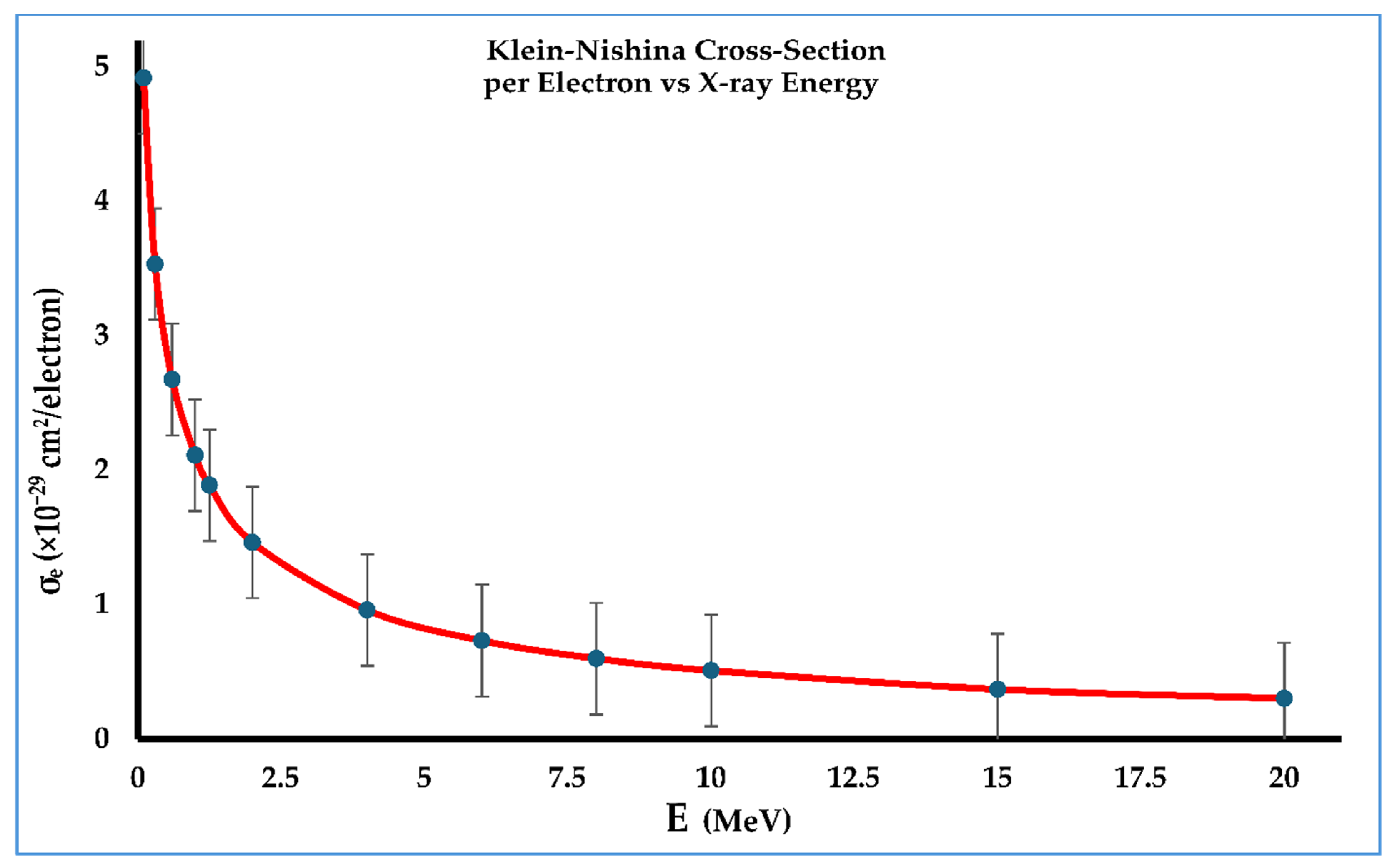
The Klein–Nishina electronic cross-section σ e represents the probability of a photon or electromagnetic radiation interacting with a single electron and is independent of the type of tissue through which the photon interacts. Figure 2 shows a relationship between the energy of X-rays in the 0.1–20 MeV range and the response of σ e . Table 1 provides data corresponding to Figure 2. It is clear from the figure that σ e decreases rapidly as the energy of the X-rays increases. This behavior can be explained based on the nature of the interaction at various X-ray energies. During the interaction of photons with a tissue, the interaction dominantly occurs following the photoelectric absorption of energy E ≤ 0.5 MeV. For energies 0.5 MeV ≤ E ≥ 10 MeV, the Compton scattering is dominant, followed by the pair production. For lower energies, photons interact through photoelectric absorption, giving up all their energy to the electron, and hence making the interaction probability the highest. At higher energies, due to the Compton scattering phenomenon, the interacting photon only gives up a part of its energy and still retains its identity and motion. This makes the interaction probability relatively lower. At very high energies where pair production is dominant, there is absorption; however, due to the occurrence of scattering at the same time, the cross-section still decreases.
Figure 3 shows the Klein–Nishina interaction cross-section of a photon with an atom of the tissue. A relationship of σ a with photon energy is provided for the breast, testis, ovary, eye lens, and blood. The Klein–Nishina atomic cross-section σ a , which is also known as the Klein–Nishina cross-section per atom, represents the probability of a photon or electromagnetic radiation interacting with a single atom, and hence depends on the type of tissue through which the photon interacts. A similar trend of decreasing σ a with increasing X-ray energy in the 0.1–20 MeV energy range is observed. However, the rate of decrease is different than the rate observed in the case of the Klein–Nishina electronic cross-section. The reason is that when an entire atom is considered, the interacting photon considers a more complicated structure due to several factors, like the size of an atom, the unoccupied space in an atom, the existence of a nucleus, and the distribution of electrons in an atom. The data is given in Table 2, Table 3, Table 4, Table 5 and Table 6.

4.3. Tissue Effective Charge Number Zeff and Klein–Nishina Atomic Cross-Section

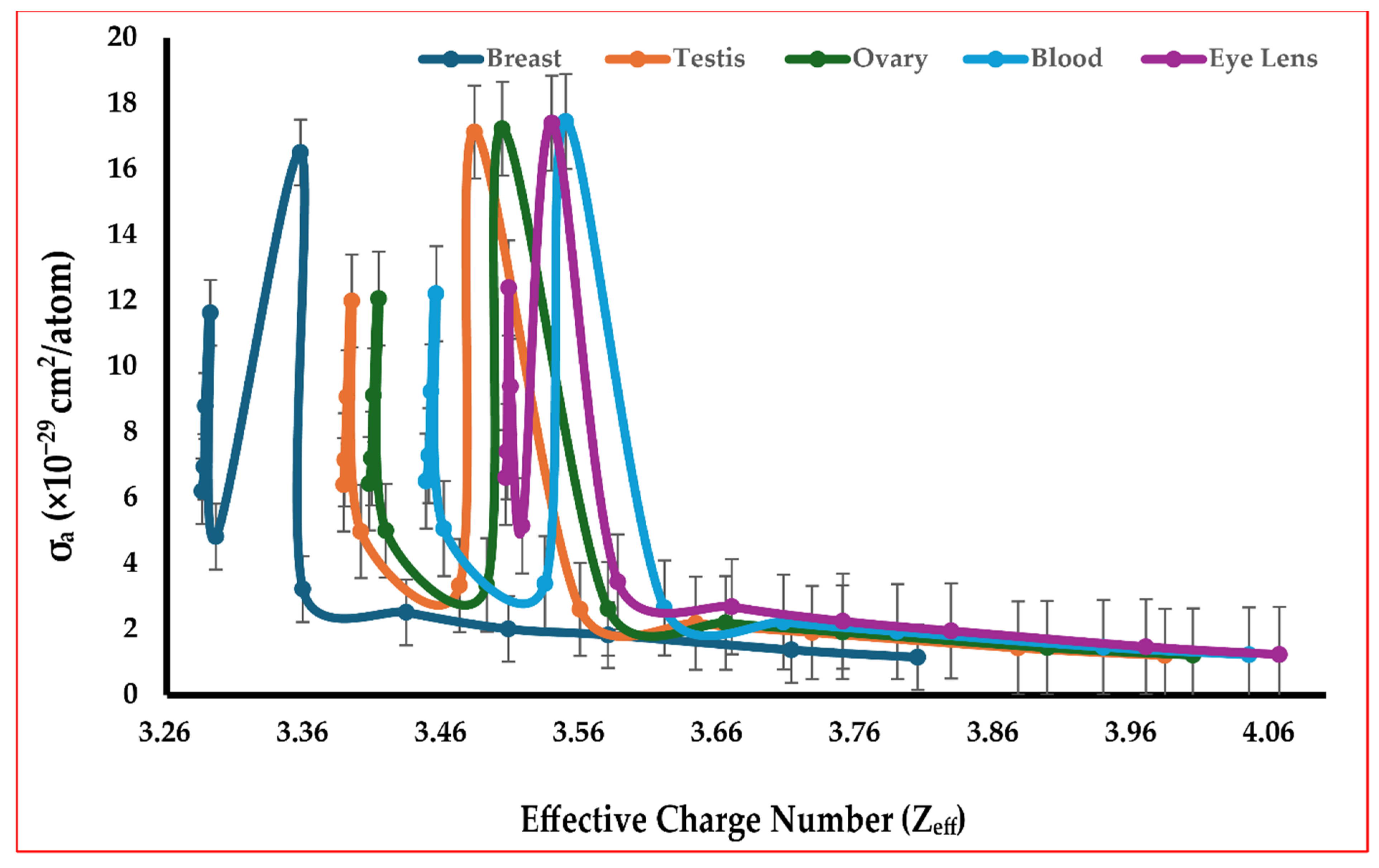
The Klein–Nishina cross-section per atom σ a is found to be dependent on several factors. Figure 4 gives information about how σ a changes with changing the effective charge number Z of a tissue. Calculations are performed for the breast, testis, ovary, blood, and eye lens. The figure shows that the σ a  changes with rise and fall for lower values of Zeff. For high values of Zeff, the Klein–Nishina cross-section per atom stays mostly the same with a very slow decrease. Overall, the changes in σ a  occur in a very complex way when Zeff is increased from 3.26 up to 4.06. The reason for this complex change could be attributed to the fact that all three major processes of photon interaction with tissues, the photoelectric absorption, Compton scattering, and pair production, depend strongly on the charge number Z of the medium, in general, and the effective charge number of a tissue. This simultaneous but different dependence contributes to the complex variation in the interaction cross-section of a photon with Zeff of each tissue. No doubt, there is a complex relationship between Zeff and σ a  but the same trend is observed on all tissues investigated.

4.4. Tissue Density and Klein–Nishina Atomic Cross-Section

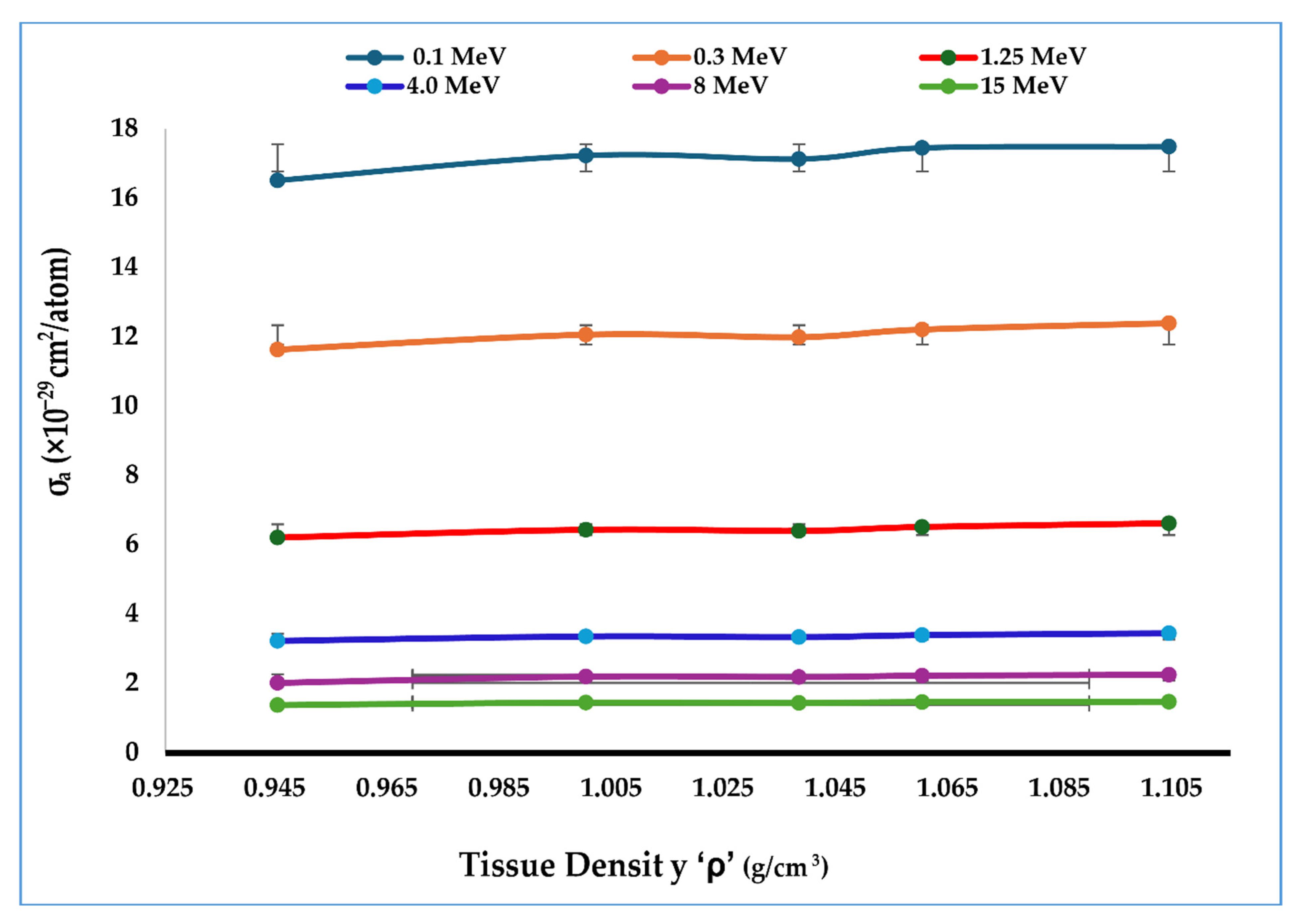
Figure 5 provides information about how the Klein–Nishina cross-section per atom σ a  changes with changing density ρ of tissues. The figure shows that a linear relation exists between σ a and ρ. Tissue density of the breast, testis, ovary, blood, and eye lens is considered. Straight lines are observed for all tissues with a small slope. The small but positive slope of each curve shows that the Klein–Nishina atomic cross-section increases at a slow rate when the density of the tissue is increased. Thus, increasing the density of a tissue gives rise to an increased probability of photon interaction with the tissue. The reason for this increase in σ a with ρ is because increasing density means more matter exists per unit volume of the tissue. The existence of more matter in the same volume will fill and occupy the space in the tissue more. This occupation of matter in the tissue provides more chances of interaction, giving rise to a high value of σ a .

4.5. Tissue Electron Density ρe and Klein–Nishina Atomic Cross-Section

The electron density ρe of a tissue represents the number of electrons present per unit volume of the tissue. Figure 6 shows the trend of changing Klein–Nishina atomic cross-section with changing electron density of the breast, testis, ovary, blood, and eye lens. An increase in the σ a is observed with increasing electron density. The rate of change of σ a varies slightly with changing electron density but the variation rate is not a lot different in different regions of ρ e . Overall, the Klein–Nishina atomic cross-section increases with increasing electron density of tissues. The reason for this increase is that high electron density means a greater number of electrons are available per unit volume in an atom of a tissue. The availability of more electrons per atom provides more chances of interaction, which gives a higher value of σ a .

4.6. Compton Mass-Attenuation Coefficient σ/ρ

The Compton mass-attenuation coefficient ‘σ/ρ’ describes the probability of a photon being attenuated through the phenomenon of Compton scattering, per unit mass of a material or tissue. Figure 7 shows the variation in σ/ρ as a function of the ratio of the effective charge number to the effective mass number of breast, testis, ovary, blood, and eye lens. Compton scattering occurs due to the interaction of a photon with an electron in an atom, in such a way that the photon still retains part of its energy after interaction. Thus, it is logical to understand that an increasing number of electrons and atoms in a tissue will boost the phenomenon of Compton scattering in a tissue, and, hence, more attenuation will occur through Compton scattering. However, in the present work, it has also been investigated that the Compton mass-attenuation coefficient not only depends upon the effective charge number Zeff due to electrons in a tissue, but it also has a strong dependency on the ratio Zeff/Aeff. The figure shows that the σ/ρ increases rapidly for lower Zeff/Aeff tissues with Zeff/Aeff ≤ 3 and then becomes almost smooth with a very slow increase for tissues with Zeff/Aeff above 3. The trend of increasing σ/ρ and then becoming smoother is quantitatively different for different tissues, but qualitatively, it is the same for all tissues. These results show that in considering the Compton mass-attenuation coefficient, one should not only look for the Zeff of a tissue, but to obtain more accurate results, it is mandatory to look at the ratio Zeff/Aeff. Additional work is needed on other tissues to check if this σ/ρ dependency on Zeff/Aeff is valid in the same way for other tissues too.

5. Discussion

The interaction of X-rays of various energies with body tissues is very important from a diagnostic imaging, radiotherapy, radiation dosimetry, and medical physics point of view. The mode and importance of such interaction in various fields and applications might be different, but without studying and understanding this radiation–tissue interaction properly, many shortcomings and cons will be visible. This study provides very important information to study the interaction of radiation with tissues properly. One must take the effect of radiation energy, tissue effective charge number, tissue effective mass number, and tissue density into account. These parameters also depend upon other characteristic properties. For example, the density of a material or tissue is related to the electron density, and the effective charge number is related to the linear attenuation coefficient, which means one should consider several other such types of factors to completely understand the interaction of radiation with tissues. The more such interaction-dependent factors are explored and investigated, the better radiation delivery, radiation treatment, and the better radiation shielding will be attained. Moreover, such interaction of radiation with tissues is also important from exposure to environmental radiation and its effects on the human body.

6. Conclusions

The interaction of X-rays with various body tissues, including human breasts, testis, ovary, blood, and eye lens, is studied to identify multiple factors upon which such interaction depends. Knowing such factors, controlled use of radiation can be achieved in various important fields and applications. The Klein–Nishina cross-section per electron, the Klein–Nishina cross-section per atom, and the Compton mass-attenuation coefficient are the parameters considered in these interactions. The important factors upon which the radiation interaction depends are the radiation energy, tissue effective charge number, tissue effective mass number, and tissue density. The results obtained in this work provide important and valuable information that can be applied to various practical applications and fields.

Author Contributions

Conceptualization, D.B., E.K., J.A., and M.M.; methodology, D.B., S.R., M.R., and M.M.; software, E.K., J.A., M.R., and S.R.; validation, M.A., S.R., and D.B.; formal analysis, D.B., E.K., J.A., M.A., and M.M.; investigation, J.A., S.R., D.B., and E.K.; resources, M.R., S.R., and M.A.; data curation, M.A., E.K., and J.A.; writing—original draft preparation, M.R., S.R., J.A., E.K., and D.B.; writing—review and editing, M.M. and M.A.; visualization, S.R., J.A., and E.K.; supervision, M.M.; project administration, M.M.; funding acquisition, no funding was received. All authors have read and agreed to the published version of the manuscript.

Funding

This research received no external funding.

Data Availability Statement

All data is available and provided within the manuscript.

Conflicts of Interest

All authors declare no conflicts of interest.

References

  1. Pereira, G.C.; Traughber, M.; Muzic, R., Jr. The Role of Imaging in Radiation Therapy Planning: Past, Present, and Future. BioMed Res. Int. 2014, 2014, 231090. [Google Scholar] [CrossRef]
  2. Fatima, Z.; Ali, N.; Williams, M.; Dhar, S.; Maqbool, M. X-ray scattering and attenuation cross-sections and coefficients of bone, brain, lung, fat, and soft tissue for applications in dosimetry, cancer detection, and treatment. Radiat. Phys. Chem. 2023, 208, 110908. [Google Scholar] [CrossRef]
  3. Huh, H.D.; Kim, S. History of radiation therapy technology. Prog. Med. Phys. 2020, 31, 124–134. [Google Scholar] [CrossRef]
  4. Baskar, R.; Lee, K.; Yeo, R.; Yeoh, K.W. Cancer and Radiation Therapy: Current Advances and Future Directions. Int. J. Med. Sci. 2012, 9, 193–199. [Google Scholar] [CrossRef]
  5. Okunieff, P.; Morgan, D.; Niemierko, A.; Suit, H.D. Radiation Dose-Response of Human Tumors. Int. J. Radiat. Oncol. Biol. Phys. 1995, 32, 1227–1237. [Google Scholar] [CrossRef]
  6. Breitkreutz, D.; Weil, M.; Bazalova-Carter, M. External beam radiation therapy with kilovoltage x-rays. Phys. Medica 2020, 79, 103–112. [Google Scholar] [CrossRef]
  7. Fisher, D.R. Radiation Dosimetry for Radioimmunotherapy. An Overview of Current Capabilities and Limitations. Cancer 1994, 73 (Suppl. S3), 905–911. [Google Scholar] [CrossRef] [PubMed]
  8. Hellevik, T.; Martinez-Zubiaurre, I. Radiotherapy and the Tumor Stroma: The Importance of Dose and Fractionation. Front. Oncol. 2014, 4, 1. [Google Scholar] [CrossRef] [PubMed]
  9. Desrosiers, M.; DeWerd, L.; Deye, J.; Lindsay, P.; Murphy, M.K.; Mitch, M.; Macchiarini, F.; Stojadinovic, S.; Stone, H. The Importance of Dosimetry Standardization in Radiobiology. J. Res. Natl. Inst. Stand. Technol. 2013, 118, 403–418. [Google Scholar] [CrossRef] [PubMed]
  10. Alenezi, M.; Stinson, K.; Maqbool, M.; Bolus, N. Klein-Nishina electronic cross-section, Compton cross sections, and buildup factor of wax, for radiation shielding and protection. J. Radiol. Prot. 2018, 38, 372–381. [Google Scholar] [CrossRef]
  11. Pratt, R. Photon absorption and photon scattering—What we do not know and why it matters. Radiat. Phys. Chem. 2014, 95, 4–13. [Google Scholar] [CrossRef]
  12. Massillon-JL, G. Future directions on low-energy radiation dosimetry. Sci. Rep. 2021, 11, 10569. [Google Scholar] [CrossRef] [PubMed]
  13. Ming, C.; Ma, C.; Chetty, I.; Deng, J.; Faddegon, B.; Jiang, S.; Li, J.; Seuntjens, J.; Siebers, J.; Traneus, E. Beam modeling and beam model commissioning for Monte Carlo dose calculation-based radiation therapy treatment planning: Report of AAPM Task Group 157. Med. Phys. 2020, 47, e1–e18. [Google Scholar]
  14. Deloch, L.; Derer, A.; Hartmann, J.; Frey, B.; Fietkau, R.; Gaipl, U.S. Modern Radiotherapy Concepts and the Impact of Radiation on Immune Activation. Front. Oncol. 2016, 6, 141. [Google Scholar] [CrossRef]
  15. Donya, M.; Radford, M.; Elguindy, A.; Firmin, E.; Yacoub, M.H. Radiation in medicine: Origins, risks, and aspirations. Glob. Cardiol. Sci. Pract. 2014, 2014, 57. [Google Scholar] [CrossRef]
  16. Barnett, G.C.; West, C.M.L.; Dunning, A.M.; Elliott, R.M.; Coles, C.E.; Pharoah, P.D.P.; Burnet, N.G. Normal tissue reactions to radiotherapy: Towards tailoring treatment dose by genotype. Nat. Rev. Cancer 2009, 9, 134–142. [Google Scholar] [CrossRef] [PubMed]
  17. Stone, H.B.; Coleman, C.N.; Anscher, M.S.; McBride, W.H. Effects of radiation on normal tissue: Consequences and mechanisms. Lancet Oncol. 2003, 4, 529–536. [Google Scholar] [CrossRef] [PubMed]
  18. Attix, F.H. Introduction to Radiological Physics and Radiation Dosimetry; Wiley: New York, NY, USA, 2004; pp. 124–157. [Google Scholar]
  19. Maqbool, M. An Introduction to Medical Physics; Springer: New York, NY, USA, 2017; pp. 43–67. [Google Scholar]
  20. Ottolenghi, A.; Smyth, V.; Trott, K.R. The risks to healthy tissues from the use of existing and emerging techniques for radiation therapy. Radiat. Prot. Dosim. 2011, 143, 533–535. [Google Scholar] [CrossRef]
  21. Lapierre, A.; Bourillon, L.; Larroque, M.; Gouveia, T.; Bourgier, C.; Mahmut Ozsahin, M.; Pèlegrin, A.; Azria, D.; Brengues, M. Improving Patients’ Life Quality after Radiotherapy Treatment by Predicting Late Toxicities. Cancers 2022, 14, 2097. [Google Scholar] [CrossRef]
  22. Cleland, M.R.; Stichelbaut, F. Radiation processing with high-energy X-rays. Radiat. Phys. Chem. 2013, 84, 91–99. [Google Scholar] [CrossRef]
  23. Momose, A. X-ray phase imaging reaching clinical uses. Phys. Medica 2020, 79, 93–102. [Google Scholar] [CrossRef] [PubMed]
  24. Ha, D.; Lee, S.; Ghergherehchi, M.; Choi, H.; Namgoong, H.; Lee, J.; Chai, J. 6 MV X-band linear accelerator for stereotactic body radiation therapy. Nucl. Eng. Technol. 2024, 56, 4502–4511. [Google Scholar] [CrossRef]
  25. Esien-Umo, E.; Obu, J.; Chiaghanam, N.; Ugbem, T.; Egbe, N. Photon interaction coefficients for the colorectal cancer tissue. J. Med. Phys. 2023, 47, 381–386. [Google Scholar] [CrossRef]
  26. Alexander, R.; Thiele, K.; Maqbool, M. Electronic cross-sections and Compton attenuation and transfer coefficients of 82Pb208, 29Cu64, 27Co59, 20Ca40, and 13Al27 for applications in radiation shielding and dosimetry. Phys. Scr. 2020, 95, 085006. [Google Scholar] [CrossRef]
  27. Kurudirek, M. Effective atomic numbers and electron densities of some human tissues and dosimetric materials for mean energies of various radiation sources relevant to radiotherapy and medical applications. Radiat. Phys. Chem. 2014, 102, 139–146. [Google Scholar] [CrossRef]
  28. Jayachandran, C. Calculated effective atomic number and kerma values for tissue-equivalent and dosimetry materials. Phys. Med. Biol. 1971, 16, 617–623. [Google Scholar] [CrossRef]
  29. Perrigin, M.; Williams, K.; Wright, B.; Maqbool, M.; Caffrey, E.; George, R.; Ali, G.; Maqbool, M. Low melting point MCP-69, MCP-96, MCP-137, and MCP-200 alloys for radiation protection in radiological and therapeutic processes. Radiat. Med. Prot. 2022, 3, 175–182. [Google Scholar] [CrossRef]
  30. De Souza, A.; Aristone, F.; Gouvea, A.; Fernandes, H. Characterization and measurement of gamma radiation shielding of a new tungsten-lignin composite. J. Compos. Mater. 2021, 55, 3579–3588. [Google Scholar] [CrossRef]
  31. Pons, D.; Pons, A.; Pons, A. Nuclear polymer explains the stability, instability, and nonexistence of nuclides. Physiother. Res. Int. 2015, 2015, 651361. [Google Scholar] [CrossRef]
  32. Salehi, D.; Sardari, D.; Jozani, M. Investigation of some radiation shielding parameters in soft tissue. J. Radiat. Res. Appl. Sci. 2015, 8, 439–445. [Google Scholar] [CrossRef]
  33. Kalyani, I.; Altowyan, A.; Edukondalu, A.; Pujari, N.; Reddy, M.; Alrowaili, Z.; Olarinoye, I.; Al-Buriahi, M. Synthesis, Thermal, Optical, and Radiation-Absorbing Properties of Bi2O3-Li2O-As2O3-B2O3 Glasses. J. Electron. Mater. 2023, 52, 569–582. [Google Scholar] [CrossRef]
Figure 1. Variation in the effective charge number Zeff of tissues with the X-ray energy.
Radiation 05 00024 g001
Figure 2. The response of Klein–Nishina electronic cross-section σ e to the X-ray energy.
Radiation 05 00024 g002
Figure 3. Variation in the Klein–Nishina atomic cross-section σ a with the X-ray energy.
Radiation 05 00024 g003
Figure 4. The response of the Klein–Nishina atomic cross-section σ a to the tissue’s effective charge number Zeff.
Radiation 05 00024 g004
Figure 5. Variations in Klein–Nishina atomic cross-section σ a with a changing density of tissue.
Radiation 05 00024 g005
Figure 6. Increase in the Klein–Nishina atomic cross-section with increasing electron density.
Radiation 05 00024 g006
Figure 7. Variations in the Compton mass-attenuation coefficient with changing Zeff/Aeff of tissues.
Radiation 05 00024 g007
Table 1. Klein–Nishina cross-section per electron and coupling constant at various energies.
Energy (MeV)Coupling Constant αKlein–Nishina Cross-Section per Electron σe (cm2/Electron)
0.10.195694.918
0.30.587083.533
0.61.17422.675
1.01.95692.112
1.252.44621.888
2.03.91391.463
4.07.82780.9596
6.011.7420.7323
8.015.6560.5987
10.019.5690.5098
15.029.3540.3696
20.039.1390.3024
Table 2. Klein–Nishina cross-section per atom and Zeff of the breast at various energies.
Energy (MeV)Zeffσa (×10−29 cm−2)
(Breast)
0.13.285616.51
0.33.28711.63
0.63.28798.795
1.03.29166.942
1.253.29566.203
2.03.35684.821
4.03.35853.223
6.03.43352.514
8.03.50752.01
10.03.57971.825
15.03.71251.372
20.03.8041.15
Table 3. Klein–Nishina cross-section per atom and Zeff of testis at various energies.
Energy (MeV)Zeffσa (×10−29 cm−2)
(Testis)
0.13.388217.13
0.33.38911.99
0.63.39069.07
1.03.39417.158
1.253.40056.397
2.03.4724.975
4.03.48293.332
6.03.55942.607
8.03.64322.181
10.03.72761.9
15.03.87671.433
20.03.98311.204
Table 4. Klein–Nishina cross-section per atom and Zeff of ovary at various energies.
Energy (MeV)Zeffσa (×10−29 cm−2)
(Ovary)
0.13.406717.23
0.33.408412.06
0.63.40989.121
1.03.41357.199
1.253.41876.432
2.03.49185.002
4.03.5033.351
6.03.57972.621
8.03.66492.194
10.03.74961.912
15.03.89791.441
20.04.00341.211
Table 5. Klein–Nishina cross-section per atom and Zeff of blood at various energies.
Energy (MeV)Zeffσa (×10−29 cm−2)
(Blood)
0.13.447917.45
0.33.449912.21
0.63.45149.232
1.03.45517.286
1.253.46076.51
2.03.5345.063
4.03.5493.391
6.03.62042.651
8.03.70672.219
10.03.78921.932
15.03.93851.456
20.04.0441.223
Table 6. Klein–Nishina cross-section per atom and Zeff of eye lens at various energies.
Energy (MeV)Zeffσa (×10−29 cm−2)
(Eye Lens)
0.13.505617.4
0.33.506512.39
0.63.50779.387
1.03.50917.406
1.253.51756.619
2.03.5395.146
4.03.58683.442
6.03.66932.687
8.03.74982.245
10.03.82821.952
15.03.96931.467
20.04.0661.23
Disclaimer/Publisher’s Note: The statements, opinions and data contained in all publications are solely those of the individual author(s) and contributor(s) and not of MDPI and/or the editor(s). MDPI and/or the editor(s) disclaim responsibility for any injury to people or property resulting from any ideas, methods, instructions or products referred to in the content.