Article

Early and Transient Formation of Highly Acidic pH Spikes in Water Radiolysis under the Combined Effect of High Dose Rate and High Linear Energy Transfer

by
Md Ibrahim Bepari
,
Jintana Meesungnoen
and
Jean-Paul Jay-Gerin
*
Département de Médecine Nucléaire et de Radiobiologie, Faculté de Médecine et des Sciences de la Santé, Université de Sherbrooke, 3001, 12ème Avenue Nord, Sherbrooke, QC J1H 5N4, Canada
*
Author to whom correspondence should be addressed.
Radiation 2023, 3(3), 165-182; https://doi.org/10.3390/radiation3030014
Submission received: 16 August 2023 / Revised: 6 September 2023 / Accepted: 7 September 2023 / Published: 11 September 2023
(This article belongs to the Topic Innovative Radiation Therapies)

Abstract

:

Simple Summary

Exposure of water to ionizing radiation (e.g., X- or gamma-rays and charged particles such as protons) leads to the rapid formation of hydronium ions, H3O+. The presence of these H3O+ ions temporarily renders the irradiated water very acidic. According to previous work, these early acidity spikes strongly depend on radiation beam parameters such as absorbed dose rate and radiation quality (or “linear energy transfer”, LET). In this study, we examine how this transient acidic response evolves under irradiation conditions combining both high-dose-rate and high-LET (i.e., densely ionizing) radiation. Given the crucial role that pH plays in many cellular functions, it is conceivable that the generation of these acidic pH spikes may possibly play a role in the current radiobiological puzzle that seeks to explain the “FLASH effect” in ultrahigh (FLASH) dose rate radio-, proton-, and hadron-therapy, which selectively spares normal tissues while maintaining tumor control.

Abstract

(1) Background: Water radiolysis leads to the formation of hydronium ions H3O+ in less than 50 fs, resulting in the formation of transient acidic pH spikes in the irradiated water. The purpose of this study is to examine the time evolution of these spikes of acidity under irradiation conditions combining both high absorbed dose rate and high-LET radiation. (2) Methods: The early space–time history of the distributions of the various reactive species was obtained using our Monte Carlo multitrack chemistry simulation code IONLYS-IRT. To simulate different LETs, we used incident protons of varying energies as radiation sources. The “instantaneous pulse” (or Dirac) model was used to investigate the effect of dose rate. (3) Results: One major finding is that the combination of high dose rates and high LETs is clearly additive, with a very significant impact on the pH of the solution. For example, at 1 ns and for a dose rate of ~107 Gy/s, the pH drops from ~4.7 to 2.7 as the LET increases from ~0.3 to 60 keV/μm. (4) Conclusions: Confirming previous work, this purely radiation chemical study raises the question of the possible importance and role of these spikes of acidity in underpinning the physical chemistry and biology of the “FLASH effect”.

1. Introduction

Living cells are composed of a variety of molecules and compounds, with water being the most abundant constituent, accounting for about 70–90% of the cell mass. Therefore, a thorough knowledge of the radiolysis of water is critical for understanding the effects of ionizing radiation on living organisms. When energetic radiation is absorbed by water, it produces chemically reactive species that can damage all biomolecules present in living cells, such as lipids, proteins, and DNA (the so-called “indirect effect” of radiation). Of all biomolecules, DNA is considered the most important in determining the radiobiological response. Ionizing radiation can randomly induce lesions in cellular DNA, which can either be repaired or result in cytotoxic and mutagenic effects and chromosomal instability that can be detrimental to the organism [1,2,3,4,5]. The severity of the damage caused by ionizing radiation depends on several factors, including the radiation absorbed dose and dose rate, radiation quality (or “linear energy transfer”, LET, also defined as the rate of energy loss per unit path length or “stopping power”, −ΔEx), and the presence of radioprotective/antioxidant agents [6,7,8].
Recently, classical radiobiological principles established during the past century have been challenged by the observation that radiation therapy using ultrahigh dose rates (mean values in excess of ~40–100 Gy/s) in short timeframes (milliseconds) selectively spares normal tissues without compromising tumor-killing efficacy. This fascinating new irradiation method in radiotherapy, known as “FLASH-RT” [9,10], results in a markedly increased therapeutic index compared to conventional radiation delivery, which is administered at much lower dose rates (~0.03 Gy/s) and over minutes. Such a FLASH normal-tissue protection effect has become a topic of great value in radiotherapy and its advantages are receiving widespread attention in fundamental and clinical research using electron irradiation, and, more recently, with megavoltage photons and cyclotron-based protons, helium, and even carbon ions (see, e.g., [11,12,13,14,15,16,17,18,19,20]). Despite all the work carried out to date, the molecular mechanisms underlying the different responses of tumor and normal tissue to FLASH remain largely unknown.
Following water radiolysis at the prevailing ultrahigh FLASH dose rates (pulses of a fraction of a second), we previously demonstrated with Monte-Carlo-based simulations the existence of strongly acidic spikes which appear transiently over the entire irradiated volume immediately after the passage of the radiation [21,22]. Let us briefly recall here the origin of this acidity.
Upon being ionized into H2O•+, water molecules undergo rapid dissociation (in times less than ~50 fs [23]) via a pseudo-first-order proton transfer reaction to yield the hydronium cation (H3O+) and the hydroxyl radical (OH) [1,24]:
H2O•+ + H2O → H3O+ + OH.
The H3O+ and OH produced in reaction (1) are located very close to the position at which ionization occurred. On the other hand, the secondary electron (also called “dry” electron) created during the ionization event is ejected with a kinetic energy of a few tens of electron-volts [25,26,27], allowing it to travel, on average, a distance of the order of ~10 nm [28] before becoming thermalized and hydrated (at about 1.3 ps [29,30]). At this point in time, the hydrated electron (eaq), on the one hand, and H3O+ and the OH radical, on the other hand, are quite far apart [31,32,33]. Such a situation precludes the possibility of an immediate recombination between these charges according to the following reactions:
eaq + H3O+ → H + H2O (k2 = 2.1 × 10¹⁰ M−1 s−1)
eaq + OH → OH (k3 = 3.5 × 10¹⁰ M−1 s−1),
where k2 and k3 are the rate constants for the two reactions [34]. As a result, this initial charge separation creates a very acidic environment around the “native” radiation track, which will persist until diffusion of the slowly moving H3O+ and OH has brought these two species to the remote position then occupied by the eaq, eventually forming H and OH through reactions (2) and (3). We previously called this transient acid response an “acid spike” [35,36] in analogy with the “thermal spike” sometimes proposed in radiation chemistry to describe the formation of a transient excess temperature region around the tracks of very-high-LET heavy ions in water [37]. Although experimental evidence of this early acidity has been obtained by several authors [24,38,39,40], these acid-spike effects have gone largely unnoticed in water exposed to ionizing radiation [41]. From a radiobiological point of view, this is rather surprising in view of the fact that many biological structures and cellular processes are highly susceptible to variations in pH [41,42].
The spikes of acidity described just above obviously involve individual, nonoverlapping radiation tracks, i.e., they apply only when the dose rates (or, in other words, the densities of absorbed energy) are sufficiently low such that no overlap between the different tracks occurs. In that case, the transient yields and the concentrations of H3O+ formed by radiolysis are independent of the dose rate, and the acid-spike response is characteristic only of the LET of the radiation [36,43]. Under conditions of high dose rates, however, track overlap starts setting in at a very early stage of radiolysis, with the adjacent tracks overlapping even before the intratrack reactions are completed. At the ultrahigh dose rates of FLASH-RT, the energy of the ionizing radiation can then be considered to be relatively uniformly distributed throughout the entire irradiated volume. As a result, the transient acid spikes around each radiation track combine due to their close physical proximity, thus producing an early acidic pH response across the whole solution [21,22].
Interestingly, these latter results are consistent with the theoretical work of Schneider et al. [44], who applied a kinetic model for water radiolysis to the high-dose-rate regime commonly encountered during liquid cell electron microscope imaging experiments. These authors found that high-dose-rate irradiation indeed strongly alters the concentration of H3O+ in (aqueous) solutions, estimating that the initial, preirradiation pH of 7 decreased to ~3.25 as the dose rate increased to ~1010 Gy/s.
In this work, we extend our Monte Carlo track chemistry simulations to study the effect of combining the action of both high absorbed dose rate and high-LET irradiations on the transient yields and concentrations of H3O+ ions and the resulting acidic pH spikes that form early in water radiolysis, in relation to their potential relevance to the FLASH effect. The present study can thus be seen as a continuation of our previous work [21,22,36].

2. Multitrack Chemistry Simulation of High-LET and High-Dose-Rate Effects in Water Irradiated by Fast Incident Protons

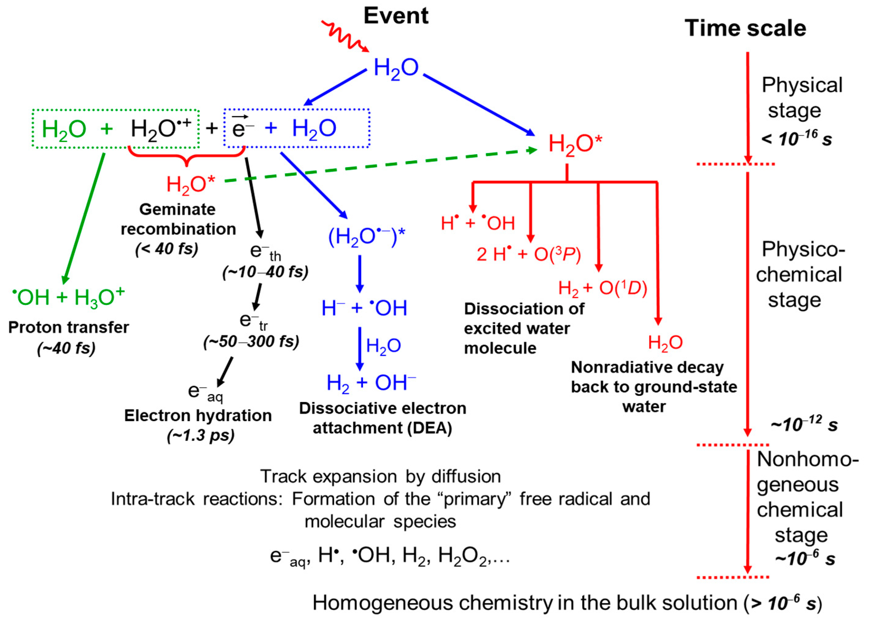
2.1. Radiolysis of Pure, Deaerated Water: Time Scale of Events and Formation of Primary Radical and Molecular Products

In a condensed phase such as liquid water, the radiolysis products (water cations H2O•+, excited water H2O*, and all ejected secondary electrons) generated in the initial or “physical” stage (~1–100 as) of radiation action [45,46] are very energetically/chemically unstable and undergo a complex sequence of fast reactions and reorganization in the second or “physicochemical” stage (≤1 ps), leading to the formation of the very first reactive chemical species which include eaq, H3O+, OH, H, OH, H2, H2O2, O(1D), O(3P), H, O•−, etc. [36,47]. Of these, H3O+, eaq, and OH are produced in the highest concentrations [36]. All these radiolytic species are spatially distributed in a specific, highly nonhomogeneous track structure that strongly depends on the type and energy of the radiation used [24,37,47,48]. In the case of radiations of low LET (such as Compton electrons produced by 60Co γ-rays, fast (e.g., MeV) electrons, or a few hundred MeV protons with typical LET values of ~0.3 keV/μm) and under conditions of lack of dose-rate effects (no interaction between tracks), the track structure is initially formed by small, well-separated Magee-type “spurs” (clusters of radiolytic species that are approximately spherical in shape) along the path of the incident radiation [49,50,51,52]. The third or “chemical” stage (>1 ps) of the radiolysis process consists of diffusion processes (obeying Fick’s laws of diffusion) and chemical reactions of these reactive species as the tracks develop in time. At 25 °C, the track expansion is essentially complete (i.e., when the spurs on expanding by diffusion touch each other) by ~0.2 μs after the first ionization event [53]. At this point in time, the radiation track structure no longer exists and the species that have escaped from spur/track reactions can be regarded as being homogeneously distributed throughout the bulk of the solution (i.e., the system at large). The main reactive species at homogeneity include eaq, H, and OH (the “radical” products) and H2 and H2O2 (the “molecular” products) [24,48,54]. They are often called, though improperly, “primary” species, and their yields—the “primary” or “escape” yields—are given by g(eaq), g(H), g(OH), g(H2), and g(H2O2), which are the numbers of molecules formed per 100 eV of radiation energy absorbed by the system (for conversion into SI units: 1 molecule/100 eV ≈ 0.10364 μmol/J [48]). Note that the hydroperoxyl radical, HO2, or its ionic form O2•−, has also been identified in deaerated water radiolysis, but is normally neglected because it has a very low yield of ~0.02 molecule/100 eV [55], which is less than 1% of the other primary radiolytic species. Finally, beyond a few microseconds, the reactions that occur in the bulk solution can usually be described with conventional homogeneous chemistry methods (see, e.g., [56]). The time scale for the various processes occurring in the low-LET radiolysis of deaerated water is given in Figure 1, which is based on a similar figure from Meesungnoen and Jay-Gerin [47].

2.2. Simulating the Effect of LET with Incident Protons of Different Energies

At low LET, tracks are clearly separated initially into spherical spurs that develop independently as time progresses. The adjacent spurs are so far apart that there is practically no overlapping among reactants. Under these conditions, the predominant effect is radical production. With increasing LET, the isolated spur structure changes to a situation in which the spurs are so close to each other that they eventually merge (due to diffusion of the reactive species) along the radiation path, then forming a dense continuous column (cylindrical in shape) of species [37,47,52,57]. This allows more radicals to be formed in close proximity with a correspondingly greater probability of radical–radical combination/recombination reactions during the track stage of radiolysis. Unlike low-LET radiation, where the radical yield is high, densely ionizing radiation therefore tends to increase the yield of the molecular products (such as H2 and H2O2) and to reform water.
In this study, to simulate on a quantitative basis the effect of LET on the yields of H3O+ ions that form early in water radiolysis via reaction (1), we used incident protons of five different energies as radiation sources, namely, 5, 1.5, 0.7, 0.4, and 0.25 MeV, whose mean LET values at 25 °C are ~8, 19.5, 33, 47, and 60 keV/μm, respectively [58,59,60]. Data for 300 MeV irradiating protons, which mimic the low-LET limit of ~0.3 keV/μm of Compton electrons ejected from the absorption of 60Co γ rays in liquid water [61], are also used here as a reference.

2.3. Modeling the Effect of Dose Rate: The “Instantaneous Pulse” (or Dirac) Model

Herein, a multitrack irradiation model recently formulated in our laboratory was applied to describe the effects of high dose rates on the radiolysis of water by incident protons at ambient [22,62,63,64] and elevated [65] temperatures. In short, this model consists of the random irradiation of water with single pulses of N incident monoenergetic protons, which simultaneously impact the water perpendicularly across the surface of a circle of radius Ro (see Figure 1 of Alanazi et al. [62]). This is the so-called “instantaneous pulse” (or Dirac) model, in which the pulse duration is assumed to be zero [66], i.e., all chemical species are created instantaneously. In this case, the absorbed dose per pulse is the only relevant parameter.
The fact that the fast protons have essentially rectilinear trajectories makes it possible to define a cylindrical geometry of the beam at the entry time, with all proton tracks parallel to the cylinder axis. Since the irradiated cylinder is embedded in nonirradiated bulk water, the various radiolytic species initially formed therein are not confined to this volume, but, rather, diffuse through the entire bulk water (infinite in fact) as time progresses. In this particular irradiation geometry, the incident “fluence” of protons (number of impacting protons per unit area) is simply given by NRo2, so that the effect of the dose rate can be studied simply by varying N, the number of irradiating protons per pulse. In the present study, N is chosen to vary from 1 to 20. Data for N = 1, which indicates the absence of dose-rate effects, serve as a reference. According to our previous calibration of N in terms of dose rate (see Figure 3B of Alanazi et al. [62]), N = 20 corresponds to an absorbed dose rate of about 107 Gy/s. Finally, time zero is chosen as the time when the N incident protons reach the front of the cylinder.

2.4. Monte Carlo Multitrack Chemistry Simulations: The IONLYS-IRT Code

In this work, we use an extended multitrack chemistry version of our Monte Carlo computer code IONLYS-IRT [47] to simulate the radiolysis of pure, deaerated water at 25 °C by incident protons of various energies (i.e., of different LETs) under high-dose-rate conditions. This version has been described in detail elsewhere [22,62,64,65].
In short, we modified our “IONLYS” step-by-step program [67], which covers the early physical and physicochemical stages of radiation action (up to ~1 ps), to simulate the space–time history of N simultaneously incident proton tracks in close spatial proximity [62]. The resulting complex spatial distribution of the reactants of the considered track system then serves directly as the starting point for the subsequent chemical stage (>1 ps), which is covered by our “IRT” program [68]. Using the “independent reaction times” (IRT) method [69,70], this program generates reaction times without having to follow the diffusive trajectories of all the reacting species. Its ability to provide reliable time-dependent chemical yields under a wide range of irradiation conditions has been well validated by comparison with Monte Carlo simulations of full random flights that do follow reactant trajectories in detail [71,72]. It has been shown that simulating N interactive proton trajectories at the same time does not present any particular problem for the implementation of our IRT program at high dose rates. The only drawback is that the computation times are obviously much longer than for the simulation of single-proton irradiation.
For pure, deaerated water at 25 °C, the reaction scheme, rate constants, and the diffusion coefficients of reactive species used in our IRT program are the same as those used previously [34,58,62,68,73,74].
All calculations were performed by simulating short track segments (typically ~1–100 μm, depending on N and LET) of incident monoenergetic protons having LETs in the range 0.3–60 keV/μm. For a given value of N (between 1 and 20) and LET, the number of simulated “histories” (i.e., the number of pulses, usually ~2–150) was chosen so that the average chemical yields could be calculated with acceptable statistical reliability while keeping reasonable computer time limits.

3. Results and Discussion

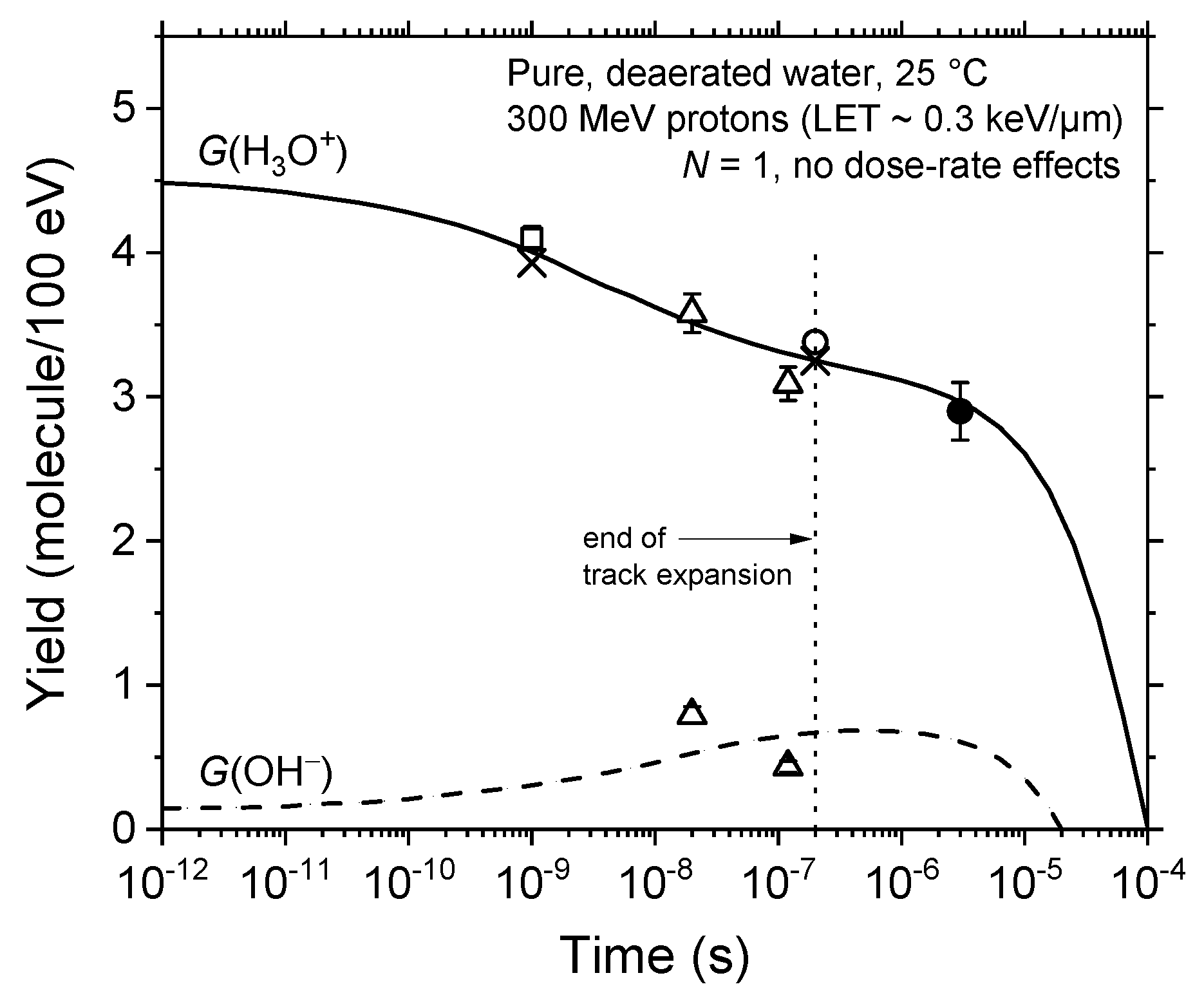
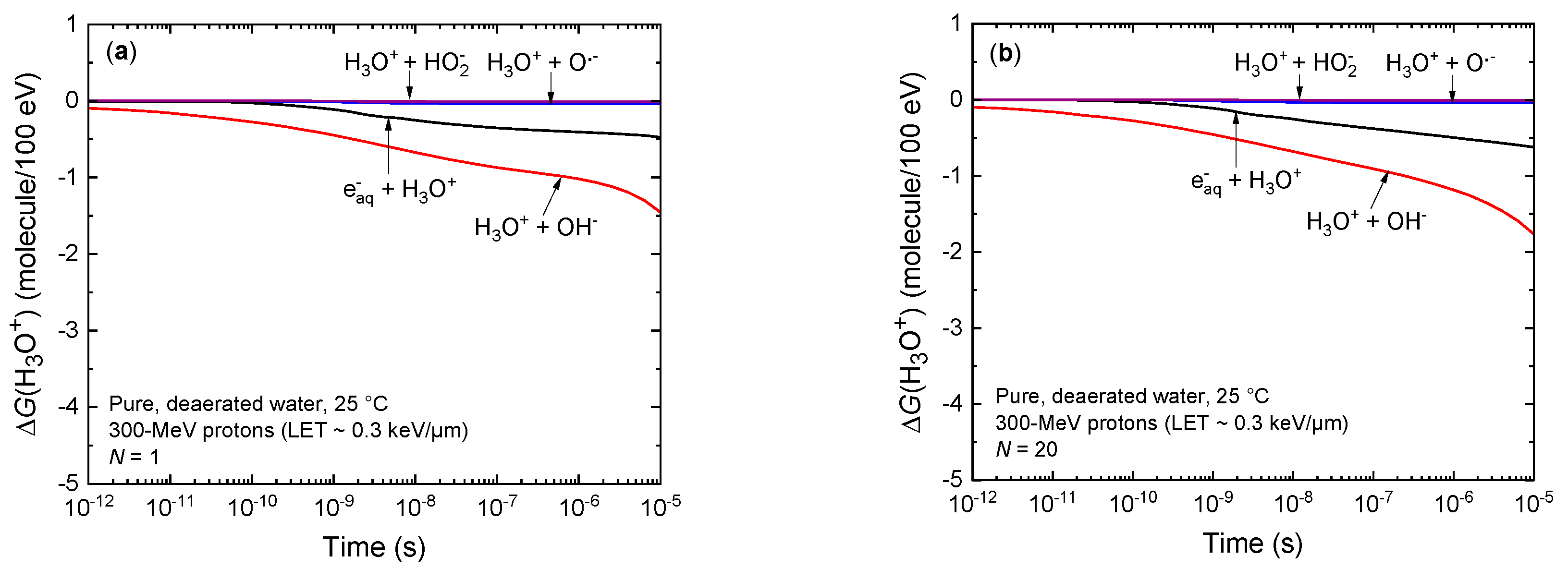
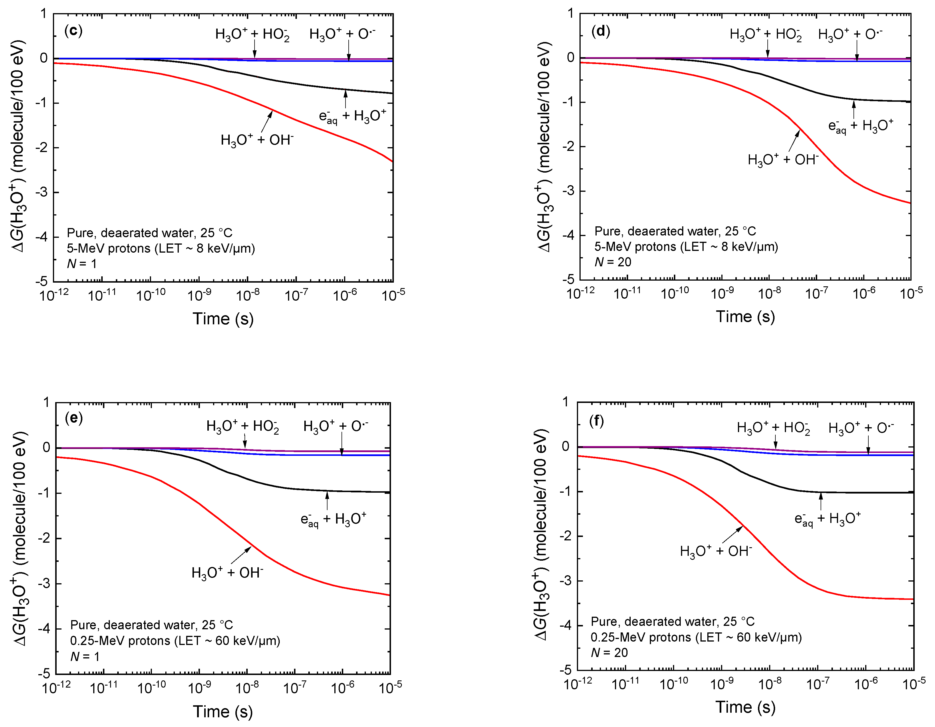
3.1. Time Evolution of the Yields of H3O+ and OH in the Absence of Dose-Rate Effects

Figure 2 shows the temporal variation of the yields of H3O+ and OH as obtained from our track chemistry simulations of the radiolysis of pure, deaerated water by 300 MeV irradiating protons (LET~0.3 keV/μm) at ambient temperature, under conditions of lack of dose-rate effects (i.e., one single-proton irradiation, N = 1). For comparison, available experimental data for 60Co γ-ray or fast electron irradiation [40,75,76,77,78] are also shown in the figure. As can be seen, our simulated values agree very well with the measured H3O+ and OH yields. This good agreement between simulated and measured yield values (without adjustable parameters) supports, a posteriori, the validity of the assumptions used in our computational approach and also demonstrates the robustness of our Monte Carlo code to adequately describe the geometry of the proton track structure and its chemical development.

3.2. Temporal Variation of the Yields of H3O+ and OH under Irradiation Conditions Combining Both High-Dose-Rate and High-LET Radiation

In Figure 3a–f, we compare the effect of dose rate (by varying N, the “number of proton tracks per pulse”, between 1 and 20) on the time evolution of G(H3O+) and G(OH), as obtained from our IONLYS-IRT multitrack chemistry simulations of the radiolysis of pure, deaerated water by the six irradiating protons under consideration, whose mean LET values at 25 °C are ~0.03, 8, 19.5, 33, 47, and 60 keV/μm, in the interval of 1 ps–10 μs. The data obtained for N = 1 (i.e., one single-proton irradiation) are shown for reference.
While the radiolytic formation of H3O+ causes the irradiated volume to become temporarily acidic, the hydroxide ion OH, which is formed largely by the (eaq + OH → OH) reaction (3) during the track stage of the radiolysis, contributes to the generation of an early, transient alkaline response, which may therefore counteract the “acid spike” effect discussed in this work. However, as can be seen in the figures, G(OH) remains much lower than G(H3O+) over the entire time range of interest, regardless of the values of N and the LET considered. As a result, it affects the quantitative features of the pH (see infra) only slightly and can be ignored to a reasonably good approximation.
As expected, the yields of H3O+ (and those of OH) are nearly the same at ~1 ps irrespective of the LET and of the dose rate. With increasing time, G(H3O+) decreases sharply. As can be seen in the figures, this decrease strongly depends on the LET; as the LET increases from ~0.3 to 60 keV/μm, the decrease in G(H3O+) becomes more pronounced as a function of time and begins at shorter times. As discussed previously [36], this decrease is mainly due to H3O+ reacting with OH via reaction (4):
H3O+ + OH → 2H2O (k4 = 1.2 × 10¹¹ M−1 s−1)
and, but to a lesser extent, with eaq, via reaction (2). Other reactions, such as H3O+ + O•−OH + H2O and H3O+ + HO2 → H2O2 + H2O, also contribute to the decay of G(H3O+) with time but only very weakly. This is illustrated in Figure 4a–f, where we compare the time profiles of the extents ΔG(H3O+) of the various components of G(H3O+) calculated from our Monte Carlo multitrack simulations of the radiolysis of deaerated water by 300, 5, and 0.25 MeV incident protons (LET~0.3, 8, and 60 keV/μm, respectively) for N = 1 and 20. As can be seen, the contribution of the (H3O+ + OH → 2H2O) reaction (4) increases strongly with increasing LET and dose rate and becomes largely predominant in the decay of G(H3O+). This reflects the increasing intervention of intratrack (when the LET increases at a fixed N) and intertrack (when N increases at a fixed LET) radical–radical (eaq and OH in the present case) reactions in the solution, as the proximity conditions that would allow these radicals to combine or recombine are favored. This results in the formation of more and more OH in the nonhomogeneous stage of radiolysis, which in turn increases the chance for hydroxide ions to react with H3O+ on the time scale considered in this work. This explains the faster decay kinetics of G(H3O+), which are observed with increasing LET and dose rate (see Figure 3a–f). To our knowledge, there are no experimental data available in the literature with which to compare our results on the time dependence of the yield of H3O+ at high LET.

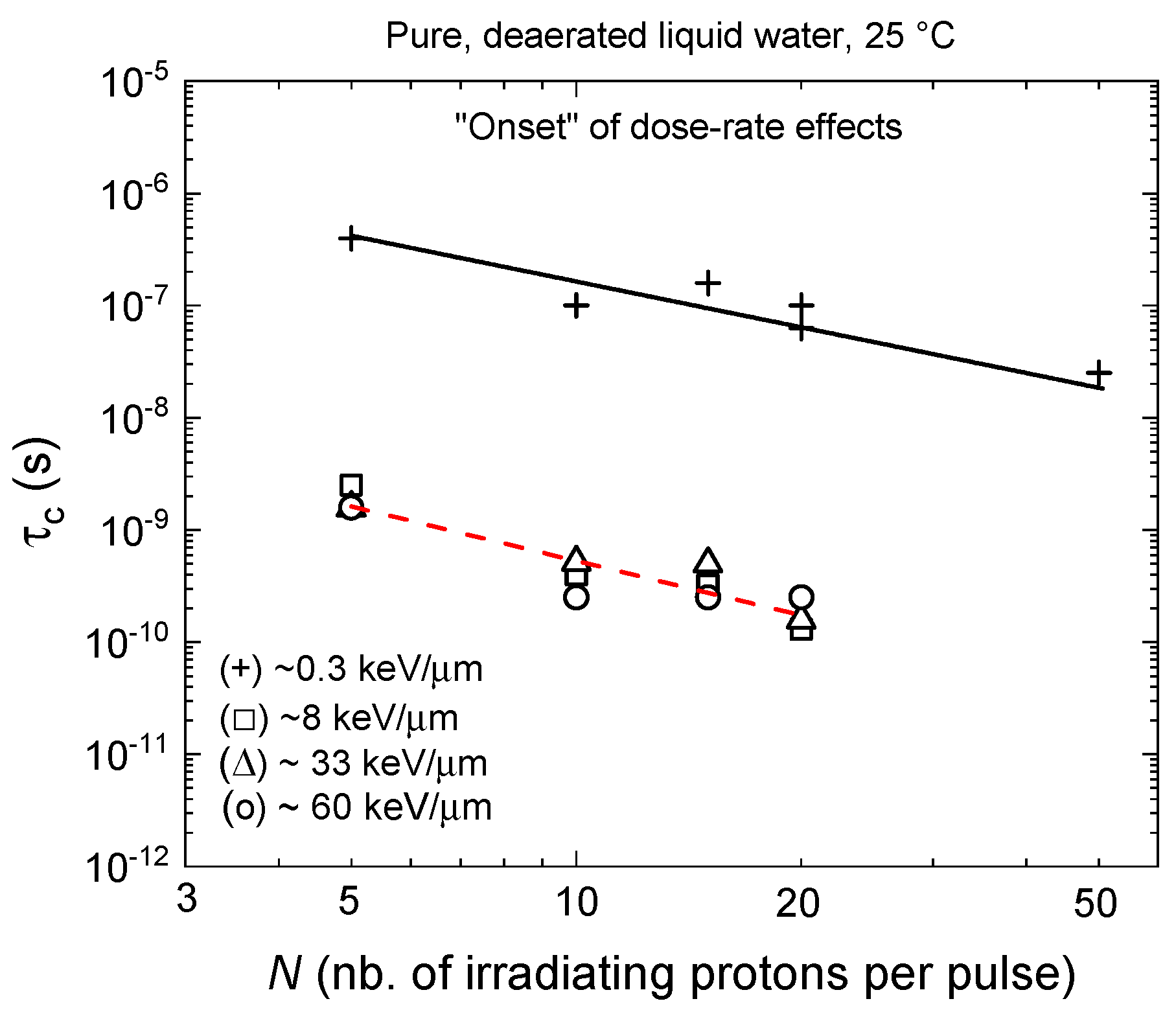
3.3. The Onset of Dose-Rate Effects

As can be seen in Figure 3a–f, for all the LET values considered, the different G(H3O+) curves initially remain unchanged as a function of time regardless of N, until a critical time (τc) is reached, after which the different yield values begin to deviate from their respective one single-proton irradiation (N = 1) reference curves. This critical time, which clearly depends on N, can be regarded as a signature for the “onset” of dose-rate effects in the solution as a whole [62,65,79]. In other words, τc corresponds to the minimum time before interactions between tracks occur. At a given LET, with increasing N, these deviations occur at ever shorter times and with a greater amplitude. To determine τc, we used the same method as that developed in Alanazi et al. [62], consisting of (1) subtracting the G(H3O+) vs. time values that correspond to the simulation of N (=5, 10, 15, and 20 in the present case) interacting proton tracks from those obtained for N = 1 (i.e., under conditions of lack of dose-rate effects), and (2) noting the time when this difference (initially near zero, as the yield curves are all superimposed) begins to increase. Figure 5 compares the variation of our values of τc so determined as a function of N in a log–log plot for different LET values, namely, ~0.3, 8, 33, and 60 keV/μm. As shown in the figure, there is a similar linear relationship between the logarithms of τc and N for the four LET values considered, with the corresponding straight lines being practically all parallel. However, Figure 5 shows that the straight lines obtained for the three high-LET 5, 0.7, and 0.25 MeV irradiating protons (~8, 33, and 60 keV/μm, respectively) nearly coincide (within their plotting accuracy) and are shifted by about two decades (two orders of magnitude) to shorter times compared with that obtained for irradiation with low-LET 300 MeV incident protons (~0.3 keV/μm) [62]. Qualitatively, this result can be explained on physical grounds as it reflects differences in the geometry of the proton track structure: with increasing LET of the radiation, the isolated, well-separated spherical spurs change into dense continuous cylindrical tracks. This favors, at shorter time scales, an increased amount of intervening intratrack reactions. In this high-LET case, the decrease in G(H3O+) is therefore expected to begin at shorter times (as compared to the low-LET case) [36]. Quantitatively, Figure 5 shows that τc remains relatively little dependent on the LET of the irradiating protons in the studied range between ~8 and 60 keV/μm, indicating that the variation of τc with N in this LET range is rather insensitive to the track structure and is mainly controlled by the intertrack interactions (i.e., by the distances between neighboring tracks).
According to the results of Figure 5, the linear relationship that is observed between the logarithms of τc and N (i.e., the dose rate) can be written as
τ c = c o n s t a n t ( N ) n ,
where the constant of proportionality depends on the LET of the radiation, and the 99% confidence interval of the exponent contains the value n ≈ 1 [62,65]. In other words, it can be concluded that, whatever the proton LET values considered in this work, τc varies inversely with the dose rate.

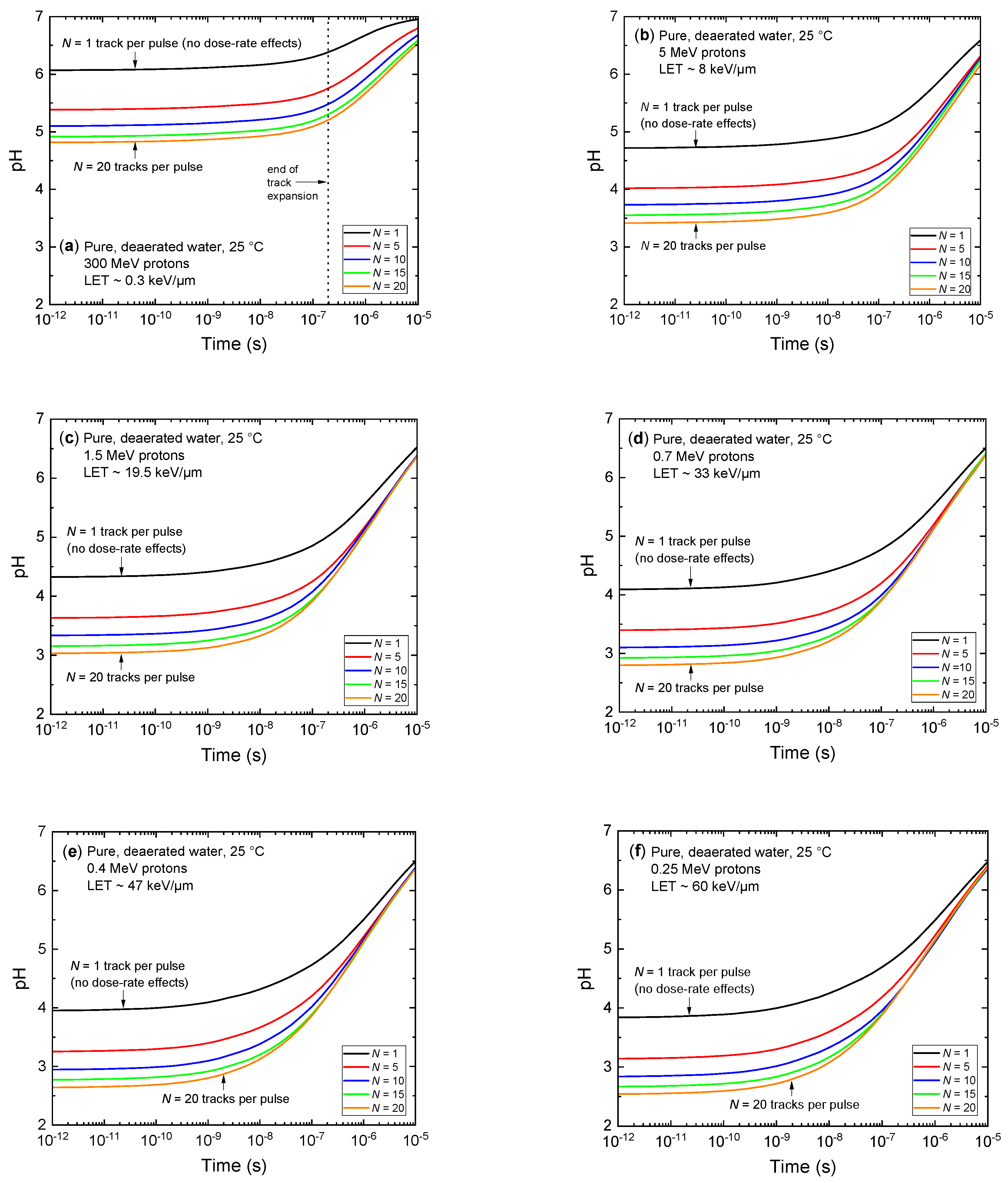
3.4. Ultrafast, Transient Acidic pH Response

In order to calculate the pH values prevailing in the solutions, we now need to estimate the concentrations of the radiolytically generated hydronium ions, [H3O+]radiolytic, as a function of time. This can readily be carried out by using our calculated G(H3O+) values reported in Figure 3a–f with the general relationship [80]
C = ρ D G ,
where C is the concentration, ρ is the density of the solution, D is the radiation dose, and G is the chemical yield. Using our cylindrical, multitrack irradiation model (see Section 2.3) and assuming that the hydronium ions generated by N incident protons are evenly distributed in the considered circular cylinder with a length of 1 μm and initial radius Ro = 0.1 μm (at 1 ps; see Figure 1 of Alanazi et al. [62]), [H3O+]radiolytic (expressed in millimolar, mM) can be derived from [47,62,81,82]
[ H 3 O + ] r a d i o l y t i c ( t ) 5.3 × 10 6 × ( N × L E T R ( t ) 2 ) × G ( H 3 O + ) ( t ) ,
where LET is in keV/μm, G(H3O+) is in molecule per 100 eV, and
R ( t ) 2 R o 2 + 4 D ( H 3 O + ) t
represents the change of Ro (in μm) over time due to the two-dimensional diffusive expansion of the tracks. Here, t is the time (in s) and D (=9.46 × 10⁻⁹ m2 s−1 [73]) is the diffusion coefficient of H3O+ in water.
The total concentration of H3O+ in the solution is thus the sum of [H3O+]radiolytic given by Equations (7) and (8) and of the nonradiolytic, preirradiation concentration [H3O+]autoprotolysis (10−7 M in the present case) due to the autoprotolysis of water:
[ H 3 O + ] total ( t ) = [ H 3 O + ] radiolytic ( t ) + [ H 3 O + ] autoprotolysis .
The pH of the irradiated volume is then simply given by
pH ( t ) = log 10 ( [ H 3 O + ] total ( t ) ) .
Figure 6a–f shows the temporal evolution of the pH values calculated from Equations (7)–(10) for N 300, 5, 1.5, 0.7, 0.4, and 0.25 MeV irradiating protons in deaerated, pure water at 25 °C for N = 1, 5, 10, 15, and 20, using the G(H3O+) values given in Figure 3a–f. As can be seen, for all LET values considered, there is an abrupt transient acid pH effect at times immediately after the initial energy deposition. This “acid spike” effect is found to be most intense at times shorter than, say, ~1–10 ns. In this time range, the pH remains nearly constant, decreasing markedly with the LET of the incident radiation. Moreover, its magnitude and duration also strongly depend on the value of N (i.e., the dose rate). For example, at ~1 ns, the pH for 300 MeV incident protons (LET~0.3 keV/μm) varies from ~6.1 for N = 1 (no dose-rate effects) to ~4.9 for N = 20. For an LET of ~60 keV/μm, the pH at ~1 ns decreases from ~4 for N = 1 to ~2.7 for N = 20. Beyond ~1–10 ns, the pH gradually increases over time in all cases and eventually reaches the constant value of 7 (corresponding to the pH of neutral water at 25 °C), i.e., when the pH of the solution equals log 10 ( [ H 3 O + ] autoprotolysis ) according to Equations (9) and (10).

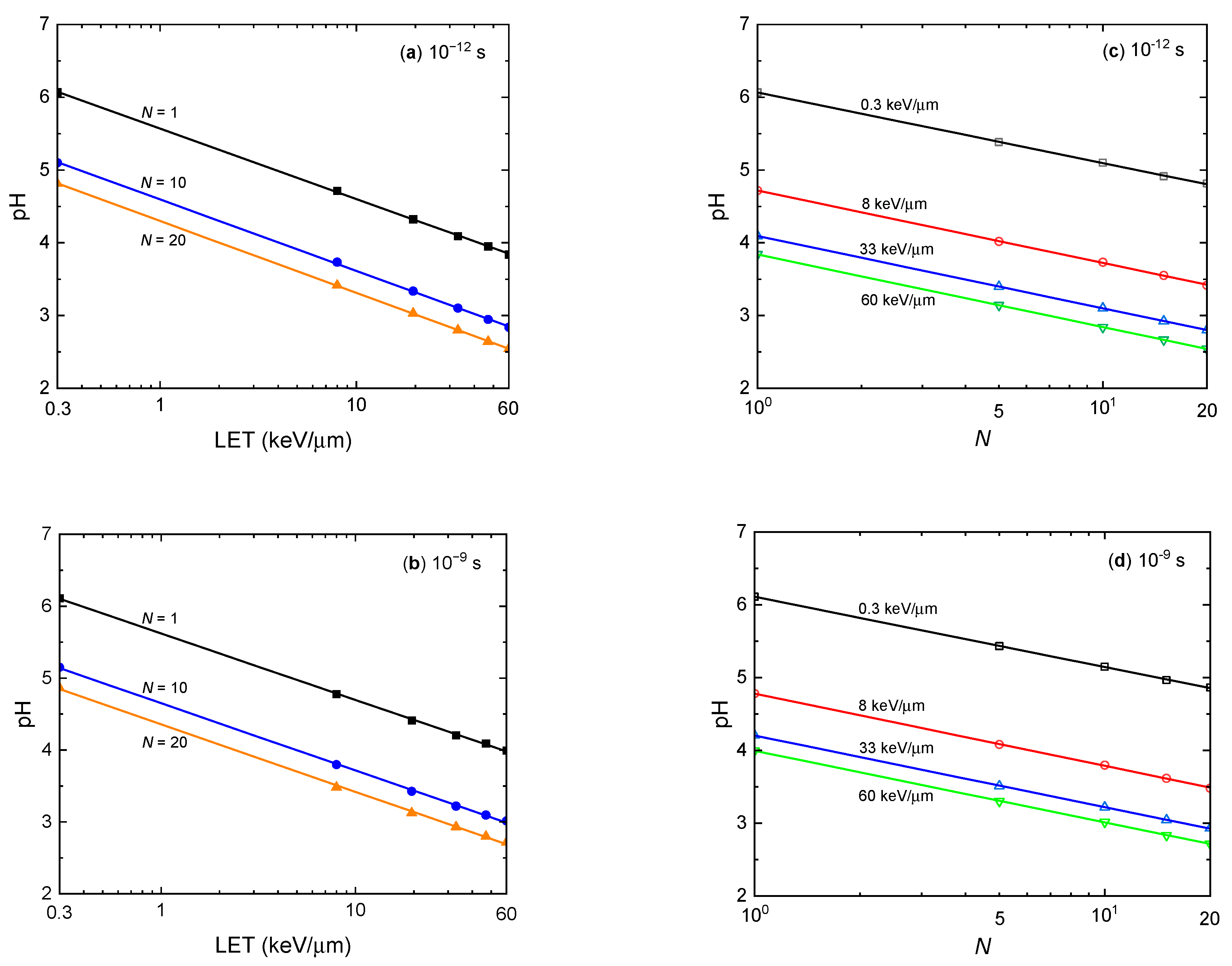
3.5. Correlation between pH, LET, and Dose Rate

Even though their underlying mechanism of action differs, high dose rates and high LETs obviously deserve special consideration as they affect radiolysis yields in a similar way: indeed, the probability of radical–radical reactions is increased in both cases, either in the bulk of the solution through intertrack reactions (for high dose rates) or in the individual radiation tracks through intratrack reactions (for high-LET irradiations) [24,62,79,83]. This is theoretically a consequence of the higher radical densities in the track stage of the radiolysis for densely ionizing radiations and/or under conditions of high dose rates. As seen above for irradiating protons, this similarity in the action of high LETs and high dose rates in the radiolysis of water is also well demonstrated here in the G value for the formation of H3O+ at early time (see Figure 3a–f) and in the acid pHs that result therefrom (see Figure 6a–f).
For the sake of illustration, Figure 7 shows semi-log plots of the pH against LET for various dose rates (N = 1, 10, and 20) (panels (a) and (b)) and against N for various LETs (~0.3, 8, 33, and 60 keV/μm) (panels (c) and (d)) at two different times (1 ps and 1 ns) after the initial energy deposition. As can be seen, a linear correlation is found in both cases, which can be expressed by the following relationships:
pH = A B ln ( LET )   where   A   is   a   function   of   N
and
pH = E F ln ( N )   where   E   is   a   function   of   LET .
It should be noted that the parameters B in Equation (11) and F in Equation (12), which are, respectively, the slopes of the pH vs. ln(LET) and pH vs. ln(N) straight lines in Figure 7, do not depend on N (for B) or on the LET (for F) since these lines are all parallel (i.e., they have the same slopes whatever the values of N or of the LET). Indeed, values of these slopes, obtained from a least-squares fit of the pH vs. ln(LET) and pH vs. ln(N) data and averaged over the two considered times, are B = 0.41 ± 0.02 (A varying from ~5.6 for N = 1 to ~4.3 for N = 20) and F = 0.42 ± 0.01 (E varying from ~6.1 for LET~0.3 keV/μm to ~3.9 for LET~60 keV/μm).

4. Conclusions

In this work, we extended our Monte Carlo track chemistry simulations to study the effect of combining the action of both high absorbed dose rate and high-LET irradiations on the transient yields and concentrations of H3O+ ions and the resulting acid pH spikes that develop at early times during the radiolysis of pure, deaerated water. Using incident protons of varying energies in the range of 300–0.25 MeV as radiation sources to simulate the effect of LET and our previously formulated cylindrical, “instantaneous pulse” (or Dirac) irradiation model to investigate the effect of dose rate, a major finding was that the combination of high dose rates and high LETs is clearly additive, with a very significant impact on the pH of the irradiated solution.
Indeed, for all LET values considered, we observe an abrupt, transient, highly acidic pH response immediately after the initial energy deposition that persists for periods of more than five orders magnitude across the entire solution. The magnitude of this response not only depends on the LET of the radiation but also on the value of its dose rate. For example, at ~1 ns, the pH for an LET of ~0.3 keV/μm (300 MeV incident protons) varies from ~6.1 in the absence of dose-rate effects to ~4.9 for an instantaneous dose rate of ~107 Gy/s. On the other hand, for an LET of ~60 keV/μm (0.25 MeV incident protons), the pH at ~1 ns decreases from ~4 for one single irradiation track to ~2.7 for a dose rate of ~107 Gy/s. At longer times, the pH gradually increases for all LET and dose-rate values, eventually reaching the constant value of 7, which corresponds to the nonradiolytic, preirradiation concentration of hydronium ions, due to the autoprotolysis of water.
This work confirms previous experimental and theoretical studies on the similarity of the action of high dose rates and high LETs in the track stage of the radiolysis. Such a similarity, which manifests itself in the various radiolysis yields, including those of the formation of H3O+, finds its origin in an increase in the radical densities. Even if their underlying mechanism of action differs, the probability of radical–radical combination or recombination reactions is increased, either in the bulk of the solution through intertrack reactions (case of high dose rates) or in the individual radiation tracks through intratrack reactions (case of high LETs), with the consequence of an increase in molecular yields and a simultaneous reduction in the radical yields.
Using the space–time history of the tracks, we were able to determine the critical time τc corresponding to the “onset” of dose-rate effects in the solution. Interestingly, we demonstrated that the variation of τc with the dose rate is rather insensitive to the track structure (i.e., to the LET of the radiation), but is mainly controlled by the intertrack interactions or, in other words, by the distances between neighboring tracks. Under the conditions of our simulation model, we found that this onset of dose-rate effects is nearly inversely proportional to the dose rate.
To the best of our knowledge, these early acidic pH responses have largely gone unnoticed in water or in a cellular environment exposed to ionizing radiation. In view of the importance of the pH for many cellular functions (e.g., it is well known that the acidity of the intracellular water strongly influences most of the enzyme functions present in the cells), the present findings raise the question of whether the generation of such spikes of acidity in cell water under high-LET and high-dose-rate irradiation conditions plays a role in relation to the “FLASH effect” in FLASH radio-, proton-, and hadron-therapy. This question is all the more relevant when we consider that the diffusion of H3O+ in cell water is ~100 to 1000 times lower than in free water [84,85]. In this case, this decrease in mobility of hydronium ions in the water of the cell should then lead to a marked increase in the period of time during which the highly acidic pH spikes are observed, further underlining the potential importance and role of pH in the FLASH effect.
Although this study is based on a pure radiation chemistry perspective, we hope that the current findings involving high-LET radiation delivered at high dose rates may provide some useful information for a better understanding of the early physicochemical steps in the chain of radiobiological events that underlie the FLASH effect.
Currently, we are continuing our present modeling work by trying to progress towards the study of a cellular medium, including its many various components. In particular, we are working to introduce the effect of the viscosity of intracellular water on the radiolysis and its consequences on the observed acidity spikes. With regard to the effect of acidity itself, we also wish to introduce the effect of quenching of the H3O+ ions produced by radiolysis by various buffers (e.g., phosphate buffer and others). All these works are in progress.

Author Contributions

Conceptualization: J.-P.J.-G., J.M. and M.I.B.; M.I.B. performed all the simulations, analyzed the results, and wrote the original draft of the manuscript based on discussions with J.M. and J.-P.J.-G.; J.M. developed the software and implemented the model; J.M. and J.-P.J.-G. supervised the work; J.-P.J.-G. and M.I.B. reviewed and edited the final version of the manuscript; project administration and funding acquisition: J.-P.J.-G. All authors have read and agreed to the published version of the manuscript.

Funding

This research was funded by the Natural Sciences and Engineering Research Council of Canada (NSERC), grant number RGPIN-2022-03972.

Institutional Review Board Statement

Not applicable.

Informed Consent Statement

Not applicable.

Data Availability Statement

Data generated or analyzed during this study are provided in full within the article.

Acknowledgments

M.I.B. is the recipient of an Abdenour-Nabid, MD graduate scholarship from the “Programme de bourses d’excellence aux études supérieures” of the Université de Sherbrooke.

Conflicts of Interest

The authors declare no conflict of interest.

References

  1. von Sonntag, C. Free-Radical-Induced DNA Damage and Its Repair. A Chemical Perspective; Springer: Berlin/Heidelberg, Germany, 2006. [Google Scholar]
  2. Becker, D.; Adhikary, A.; Sevilla, M.D. Physicochemical mechanisms of radiation-induced DNA damage. In Charged Particle and Photon Interactions with Matter. Recent Advances, Applications, and Interfaces; Hatano, Y., Katsumura, Y., Mozumder, A., Eds.; CRC Press (Taylor & Francis Group): Boca Raton, FL, USA, 2011; Chapter 19; pp. 503–541. [Google Scholar]
  3. Cadet, J.; Davies, K.J.A.; Medeiros, M.H.; Di Mascio, P.; Wagner, J.R. Formation and repair of oxidatively generated damage in cellular DNA. Free Radic. Biol. Med. 2017, 107, 13–34. [Google Scholar] [CrossRef] [PubMed]
  4. O’Neill, P. Radiation-induced damage in DNA. In Radiation Chemistry: Present Status and Future Trends; Jonah, C.D., Rao, B.S.M., Eds.; Elsevier: Amsterdam, The Netherlands, 2001; Chapter 21; pp. 585–622. [Google Scholar]
  5. Azzam, E.I.; Jay-Gerin, J.-P.; Pain, D. Ionizing radiation-induced metabolic oxidative stress and prolonged cell injury. Cancer Lett. 2012, 327, 48–60. [Google Scholar] [CrossRef] [PubMed]
  6. Priyadarsini, K.I. Redox reactions of antioxidants: Contributions from radiation chemistry of aqueous solutions. In Charged Particle and Photon Interactions with Matter. Recent Advances, Applications, and Interfaces; Hatano, Y., Katsumura, Y., Mozumder, A., Eds.; CRC Press (Taylor & Francis Group): Boca Raton, FL, USA, 2011; Chapter 22; pp. 595–622. [Google Scholar]
  7. Hall, E.J.; Giaccia, A.J. Radiobiology for the Radiologist, 8th ed.; Wolters Kluwer: Philadelphia, PA, USA, 2019. [Google Scholar]
  8. Penabeï, S.; Meesungnoen, J.; Jay-Gerin, J.-P. Assessment of cystamine’s radioprotective/antioxidant ability under high-dose-rate irradiation: A Monte Carlo multi-track chemistry simulation study. Antioxidants 2023, 12, 776. [Google Scholar] [CrossRef]
  9. Favaudon, V.; Caplier, L.; Monceau, V.; Pouzoulet, F.; Sayarath, M.; Fouillade, C.; Poupon, M.-F.; Brito, I.; Hupé, P.; Bourhis, J.; et al. Ultrahigh dose-rate FLASH irradiation increases the differential response between normal and tumor tissue in mice. Sci. Transl. Med. 2014, 6, 245ra93. [Google Scholar] [CrossRef]
  10. Favaudon, V.; Fouillade, C.; Vozenin, M.-C. La radiothérapie FLASH pour épargner les tissus sains. Médecine/Sciences 2015, 31, 121–123. [Google Scholar] [CrossRef] [PubMed]
  11. Wilson, J.D.; Hammond, E.M.; Higgins, G.S.; Petersson, K. Ultra-high dose rate (FLASH) radiotherapy: Silver bullet or fool’s gold? Front. Oncol. 2020, 9, 1563. [Google Scholar] [CrossRef]
  12. Esplen, N.; Mendonca, M.S.; Bazalova-Carter, M. Physics and biology of ultrahigh dose-rate (FLASH) radiotherapy: A topical review. Phys. Med. Biol. 2020, 65, 23TR03. [Google Scholar] [CrossRef]
  13. Wardman, P. Radiotherapy using high-intensity pulsed radiation beams (FLASH): A radiation-chemical perspective. Radiat. Res. 2020, 194, 607–617. [Google Scholar] [CrossRef]
  14. Marcu, L.G.; Bezak, E.; Peukert, D.D.; Wilson, P. Translational research in FLASH radiotherapy—From radiobiological mechanisms to in vivo results. Biomedicines 2021, 9, 181. [Google Scholar] [CrossRef]
  15. Vozenin, M.-C.; Bourhis, J.; Durante, M. Towards clinical translation of FLASH radiotherapy. Nature Rev. Clin. Oncol. 2022, 19, 791–803. [Google Scholar] [CrossRef]
  16. Gao, Y.; Liu, R.; Chang, C.-W.; Charyyev, S.; Zhou, J.; Bradley, J.D.; Liu, T.; Yang, X. A potential revolution in cancer treatment: A topical review of FLASH radiotherapy. J. Appl. Clin. Med. Phys. 2022, 23, e13790. [Google Scholar] [CrossRef] [PubMed]
  17. Kacem, H.; Almeida, A.; Cherbuin, N.; Vozenin, M.-C. Understanding the FLASH effect to unravel the potential of ultra-high dose rate irradiation. Int. J. Radiat. Biol. 2022, 98, 506–516. [Google Scholar] [CrossRef]
  18. Schüler, E.; Acharya, M.; Montay-Gruel, P.; Loo, B.W., Jr.; Vozenin, M.-C.; Maxim, P.G. Ultra-high dose rate electron beams and the FLASH effect: From preclinical evidence to a new radiotherapy paradigm. Med. Phys. 2022, 49, 2082–2095. [Google Scholar] [CrossRef]
  19. Friedl, A.A.; Prise, K.M.; Butterworth, K.T.; Montay-Gruel, P.; Favaudon, V. Radiobiology of the FLASH effect. Med. Phys. 2022, 49, 1993–2013. [Google Scholar] [CrossRef] [PubMed]
  20. Hageman, E.; Che, P.-P.; Dahele, M.; Slotman, B.J.; Sminia, P. Radiobiological aspects of FLASH radiotherapy. Biomolecules 2022, 12, 1376. [Google Scholar] [CrossRef]
  21. Jay-Gerin, J.-P. Ultra-high dose-rate (FLASH) radiotherapy: Generation of early, transient, strongly acidic spikes in the irradiated tumor environment. Cancer Radiother. 2020, 24, 332–334. [Google Scholar] [CrossRef] [PubMed]
  22. Sultana, A.; Alanazi, A.; Meesungnoen, J.; Jay-Gerin, J.-P. Generation of ultrafast, transient, highly acidic pH spikes in the radiolysis of water at very high dose rates: Relevance for FLASH radiotherapy. Can. J. Chem. 2022, 100, 272–279. [Google Scholar] [CrossRef]
  23. Loh, Z.-H.; Doumy, G.; Arnold, C.; Kjellsson, L.; Southworth, S.H.; Al Haddad, A.; Kumagai, Y.; Tu, M.-F.; Ho, P.J.; March, A.M.; et al. Observation of the fastest chemical processes in the radiolysis of water. Science 2020, 367, 179–182. [Google Scholar] [CrossRef]
  24. Spinks, J.W.T.; Woods, R.J. An Introduction to Radiation Chemistry, 3rd ed.; Wiley: New York, NY, USA, 1990. [Google Scholar]
  25. Garrett, B.C.; Dixon, D.A.; Camaioni, D.M.; Chipman, D.M.; Johnson, M.A.; Jonah, C.D.; Kimmel, G.A.; Miller, J.H.; Rescigno, T.N.; Rossky, P.J.; et al. Role of water in electron-initiated processes and radical chemistry: Issues and scientific advances. Chem. Rev. 2005, 105, 355–390. [Google Scholar] [CrossRef]
  26. Cobut, V.; Frongillo, Y.; Jay-Gerin, J.-P.; Patau, J.P. A Monte Carlo calculation of subexcitation and vibrationally-relaxing electron spectra in irradiated liquid water. Radiat. Phys. Chem. 1992, 40, 589–591. [Google Scholar] [CrossRef]
  27. LaVerne, J.A.; Pimblott, S.M. Electron energy-loss distributions in solid, dry DNA. Radiat. Res. 1995, 141, 208–215. [Google Scholar] [CrossRef]
  28. Meesungnoen, J.; Jay-Gerin, J.-P.; Filali-Mouhim, A.; Mankhetkorn, S. Low-energy electron penetration range in liquid water. Radiat. Res. 2002, 158, 657–660. [Google Scholar] [CrossRef] [PubMed]
  29. LaForge, A.C.; Michiels, R.; Bohlen, M.; Callegari, C.; Clark, A.; von Conta, A.; Coreno, M.; Di Fraia, M.; Drabbels, M.; Huppert, M.; et al. Real-time dynamics of the formation of hydrated electrons upon irradiation of water clusters with extreme ultraviolet light. Phys. Rev. Lett. 2019, 122, 133001. [Google Scholar] [CrossRef]
  30. Low, P.J.; Chu, W.; Nie, Z.; Mohd Yusof, M.S.B.; Prezhdo, O.V.; Loh, Z.-H. Observation of a transient intermediate in the ultrafast relaxation dynamics of the excess electron in strong-field-ionized liquid water. Nature Commun. 2022, 13, 7300. [Google Scholar] [CrossRef] [PubMed]
  31. Lea, D.E. Actions of Radiations on Living Cells; Cambridge University Press: Cambridge, UK, 1946; Chapter 2. [Google Scholar]
  32. Lea, D.E. The action of radiations on dilute aqueous solutions: The spatial distribution of H and OH. Br. J. Radiol. 1947, 1, 59–64. [Google Scholar]
  33. Morrison, P. Radiation in living matter: The physical processes. In Symposium on Radiobiology. The Basic Aspects of Radiation Effects on Living Systems; Nickson, J.J., Ed.; Wiley: New York, NY, USA, 1952; pp. 1–12. [Google Scholar]
  34. Elliot, A.J.; Bartels, D.M. The Reaction Set, Rate Constants and g-Values for the Simulation of the Radiolysis of Light Water over the Range 20 to 350 °C Based on Information Available in 2008; Report No. 153-127160-450-001; Atomic Energy of Canada Limited: Mississauga, ON, Canada, 2009. [Google Scholar]
  35. Kanike, V.; Meesungnoen, J.; Jay-Gerin, J.-P. Transient acid pH effect in tracks in the radiolysis of water: Does this effect contribute to biological damage caused by ionizing radiation? Austin J. Nucl. Med. Radiother. 2015, 2, 1011. [Google Scholar]
  36. Kanike, V.; Meesungnoen, J.; Jay-Gerin, J.-P. Acid spike effect in spurs/tracks of the low/high linear energy transfer radiolysis of water: Potential implications for radiobiology. RSC Adv. 2015, 5, 43361–43370. [Google Scholar] [CrossRef]
  37. LaVerne, J.A. Track effects of heavy ions in liquid water. Radiat. Res. 2000, 153, 487–496. [Google Scholar] [CrossRef]
  38. Smith, D.R.; Stevens, W.H. Radiation-induced hydrolysis of acetal: Evidence for the reaction of H3O+ ions in spurs in the radiolysis of water. Nature 1963, 200, 66–67. [Google Scholar] [CrossRef]
  39. Anbar, M.; Thomas, J.K. Pulse radiolysis studies of aqueous sodium chloride solutions. J. Phys. Chem. 1964, 68, 3829–3835. [Google Scholar] [CrossRef]
  40. Pikaev, A.K.; Kabakchi, S.A.; Zansokhova, A.A. Yields and reactions of hydrogen ions on radiolysis of water and aqueous solutions. Faraday Discuss. Chem. Soc. 1977, 63, 112–123. [Google Scholar] [CrossRef]
  41. Byakov, V.M.; Stepanov, S.V. The mechanism for the primary biological effects of ionizing radiation. Phys. Usp. 2006, 49, 469–487. [Google Scholar] [CrossRef]
  42. Kanike, V. “Acid-Spike” Effect in Spurs/Tracks of the Low/High Linear Energy Transfer Radiolysis of Water: Potential Implications for Radiobiology and Nuclear Industry. Master’s Thesis, Université de Sherbrooke, Sherbrooke, QC, Canada, 2016. Available online: https://savoirs.usherbrooke.ca/handle/11143/9711 (accessed on 17 November 2016).
  43. Islam, M.M.; Kanike, V.; Meesungnoen, J.; Lertnaisat, P.; Katsumura, Y.; Jay-Gerin, J.-P. In Situ generation of ultrafast transient “acid spikes” in the 10B(n,α)7Li radiolysis of water. Chem. Phys. Lett. 2018, 693, 210–215. [Google Scholar] [CrossRef]
  44. Schneider, N.M.; Norton, M.M.; Mendel, B.J.; Grogan, J.M.; Ross, F.M.; Bau, H.H. Electron-water interactions and implications for liquid cell electron microscopy. J. Phys. Chem. C 2014, 118, 22373–22382. [Google Scholar] [CrossRef]
  45. Platzman, R.L. The physical and chemical basis of mechanisms in radiation biology. In Radiation Biology and Medicine. Selected Reviews in the Life Sciences; Claus, W.D., Ed.; Addison-Wesley: Reading, MA, USA, 1958; pp. 15–72. [Google Scholar]
  46. Kuppermann, A. Theoretical foundations of radiation chemistry. J. Chem. Educ. 1959, 36, 279–285. [Google Scholar] [CrossRef]
  47. Meesungnoen, J.; Jay-Gerin, J.-P. Radiation chemistry of liquid water with heavy ions: Monte Carlo simulation studies. In Charged Particle and Photon Interactions with Matter. Recent Advances, Applications, and Interfaces; Hatano, Y., Katsumura, Y., Mozumder, A., Eds.; CRC Press (Taylor & Francis Group): Boca Raton, FL, USA, 2011; Chapter 14; pp. 355–400. [Google Scholar]
  48. Ferradini, C.; Jay-Gerin, J.-P. Radiolysis of water and aqueous solutions: History and present state of the science. Can. J. Chem. 1999, 77, 1542–1575. (In French) [Google Scholar] [CrossRef]
  49. Magee, J.L. Radiation chemistry. Annu. Rev. Nucl. Sci. 1953, 3, 171–192. [Google Scholar] [CrossRef]
  50. Freeman, G.R. Basics of radiation chemistry. In The Study of Fast Processes and Transient Species by Electron Pulse Radiolysis; Baxendale, J.H., Busi, F., Eds.; Reidel Publishing: Dordrecht, The Netherlands, 1982; pp. 19–34. [Google Scholar]
  51. Plante, I.L.; Filali-Mouhim, A.; Jay-Gerin, J.-P. SIMULRAD: A JAVA interface for a Monte Carlo simulation code to visualize in 3D the early stages of water radiolysis. Radiat. Phys. Chem. 2005, 72, 173–180. [Google Scholar] [CrossRef]
  52. Mozumder, A. Fundamentals of Radiation Chemistry; Academic Press: San Diego, CA, USA, 1999. [Google Scholar]
  53. Sanguanmith, S.; Meesungnoen, J.; Muroya, Y.; Lin, M.; Katsumura, Y.; Jay-Gerin, J.-P. On the spur lifetime and its temperature dependence in the low linear energy transfer radiolysis of water. Phys. Chem. Chem. Phys. 2012, 14, 16731–16736. [Google Scholar] [CrossRef]
  54. Buxton, G.V. Radiation chemistry of the liquid state: (1) Water and homogeneous aqueous solutions. In Radiation Chemistry. Principles and Applications; Farhataziz, Rodgers, M.A.J., Eds.; VCH Publishers: New York, NY, USA, 1987; pp. 321–349. [Google Scholar]
  55. Bjergbakke, E.; Hart, E.J. Oxygen formation in the γ-ray irradiation of Fe2+-Cu2+ solutions. Radiat. Res. 1971, 45, 261–273. [Google Scholar] [CrossRef]
  56. Pastina, B.; LaVerne, J.A. Effect of molecular hydrogen on hydrogen peroxide in water radiolysis. J. Phys. Chem. A 2001, 105, 9316–9322. [Google Scholar] [CrossRef]
  57. Ferradini, C. Heterogeneous aspect of radiolytic phenomena (Article in French). In Actions Biologique et Chimique des Radiations Ionisantes; Tilquin, B., Ed.; Éditions Ciaco: Brussels, Belgium, 1990; Volume 1, pp. 52–63. [Google Scholar]
  58. Alanazi, A.; Meesungnoen, J.; Jay-Gerin, J.-P. Linear energy transfer dependence of transient yields in water irradiated by 150 keV–500 MeV protons in the limit of low dose rates. Can. J. Chem. 2020, 98, 427–433. [Google Scholar] [CrossRef]
  59. Watt, D.E. Quantities for Dosimetry of Ionizing Radiations in Liquid Water; Taylor & Francis: London, UK, 1996. [Google Scholar]
  60. Stopping Powers and Ranges for Protons and Alpha Particles; ICRU Report No. 49; International Commission on Radiation Units and Measurements: Bethesda, DC, USA, 1993.
  61. Mustaree, S.; Meesungnoen, J.; Butarbutar, S.L.; Causey, P.; Stuart, C.R.; Jay-Gerin, J.-P. Self-radiolysis of tritiated water. 3. The OH scavenging effect of bromide ions on the yield of H2O2 in the radiolysis of water by 60Co γ-rays and tritium β-particles at room temperature. RSC Adv. 2014, 4, 43572–43581. [Google Scholar] [CrossRef]
  62. Alanazi, A.; Meesungnoen, J.; Jay-Gerin, J.-P. A computer modeling study of water radiolysis at high dose rates. Relevance to FLASH radiotherapy. Radiat. Res. 2021, 195, 149–162. [Google Scholar] [CrossRef]
  63. Sultana, A.; Alanazi, A.; Meesungnoen, J.; Jay-Gerin, J.-P. On the transient radiolytic oxygen depletion in the ultra-high (FLASH) dose-rate radiolysis of water in a cell-like environment: Effect of eaq and OH competing scavengers. Radiat. Res. 2022, 197, 566–567. [Google Scholar] [CrossRef]
  64. Alanazi, A.; Sultana, A.; Meesungnoen, J.; Jay-Gerin, J.-P. The effect of linear energy transfer on the early, transient radiolytic oxygen depletion in the radiolysis of water by high-dose-rate irradiating protons. Can. J. Chem. 2023, 101, 70–80. [Google Scholar] [CrossRef]
  65. Sultana, A.; Meesungnoen, J.; Jay-Gerin, J.-P. High-dose-rate effects in the radiolysis of water at elevated temperatures. Can. J. Chem. 2021, 99, 594–602. [Google Scholar] [CrossRef]
  66. The Dosimetry of Pulsed Radiation; ICRU Report No. 34; International Commission on Radiation Units and Measurements: Bethesda, MD, USA, 1982.
  67. Cobut, V.; Frongillo, Y.; Patau, J.P.; Goulet, T.; Fraser, M.-J.; Jay-Gerin, J.-P. Monte Carlo simulation of fast electron and proton tracks in liquid water—I. Physical and physicochemical aspects. Radiat. Phys. Chem. 1998, 51, 229–243. [Google Scholar]
  68. Frongillo, Y.; Goulet, T.; Fraser, M.-J.; Cobut, V.; Patau, J.P.; Jay-Gerin, J.-P. Monte Carlo simulation of fast electron and proton tracks in liquid water—II. Nonhomogeneous chemistry. Radiat. Phys. Chem. 1998, 51, 245–254. [Google Scholar]
  69. Tachiya, M. Theory of diffusion-controlled reactions: Formulation of the bulk reaction rate in terms of the pair probability. Radiat. Phys. Chem. 1983, 21, 167–175. [Google Scholar] [CrossRef]
  70. Pimblott, S.M.; Pilling, M.J.; Green, N.J.B. Stochastic models of spur kinetics in water. Radiat. Phys. Chem. 1991, 37, 377–388. [Google Scholar] [CrossRef]
  71. Goulet, T.; Fraser, M.-J.; Frongillo, Y.; Jay-Gerin, J.-P. On the validity of the independent reaction times approximation for the description of the nonhomogeneous kinetics of liquid water radiolysis. Radiat. Phys. Chem. 1998, 51, 85–91. [Google Scholar] [CrossRef]
  72. Plante, I. Développement de Codes de Simulation Monte Carlo de la Radiolyse de l’Eau par des Électrons, Ions Lourds, Photons et Neutrons. Applications à Divers Sujets d’Intérêt Expérimental. Ph.D. Thesis, Université de Sherbrooke, Sherbrooke, QC, Canada, 2009. [Google Scholar]
  73. Tippayamontri, T.; Sunuchakan, S.; Meesungnoen, J.; Sunaryo, G.R.; Jay-Gerin, J.-P. Fast neutron radiolysis of the ferrous sulfate (Fricke) dosimeter: Monte Carlo simulations. In Recent Research Developments in Physical Chemistry; Pandalai, S.G., Ed.; Transworld Research Network: Trivandrum, Kerala, India, 2009; Volume 10, pp. 143–211. [Google Scholar]
  74. Mirsaleh Kohan, L.; Sanguanmith, S.; Meesungnoen, J.; Causey, P.; Stuart, C.R.; Jay-Gerin, J.-P. Self-radiolysis of tritiated water. 1. A comparison of the effects of 60Co γ-rays and tritium β-particles on water and aqueous solutions at room temperature. RSC Adv. 2013, 3, 19282–19299. [Google Scholar] [CrossRef]
  75. Čerček, B.; Kongshaug, M. Hydrogen ion yields in the radiolysis of neutral aqueous solutions. J. Phys. Chem. 1969, 73, 2056–2058. [Google Scholar] [CrossRef] [PubMed]
  76. Barker, G.C.; Fowles, P.; Sammon, D.C.; Stringer, B. Pulse radiolytic induced transient electrical conductance in liquid solutions. Part 1. Technique and the radiolysis of water. Trans. Faraday Soc. 1970, 66, 1498–1508. [Google Scholar] [CrossRef]
  77. Anderson, R.F.; Vojnovic, B.; Michael, B.D. The radiation-chemical yields of H3O+ and OH as determined by nanosecond conductimetric measurements. Radiat. Phys. Chem. 1985, 26, 301–303. [Google Scholar] [CrossRef]
  78. Schmidt, K.H.; Ander, S.M. Formation and recombination of H3O+ and hydroxide in irradiated water. J. Phys. Chem. 1969, 73, 2846–2852. [Google Scholar] [CrossRef]
  79. Kuppermann, A. Diffusion kinetics in radiation chemistry. In Actions Chimiques et Biologiques des Radiations; Haïssinsky, M., Ed.; Masson: Paris, France, 1961; Volume 5, pp. 85–166. [Google Scholar]
  80. Hummel, A. Radiation Chemistry: The Chemical Effects of Ionizing Radiation and their Applications; Interfaculty Reactor Institute-Technische Universiteit Delft (IRI-FUT): Delft, The Netherlands, 1995. [Google Scholar]
  81. Schardt, D.; Elsässer, T.; Schulz-Ertner, D. Heavy-ion tumor therapy: Physical and radiobiological benefits. Rev. Mod. Phys. 2010, 82, 383–425. [Google Scholar] [CrossRef]
  82. Baverstock, K.F.; Burns, W.G. Primary production of oxygen from irradiated water as an explanation for decreased radiobiological oxygen enhancement at high LET. Nature 1976, 260, 316–318. [Google Scholar] [CrossRef]
  83. Draganić, I.G.; Draganić, Z.D. The Radiation Chemistry of Water; Academic Press: New York, NY, USA, 1971. [Google Scholar]
  84. Negendank, W.; Edelmann, L. The State of Water in the Cell; Scanning Microscopy International: Chicago, IL, USA, 1988. [Google Scholar]
  85. Swietach, P.; Vaughan-Jones, R.D. Relationship between intracellular pH and proton mobility in rat and guinea-pig ventricular myocytes. J. Physiol. 2005, 566, 793–806. [Google Scholar] [CrossRef]
Figure 1. Time scale of events in radiolysis of oxygen-free water at low LET. The figure is divided into three, more or less distinct, consecutive, temporal stages: physical, physicochemical, and (nonhomogeneous and homogeneous) chemical, as suggested by Platzman [45] and Kuppermann [46].
Radiation 03 00014 g001
Figure 2. Time evolution of the yields (in molecule per 100 eV) of radiolytically produced hydrogen ions, G(H3O+) (solid line), and hydroxide ions, G(OH) (dashed line), obtained from our Monte Carlo simulations of the radiolysis of pure, deaerated water by 300 MeV incident protons (LET ~0.3 keV/μm) at 25 °C, in the interval of ~1 ps to 100 μs. Experimental data for 60Co γ/fast electron irradiation: (×) Čerček and Kongshaug [75], (○) Barker et al. [76], (□) Pikaev et al. [40], (∆) Anderson et al. [77], and (●) Schmidt and Ander [78]. The thin vertical (dotted) line shown at ~0.2 μs indicates the end of spur/track expansion (in the absence of dose-rate effects), i.e., the time marking the end of the nonhomogeneous radiation chemical stage (or, in other words, the transition from nonhomogeneous track kinetics to homogeneous kinetics in the bulk solution) [53].
Radiation 03 00014 g002
Figure 3. Calculated yields (in molecule per 100 eV) of radiolytically produced H3O+ (solid lines) and OH (dashed lines) ions as functions of time, LET, and N (the number of irradiating protons per pulse), obtained from our Monte Carlo multitrack chemistry simulations of the radiolysis of pure, deaerated water by incident protons of varying energies: 300 MeV (LET~0.3 keV/μm) (panel (a)), 5 MeV (LET~8 keV/μm) (panel (b)), 1.5 MeV (LET~19.5 keV/μm) (panel (c)), 0.7 MeV (LET~33 keV/μm) (panel (d)), 0.4 MeV (LET~47 keV/μm) (panel (e)), and 0.25 MeV (LET~60 keV/μm) (panel (f)) at 25 °C, in the interval of ~1 ps to 10 μs. The values of N chosen to illustrate the dose-rate effects vary from 1 to 20, with N = 20 corresponding to an absorbed dose rate of about 107 Gy/s (Alanazi et al. [62]). Data for N = 1 correspond to the absence of dose-rate effects and are shown here as a reference.
Radiation 03 00014 g003
Figure 4. Time dependence of the cumulative yield variations ΔG(H3O+) (in molecule per 100 eV) for each of the reactions that contribute to the yield of H3O+ on the ~1 ps–10 μs time scale, calculated from our Monte Carlo simulations of the radiolysis of pure, deaerated water by incident protons of different energies and dose rates (see text): 300 MeV (LET~0.3 keV/μm) for N = 1 (panel (a)) and N = 20 (panel (b)), 5 MeV (LET~8 keV/μm) for N = 1 (panel (c)) and N = 20 (panel (d)), and 0.25 MeV (LET~60 keV/μm) for N = 1 (panel (e)) and N = 20 (panel (f)) at 25 °C. The value of N = 20 chosen here to illustrate the dose-rate effects corresponds to an absorbed dose rate of ~107 Gy/s (Alanazi et al. [62]). Data for N = 1 (i.e., for one single-proton track) correspond to the absence of dose-rate effects and are shown as a reference.
Radiation 03 00014 g004aRadiation 03 00014 g004b
Figure 5. Log–log plot showing the variation of the time τc (in second) at which intertrack interactions (or dose-rate effects) start to occur in the radiolysis of deaerated, pure water by 300 MeV (LET~0.3 keV/μm) (+), 5 MeV (LET~8 keV/μm) (□), 0.7 MeV (LET~33 keV/μm) (∆), and 0.25 MeV (LET~60 keV/μm) (o) incident protons at 25 °C, as a function of N, the “number of incident protons per pulse” in our cylindrical irradiation model (see text). The two straight lines were obtained from a least-squares fit of the different log(τc) vs. log(N) data: the solid black line is from data for 300 MeV irradiating protons, and the dashed red line is from the combined data for 5, 0.7, and 0.25 MeV incident protons.
Radiation 03 00014 g005
Figure 6. Time evolution of pH calculated from Equations (7)–(10) for pure, deaerated water irradiated by protons of varying energies: 300 MeV (LET~0.3 keV/μm) (panel (a)), 5 MeV (LET~8 keV/μm) (panel (b)), 1.5 MeV (LET~19.5 keV/μm) (panel (c)), 0.7 MeV (LET~33 keV/μm) (panel (d)), 0.4 MeV (LET~47 keV/μm) (panel (e)), and 0.25 MeV (LET~60 keV/μm) (panel (f)) at 25 °C, in the interval of ~1 ps to 10 μs. The values of N chosen to illustrate the dose-rate effects vary from 1 to 20, with N = 20 corresponding to an absorbed dose rate of about 107 Gy/s (Alanazi et al. [62]). Data for N = 1 (i.e., for one single-proton track) correspond to the lack of dose-rate effects and are shown as a reference. The thin vertical (dotted) line shown at ~0.2 μs in panel (a) indicates the end of spur/track expansion, i.e., the time marking the end of the nonhomogeneous radiation chemical stage [53].
Radiation 03 00014 g006
Figure 7. Plot of pH against LET for pure, deaerated water irradiated by protons of varying energies: 300 MeV (LET~0.3 keV/μm), 5 MeV (LET~8 keV/μm), 1.5 MeV (LET~19.5 keV/μm), 0.7 MeV (LET~33 keV/μm), 0.4 MeV (LET~47 keV/μm), and 0.25 MeV (LET~60 keV/μm) at 25 °C, at two different times after the initial energy deposition: ~1 ps (panel (a)) and 1 ns (panel (b)). Dose-rate effects are illustrated by three values of N, the “number of incident protons per pulse”, namely, 1, 10, and 20. Similarly, panels (c,d) show, at the same two times, the plot of pH against N (N = 1, 5, 10, 15, and 20) for four different values of the LET: ~0.3, 8, 33, and 60 keV/μm. A clear correlation is observed between pH, LET, and N (see text).
Radiation 03 00014 g007
Disclaimer/Publisher’s Note: The statements, opinions and data contained in all publications are solely those of the individual author(s) and contributor(s) and not of MDPI and/or the editor(s). MDPI and/or the editor(s) disclaim responsibility for any injury to people or property resulting from any ideas, methods, instructions or products referred to in the content.

