Article

The Effect of Pyrantel Pamoate Treatment on Fecal Pinworm (Leidynema appendiculata) Parasites of Dietary Dubia Roaches (Blaptica dubia): Efforts to Eliminate Passthrough Fecal Pseudoparasites in Lesser Hedgehog Tenrecs (Echinops telfairi)

by
Elizabeth Browder
1,
Sabrina Kapp
1,
Kimberly Ange-van Heugten
2,*,
James Flowers
1,
Larry S. Christian
3 and
Daniel S. Dombrowski
3
1
North Carolina State University College of Veterinary Medicine, Raleigh, NC 27606, USA
2
Department of Animal Science, North Carolina State University, Raleigh, NC 27695, USA
3
North Carolina Museum of Natural Sciences, Raleigh, NC 27601, USA
*
Author to whom correspondence should be addressed.
J. Zool. Bot. Gard. 2023, 4(1), 146-157; https://doi.org/10.3390/jzbg4010015
Submission received: 30 December 2022 / Revised: 21 January 2023 / Accepted: 7 February 2023 / Published: 10 February 2023
(This article belongs to the Special Issue Innovative Techniques Used to Improve Animal Health and Welfare)

Abstract

:
Pinworm ova were discovered on lesser hedgehog tenrec (Echinops telfairi) fecal exams. Ova were passthrough pseudoparasite pinworms originating from feeder roaches (Blaptica dubia). Roaches were maintained as a feeder colony and offered to tenrecs as a portion of their diet. Pinworms were identified as Leidynema appendiculata. This study aimed to determine if these pinworms could be eliminated from the roaches. Roaches were randomly assigned into groups (n = 24), including a control (A) and four treatment groups (B–E). Treatment group roaches received oral dosing of anthelmintic pyrantel pamoate at four concentrations (mg/g as offered): 3.5 (Group B), 14.0 (Group C), 26.0 (Group D), and 35.0 (Group E). Roach diets were made weekly and offered to roaches 2 consecutive days per week for 3 consecutive weeks. The total pinworm ova per gram of roach feces examined were visually reduced in all treatment groups compared to controls at the end of the feed dosing period (Day 23). Post-treatment pinworm numbers were visually reduced in all treatment groups compared to controls on Day 29 and Day 65. Groups receiving higher concentrations of the oral dosing (C–E) significantly differed from controls at Day 29 (p = 0.0086, p = 0.0045, and p = 0.0013, respectively) with a concentration-dependent response. Parasites were not eliminated in any group at Day 29 or 65 post-treatment, with an increasing visual trend indicating recontamination. This is the first report confirming a passthrough pseudoparasite in tenrecs from dubia roaches, and anthelmintic dosage research is warranted.

1. Introduction

Tenrecs (family Tenrecidae) are small to medium-sized (130–180 g) insectivorous mammals native to Madagascar and the tropics of Africa [1] and are a species used as educational ambassadors at zoos and museums [2]. The North Carolina Museum of Natural Sciences (NCMNS) housed eight lesser hedgehog tenrecs (Echinops telfairi) during the spring and summer of 2021. During routine health evaluations of the lesser hedgehog tenrecs, fecal diagnostics revealed the presence of pinworm ova. After 4 months of tenrec treatment and monitoring with continued consistent detection of pinworm ova without any correlated clinical signs, a passthrough pseudoparasite was suspected.
Necropsy of randomly selected NCMS feeder colony dubia roaches (Blaptica dubia) revealed the presence of the oxyurid Leidynema appendiculata [3]. Adult L. appendiculata reside in the anterior hindgut of the roach [4] and have a simple direct life cycle in which eggs laid by adult females are deposited in roach feces and, after development, may be ingested by a new roach or the same roach leading to infection or re-infection, respectively [5,6]. The transmission cycle is shown in Figure 1.
The diets of many of the NCMNS-maintained insectivore species include dubia roaches from the parasite-tested feeder roach breeder colony. These insects served as a primary daily nutrient source in the lesser hedgehog tenrec diet. Once the roaches were removed from the diet and replaced by an alternative nutrient source, fecal pinworm ova were no longer detected on tenrec fecal examination.
To better understand future confounding fecal diagnostic results for tenrecs and potentially other insectivores due to dietary passthrough pseudoparasite pinworm ova, the current study was conducted to determine the efficacy of medically deworming the NCMNS dubia feeder roach diet source. There have been few studies evaluating the effectiveness of anthelmintics in roaches or other invertebrates [4,7,8,9]. In 2020, Kobayashi et al. published a study evaluating the effects of anthelmintics on pinworms in lab-reared German cockroaches (Blatella germanica) [4]. Kobayashi et al., 2020 confirmed that pyrantel pamoate added to drinking water (100–1000 ppm) was effective in eliminating pinworms when reevaluated at days 3 and 17 after treatment. To date, no studies have investigated the effects of oral anthelmintic treatment via a medicated gel diet on L. appendiculata-infected B. dubia colonies for any time period.
Pinworms and other gastrointestinal parasitic nematodes are commonly found in both vertebrate and invertebrate hosts. Pinworm parasites are generally considered to be host limited or host-specific. However, some, like Leidynema appendiculatum, have been found to demonstrate broad infectivity, at least between related invertebrate species. In this study, Leidynema appendiculatum was found infecting the gastrointestinal tract of Blaptica dubia (dubia roaches). Leidynema appendiculatum has been reported as primarily associated with the Periplaneta fuliginosa (smokybrown cockroach) [9]. However, they have also found successfully parasitizing at least five other roach species, including two discovered already parasitized: Pycnoscelus surinamensis (Surinam cockroach, sold as feeder insects) and Blatta lateralis (Turkestan cockroach, field-collected), and three infected via laboratory inoculation: Periplaneta japonica (Japanese cockroach), Blattella nipponica (forest cockroach), and Pycnoscelus surinamensis (burrowing cockroach) [9]. Although Leidynema appendiculatum may have broad infectivity among invertebrates, particularly roaches, it has not been observed or described as a vertebrate parasite or considered a public health or zoonotic risk. In the case of the tenrecs associated with this study, it is considered a passthrough pseudoparasites only, passing from the infected roaches through to the feces of the tenrecs without infecting the tenrecs.
The prepatent period (i.e., from ingestion of infective ova to egg-producing adult worms) in dubia roaches for L. appendiculata is 21 to 35 days [9]. Therefore, the objective of the current study was to treat the roaches for two days a week for three weeks to successfully eliminate all adult pinworms from the B. dubia hosts. This objective led to the hypothesis that successful treatment would eliminate oxyurid parasites from the museum’s feeder roach colony and consequently as a pseudoparasite in the insectivore species.

2. Materials and Methods

2.1. Pilot Study: Preliminary Roach Necropsy Findings

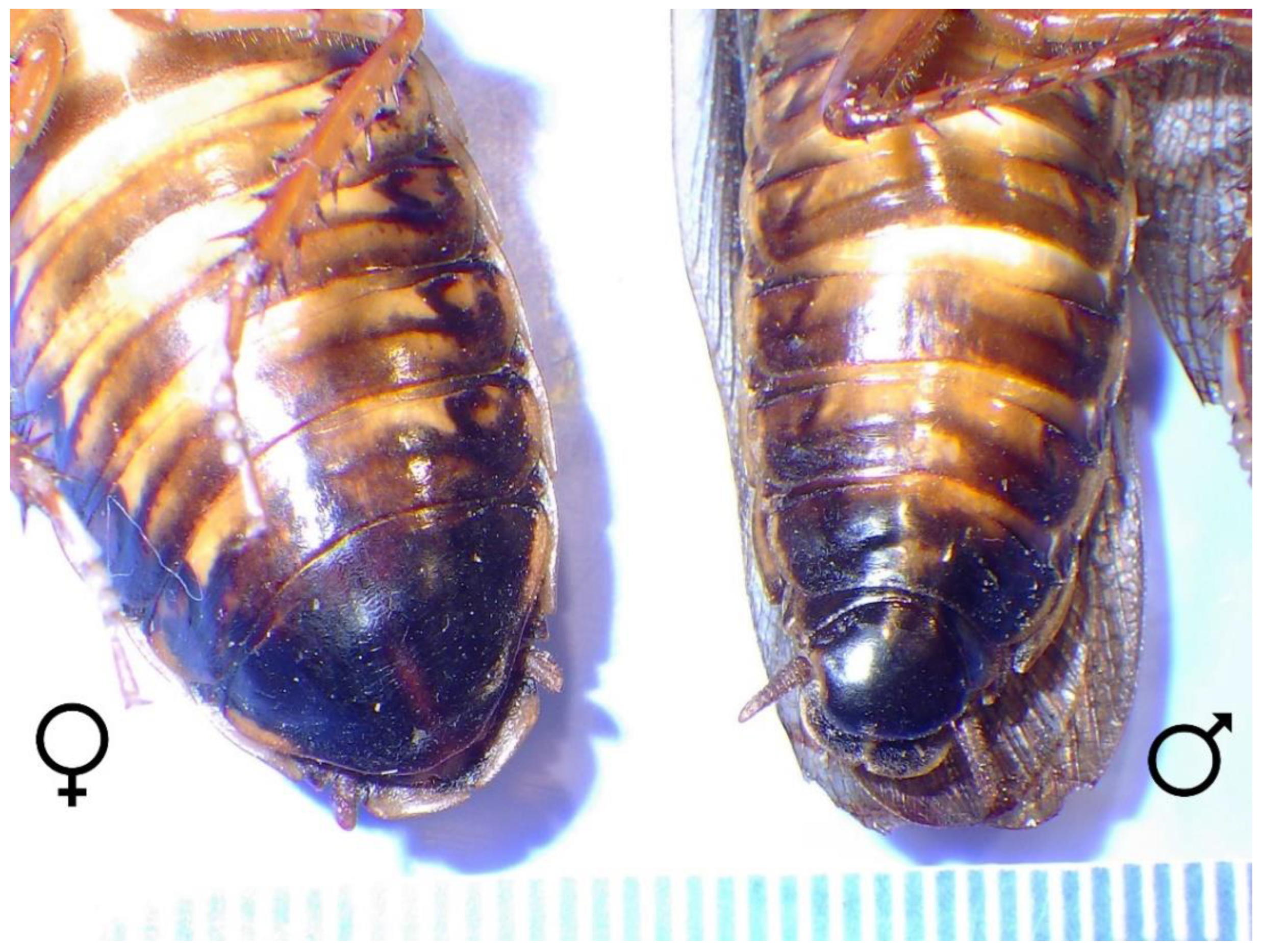
Fourteen B. dubia roaches, including males (n = 5) and females (n = 9), were randomly selected from the NCMNS feeder roach colony using the random number generator: https://www.random.org (accessed on 31 December 2022). Male and female dubia roaches are easily distinguished by the female’s lack of wing development or developing wings, a wider abdominal base, and a single enlarged terminal sternite (caudal-most ventral abdominal plate) as compared to the male (Figure 2). These roaches were individually housed for 24 h, and feces were collected and weighed. A centrifugation technique [10] was utilized to recover pinworm eggs. After centrifugation, all pinworm eggs were counted and recorded for each roach fecal sample. There is not an IACUC (Institutional Animal Care and Use Committee) at NCMNS, and the NC State University (Raleigh, NC, USA) IACUC does not review the use of invertebrates as it is not required by the Animal Welfare Act, Public Health Service Policy, or NC State policies. However, measures were taken to try to reduce pain and stress.
Two days after fecal analysis, all 14 roaches were euthanized, and necropsies were performed. As part of a two-step euthanasia process: the roaches were: (1) temporality anesthetized with 5% isoflurane-soaked cotton balls until 3 min post-movement in a method adapted from Lewbart et al. [11] and then (2) the roaches’ central nerve cords were longitudinally bisected (mechanical destruction) via ventral midline incision through the ventral aspect of the head, thorax, and abdomen using 4.5 in iris dissection scissors.
The euthanized roaches were delivered to the parasitology lab of NC State University’s College of Veterinary Medicine, Raleigh, NC, USA, for immediate necropsy. The hindgut of each roach was individually removed and placed in a dish filled with sterile 0.9% saline. Then hindgut lumens were incised longitudinally, and tissue was teased apart and shaken in the saline bath to free pinworms. Pinworms were counted, preserved, and processed for species identification. Pinworm specimens were collected, fixed, processed, and mounted utilizing standard parasitological techniques [12,13] and were identified by comparison with published descriptions [4,6,14].

2.2. Pyrantel Pamoate Efficacy Study

2.2.1. Group Randomization and Pre-Treatment Fecal Worm Ova Counts (Pre-FWOC)

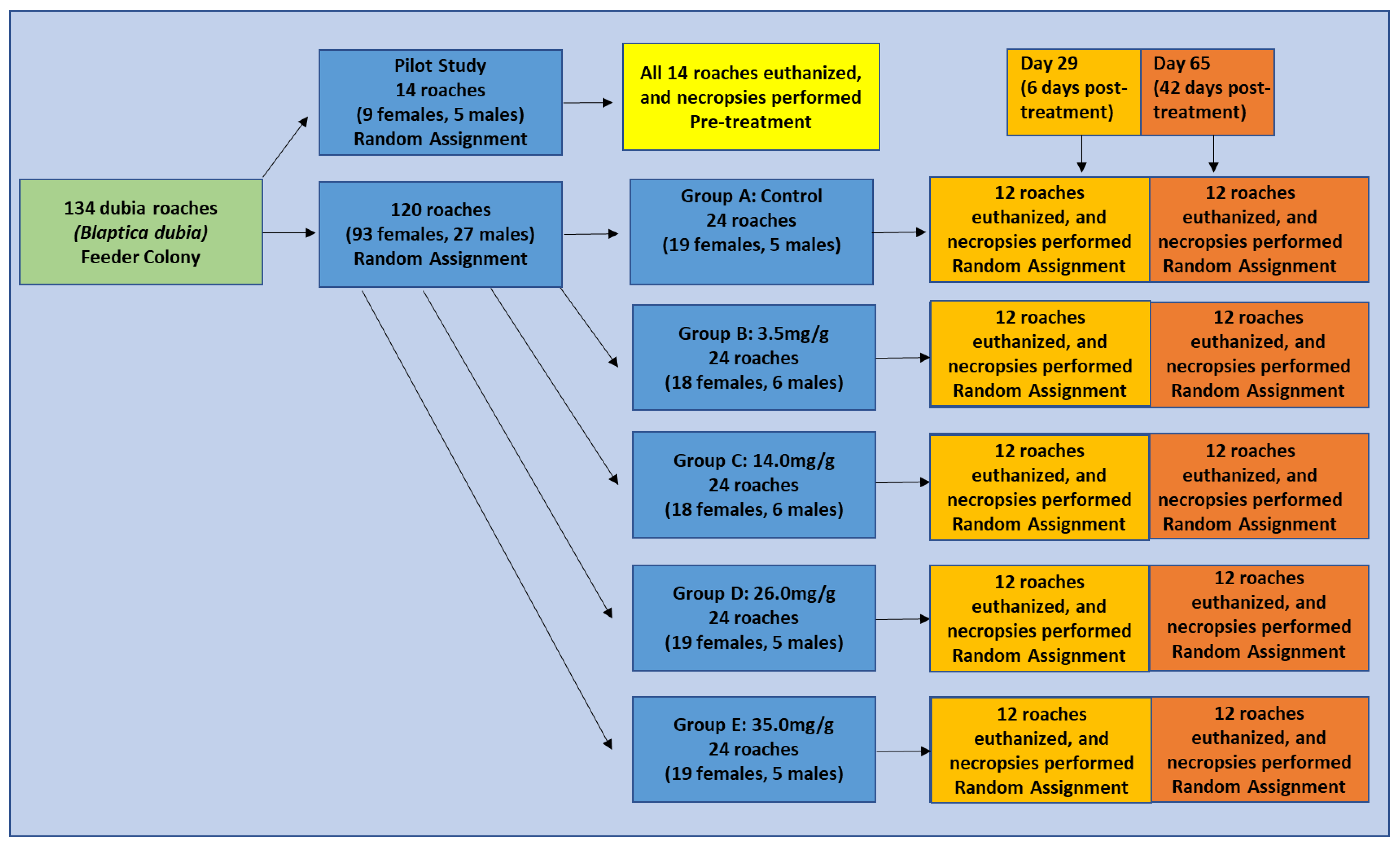
Dubia roaches (n = 120) were isolated from the feeder roach colony. On day 0 of the efficacy study, these roaches were divided into groups of males and females (males n = 27 and females n = 93). Counting the roaches discussed in Section 2.1, the total numbers in this study were based on using all of the roaches in the size class needed from the museum colony. The entire feeder colony availability (small ratio of male: female roaches) was comprised of the total 134 roaches used (Figure 3). The roaches were then randomly divided (males and females separately) into 5 groups, with 24 roaches total in each group. The groups were labeled A (control) and B–E (medicated treatment groups). Randomization of group assignments was achieved by using the random number generator: https://www.random.org/ (accessed 31 December 2022) with 18–19 females and 5–6 males total assigned per group. All 5 groups were separately housed in 190 oz (10 in diameter) round plastic deli containers (TSK Supply, Spanish Fork, UT, USA). Included in each container were sections of clean paper egg crate (Josh’s Frogs, Owosso, MI, USA) for hiding and enrichment and a small dish of fresh drinking water. All five groups were kept in a controlled 35 in × 28 in × 82 in Darwin chamber (Darwin Chambers Co., St. Louis, MO, USA) with a 12-h light and 12-h dark photoperiod cycle at approximately 27 °C and approximately 70% relative humidity.
After approximately 24 h (Day 1), prior to offering medicated or nonmedicated gel diet, roaches were transferred to clean containers with fresh sections of egg carton and fresh water. Fecal samples were collected from each treatment group for pre-treatment fecal worm egg counts (pre-FWOC). The modified centrifugation fecal floatation technique [10] described above was used to determine pre-FWOC for each sample.

2.2.2. Medicated Diet

All medicated roach diets were made by mixing a combination of Repashy Bug Burger Insect Gel Food (Repashy Ventures Inc., Oceanside, CA, USA), Knox unflavored gelatin (The Kraft Heinz Company, Chicago, IL, USA), and a total of 25 mL deionized water for unmedicated diet (group A) or 25 mL mixture of water and pre-determined volume pyrantel pamoate (Zoetis Inc., Kalamazoo, MI, USA) for treatment groups (B–E). Volumes of pyrantel pamoate added to each gel diet preparation for treatment groups (B–E) were calculated to achieve approximate final concentrations of drug per gram (wet weight) of food at 3.5 mg/g, 14.0 mg/g, 26.0 mg/g, and 35.0 mg/g, respectively. Food gel was made fresh each week of the study and first offered to roaches within 48 h of preparation. Any food not offered immediately was stored in light-resistant brown bags and refrigerated at 4 °C.

2.2.3. Anthelmintic Oral Treatments via Medicated Gel Diet

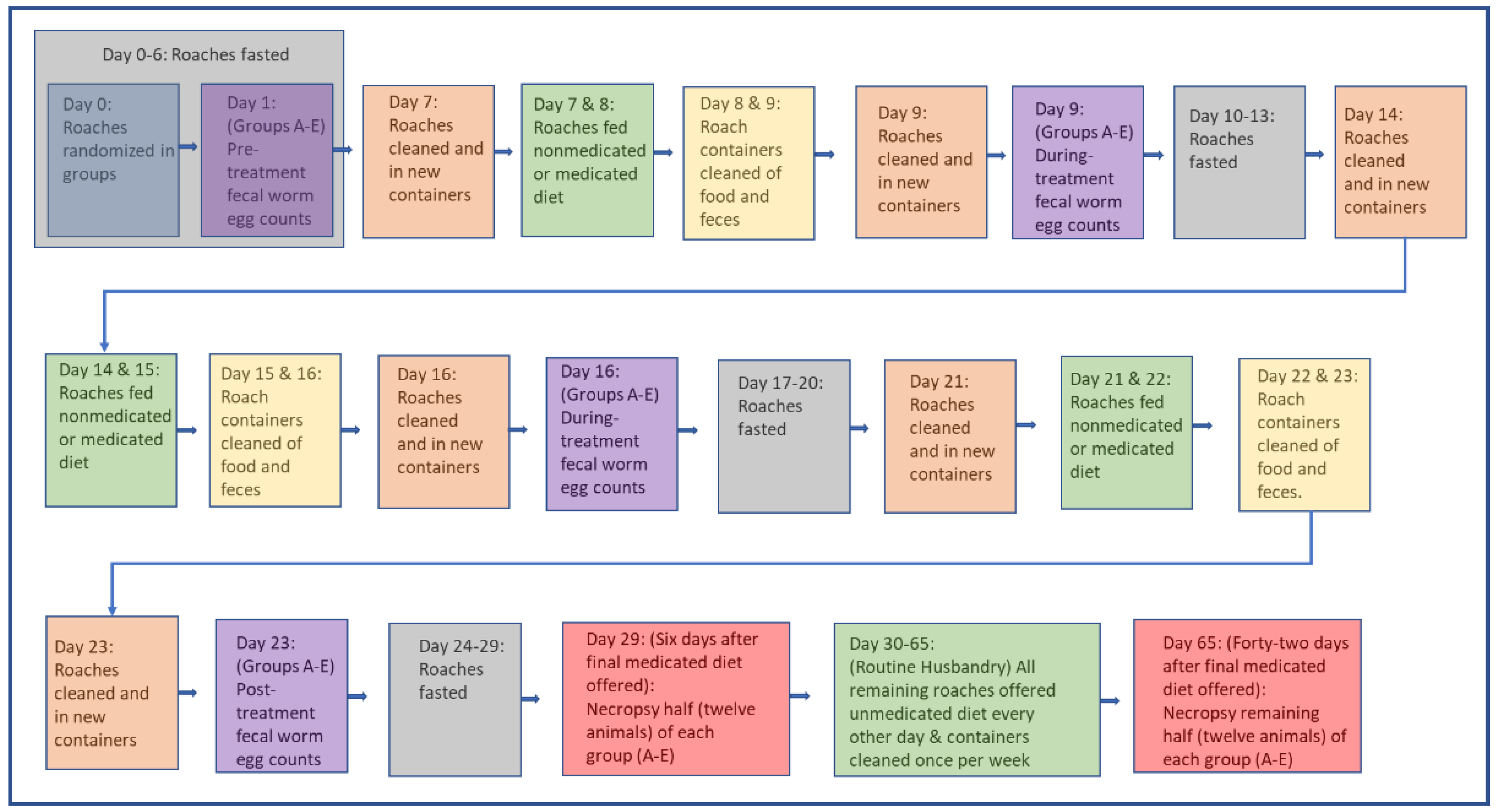
Roaches were fasted for the initial 7 days (Days 0–6) of the study prior to being offered their gel diet. Figure 4 provides a schematic of the process followed. The gel diet (nonmedicated control or medicated) food was offered to the groups (A–E) on Days 7 and 8, Days 14 and 15, and then Days 21 and 22. Each feeding consisted of offering each group (A–E) approximately 6.0 g (wet weight) of gel diet food.
Roaches were provided continual access to the gel diet for approximately 24 h after each feeding. Leftover food was removed after approximately 24 h of each feeding (uneaten removed) on Days 8 and 9, 15 and 16, and Days 22 and 23. Roaches were fasted in between treatments (Days 10–13 and Days 17–20) and for approximately 6 days (Days 24–29) following the final treatment. From Day 30 until the end of the study (Day 65), all groups of roaches were returned to normal feeding (unmedicated gel diet offered every other day). During the study period and treatments, the feeding and cleaning schedule, as described in detail, was used from day 0–29 (Figure 4). Day 30- 65, after all the treatments, animals returned to their normal husbandry routine and were changed to being fed every other day and cleaned weekly.

2.2.4. Post-Treatment Fecal Worm Ova Counts (Post-FWOC)

Prior to each weekly 2-day feeding session (Days 7, 14, and 21) and 24 h after each weekly 2-day feeding session (Days 9, 16, and 23), roaches were transferred to clean containers with fresh sections of egg carton and fresh water. To minimize contamination with fecal ova (and a potential source of reinfection throughout the study), before and after weekly treatment sessions on Days 7, 9, 14, 16, 21, and 23, each roach was carefully examined, and any loose feces was removed (and saved) prior to moving to a clean container. Fecal samples (including feces brushed from roaches at the time of transfer to the new enclosure) were collected from each treatment group (after roaches were moved) for post-fecal worm ova counts (post-FWOC). The modified centrifugation fecal floatation technique [10], previously described, was used to determine post-FWOC for each sample.

2.2.5. Post-Treatment Necropsies

Necropsies and ova and pinworm counts (as described above in Pilot necropsy details) were performed on roaches from each group (A–E) at 2 time points following the treatment portion of the study (on Day 29 (6 days post-treatment) and Day 65 (42 days post-treatment)). On Day 29, a random selection of 12 roaches from each group (using the random number generator: https://www.random.org/ (accessed 31 December 2022)) was euthanized, and necropsies were performed as previously described. The remaining 12 roaches from each group were euthanized, and necropsies were similarly performed on Day 65. The hindgut lumens were inspected following the protocol described in Section 2.1.

2.3. Data Analysis

Pinworm post-treatment data were analyzed using unpaired t-tests with significance at p ≤ 0.05 via GraphPad by Dotmatics, San Diego, CA, USA, www.graphpad.com/quickcalcs/ttest1/ (accessed 31 December 2022).
Optical (qualitative) numerical comparisons instead of quantitative statistics were utilized for group ova number comparisons.

3. Results

3.1. Pilot Roach Fecal Examinations and Necropsies

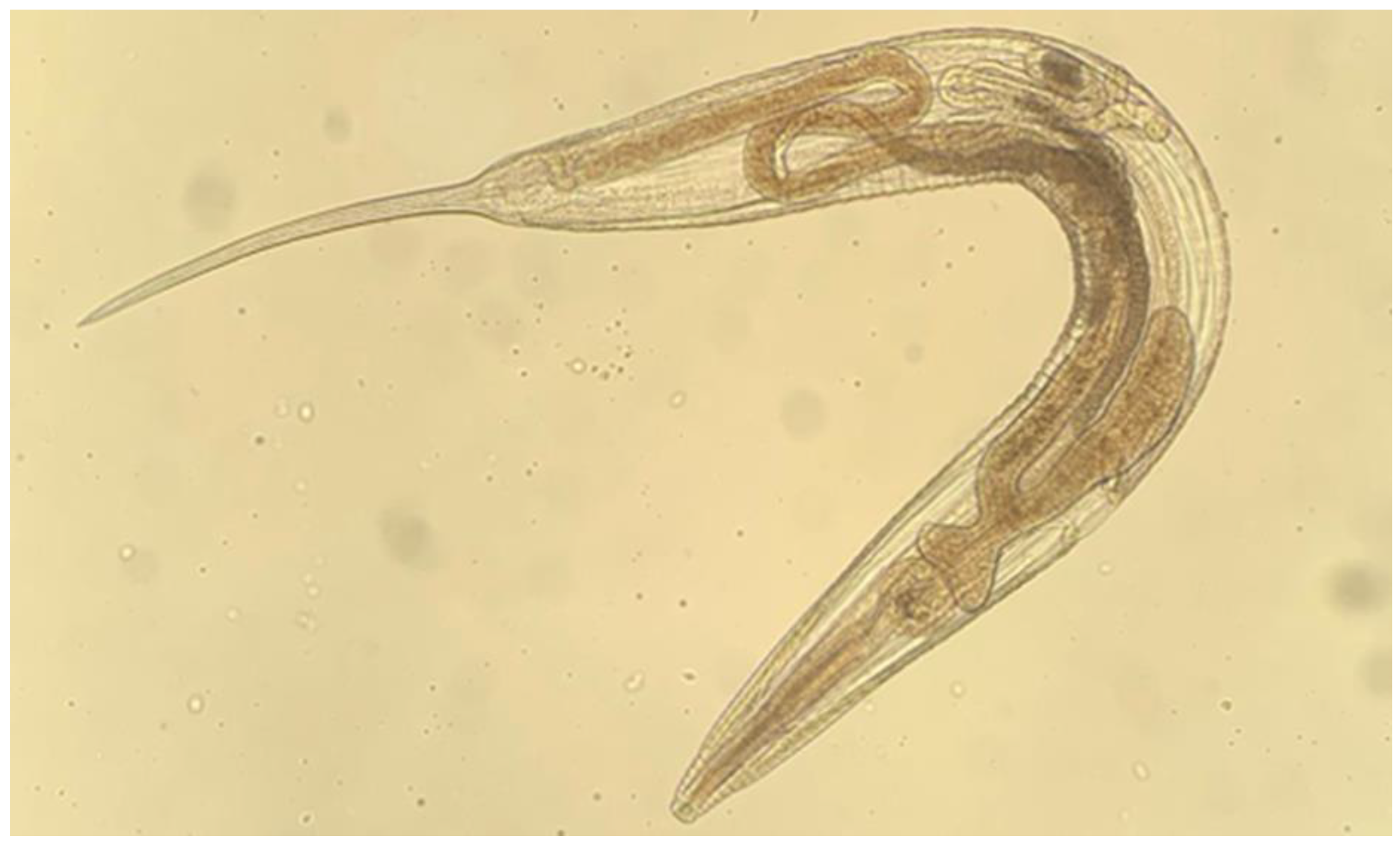
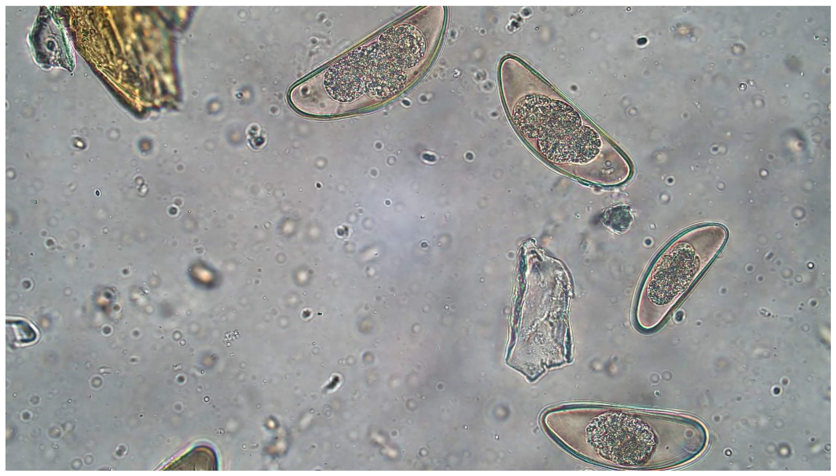
Fecal examinations revealed pinworm ova (Leidynema appendiculata) (Figure 5 and Figure 6) present in eight of the 14 preliminary dubia roaches evaluated (Table 1). Feces were uncollectable from two individuals in this group. The number of ova observed per animal ranged from zero to 777 in total. All roaches with zero ova observed on fecal examination also had zero pinworms identified on roach gut examination following euthanasia and necropsy.
The highest number of pinworms observed in any individual was seven. Five of the fourteen roaches had zero pinworms. Three roaches, all with even pinworms found on necropsy, had 777, 38, and 416 ova, respectively, found 2 days prior to necropsy.

3.2. Pyrantel Pamoate Efficacy Study

3.2.1. Pyrantel Pamoate Consumed

Dry weights of roach diet remaining 24 h after each feeding were obtained, and the total diet consumed at each treatment was calculated. Based on the percent of diet consumed, the estimated total amount of pyrantel pamoate (mg) consumed on each treatment day was calculated for each treatment group. Group A received no medication and served as a control. Groups B–E consumed an average of 3.5 mg (0.8–9.9 mg), 14 mg (3.4–32.3 mg), 19.8 mg (4.0–56.9 mg), and 54.7 mg (2.0–210.0 mg) total per feeding, respectively.

3.2.2. Fecal Pinworm Ova Counts

Pre-treatment fecal pinworm ova counts were determined for each group on Day 1 of the study. Group A had 620 ova/g of feces, group B had 509 ova/g, group C had 461 ova/g, group D had 1501 ova/g, and group E had 510 ova/g (Table 2). Mid-treatment ova counts were recorded for all groups on Days 9, 16, and 23 (24-h following each 2 day oral gel diet/ treatment session) in addition to a fecal ova count immediately following the final treatment session (24-h after offering the gel diet on the second day of treatment in the third week) on Day 23 of the study (Table 2). On Day 23, Group A, the control group, had higher ova counts than all treatment groups. The ova counts from Day 1 of the study compared to Day 23 of the study decreased in all groups except for the control. No group had zero eggs at any point in the study. A qualitative numerical review of the fecal ova numbers shows considerable variability across the groups and Days. Quantitative statistics were not used for these data.

3.2.3. 6 Days Post-Treatment Hindgut Examination for Pinworms

Six days following the end of the final treatment (Day 29), 12 roaches from each group were euthanized for necropsy with excision and evaluation of the gastrointestinal tract for pinworms. In total, roaches from Group A, the control group, had 143 pinworms found in their hindgut; Group B, offered pyrantel pamoate medicated gel diet at 3.5 mg/g, had 130 pinworms; Group C, offered the 14.0 mg/g medicated gel diet, had 34 pinworms; Group D, offered the 26.0 mg/g medicated diet had 25 pinworms; and Group E, the group offered 35.0 mg/g medicated gel diet had 10 pinworms. The roaches in groups C, D, and E had lower parasite burdens than the control group (Table 3). Some individual roaches in groups D and E had zero pinworms; however, no group had zero worms found. In both groups D and E, 41.6% of the roaches had zero pinworms, while 25% of the roaches in groups A and C had zero worms. Group B had zero roaches with zero worms.

3.2.4. Forty-Two Day Post-Treatment Hindgut Examination for Pinworms

The remaining 12 roaches from all groups (A–E) were euthanized, and necropsies were performed on day 65 of the study. Hindguts of all remaining roaches were excised and evaluated for pinworms. Group A, the control group, had a total of 106 pinworms found in their hindguts, Group B had 49 pinworms total, Group C had 74 pinworms total, Group D had 69 pinworms total, and group E had 36 pinworms total (Table 3). No significant differences were noted between Groups B–E and the control Group A. All groups had at least one roach with zero pinworms observed.
No roaches died over the course of the study prior to euthanasia.

4. Discussion

The evaluation of fecal parasites is a common procedure performed as a part of veterinary medical examinations. The detection of nematode ova in a fecal sample often directs medical management decisions and leads to treatments with anthelmintics. Anytime potential parasite ova are detected on fecal examination for tenrecs or any other species, it must be determined, prior to treatment, if they are actually parasites or are, in fact, pass-through pseudoparasites. It is recommended that multiple fecal samples are evaluated over time, and that diet is adjusted appropriately over this period to help rule out pass-through pseudoparasites. Otherwise, periodical screening of feeder insects (or other feeders) is recommended. It can be difficult and a timely procedure to distinguish passthrough pseudoparasites from true evidence of gastrointestinal parasite infection. Even when not likely to pose an infective risk to the animals that eat them, as in this case with tenrecs, elimination of parasites from feeder roach colonies that may present as passthrough pseudoparasites is important to ensure appropriate health management. Unnecessary treatment with anthelmintics can have adverse resistance effects and be resource wasteful [15].
This is the first report describing a confirmed passthrough pseudoparasite in tenrecs from feeder dubia roaches. Documentation of anthelmintic treatment of arthropods, specifically dubia roaches, is scant. While pinworms were not fully eliminated by anthelmintic treatment of dubia roaches in this study, there was a documented reduction in the number of infective worms and ova with a response to treatment in groups offered gel diets with pyrantel pamoate at approximate concentrations of 3.5 mg/g, 14.0 mg/g, 26.0 mg/g, and 35.0 mg/g.
Pyrantel pamoate was determined to be non-lethal in B. dubia for concentrations in a gel diet up to 35.0 mg/g. Pyrantel pamoate is a common anthelmintic agent used to treat intestinal nematodes. It is believed to work by depolarizing neuromuscular junctions leading to muscular contractions and paralysis in target helminths. The drug causes worms to release from their hold on the inner gastrointestinal luminal wall to be passed in the feces [16]. Based on the failure to eliminate pinworms at any of the dietary anthelmintic concentrations offered, it is suspected that remaining pinworms and ova may provide a source for reinfection. At Day 65 (42 days after the last treatment), pinworm numbers were lower in all treatment groups (B–E) as compared to the control group A. However, none of the treatment groups were completely cleared of pinworms. The numbers of pinworms and pinworm ova varied greatly throughout all groups at all time points in the study. The detection of pinworm-free B. dubia individuals in all groups throughout the study may suggest that some roaches are resistant to oxyurid infection or that they have natural ways to clear worms.
After the eggs of L. appendiculata have been discharged with the feces, it takes 24–48 h for the eggs to develop into an infective larvated (or embryonated) stage [14]. In addition, the gut passage in another species of roach (Rutilus rutilus) at room temperature was determined to be less than 10 h [17]. Therefore, if the pyrantel pamoate worked on all adult pinworms present in the roach host on a single day of treatment, the pinworms should have been passed along with any egg-containing feces before the cages were cleaned the next day, limiting the possibility of re-infection. Given that only 41.6% (5/12 roaches evaluated) of the roaches in Group E, the highest pyrantel pamoate treatment group (35.0 mg/g concentration in gel diet) were cleared of adult pinworms by Day 65 of the study, reinfection is suspected. However, based on the known life cycle of the pinworm and the timing of treatments and cage cleaning noted above, this reinfection was likely not simply from ova passed from treated worms prior to treatment death or ova that passed as anticipated via normal timing in feces. Reinfections, in this case, are suspected to be from pinworms in individual roaches that did not eat any (or enough) diet to receive an effective dose of anthelmintic during some part of the trial. Because roaches were maintained and offered medicated diet in groups for this study, individual feeding data was not able to be observed. Individual roaches may not have eaten diet at one or more medicated feedings allowing for some pinworms in some individuals to remain through each feeding of medicated gel and continue to pass infective ova.
A previous study treated smoky brown cockroaches (Periplaneta fuliginosa) with oral pyrantel pamoate via suspension in the drinking water source [9]. However, our current dietary research found great variability in the amount of gel diet food eaten from week to week in all groups, even the nonmedicated control group. This may have been due to the palatability of the drug, the diet, or the gelatin products used. Oral treatment with pyrantel pamoate via addition to water could potentially provide a more consistent dosing regimen. In addition, the limitations of group housing may have affected the results. Some roaches may not have eaten the food provided on some, or all, the treatment days. NCMNS invertebrate caretakers have noticed the on-site Blaptica dubia are less inclined to eat immediately following a molt as well, and this data was not recorded during the current research.
As passthrough pseudoparasites, the pinworms are a confounding issue with the health management of the tenrecs. The intent in attempting to eliminate them from the feeder roaches is to improve care for the tenrecs. However, it is unknown if the pinworms are causing harm to the host roaches. It is likely that eliminating the worms from the roaches will impact the balance of the gastrointestinal microbiome [18]. The role of these worms in the overall population health and ecosystem balance of the roaches should be studied.
In addition, future studies are needed to determine if an anthelmintic treatment method can be developed to completely eliminate pinworms and Leidynema appendiculata from dubia roach and Blaptica dubia feeder insect colonies. Moving forward, studies may include the evaluation of other anthelmintic medications or alternative methods of drug delivery. In this study, the amount of medicated food consumed among groups was found to be variable. Groups were offered food with a pre-calculated concentration of medication, but consumption by each individual roach was not confirmed. Some individuals may not have eaten any or enough of the medicated diet during the study to achieve the desired outcome. This may be the explanation for not fully eliminating the parasites from any of the groups. Instead of offering the medication premixed in food, single-dose injectable medications of a colony founder group of roaches may offer a more effective and reliable method for assuring all roaches receive a full pre-determined dose of medication. In addition, histological evaluation of hindgut tissue following treatment could help in confirming the successful elimination of parasites. Evaluating the treatment of male and female roaches separately or in equal numbers across all groups may reduce variability and produce more consistent results. Other improvements to experimental design may include better-aligned life stages of all roaches- and documenting molting cycles.
Decreasing the occurrence of confounding fecal diagnostics in insectivores such as the lesser hedgehog tenrecs at the NCMNS (and all human management facilities) will universally benefit animals and museum and zoological veterinary staff by reducing unnecessary treatment with anthelmintics and use of unnecessary resources for repeated examinations.

Author Contributions

Conceptualization, D.S.D., L.S.C. and E.B.; methodology, E.B., D.S.D., L.S.C., J.F. and K.A.-v.H.; formal analysis, E.B.; investigation, E.B., S.K., J.F. and K.A.-v.H.; resources, D.S.D., J.F. and K.A.-v.H.; writing—original draft preparation, E.B. and D.S.D.; writing—review and editing, J.F., K.A.-v.H. and D.S.D.; visualization, E.B.; supervision, D.S.D.; project administration, L.S.C.; funding acquisition, D.S.D. All authors have read and agreed to the published version of the manuscript.

Funding

This research was funded by the North Carolina Museum of Natural Sciences, Raleigh, NC, USA.

Institutional Review Board Statement

This population health project was completed under the direction of the NCMNS Veterinary Science department.

Data Availability Statement

The data presented in this study are available from the corresponding author upon reasonable request.

Acknowledgments

The authors thank all NCMNS individuals responsible for the daily care of both the dubia roaches and the tenrecs participating in this health study. We also thank the NC State Department of Animal Science (primarily Matt Poore) for lab space. Finally, we sincerely appreciate Molly Kittson and April Shaeffer for laboratory analyses and expertise, respectively, during the very difficult pandemic timing of this research.

Conflicts of Interest

The authors declare no conflict of interest.

References

  1. Olson, L.E. Tenrecs. Curr. Biol. 2013, 23, R5–R8. Available online: https://www.cell.com/current-biology/pdf/S0960-982201324-3.pdf (accessed on 31 December 2022). [CrossRef]
  2. Harrison, T.M.; Harrison, S.H. Evaluation of husbandry and mortality in lesser hedgehog tenrecs (Echinops telfairi). J. Zoo Wildl. Med. 2017, 48, 440–445. [Google Scholar] [CrossRef] [PubMed]
  3. Leidy, J. Description of some nematoid entozoa infesting insects. Proc. Natl. Acad. Sci. USA 1850, 5, 100–102. [Google Scholar]
  4. Kobayashi, M.; Ooi, H.; Taira, K. Effects of anthelmintics on the pinworm Blatticola blattae in laboratory-reared German cockroaches Blatella germanica. Parasitol. Res. 2020, 119, 3093–3097. [Google Scholar] [CrossRef] [PubMed]
  5. Adamson, M.L.; Van Waerebeke, D. Revision of the Thelastomatoidea, Oxyurida of invertebrate hosts. I. Thelastomatidae. Syst. Parasitol. 1992, 21, 21–63. [Google Scholar] [CrossRef]
  6. Ozawa, S.; Vicent, C.S.L.; Sato, K.; Yoshiga, T.; Kanzaki, N.; Hasegawa, K. First report of the nematode Leidynema appendiculata from Periplaneta fuliginosa. Acta Parasitol. 2014, 59, 219–228. [Google Scholar] [CrossRef] [PubMed]
  7. Pawlik, J. Control of the nematode Leidynema appendiculata (Leidy) (Nemata: Rhabditida: Thelastomatidae) in laboratory cultures of the American cockroach. J. Econ. Entomol. 1966, 59, 468–469. [Google Scholar] [CrossRef]
  8. Pizzi, R.; Carta, L.; George, S. Oral nematode infection of tarantulas. Vet. Rec. 2003, 152, 695. Available online: https://pubmed.ncbi.nlm.nih.gov/12803400/ (accessed on 31 December 2022). [PubMed]
  9. Ozawa, S.; Hasegawa, K. Broad infectivity of Leidynema appendiculatum (Nematoda: Oxyurida: Thelastomatidae) parasite of the smoky brown cockroach Periplaneta fuliginosa (Blattodea: Blattidae). Ecol. Evol. 2018, 8, 3908–3918. [Google Scholar] [CrossRef] [PubMed]
  10. Dryden, M.W.; Payne, P.A.; Ridley, R.; Smith, V. Comparison of common fecal flotation techniques for the recovery of parasite eggs and oocysts. J. Vet. Pharmacol. 2005, 6, 15–28. Available online: https://pubmed.ncbi.nlm.nih.gov/15906267/ (accessed on 31 December 2022).
  11. Lewbart, G.A.; Mosley, C. Clinical anesthesia and analgesia in invertebrates. J. Exot. Pet Med. 2012, 21, 59–70. [Google Scholar] [CrossRef]
  12. Pritchard, M.H.; Kruse, G.O.W. The Collection and Preservation of Animal Parasites; University of Nebraska Press: Lincoln, NE, USA, 1982. [Google Scholar]
  13. Chitwood, B.G. A synopsis of the nematodes parasitic in insects of the family Blattidae. Z. Parasitenkd. 1932, 5, 14–50. [Google Scholar] [CrossRef]
  14. Dobrovolny, C.G.; Ackert, J.E. The life history of Leidynema appendiculata (Leidy), a nematode parasite of cockroaches. Parasitol. Res. 1934, 26, 468–480. [Google Scholar] [CrossRef]
  15. Nixon, S.A.; Welz, C.; Woods, D.J.; Costa-Junior, L.; Zamanian, M.; Martin, R.J. Where are all the anthelmintics? Challenges and opportunities on the path to new anthelmintics. Int. J. Parasitol. Drugs Drug. Resist. 2020, 14, 8–16. [Google Scholar] [CrossRef] [PubMed]
  16. Pyrantel. In LiverTox: Clinical and Research Information on Drug-Induced Liver Injury [Internet]; National Institute of Diabetes and Digestive and Kidney Diseases: Bethesda, MD, USA, 2012. Available online: https://www.ncbi.nlm.nih.gov/books/NBK548238/ (accessed on 20 July 2020).
  17. Hofer, R.; Forstner, H.; Rettenwander, R. Duration of gut passage and its dependence on temperature and food consumption in roach, Rutilus rutilus L: Laboratory and field experiments. J. Fish Biol. 1982, 20, 289–299. [Google Scholar] [CrossRef]
  18. Vicente, C.S.; Ozawa, S.; Hasegawa, K. Composition of the cockroach gut microbiome in the presence of parasitic nematodes. Microbes Environ. 2016, 31, 314–320. [Google Scholar] [CrossRef] [Green Version]
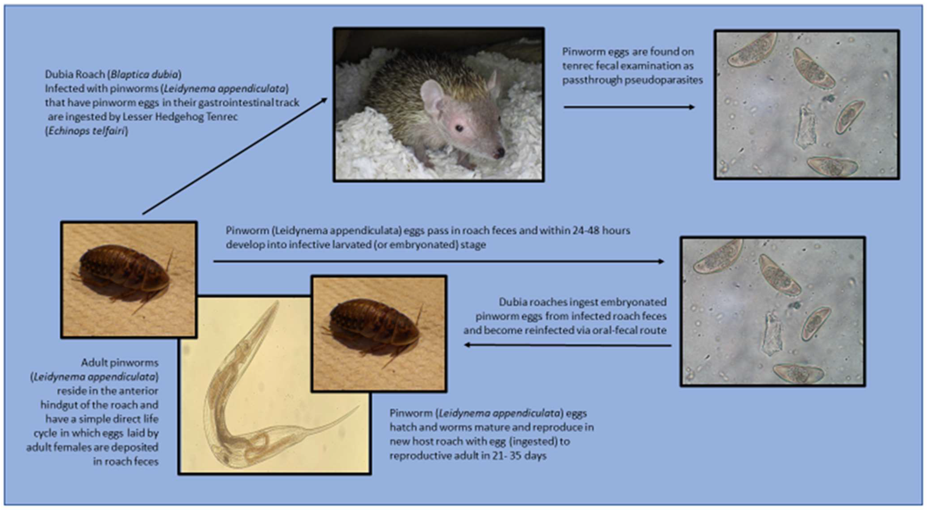
Figure 1. Transmission cycle of Leidynema appendiculata through the dubia roach (Blaptica dubia) host and lesser hedgehog tenrec (Echinops telfairi) as a passthrough pseudoparasite.
Jzbg 04 00015 g001
Figure 2. Dubia roaches (Blaptica dubia) image indicating sexual dimorphism in the species. The caudal most visible sternite in females (animal on the left) is larger & wider than in males (animal on the right), with males appearing to have an extra visible sternite.
Jzbg 04 00015 g002
Figure 3. Dubia roach (Blaptica dubia) assignment into both the pilot study and the main study research groups (control, A–E).
Jzbg 04 00015 g003
Figure 4. Study design schematic to show workflow from Day 0 to Day 65 of the dubia roach (Blaptica dubia) pyrantel pamoate efficiency on passthrough fecal pinworm (Leidynema appendiculata) parasites research study.
Jzbg 04 00015 g004
Figure 5. Leidynema appendiculata image pinworm found in the hindgut of dubia roaches (Blaptica dubia) at The North Carolina Museum of Natural Science (Raleigh, NC, USA). Image taken at 400× during pre-treatment necropsy.
Jzbg 04 00015 g005
Figure 6. Leidynema appendiculata pinworm eggs found on a floatation of fresh feces from dubia roaches (Blaptica dubia) at The North Carolina Museum of Natural Science (Raleigh, NC, USA). Image taken at 400× during pre-treatment screening.
Jzbg 04 00015 g006
Table 1. Dubia roach, Blaptica dubia, 24 hr pre-treatment fecal total wet weight (g) and pinworm ova (#) and adult Leidynema appendiculata (#) confirmed from each roach on necropsy 2 days after fecal collections.
Roach Number (n = 14)Weight of Feces (g)Oxyurid Ova (#)Pinworms (#)
10.011902
20.014714
30.02200
40.03900
50.016622
60.0171174
70.0097777
80.026387
90.02700
100.0124167
1101NA3
120.00800
130.0127502
140 1NA0
1 Feces was liquid and absorbed into the substrate, unable to collect. # = The count of observations. NA = Not available.
Table 2. Dubia roach group (n = 24), Blaptica dubia, hindgut examination results of pinworm, Leidynema appendiculata fecal ova count (#/gram feces) at Days 1, 9, 16 and 23 after oral pyrantel pamoate anthelmintic treatment via medicated or control gel diet.
Pyrantel Pamoate (mg/g)Day 1: Total Fecal OvaDay 9: Total Fecal OvaDay 16: Total Fecal OvaDay 23: Total Fecal Ova
Group A—0.0 (control)620596444636
Group B—3.5509210390160
Group C—14.0461553480172
Group D—26.0150116046225
Group E—35.0510176358194
Table 3. Dubia roach, Blaptica dubia, hindgut examination results of pinworm (Leidynema appendiculata) counts (#) at Day 29 (6 days after oral pyrantel pamoate anthelmintic medicated gel diet treatments ended) and Day 65 (42 days after treatment ended).
Group (n = 12 Roaches Each)Pyrantel Pamoate in Gel Diet (mg/g)Day 29: 6 Days Post-Treatment Total PinwormsDay 65: 42 Days Post-Treatment Total Pinworms
A (control)0.0143106
B3.513049
C14.034 174
D26.025 269
E35.010 336
1,2,3 Indicates the significances (p ≤ 0.05) when Day 29 and Day 65 Groups B–E were compared via unpaired t-test to the Control Group A. 1 Two-tailed p = 0.0086 comparison with control group A; Day 29 95% confidence interval from 2.6–5.6. 2 Two-tailed p = 0.0045 comparison with control group A; Day 29 95% confidence interval from 3.4–16.3. 3 Two-tailed p = 0.0013 comparison with control group A; Day 29 95% confidence interval from 4.8–17.3.
Disclaimer/Publisher’s Note: The statements, opinions and data contained in all publications are solely those of the individual author(s) and contributor(s) and not of MDPI and/or the editor(s). MDPI and/or the editor(s) disclaim responsibility for any injury to people or property resulting from any ideas, methods, instructions or products referred to in the content.