Article

Training a Giraffe (Giraffa camelopardalis reticulata) for Voluntary Foot Radiographs at Dubai Safari Park

1
Zoology Department, Dubai Safari Park, Dubai P.O. Box 67, United Arab Emirates
2
Veterinary Department, Dubai Safari Park, Dubai P.O. Box 67, United Arab Emirates
*
Author to whom correspondence should be addressed.
J. Zool. Bot. Gard. 2022, 3(4), 688-698; https://doi.org/10.3390/jzbg3040051
Submission received: 27 October 2022 / Revised: 2 December 2022 / Accepted: 5 December 2022 / Published: 8 December 2022
(This article belongs to the Special Issue Innovative Techniques Used to Improve Animal Health and Welfare)

Abstract

:
When 0.1 Reticulated Giraffe (Giraffa camelopardalis reticulata) began displaying signs of lameness and a persistent swelling of the front left pastern joint, an operant based training program was created for front foot radiographs to diagnose and better manage lameness. Using a protected contact set up, behaviour was shaped using a positive reinforcement procedure. A series of cued behaviors were trained and used to obtain dorsolateral palmar medial oblique front foot radiographs to aid vets in diagnosing the cause of lameness. By training giraffes for foot radiographs, potential causes of lameness could be identified and better managed. Long-term, the results may help zoos identify best practices for managing and preventing lameness in giraffe and subsequently improve welfare. Training animals to participate in their own medical care can improve welfare by giving them a sense of control within the situation. Training for medical procedures helps to reduce the stress imposed on the giraffe and the associated risks compared to alternative methods.

1. Introduction

Giraffes (Giraffa spp.) are a popular, charismatic species to house within zoological institutions. However, due to their size and longevity, hoof problems and arthritis are some of the most prevalent concerns for zoo housed giraffes [1]. Up to 80% of giraffe immobilizations performed are to address hoof overgrowth and limping. This statistic indicates that lameness is an important health and welfare concern for this species [2]. Physical examination of the hooves is often limited or not possible without training or anesthesia, however, sedation carries a risk, with giraffe anesthesia having a 10% mortality rate [2].

1.1. Individual

In April 2022, Dubai Safari housed a herd of 10 giraffes. The herd consisted of 2 female reticulated giraffes (Giraffa camelopardalis reticulata) and 8 southern giraffes (Giraffa giraffa), consisting of 3 bulls and 5 females. After 0.1 Reticulated Giraffe, aged 22 years, began displaying signs of lameness and a persistent swelling of the front left pastern joint, an operant conditioning program was created for front foot radiographs and hoof trims to diagnose and better manage lameness. This giraffe had no prior background in operant conditioning. The calm temperament of this animal meant that she often participated in visitor feeding interactions until her condition declined. When she began showing signs of lameness, she was subsequently taken off display.

1.2. Housing

The giraffes at Dubai Safari were housed in an open ventilated holding with ten stalls, each measuring 6 metres by 6 metres. The stall flooring was concrete with lined grooves, with woodchips provided as a flooring substrate. A detailed description of the training area is provided in Section 2.2. The giraffes had access to three adjoining sand-filled yards, as well as a large multi-terrain (grass, concrete, sand) drive-through exhibit. An additional yard was used for public interactions such as hand-feeding the giraffes. Each of the giraffes was rotated between different areas and exhibits.

1.3. Diet

The giraffes were fed a diet consisting of alfalfa hay, fresh browse (Acacia spp.), and a formulated grain pellet (Antelope Pellet by Fujairah Feed). Food was provided three times per day, with hay and browse provided ad libitum. Additionally, all giraffes could be hand-fed romaine lettuce or carrots by visitors at set feeding times throughout the day. Visitor feeding sessions occurred at 10:30, 12:30, 14:30, and 16:00 h. After being taken off display, the lame giraffe was offered food by hand to maintain conditioning for public interactions and to maintain a bond with the keepers. Additional lettuce and carrots were occasionally used for enrichment purposes. Enrichment was provided at varied times throughout the day to ensure unpredictability. For training purposes, positive reinforcers were either fresh browse or romaine lettuce, which was based on keeper observations of the individual’s preference. Keepers observed that the giraffe preferred certain parts of the lettuce, such as the stem and the inner blanched leaves. The keepers ensured that they offered multiple parts of the lettuce as reinforcement. Training sessions took place between 7:00 and 8:00 am; after the giraffes had received their first feed but had not yet been given browse. During the training session, the keeper had both lettuce and browse on hand and offered the giraffe the choice of reinforcement to maintain interest.

2. Materials and Methods

2.1. Safety Considerations

Despite their size and status as the tallest land mammal, giraffes are still a prey species. In a captive situation, giraffes are known to defensively kick or bolt from any perceived threat [3]. Therefore, the safety of both animal keepers and trainers within a captive environment is paramount.
Prior to initiating the training program, the head keeper and the training coordinator carefully evaluated the temperament of the individual animal, the keepers who were to conduct the training, and the training set-up. Each session began with a debriefing from the primary trainer regarding the giraffe’s behaviour and temperament and any safety concerns or important notes. Training took place in a double stall, separated by a sliding door, with the giraffe and keepers on adjacent sides. The adjacent stalls were separated by a primary barrier of firehose strips. The training set-up and containment area are further described in Section 2.2. All keepers involved in the training session were aware of the emergency egress if the giraffe breached the containment area. Written safety protocols and protected contact setups are strongly recommended to help ensure both human and animal safety.
Flies in the area were a distraction, especially during the hotter months, as they would occasionally cause twitching of the upper legs or loss of concentration. Keepers would monitor leg movements due to flies throughout the session in case it led to any dangerous behaviour or situations for the keepers, especially the secondary keeper in close proximity to the foot.

2.2. Protected Contact

Training was conducted in a protected contact set-up. The size of the training stall was 6 × 6 m. This set-up consisted of a primary containment barrier made up of firehose strips fixed at chest height to both sides of a sliding gate (Figure 1). This set-up created a temporary training area for keepers to work with the giraffe in the adjacent stall. The second keeper, focusing on the giraffe’s feet, was protected by the mesh wall of the gate while the giraffe was in the training area. If the giraffe lowered her head below or between the straps, it was cued to back up from the area, reinforced, and then targeted into the correct position. Training sessions were always voluntary. The giraffe chose to participate in all but two sessions.

2.3. Training Sessions

Each training session involved one giraffe and at least two keepers, hereafter referred to as trainers. The primary trainer led the training session by delivering the cues, bridge, and reinforcements, and the secondary trainer oversaw the desensitization of touching hooves and counterconditioning of the radiograph equipment. Once the primary trainer felt the behaviour was under stimulus control, it was then generalized to other trainers. The training session only proceeded if the giraffe displayed comfortable body language, such as an interest in training, relaxed ears, and nostrils. The duration of daily training sessions varied, averaging around 15–20 min. The duration of the session was dependent on the giraffe’s concentration level of the day and ended with a successful response or outcome. Sessions during radiographs could last up to an hour depending on the participation of the giraffe. This duration was opportunistically achieved due to the giraffe’s engagement in the session.
The differential reinforcement of successive approximations (shaping) was used to teach new behaviours by successively reinforcing subtle variations in responses along a continuum that led to the final target approximation [4,5,6,7]. At this stage, every instance of the completed target approximation was reinforced using a continuous reinforcement schedule. For example, when shaping the “lift” behaviour, the giraffe was initially reinforced for touching the wooden foot block (see Figure 1), and the behaviour was shaped until the foot was lifted to a sufficient height and was placed flat on the block as the final target approximation. Once new behaviours were established, an intermittent reinforcement schedule was used to strengthen them.
All target approximations were trained using positive reinforcement, with a clicker to reinforce the instances that the “correct” behaviour was offered, and unwanted behaviours were ignored or redirected. When the giraffe did not provide the cued behaviour, training momentum was re-established by cuing and reinforcing a high-probability behaviour before asking again for the desired behaviour.

3. Goal Behaviors

The giraffe was taught six behaviours to complete the radiograph training program.

3.1. Target Training

The goal was for the giraffe to touch its nose to a target (a tennis ball attached to the end of a section of broomstick). The giraffe was trained to hold its nose to the target for a set duration of time (seconds) when given the cue “Hold.” The durations for this behaviour varied. The giraffe was asked to hold this behaviour until the clicker sounded.

3.2. Recall

The goal was for the giraffe to walk toward the trainer when given vocal and hand cues.

3.3. Back Up

The goal was for the giraffe to walk a few steps backwards from the trainer when given vocal and hand cues. The giraffe should not completely turn its back on the trainer.

3.4. Station

The giraffe was considered to be at station (or stationing) when its chest was fully in contact with the straps, holding the strap in a horizontal position, and leaning into the strap slightly.

3.5. Lift Foot

The goal was for the giraffe to lift the cued front foot and rest it on a low block in the doorway, with a keeper kneeling next to the foot from behind a mesh gate.
Necessary duration was achieved slowly with using a continuous reinforcement schedule and the verbal cue ‘Hold’. The bridge was used to reinforce the correct behaviour of maintaining the foot on the block for a predetermined amount of time, starting with small increments to build duration. The giraffe was expected to maintain its current behaviour of resting its foot on the block.

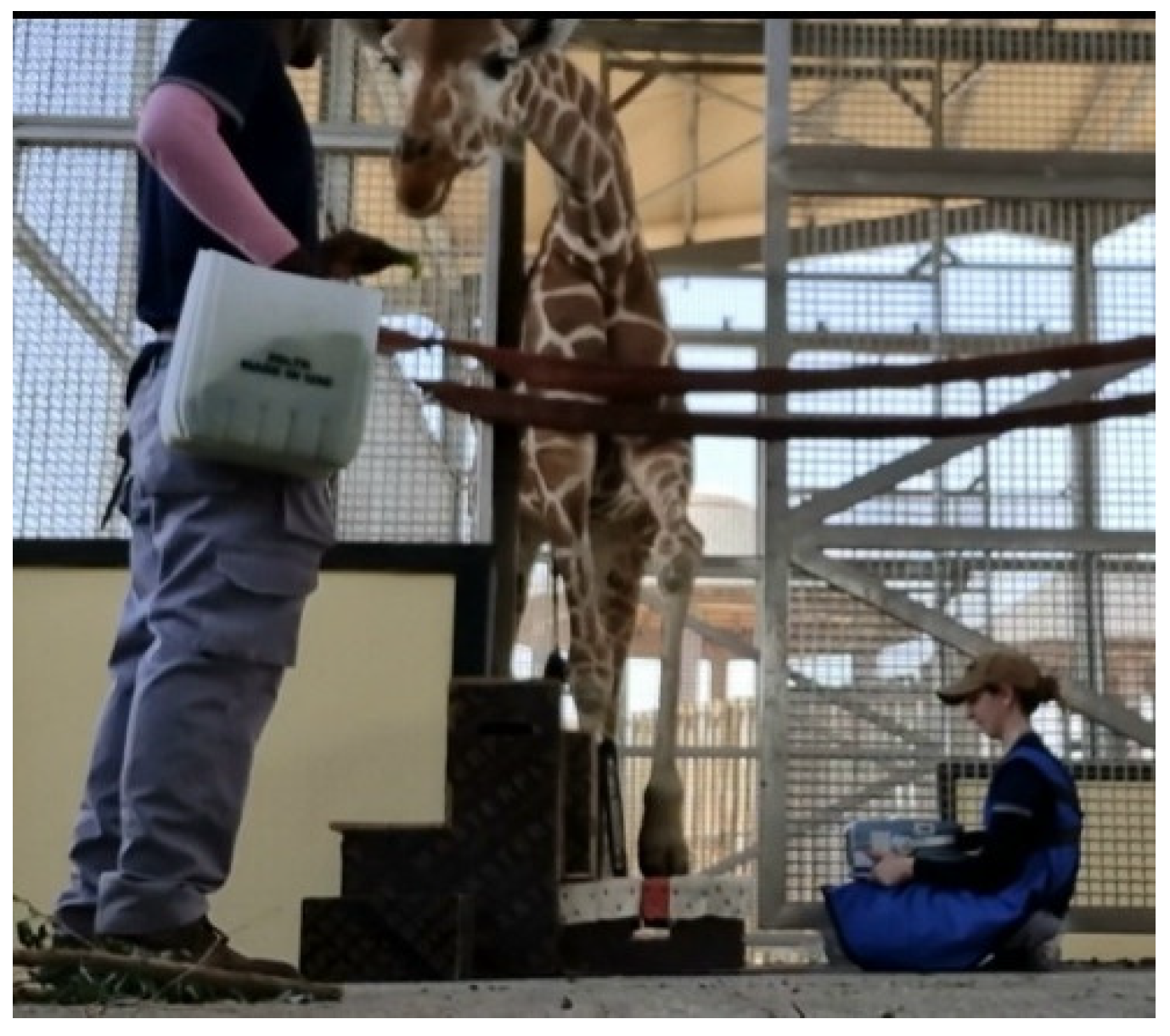
3.6. Front Foot Radiograph

The goal was for the giraffe to stand with its foot on a block for lateral and lateral oblique radiographs. Prior to this step, the giraffe needed to be desensitized to machines and staff being close to her feet while on the block.
The trainers desensitized the giraffe to a second keeper kneeling near the foot block but still semi-protected by the mesh gate barrier. The trainers slowly increased the duration that the foot was on the block. Additionally, the giraffe was desensitized to allowing keepers to move their hands in close proximity to the foot and eventually touch the foot. Throughout this process, the giraffe was reinforced for remaining calm and comfortable.
A combination of desensitization and counter conditioning was used at this stage of the training program. To desensitize the radiograph equipment, the trainers created a mock-up radiograph cassette and unit, which were made to the same measurements as the real devices (Figure 2). This equipment was desensitized slowly over time by having it in view of the giraffe and gradually introducing it during training sessions. This mock cassette and unit were used by trainers during the initial stages of their daily training procedures, allowing the giraffe to become comfortable with the trainer holding the radiograph cassette close to the foot at various heights and angles to obtain different views (Figure 3). During this stage, the giraffe was reinforced for staying in a stationary position and holding the foot on the block while the trainer manipulated the objects.
Initially, an object near the foot or a touch to the foot caused the giraffe to withdraw its foot from the block. After desensitization and counterconditioning, the giraffe learned that allowing objects near the foot and touching the foot were good predictors of food. This led to a positive training experience and a lack of fear response.
Once the giraffe was desensitized to the mock-up equipment, the next step was to introduce the real radiograph equipment. Trainers first desensitized the giraffe to the lead jackets by hanging them on the wall of the stall adjacent to the training area, where they were visible to the giraffe. The trainers then counter conditioned the jackets by holding them while hand-feeding the giraffe, allowing her to investigate the new object while being reinforced. This progressed to the trainers wearing the jackets and reinforcing the giraffe. When the giraffe was comfortable with the jackets, the radiograph equipment was incrementally moved closer to the giraffe while it was continuously given food reinforcement. This created a positive association with the stimulus.
Front foot radiographs were taken once the giraffe was comfortable with people kneeling on either side of the fencing with the necessary equipment and when the foot could be positioned on the block with all necessary staff present (Figure 2). With the giraffe foot in position on the foot block, the radiograph cassette was positioned on or next to the block, and the portable radiograph unit was held from the opposite side of the training doorway. Radiographs did not require any foot handling, as the giraffe would reposition the foot on cue if directed by the trainer.

4. Radiograph Positioning and Techniques

The clinical appearance and severity of lameness were varying, and the giraffe was able to bear weight on its foot. The lameness was mild, with inflammation around the pastern joint of the left forelimb. The giraffe had reduced activity in the enclosure and was observed to be shifting weight repeatedly off the foot, with a head hike associated with each step. To prevent the need for chemical immobilization, the giraffe was trained for front foot radiographs. The trainer holding the radiograph unit knelt on one side of the foot with their body positioned behind the fence, and the radiograph cassette was positioned on the foot block, supported by a wooden structure. A foot block was used to elevate the foot so the hoof sole and the plantar aspects of the distal phalangeal bones could be imaged on the radiograph. Multiple dorsolateral palmaromedial oblique (DMPLO) images of the affected leg were taken in a standing position. The X-ray beam was horizontal and at 90° angle to the dorsopalmar axis of the leg, aiming from dorsolateral position towards palmaromedial position. Radiographs were captured using the PXP-16HF XPRIME Portable Radiograph Machine (NUCTECH™, Burlington, MA, USA) (Figure 3). The distance between the radiograph cassette and the unit was 70 cm. For an adult giraffe, the settings were 76 kVp and 1.6 mAs, but they were adjusted based on image quality.
Figure 3. The PXP-16HF XPRIME Portable Radiograph Machine used to obtain radiographs.
Jzbg 03 00051 g003

Lateral Radiograph (Lateromedial)

The foot was placed flat (palmar) on a foot block with the lateral claw in line with the edge of the block. The secondary keeper kneeled behind a fence lateral to this foot, wearing a lead-lined apron. The radiograph cassette was placed lateral to the foot, perpendicular to the floor. The radiograph beam was directed from the opposite side of the body, parallel to the floor, and was centred at the coronary band (Figure 4).

5. Results

5.1. Animal Participation

The giraffe in this study was trained for radiographs within a period of 3 months. Training sessions were conducted by the primary trainer between 07:00–08:00 h, 4–5 times weekly. Table 1 displays a breakdown of the training results and how many sessions it took to complete each approximation of behaviour. Multiple behaviours were often trained within one session. Trainers recorded the behaviour as complete once they felt that the behaviour was under stimulus control. Training sessions were voluntary, giving the giraffe the option of participating or leaving the session. Trainers aimed to ensure that the sessions were positive for the giraffe, adjusting training criteria to allow the giraffe to succeed and avoid confusion.

5.2. Diagnosis

Lateral front foot radiographs are useful to evaluate hoof overgrowth, distal interphalangeal joint osteoarthritis, and sole foreign bodies [2]. In a normal giraffe foot, the dorsal hoof wall is parallel to the dorsal aspect of the distal phalangeal bones, the bone surfaces are homogenous and smooth, and no debris is embedded in the sole [2]. Lateral and dorsolateral palmaromedial (DLPMO) radiographs were the most diagnostic views. This view is useful for evaluating hoof overgrowth and for diagnosing osteoarthritis of the distal interphalangeal joint, fractures and osteitis of the distal phalangeal bone, rotation of the distal phalangeal bones, sesamoid bone cysts, and sole foreign bodies [2]. There were some limitations in this view due to the positioning of the radiograph generator; future radiographs taken at an angle of 30 degrees can show good radiographs of the hoof with separated paired digits of the foot and pedal osteitis of the solar margins.
The radiographs achieved during this training program (Figure 5), were reviewed by the veterinarians at Dubai Safari Park for evidence of foot pathology and sites of inflammation at the pastern and coffin joints. Results were interpreted together to ensure the homogeneity of the data. A complete clinical examination of the giraffe with the lameness was performed. The images of the pastern joints were assigned as minor abnormal radiographic findings. There were small osteophytes at the border of the pastern joint, and those have little clinical significance and didn’t affect either the appearance or the locomotion. The lameness was resolved within three weeks with joint supplements and soft bedding in the holding.

6. Discussion

6.1. Challenges

6.1.1. Motivation to Train

This animal learned visual and vocal cues very quickly; however, there were variances between the giraffe’s motivation to train and appetite on a particular day. It was hypothesized by trainers that weather may have had an affect on the giraffes’ motivation to train. It was noted that when the temperature and humidity were higher, the giraffe seemed to have less motivation to participate in training. Training motivation was reduced when the giraffe was in estrus. Given that this training program was undertaken to identify causes of lameness, pain may also have decreased this animal’s motivation to participate in the training.
While this giraffe was quick to learn the behaviors, she was easily distracted by other stimuli (keeper vehicles driving past the giraffe holding, other activities occurring inside the holding) during training sessions. The trainers practiced patience with the giraffe and were always able to resume engagement during the session.

6.1.2. Timing and Reinforcement

The success of the training session depended on the skill of the primary trainer. The primary trainer was responsible for leading the session, providing cues for the giraffe, bridging correct behaviours, and reinforcing the giraffe. The primary trainer varied due to the availability of staff. The timing of the bridge was critical when shaping the behaviours; however, incorrect behaviours were occasionally reinforced due to trainer latency with the bridge. When training duration for the target behaviour, the criteria required was for the giraffe to touch the target with the nose and hold this for a set duration. Occasionally the giraffe would lick the target while holding the duration and was sometimes reinforced for this behaviour. This reinforcement varied between primary trainers, and the giraffe became confused. Therefore, maintaining consistent criteria for reinforcement is critical to prevent confusion and facilitate learning.

6.2. Operant Conditioning for Medical Procedures

The husbandry training of zoo animals has been associated with many benefits and is indisputably a valuable tool [8]. Training animals to participate in necessary husbandry and medical procedures can improve the welfare of captive animals [9]. Lameness and hoof overgrowth are common in zoo-housed giraffes [1]. Without training or anaesthesia, a physical examination is limited. Due to large great size and sensitivity to medications, giraffes have a significantly higher risk under anaesthesia [10]. Estimated mortality rates associated with giraffe immobilisations range from 25 to 35% [11]. In zoos, immobilisations to assist with diagnostic radiographs and hoof trims, have led to an estimated 10% mortality rate [12]. Their unique anatomy creates handling problems and puts them at risk of subluxation of cervical vertebrae and their large respiratory dead space adds a physiological disadvantage to safe anaesthesia administration [13]. Furthermore, immobilised giraffes may be exposed to myopathy which is brought on by an anaerobic respiration and build-up of lactic acid, causing excessive exhaustion, stress, and hypothermia [14,15]. Myopathy can be exacerbated by underlying factors such as disease, old age, pregnancy, nutritional deficiencies, and weaknesses caused by parasites [15]. Further stress responses can cause immunosuppression, weight loss, and death [16]. Therefore, trained medical behaviours, using positive reinforcement strategies are key to improving giraffe care. Training animals to “target” and “station” is an important foundation for a variety of health care related tasks [9]. Animals that are reliably trained to target and station can be easily examined both visually and tactilely when they are calmly stationed and positioned at a target [9]. Animals can be trained to present body parts and can be desensitized to objects and apparatus. This allows for examination and the use of non-invasive imaging techniques. Most importantly to the welfare of the giraffe, it allows for repeat treatments without the use of chemical or physical restraint, therefore minimizing stress and the associated risks of anaesthesia [2,9].

6.3. Training as Enrichment

Managing animals under human care within a modern zoo has led to two major behavioural welfare advances: animal training for husbandry procedures and the implementation of animal enrichment to promote a natural behavioural repertoire or reduce undesirable behaviours [17]. Westlund [18] and Fernandez [17] have proposed that animal training is enriching and beneficial to captive animal welfare. Training enriches by providing animals with greater choices and control over their environment, by affording learning opportunities, by expanding behavioural repertoires, and by increasing positive human-animal interactions, as well as by providing a dynamic change in the giraffe’s daily routine in ways that enrich the welfare of those animals. The giraffe in this case study was interested in participating in the training sessions with the keeper and seemed mentally engaged throughout the session, learning the behaviours and cues in a short time span. Training should be recognized as a form of enrichment for giraffes, as it is for other zoo species [18]. Given the choice between working for food during a training session and receiving the identical food item from public feedings, most giraffe chose to train. This preference to work for food is consistent with the contra-freeloading literature [19,20,21].

6.4. Human and Animal Safety

Operant training has been used for hoof care for giraffes at several zoos [1]. Training set-ups included full contact [3] and protected contact in a chute [11], both undertaken without sedation. While working with large animals always has risks and should only be done by trained professionals, the protected contact positive reinforcement training techniques described in this paper provide an alternative way of working with giraffe feet that have safety benefits for both the giraffe and caretakers when compared with full contact or chute-trained hoof work [2]. With a protected contact set up, using mesh and firehose barriers between the keepers and the giraffe’s feet, risk was minimized by having the giraffe’s foot positioned on the foot block. The benefits of a protected contact training set-up may reduce a significant amount of stress when compared to using a restraint chute, as the giraffe is not confined and is allowed the freedom of walking away [3]. During the session, the trainers always monitored for and respected any discomfort or precursors to aggression, thereby averting aggression itself. Indications of discomfort included a weight shift away from the foot on the block (required before the giraffe could kick with either a front or hind leg), tail swishing, skin twitching, ear flapping, or flared nostrils. If these signs were seen, then the training criteria were immediately relaxed, and the giraffe was reinforced for previous approximations within the current session. If necessary, the keepers ended the session early, re-evaluated the criterion, breaking it down into smaller approximations if necessary, and spent more time building confidence with the behaviours that were already trained. This helped to ensure that any discomfort was not allowed to escalate, and that the keeper-giraffe relationship was positive throughout the experience. If there was any poor criteria or signs of regression in behaviour, especially when introducing new criteria, behavioural momentum was re-established by cueing and reinforcing a few high probability behaviours (e.g., target & hold) to allow the giraffe a ‘win’ before asking for the target approximation. Written safety protocols and protected contact setups are strongly recommended to help ensure both human and animal safety throughout the training program.

7. Conclusions

Training medical behaviours using positive reinforcement strategies is key to improving giraffe care. The use of operant conditioning increases patient access for medical treatments and diagnostic imaging techniques, which could aid in the early diagnosis of lameness and allow for early corrective intervention to prevent further progression. Building upon the current training procedures in place, animal keepers have been establishing a foot care program. This will involve removing hoof overgrowth from both the toe tip and sole to help normalize weight distribution in the foot and remove foreign bodies from the sole. The giraffe team at Dubai Safari plans to replicate this training with all individuals within the herd.

Author Contributions

Conceptualization, D.B.; methodology, D.B.; A.K.; H.K. and M.W.A.; resources, D.B. and M.W.A.; writing—original draft preparation, D.B. and M.W.A.; writing—review and editing, D.B.; supervision, A.M.S.; project administration, D.B. All authors have read and agreed to the published version of the manuscript.

Funding

This research received no external funding.

Institutional Review Board Statement

The animal study protocol was approved by the Ethics Committee of DUBAI SAFARI PARK (DSP-RP-2022-001, Approved: 15/04/22).

Informed Consent Statement

Informed consent was obtained from all subjects involved in the study.

Data Availability Statement

Not applicable.

Acknowledgments

We would like to thank our Curator of Ungulates, Anna Mae Sumaya, Head Keeper of Ungulates, Felix Chabaya, Head of Zoology, Frank Rietkerk, Head of Veterinary, Murad Basheer Mustafa and the Dubai Safari Veterinary team for their support and advice throughout this training program. Special thanks to the Giraffe team for their enthusiasm, positivity, and awesome training skills.

Conflicts of Interest

The authors declare no conflict of interest.

References

  1. Bush, M. Giraffidae. In Zoo and Wild Animal Medicine; Miller, R.E., Fowler, M.E., Eds.; Saunders: St. Louis, MO, USA, 2003; Volume 5, pp. 625–633. ISBN 978-072-169-499-3. [Google Scholar]
  2. Dadone, L. Lameness Diagnosis and Management in Zoo Giraffe. In Zoo and Wild Animal Medicine; Miller, R.E., Fowler, M.E., Eds.; Saunders: St. Louis, MO, USA, 2018; Volume 9, pp. 623–629. ISBN 978-032-355-228-8. [Google Scholar]
  3. Phelps, A.; McCartney, M. Using a fusion of operant conditioning and TTEAM to train giraffe calves. TTEAM1 Connect. 2007, 9, 10–16. Available online: https://library.giraffeconservation.org/download/using-a-fusion-of-operant-conditioning-and-tteam-to-train-giraffe-calves/ (accessed on 1 October 2020).
  4. Pryor, K. Don’t Shoot the Dog! In The New Art of Teaching and Training, 1st ed.; Bantam Books: New York, NY, USA, 1999; ISBN 978-055-325-388-7. [Google Scholar]
  5. Peterson, G.B. A day of great illumination: B. F. Skinner’s discovery of shaping. J. Exp. Anal. Behav. 2004, 82, 317–328. [Google Scholar] [CrossRef] [PubMed]
  6. Lind, J.; Ghirlanda, S.; Enquist, M. Insight learning or shaping? Proc. Nat. Acad. Sci. USA 2009, 106, E76. [Google Scholar] [CrossRef] [PubMed] [Green Version]
  7. Fernandez, E.J. Training Petting Zoo Sheep to Act Like Petting Zoo Sheep: An Empirical Evaluation of Response-Independent Schedules and Shaping with Negative Reinforcement. Animals 2020, 10, 1122. [Google Scholar] [CrossRef] [PubMed]
  8. Melfi, V. Is training zoo animals enriching? Appl. Anim. Behav. Sci. 2013, 147, 299–305. [Google Scholar] [CrossRef]
  9. Schapiro, S.J.; Magden, E.R.; Reamer, L.A. Behavioural Training as Part of the Health Care Program. In Management of Animal Care and Use Programs in Research, Education, and Testing, 2nd ed.; Weichbrod, R.H., Thompson, G.A.H., Norton, J.N., Eds.; CRC Press/Taylor & Francis: Boca Raton, FL, USA, 2018; Volume 1, pp. 771–792. [Google Scholar] [CrossRef]
  10. Vitali, F.; Karuiki, E.K.; Mijele, D.; Kaitho, T.; Faustini, M.; Preziosi, R.; Gakuya, F.; Ravasio, G. Etorphine–Azaperone immobilization for translocation of free–ranging Masai Giraffes (Giraffa camelopardalis tippelskirchi): A pilot study. Animals 2020, 10, 322. [Google Scholar] [CrossRef] [PubMed] [Green Version]
  11. Calle, P.P.; Bornmann, J.C. Giraffe restraint, habituation, and desensitization at the Cheyenne Mountain Zoo. Zoo Biol. 1988, 7, 243–252. [Google Scholar] [CrossRef]
  12. Dadone, L.; Olea-Popelka, F.; Stout, E.; Klaphake, E.; Johnston, M.; Barrett, M. A wake-up call: Radiographic evidence of front foot fractures and osteoarthritis in relatively young reticulated giraffe (Giraffa camelopardalis reticulata). In Proceedings of the Annual Conference American Association of Zoo Veterinarians, AAZV, Atlanta, Georgia, 16–22 July 2016; p. 130. Available online: https://www.researchgate.net/publication/328108103_A_wake-up_call_radiographic_evidence_of_front_foot_fractures_and_osteoarthritis_in_relatively_young_reticulated_giraffe (accessed on 18 October 2022).
  13. Muller, Z.; Bercovitch, F.; Brand, R.; Brown, D.; Brown, M.; Bolger, D.; Carter, K.; Deacon, F.; Doherty, J.B.; Fennessy, J.; et al. Giraffa camelopardalis (amended version of 2016 assessment). IUCN Red List. Threat. Species 2018, 6, 134–139. [Google Scholar] [CrossRef]
  14. Paterson, J. Capture Myopathy. In Zoo Animal and Wildlife Immobilization and Anesthesia, 2nd ed.; West, G., Heard, D., Caulkett, N., Eds.; Blackwell Publishing: Hoboken, NJ, USA, 2007; pp. 115–121. [Google Scholar] [CrossRef]
  15. Deacon, F.; Daffue, W.; Nell, P.; Higgs, R. Effective Field Immobilisation and Capture of Giraffe (Giraffa camelopardalis). Animals 2022, 12, 1290. [Google Scholar] [CrossRef] [PubMed]
  16. Kagan, R.; Veasey, J. Challenges of zoo animal welfare. In Wild mammals in Captivity: Principles and Techniques for Zoo Management; Kleiman, D.G., Thompson, K.V., Baer, C.K., Eds.; University of Chicago Press: Chicago, IL, USA, 2010; pp. 11–21. ISBN 978-022-644-010-1. [Google Scholar]
  17. Fernandez, E.J.; Upchurch, B.; Hawkes, N.C. Public Feeding Interactions as Enrichment for Three Zoo-Housed Elephants. Animals 2021, 11, 1689. [Google Scholar] [CrossRef] [PubMed]
  18. Westlund, K. Training is enrichment—And beyond. Appl. Anim. Behav. Sci. 2014, 152, 1–6. [Google Scholar] [CrossRef]
  19. Coulton, L.E.; Warren, N.K.; Young, R.J. Effects of foraging enrichment on the behavior of parrots. Anim. Welf. 1997, 6, 357–363. Available online: https://www.academia.edu/33996118/Parrot_enrichment_animal_welfare_pdf (accessed on 1 October 2022).
  20. Gilbert-Norton, L. Captive birds and freeloading: The choice to work. Res. News 2003, 4, 1. [Google Scholar]
  21. Tresz, H. Contra freeloading (working for food) at the Phoenix Zoo. ABMA Wellspring 2010, 11, 8–21. Available online: https://www.researchgate.net/publication/264788873_Contra_Freeloading_Working_for_Food_at_the_Phoenix_Zoo (accessed on 2 October 2022).
Figure 1. The Protected Contact set-up for giraffe footcare at Dubai Safari Park.
Jzbg 03 00051 g001
Figure 2. The Mock-up Radiograph Cassette and Unit set up with animal keepers demonstrating use during the early stages of desensitization.
Jzbg 03 00051 g002
Figure 4. The direction of the radiograph beam, centred on the coronary band.
Jzbg 03 00051 g004
Figure 5. Radiographs (DLPMO view) taken for a 22-year-old female reticulated giraffe (Giraffa camelopardalis reticulata) left forelimb in the United Arab Emirates. (A,B) shows abnormal growth of osteophytes at the border of the shorter pastern bone. All articulations of the joints are in good condition.
Jzbg 03 00051 g005
Table 1. Behaviour steps, descriptions and sessions as described in the Shaping Plan.
Behaviour StepsDefinitionNumber of Sessions
TargetInstant Touch9
Target—Duration 2 s15
Target—Duration 4 s7
Target—Duration 6 s9
Target—Duration 8 s7
Target—Duration 10 s9
RecallGiraffe walks towards Trainer10
Back UpGiraffe walks backwards from Trainer12
StationGiraffe stands at the stationing area, slightly leaning into
chest straps
10
Lift FootGiraffe Lifts foot and places it onto the foot block15
Lift Foot & HoldGiraffe holds foot on block for 2 s6
Giraffe holds foot on block for 5 s8
Giraffe holds foot on block for 10 s11
Giraffe holds foot on block for 15 s10
Giraffe holds foot on block for 20 s11
Giraffe holds foot on block for 30 s15
Front Foot RadiographGiraffe holds foot on block while Trainer moves radiograph machine close to foot12
Publisher’s Note: MDPI stays neutral with regard to jurisdictional claims in published maps and institutional affiliations.

