Article

Coordinated Th1- and Th17-Related Responses Support Antibody- and Neutrophil-Mediated Protection Against Pneumococcal Pneumonia


by
Analía Rial
*,
María Paula Céspedes
,
Victoria Comas
,
Mariana Rivera-Patrón
,
Juan Martín Marqués
and
José Alejandro Chabalgoity
Biotechnology Department, Instituto de Higiene, Facultad de Medicina, Universidad de la República, Montevideo C.P. 11600, Uruguay
*
Author to whom correspondence should be addressed.
Immuno 2026, 6(2), 41; https://doi.org/10.3390/immuno6020041
Submission received: 22 March 2026 / Revised: 14 May 2026 / Accepted: 26 May 2026 / Published: 9 June 2026
(This article belongs to the Section Infectious Immunology and Vaccines)

Abstract

Streptococcus pneumoniae is a leading cause of community-acquired pneumonia, yet the immune mechanisms required for protection against invasive pulmonary infection remain inadequately understood. Using a murine model of homologous protection against invasive pneumococcal pneumonia, we explored the relative contributions of humoral and cellular immunity using adoptive serum transfer, immune cell depletion, and lung transcriptional profiling. Our findings indicated that passive transfer of immune serum provided robust protection, while neutrophil depletion significantly compromised bacterial control, highlighting that both antibodies and neutrophils are key mediators of protection. In contrast, depletion of CD4+ T cells or NK cells did not compromise survival. Although IL-17A has been widely implicated in host defense against pneumococcal infection, IL-17A-deficient mice remained protected, albeit with delayed clearance and reduced early antibody responses. We associate this delay with compensatory upregulation of IL-17F and increased expression of Th1-associated genes in the lungs. Together, these findings indicate that IL-17A is not essential for protection and support a model in which coordinated Th1- and Th17-related cytokine responses collectively promote neutrophil recruitment and effective antibody-mediated defense. These results highlight functional redundancy within the IL-17 cytokine axis and suggest that integrated cytokine networks, rather than individual mediators, underpin protective immunity to pneumococcal pneumonia, with implications for next-generation vaccine design.

1. Introduction

Streptococcus pneumoniae (Pn) is a leading cause of community-acquired pneumonia and remains a major global health concern, particularly among vulnerable populations such as children under five years of age [1,2,3]. The World Health Organization (WHO) has classified S. pneumoniae as one of the twelve priority pathogens [4], and pneumococcal infections account for more than one million deaths annually worldwide [5]. Pneumococcal diseases encompass a wide spectrum of clinical manifestations, including pneumonia, meningitis, septicemia, and otitis media [6].
S. pneumoniae frequently colonizes the human nasopharynx, especially in children. In most cases, asymptomatic colonization is cleared by the host immune response within weeks in adults or months in children [7]. However, under certain conditions, such as viral infections or immunosuppression, bacteria may disseminate from the nasopharynx to other anatomical sites. This dissemination can result in pneumococcal pneumonia when bacteria invade the lungs or, in some cases, lead to meningitis after spreading to the central nervous system [8,9]. More than 100 serotypes have been identified, which differ in their capsular polysaccharides and their ability to colonize or cause invasive pneumococcal disease (IPD) [10,11].
In particular, S. pneumoniae serotype 1 (Pn1) remains one of the most prevalent invasive serotypes globally, especially in West and Sub-Saharan Africa [10,12] and Asia [13]. In South America, particularly in Uruguay, this serotype was among the most prevalent associated with IPD, at least prior to widespread vaccination [14,15,16,17,18]. The introduction of pneumococcal conjugate vaccines (PCVs), which target capsular polysaccharides, has substantially reduced the incidence of invasive pneumococcal disease caused by vaccine serotypes, including serotype 1 [19]. Although vaccine-induced protection has traditionally been attributed to serotype-specific opsonizing antibodies, increasing evidence suggests that CD4+ T cell-mediated immunity also contributes to protection against pneumococcal infection, especially at mucosal sites such as the lungs [20].
Historically, protective immunity against pneumococcus has been largely attributed to antibody-mediated opsonophagocytosis. While this mechanism is essential for clearing bacteremia, it may be insufficient to fully control infection at mucosal surfaces, where host–pathogen interactions are initiated [8]. This limitation has prompted growing interest in T helper cell responses that coordinate local inflammatory and antimicrobial defenses in the lungs.
CD4+ T cells differentiate into multiple subsets characterized by distinct cytokine profiles and effector functions. T helper 1 (Th1) cells, defined by their production of interferon-gamma (IFN-γ), activate macrophages and promote IgG2a/c class-switching, thereby enhancing opsonophagocytic killing [21]. Although capsular polysaccharides are generally considered T cell-independent antigens, the serotype 1 capsular polysaccharide (PnPS1) displays zwitterionic properties that allow for presentation on MHC class II molecules and activation of CD4+ T cells [22,23]. In contrast, T helper 17 (Th17) cells, which secrete interleukin-17A (IL-17A) and IL-17F, play a central role in mucosal host defense. These cells promote neutrophil recruitment by inducing the production of CXC chemokines such as CXCL1 and CXCL2, and by stimulating epithelial cells to produce antimicrobial peptides that reinforce the mucosal barrier [24,25,26].
Growing evidence suggests that effective immunity to pneumococcal infection involves coordinated activity among multiple T helper cell pathways. Th1 and Th17 responses have been shown to act synergistically in several models of bacterial infection [27,28]. The importance of IL-17A in protection against nasopharyngeal pneumococcal carriage is well established [21,29,30,31], though its role in protection against invasive pneumococcal pneumonia is less clearly defined [32]. Several studies suggest that IL-17A can exert context-dependent effects, being either protective or detrimental depending on bacterial strain characteristics and capsule thickness [33]. A pathogenic role for IL17A has been described in inflammatory lung diseases such as cystic fibrosis and severe asthma [34,35], where it can amplify inflammatory signaling pathways, including those driven by IL-13 [36].
Based on these observations, we hypothesize that homologous protection against invasive pneumococcal pneumonia requires a coordinated T cell response that integrates Th1-mediated macrophage activation with Th17-associated mechanisms that promote neutrophil recruitment and mucosal defense. Here, we demonstrate that IL-17A alone is not required for protection against homologous invasive pneumococcal pneumonia. Instead, our results indicate that IL-17-associated immune responses, even in the absence of IL-17A, contribute to protection through enhanced neutrophil recruitment and the induction of anti-capsular antibodies.

2. Materials and Methods

2.1. Animal Studies

Female C57BL/6J mice (6–8 weeks old) were obtained from the National Division of Veterinary Laboratories (Montevideo, Uruguay). Il17a−/− and Casp1/11−/− mice on a C57BL/6J background were provided by Institut Pasteur Montevideo, Uruguay (IPMont). All animals were housed in individually ventilated cages and handled in a class II A2 vertical laminar flow cabinet (ESCO), as previously described [27,37]. Anesthesia was induced by intraperitoneal (i.p.) injection of ketamine (2.2 mg) and xylazine (0.11 mg), as previously described [27,37]. Bacteria were administered intranasally (i.n.) in 50 μL of physiological saline (25 µL per nostril). Survival was monitored daily. For lung sampling, euthanasia was performed by cervical dislocation at the specified time points. Lungs were collected and homogenized using a cell dissociation sieve–tissue grinder kit (Sigma-Aldrich, St. Louis, MO, USA), and serial dilutions were plated on blood agar to determine bacterial loads. S. pneumoniae colonies were identified by their characteristic α-hemolytic green halo. For transcriptional analysis, lungs were stored in TRIzol reagent (Life Technologies, Carlsbad, CA, USA); for flow cytometry, lungs were kept on ice until processing, as described below. All animal experiments were conducted in compliance with national and institutional regulations and approved by the Comisión Honoraria de Experimentación Animal (CHEA)—Universidad de la República, Uruguay (Exp. Nº 07015300081914), in accordance with ARRIVE guidelines.

2.2. Bacterial Strains and Culture Conditions

Streptococcus pneumoniae serotype 1 (Pn1, clinical isolate E1585) was obtained from the National Reference Laboratory (Ministry of Health, Uruguay). Working stocks were prepared as previously described [27,38]. Briefly, Todd Hewitt Yeast Broth (THYB) was inoculated with fresh colonies of S. pneumoniae grown on blood agar and incubated at 37 °C until the optical density at 600 nm (OD600) reached 0.7–0.9. Cultures were stored at −80 °C in THYB with 12% (v/v) glycerol for up to 3 months. For infections, frozen stocks were thawed, centrifuged (5 min at 2500× g), washed, and diluted in saline to obtain 2 × 10⁴ CFU/50 µL for the Pn1 sublethal dose (priming) or 2 × 10⁷ CFU/50 µL for the Pn1 lethal dose (challenge). Bacterial counts in the inoculum were determined for every experiment by plating serial dilutions onto blood agar plates.

2.3. Adoptive Transfer of Immune Sera to Naïve Mice

Blood was collected from the facial veins or retro-orbital sinus of mice 7 days after the administration of saline (control serum) or a sublethal dose of Pn1 (immune serum). Serum from each group (n = 15/group) was pooled, aliquoted, and stored at −80 °C until use. For adoptive transfer in vivo assays, groups of seven naïve mice were intravenously (i.v.) injected with 0.5 mL of immune or control serum on day 0 and challenged with a lethal Pn1 dose (2 × 10⁷ CFU) on day 1. Survival was monitored daily for one week. Experiments were independently performed three times.

2.4. Depletion of Gr-1+, NK or CD4+

For granulocyte depletion, mice received an i.p. injection of 100 μg purified anti-Gr-1 antibody (clone RB6-8C5) or an isotype control antibody (clone HB152), 24 h before intranasal challenge with S. pneumoniae (2 × 10⁷ CFU), as previously described [27,39]. Similarly, depletion of natural killer (NK) or CD4 cells was achieved by i.p. injection of 100 µg of purified anti-NK (clone PK136) antibody, anti-CD4 (clone GK1.5) antibody, or isotype control (clone HB152) 24 h before challenge. Purified antibodies were kindly provided by Dr. Jean Claude Sirard (Institut Pasteur, Lille, France) and used as instructed. Depletion efficiency was confirmed by flow cytometry (Figure S1).

2.5. Detection of IgG and IgM Antibodies by ELISA

Antibodies specific to pneumococcal polysaccharide type 1 (PnPS1) were measured using a mouse-adapted version of the 3rd-generation WHO ELISA protocol to quantify serotype-specific IgG levels, as previously described [27]. Briefly, 96-well microtiter plates were coated overnight at 4 °C with PnPS1 antigen (American Type Culture Collection [ATCC], Manassas, VA, USA; 2 µg/mL). Serum samples were pre-absorbed with cell wall polysaccharide (C-PS, Statens Serum Institute, Copenhagen, Denmark; 5 µg/mL) and type 22F polysaccharide (ATCC; 5 µg/mL) for 30 min at room temperature. After extensive washing with Tris-buffered saline containing 0.01% Brij-35, HRP-conjugated antibodies against mouse IgG, IgM, or IgA were added. The reaction was developed using TMB substrate (3,3′,5,5′–tetramethylbenzidine, Sigma-Aldrich, St. Louis, MO, USA), stopped with 1M H2SO4, and absorbance was measured at 450 nm (reference 630 nm). Standard curves generated from in-house reference sera were used for quantification.

2.6. RNA Extraction and Reverse Transcription–Quantitative Real-Time PCR

Lungs were homogenized in TRIzol reagent (Life Technologies, Carlsbad, CA, USA) as previously described [27,40,41]. Total RNA was extracted according to the manufacturer’s instructions, quantified, and 1 µg was treated with DNase I (Life Technologies, Carlsbad, CA, USA). First-strand cDNA was synthesized using random primers (Life Technologies, Carlsbad, CA, USA) and M-MLV reverse transcriptase (Life Technologies, Carlsbad, CA, USA) (cycling: 10 min at 25 °C; 50 min at 37 °C; 15 min at 70 °C). Real-time PCR was performed using the QuantiTect SYBR Green PCR kit (Qiagen, Venlo, The Netherlands) and a 7900HT instrument (Applied Biosystems, Foster City, CA, USA) (cycling: 15 min at 95 °C, then 40 cycles of 95 °C for 15 s and 60 °C for 1 min; fluorescence acquisition at 60 °C) with specific primers (0.9 μM, sequences available upon request). Gene expression was normalized to Actb (β-actin) using the 2−ΔΔCt method [42], with a cut-off of 33 cycles. Results were expressed as mRNA fold increase relative to the saline-treated WT group. Experiments were independently performed three times.

2.7. Flow Cytometry Analysis of Lung Cells

At the indicated time points after challenge, mice were euthanized, and the pulmonary vasculature was perfused with saline containing 1 mM EDTA to remove intravascular cells. Lung cells were isolated following collagenase/DNase digestion, as previously described [27,40]. Cells were resuspended in FACS EDTA buffer (PBS, 0.1% azide, 1% fetal calf serum, 5 mM EDTA) and counted using a Countess Automated Cell Counter (Invitrogen, Waltham, MA, USA) prior to immunophenotypic analysis by flow cytometry. Neutrophils were defined by forward scatter area (FSC-A) and side scatter area (SSC-A) profile, high Ly6G and CD11b expression, and absence of CD11c expression. Samples were acquired on a FACS Canto II (BD) flow cytometer equipped with 488 nm and 635 nm excitation lasers. Data acquisition and analysis were performed using FACS Diva software (BD) (v 6.0). The experiments were conducted independently three times.

2.8. Statistical Analysis

Statistical analyses were performed using GraphPad Prism (v11.0.0). Differences between groups were evaluated using the non-parametric Mann–Whitney U test, Student’s t-test, or Kruskal–Wallis test with Dunn’s post-test, as appropriate and as described in each figure legend. Differences were considered statistically significant at p < 0.05 (*) and highly significant at p < 0.01 (**).

3. Results

3.1. Immune Serum and Neutrophils Are Independently Essential for Protection Against Lethal Pneumococcal Pneumonia

Anti-capsular antibodies facilitate opsonization of S. pneumoniae, promoting bacterial clearance via phagocytosis. We previously reported that wild-type (WT) mice primed with a sublethal dose of S. pneumoniae serotype 1 (Pn1) were completely protected against a homologous lethal challenge given 1 or even 8 weeks later. In contrast, naïve WT mice treated with saline (control) and subsequently challenged with the lethal Pn1 dose developed invasive pneumonia and died within 48–72 h [27].
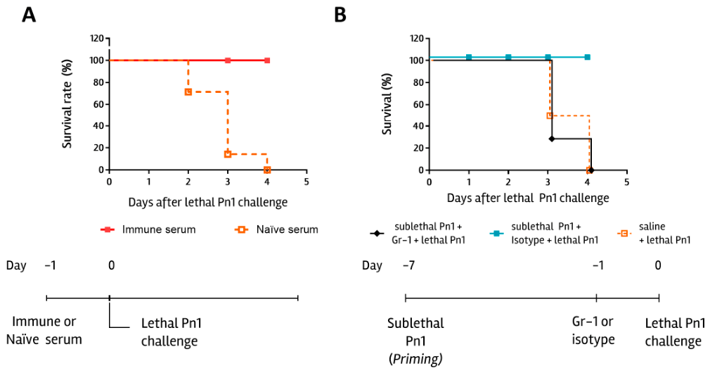
To further elucidate the mechanisms underlying protection against homologous invasive pneumococcal pneumonia, we performed adoptive serum transfer experiments. As previously described, priming with a sublethal dose of Pn1 induced serum-specific anti-capsular IgG and IgM antibodies, as well as antibodies against whole-cell pneumococcal antigens [27]. Serum collected 7 days after priming was designated as immune serum. Here, we show that naïve mice receiving immune serum prior to a lethal Pn1 challenge achieved complete survival (Figure 1A). These findings demonstrate that antibodies generated 7 days after sublethal pneumococcal infection are sufficient to confer protection against homologous invasive pneumonia, at least when administered 24 h before the challenge.
Neutrophil (PMN)-mediated opsonophagocytosis is a key mechanism in controlling S. pneumoniae infection [43,44,45]. Consistent with previous studies [27], we confirmed massive neutrophil recruitment to the lungs following a lethal pneumococcal challenge. To determine whether PMNs were required for protection in this model, neutrophils were depleted using anti-Gr-1 antibodies in sublethally Pn1-primed WT mice before the homologous lethal challenge. PMN depletion resulted in complete loss of protection, with no survival in depleted mice (Figure 1B). These findings confirm that neutrophils are essential mediators of homologous protection against invasive pneumococcal pneumonia.

3.2. CD4+, Il17a, and Casp-1/11 Are Not Exclusively Essential for Protection

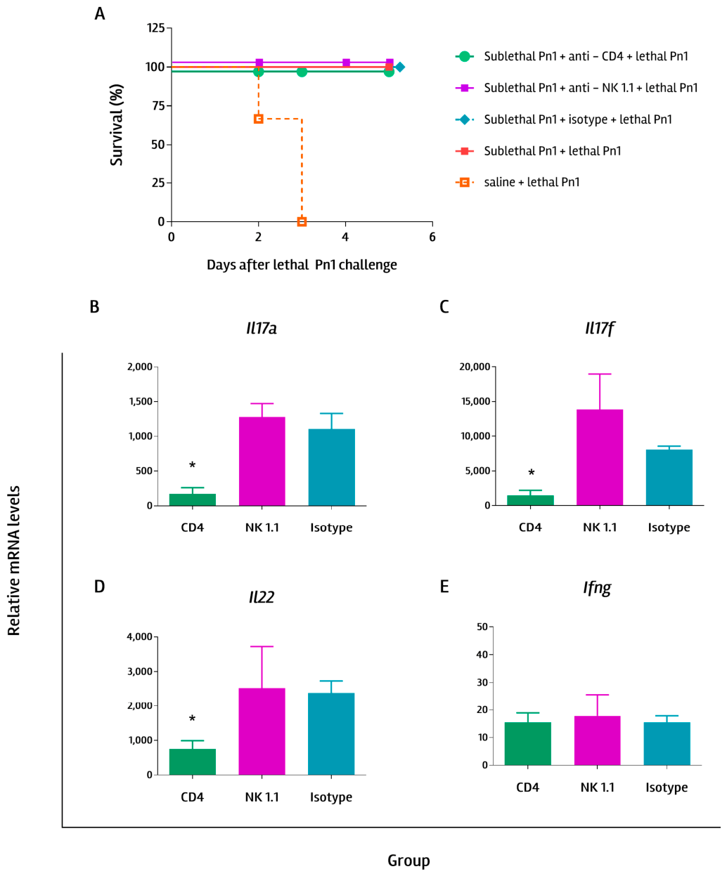
Additionally, we found that administration of anti-CD4 or anti-NK-1.1 antibodies before the lethal Pn1 challenge did not eliminate protection in WT mice (Figure 2A). However, CD4 depletion before the challenge resulted in a significant decrease in Il17a (Figure 2B), Il17f (Figure 2C), and Il22 (Figure 2D) lung mRNA levels, with no change in Ifng mRNA levels (Figure 2E), compared with sublethally Pn1-primed mice receiving isotype control. These findings confirm our previous observation that CD4+ cells are a major source of the rapid IL-17A production detected in the lungs of protected mice [27]. Studies using a serotype 23F strain of S. pneumoniae showed that homologous protection against non-invasive pneumonia depends on antibodies [28,46] and T-helper type 17 (Th17) CD4+ cells [28]. Our data indicate that, while CD4+ cells can be considered major producers of Il17a, Il17f, and Il22, they are not required for homologous protection against invasive Pn1 challenge. To further validate these findings, future studies should consider sustained depletion of CD4+ T cells and/or NK cells, starting before the sublethal Pn1 dose and maintained throughout the experiment via subsequent antibody injections, rather than only a single dose before the challenge, as used here.
Inflammasomes have been described as critical components of the innate immune response during pneumococcal infection, in part due to NLRP3 sensing of hemolytic pneumolysin [47,48] or NLRP6 sensing of lipoteichoic acid, both of which are key virulence factors of S. pneumoniae. At the post-translational level, assembly of these complexes triggers caspase-1 activation, which mediates the maturation of IL-1β and IL-18 while concurrently initiating gasdermin D-dependent pyroptosis [49]. The non-canonical caspase-11 pathway often augments this canonical signaling. We assessed Casp1 expression in protected WT mice and found that they exhibited higher Casp1 mRNA levels than naïve or non-protected mice (Supplementary Figure S2A). We then evaluated protection in syngeneic Casp1/11−/− mice. Both WT and Casp1/11−/− mice primed with a sublethal Pn1 dose and subsequently challenged with a homologous lethal dose showed identical survival rates (Supplementary Figure S2B). Consequently, these results indicate that inflammasome-mediated signaling is dispensable for this robust homologous protection.
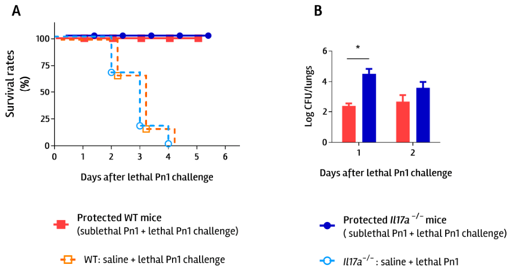
Il17a was highly upregulated in mice protected against invasive pneumococcal pneumonia, suggesting a potential association between increased IL-17A levels and protection in this model [27]. Th17 responses have been widely associated with protection against pneumococcal infection [50]. To further evaluate the role of IL-17A in homologous protection against invasive pneumococcal pneumonia, we used syngeneic Il17a-deficient mice (Il17a−/−). Il17a−/− control mice (treated with saline) developed acute invasive pneumonia, similar to WT control mice, when challenged with a lethal Pn1 dose, and also died 48–72 h after infection (Supplementary Figure S3A). Interestingly, Il17a−/− mice previously primed with a sublethal dose of Pn1 exhibited the same survival rates as primed WT mice when subsequently challenged with a homologous lethal Pn1 dose (protected WT and Il17a−/− mice; Figure 3A). However, protected WT mice showed significantly lower lung bacterial loads than protected Il17a−/− mice when evaluated 24 h after the lethal challenge (Figure 3B). These findings suggest that the absence of IL17A delays bacterial clearance from the lungs, although both WT and Il17a−/− mice ultimately achieved 100% survival.

3.3. IL-17f Could Be Compensating for the Lack of IL17A for Protection

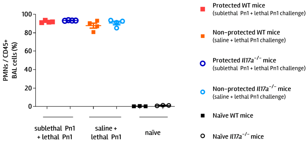
A well-described function of IL-17A is the recruitment of PMNs, so we assessed their recruitment into alveolar spaces. Flow cytometry analysis of bronchoalveolar lavage (BAL) samples from Il17a−/− and WT mice revealed similar PMN recruitment in both protected groups (Figure 4), suggesting that mediators other than IL-17A may compensate for its absence in Il17a−/− mice.
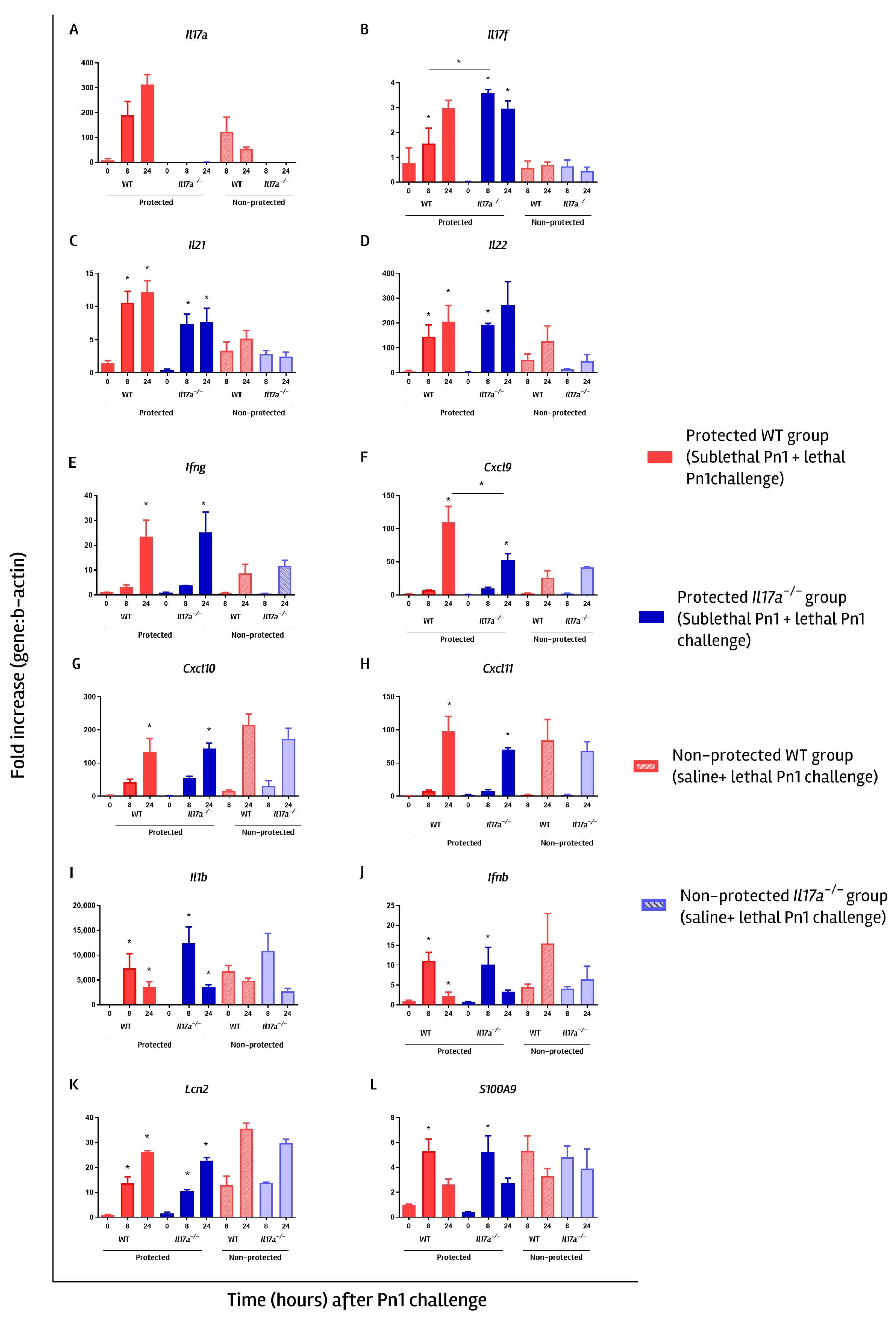
We next compared the lung transcriptional profiles between protected WT and Il17a−/− mice, defined as those primed with a sublethal Pn1 dose and surviving the subsequent challenge with a lethal Pn1 dose on day 7. Overall, both strains displayed highly similar expression patterns. However, protected Il17a−/− mice showed a more rapid increase in Il17f mRNA levels than wild-type (WT) mice, as measured 8 h after lethal challenge, although both reached similar levels at 24 h (Figure 5A). For other Th17-associated cytokines, similar increases in Il21 (Figure 5C) and Il22 (Figure 5D) mRNA levels were observed, while Il17a mRNA transcripts were absent in Il17a−/− mice (Figure 5A). Additionally, both groups of protected mice showed similar increments in relative mRNA levels for Ifng (Figure 5E) and Ifng-related genes (Cxcl9, Cxcl10, Cxcl11; Figure 5F–H) as well as for the antimicrobial peptide Lipocaline 2 (Lcn2; Figure 5K). For S100A9, both mouse strains exhibited a rapid, significant increase at 8 h, followed by a decrease by 24 h (Figure 5L), similar to the pattern observed for Il1b and Ifnb mRNA levels (Figure 5I,J). Previous studies suggest that type I interferons contributed to host defense against pneumococcal infection by limiting bacterial dissemination from the lungs to the bloodstream [51]. Accordingly, the rapid induction of Ifnb mRNA levels observed in both protected WT and Il17a−/− mice (Figure 5J) may help prevent systemic dissemination.
Overall, protected Il17a−/− and WT mice exhibited highly similar transcriptional profiles, characterized by increased expression of Th17-associated genes, Ifng and Ifng-related genes, and antimicrobial peptides. Thus, despite the absence of IL-17A, our data suggest that Il17a−/− mice mount compensatory “Th17-like” responses together with Th1 responses that may account for the protective immunity observed after homologous lethal challenge.

3.4. Il17a−/− Mice Showed a Delayed Anti-Capsular Antibody Response

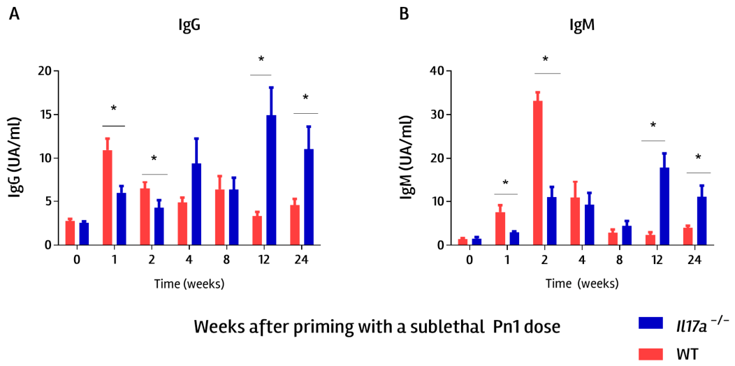
Our results suggest that serum antibodies are protective against invasive pneumonia, at least in our mouse model (Figure 1A). We then compared the induction of specific antibodies in serum from Il17a−/− and WT mice, both of which were primed with the sublethal Pn1 dose. Results showed significant induction of IgG and IgM anti-capsular antibodies in primed Il17a−/− mice, though with some differences in kinetics compared to WT (Figure 6). At earlier time points (1 and 2 weeks after sublethal Pn1 priming), WT mice showed higher levels of anti-capsular IgG and IgM antibodies compared to Il17a−/− (Figure 6). These results suggest that Il17a−/− mice are less efficient than WT in rapidly generating anti-capsular antibodies after priming, which could explain the delayed clearance of bacteria from the lungs in Il17a−/− mice (Figure 3B). However, at later time points (12 and 24 weeks after sublethal Pn1 dose), Il17a−/− mice showed increased antibody levels compared to WT, indicating that Il17a−/− mice are not impaired in their ability to mount an antibody response, consistent with findings in other studies using this deficient mouse strain [52]. Further studies are required to elucidate the mechanism underlying the delayed anti-capsular response observed in Il17a−/− mice.

4. Discussion

S. pneumoniae is a major human pathogen whose principal virulence factor is its capsular polysaccharide, largely due to its anti-phagocytic properties. Consequently, current pneumococcal vaccines are based on capsular polysaccharides from the most clinically significant serotypes and are effective at preventing invasive manifestations of pneumococcal disease [53]. While the immune mechanisms underlying protection against S. pneumoniae infection have been extensively investigated, the relative contributions of humoral and cellular immunity to defense against invasive pneumococcal pneumonia are not fully defined [28]. In this study, we further elucidated these mechanisms using a well-established murine model of homologous protection against invasive pneumococcal pneumonia [27], with a particular focus on the roles of anti-capsular antibodies, neutrophils (PMNs), CD4+ and NK cells, inflammasome activation, and IL-17A.
Our results reinforce the pivotal roles of antibodies and neutrophils in homologous protection against invasive pneumococcal pneumonia. Sublethal pneumococcal infection, which is rapidly cleared from the lungs, induces serum-specific antibodies that are sufficient to confer protection against a subsequent lethal homologous challenge, as demonstrated by passive serum transfer (Figure 1A). In parallel, neutrophil depletion completely abrogated protection, as Gr-1-treated mice failed to survive the homologous lethal Pn1 challenge (Figure 1B). Lethal Pn1 challenge induced invasive pneumonia and rapid PMN recruitment to the lungs (Figure 4). In mice without prior sublethal Pn1 priming, bacteria were not efficiently cleared from the lungs, and animals succumbed to infection. While neutrophils are essential for bacterial clearance, they can also contribute to tissue damage by releasing inflammatory mediators such as superoxide and neutrophil elastase [54]. Excessive lung inflammation is partly responsible for the lethality observed during pneumococcal pneumonia [8] and may promote bacterial dissemination to the bloodstream, exacerbating disease severity [55,56]. In our model, antibodies in immune serum likely facilitated opsonization and efficient phagocytosis of pneumococci by recruited neutrophils. In contrast, in mice receiving naïve serum, recruited PMNs failed to clear bacteria, and the resulting inflammation likely contributed to mortality. Rapid clearance of bacteria from the lungs is a key determinant of outcome. Moreover, in Gr-1-treated mice, despite the presence of anti-pneumococcal antibodies (induced by prior sublethal Pn1 priming), the absence of functional neutrophils is directly associated with loss of protection.
Overall, our findings and those of others [57] highlight that neutrophils are central determinants of both protection and pathogenesis against S. pneumoniae infection. Our data specifically demonstrate that their recruitment is essential for protection against homologous invasive pneumococcal pneumonia.
Previous studies have shown that Th17 cells play a protective role in pulmonary infections [58]. With respect to S. pneumoniae serotype 1, we previously described an association between IL17A and homologous protection against invasive pneumococcal pneumonia [27]. However, other authors have described a context-dependent, dual role for IL-17A during pneumococcal pneumonia that may depend on serotype, particularly on capsule thickness [33]. These authors described a detrimental role for IL17A in a mouse model of pneumococcal pneumonia induced by a serotype 3 strain. They propose that PMNs cannot effectively phagocytose bacteria because of the thickness of the bacteria’s capsule. Hence, recruited lung PMNs are unable to effectively phagocytose bacteria, resulting in lung damage without efficient bacterial clearance [33]. In contrast, serotype 1 expresses less capsular polysaccharide [59,60], which may enable more effective antibody-mediated opsonophagocytosis.
Importantly, our results indicate that IL17A is not strictly required for protection: sublethally Pn1-primed Il17a−/− mice, as well as WT mice, were completely protected against a homologous lethal challenge. Protected Il17a−/− mice exhibited what we consider Th17-like responses, with increased expression of Il17f, Il21, and Il22, as well as Th1-associated genes (including Ifng and other Ifng-related genes) (Figure 5). These observations support the existence of compensatory cytokine pathways that can maintain protective immunity in the absence of IL-17A.
IL-17A and IL-17F are both members of the IL-17 family of cytokines, sharing the highest sequence homology (58%) among family members, and are primarily produced by Th17 cells [25,58,61,62]. It has been shown that IL-17F can induce PMN recruitment and activation similarly to IL-17A [63], which could explain the comparable PMN recruitment observed in protected Il17a−/− and WT. Administration of IL-17F has been demonstrated to protect against S. pneumoniae infection [43]. Interestingly, other studies have reported decreased PMN recruitment in Il17ra−/− mice during pneumococcal infection [33], which contrasts our findings. Future studies assessing protection in Il17a−/− Il17f−/− double knockout mice or Il17ra−/− mice would clarify these mechanisms. Additionally, IL-22, another Th17 cytokine upregulated in Il17a−/− mice, acts on non-hematopoietic cells such as lung epithelial cells and fibroblasts, further contributing to mucosal defense [58].
Our data also suggest that IL-17A contributes to the early clearance of bacteria from the lungs, as protected Il17a−/− mice exhibited higher lung bacterial loads than protected WT mice (Figure 3B). Additionally, IL-17A may facilitate the rapid induction of anti-capsular antibodies, since Il17a−/− mice showed a delayed anti-capsular response (Figure 6). There is a very interesting report suggesting a still-unappreciated role for Th17 cells and their signature cytokines in mediating B-cell differentiation and class switch recombination [64]. Based on their results, they propose that Th17 cells not only promote B cell proliferation but also support germinal center (GC) formation and isotype switching to IgG subclasses [64]. Specifically, IL-17A drives class-switch recombination to IgG2a and IgG3, while IL-21 promotes switching to IgG2b and IgG1 [64]. Notably, pneumococcal capsular polysaccharide 1 (PnPS1) is zwitterionic [65], enabling its presentation on MHC class II molecules and activation of CD4+ T cells [22,23,66]. Although polysaccharide antigens are typically considered T-independent type 2 antigens, the zwitterionic properties of PnPS1 may be related to the delayed anti-capsular response observed in Il17a−/− mice following priming with a sublethal dose of Pn1. We therefore hypothesize that IL17A might be directly involved in this anti-capsular response. The initial deficit in anti-capsular antibody responses observed in Il17a−/− mice may represent a signaling bottleneck. In WT mice, early IL17A production orchestrates rapid leukocyte recruitment and optimizes the lymphoid microenvironment, facilitating prompt extrafollicular B-cell activation and early antibody secretion. In the absence of IL-17A, this early spatial organization is disrupted, and this could lead to a localized lag phase in polysaccharide antigen recognition and subsequent B-cell activation. Anti-capsular antibody titers in Il17a−/− reached levels comparable to those in WT mice at later time points after priming with a sublethal Pn1 dose (Figure 6). This delayed resolution is consistent with compensatory upregulation of alternative Th17-associated cytokines—namely Il17f, Il21, and Il22—alongside Ifng, as observed in our Il17a-deficient model. While these alternative pathways require more time to accumulate and reach functional thresholds than the immediate primary IL-17A response, they ultimately provide the necessary cooperative signaling to rescue the humoral response. Specifically, the compensatory rise in Il21, a potent driver of B-cell proliferation and plasma cell differentiation, would likely serve as a critical mechanism that forces the delayed anti-capsular antibody production to match WT levels over time. This hypothesis still has to be demonstrated and opens a new interesting area of research. We are deepening our investigation of IL-17A-mediated polysaccharide responses through ongoing experiments to uncover the precise mechanisms involved.
Taken together, our findings demonstrate that protection against invasive pneumococcal pneumonia is mediated not by a single cytokine pathway, but by a coordinated immune network that integrates antibody-mediated opsonization, neutrophil recruitment, and overlapping Th1- and Th17-related responses. Within this network, we propose that IL-17A enhances early bacterial clearance and antibody responses but is not strictly required for survival, highlighting the functional redundancy of IL-17–related pathways in host defense against pneumococcal infection.

Supplementary Materials

The following supporting information can be downloaded at: https://www.mdpi.com/article/10.3390/immuno6020041/s1, Figure S1: Depletion controls for PMNs, CD4, or NK depletion studies; Figure S2: Caspase-1 is not required for homologous protection against pneumococcal pneumonia; Figure S3: Survival and bacterial loads in WT and Il17a−/− mice after being challenged with Streptococcus pneumoniae serotype 1.

Author Contributions

Conceptualization, J.A.C., J.M.M. and A.R.; methodology, A.R., M.P.C., V.C. and J.M.M.; writing—original draft preparation, A.R. and J.M.M.; writing—review and editing, A.R., M.R.-P., J.M.M. and J.A.C.; supervision, A.R., J.A.C. and J.M.M.; funding acquisition, J.A.C., J.M.M. and A.R. All authors have read and agreed to the published version of the manuscript.

Funding

This research was funded by PEDECIBA, CSIC, and ANII from Uruguay.

Institutional Review Board Statement

The protocols for animal studies were conducted according to procedures authorized by the University’s Ethical Committee for Animal Experimentation, Uruguay (CHEA, Exp N° 070151-000031-25, 2025).

Data Availability Statement

The original contributions presented in this study are included in the article/Supplementary Material. Further inquiries can be directed to the corresponding author.

Acknowledgments

During the preparation of this manuscript/study, the authors used Grammarly (v 1.2.240.1851) for English corrections. The authors have reviewed and edited the output and take full responsibility for the content of this publication.

Conflicts of Interest

The authors declare no conflicts of interest.

References

  1. Bender, R.G.; Sirota, S.B.; Swetschinski, L.R.; Dominguez, R.M.V.; Novotney, A.; Wool, E.E.; Ikuta, K.S.; Vongpradith, A.; Rogowski, E.L.B.; Doxey, M.; et al. Global, Regional, and National Incidence and Mortality Burden of Non-COVID-19 Lower Respiratory Infections and Aetiologies, 1990–2021: A Systematic Analysis from the Global Burden of Disease Study 2021. Lancet Infect. Dis. 2024, 24, 974–1002. [Google Scholar] [CrossRef] [Scilit]
  2. Gadsby, N.J.; Musher, D.M. The Microbial Etiology of Community-Acquired Pneumonia in Adults: From Classical Bacteriology to Host Transcriptional Signatures. Clin. Microbiol. Rev. 2022, 35, e00015–e00022. [Google Scholar] [CrossRef] [Scilit] [PubMed]
  3. Grijalva, C.G.; Johnson, K.D.; Resser, J.J.; Whitney, C.G.; Baughman, A.; Kio, M.; Traenkner, J.; Johnson, J.; Miller, K.F.; Rostad, C.A.; et al. All-Cause and Pneumococcal Community-Acquired Pneumonia Hospitalizations Among Adults in Tennessee and Georgia. JAMA Netw. Open 2025, 8, e2524783. [Google Scholar] [CrossRef] [Scilit] [PubMed]
  4. Asokan, G.V.; Ramadhan, T.; Ahmed, E.; Sanad, H. WHO Global Priority Pathogens List: A Bibliometric Analysis of Medline-Pubmed for Knowledge Mobilization to Infection Prevention and Control Practices in Bahrain. Oman Med. J. 2019, 34, 184–193. [Google Scholar] [CrossRef] [Scilit] [PubMed]
  5. WHO (World Health Organisation). WHO Position Paper: Pneumococcal Conjugate Vaccines in Infants and Children Aged <5 Years–September 2025. Available online: https://www.who.int/publications/i/item/who-wer10039-411-437 (accessed on 25 May 2026).
  6. Donkor, E.S. Understanding the Pneumococcus: Transmission and Evolution. Front. Cell. Infect. Microbiol. 2013, 3, 7. [Google Scholar] [CrossRef] [Scilit]
  7. Bogaert, D.; De Groot, R.; Hermans, P.W. Streptococcus pneumoniae Colonisation: The Key to Pneumococcal Disease. Lancet Infect. Dis. 2004, 4, 144–154. [Google Scholar] [CrossRef] [Scilit]
  8. Weiser, J.N.; Ferreira, D.M.; Paton, J.C. Streptococcus Pneumoniae: Transmission, Colonization and Invasion. Nat. Rev. Microbiol. 2018, 16, 355–367. [Google Scholar] [CrossRef] [Scilit]
  9. Audshasai, T.; Coles, J.A.; Panagiotou, S.; Khandaker, S.; Scales, H.E.; Kjos, M.; Baltazar, M.; Vignau, J.; Brewer, J.M.; Kadioglu, A.; et al. Streptococcus pneumoniae Rapidly Translocate from the Nasopharynx through the Cribriform Plate to Invade the Outer Meninges. mBio 2022, 13, e01024-22. [Google Scholar] [CrossRef] [Scilit]
  10. Chaguza, C.; Yang, M.; Jacques, L.C.; Bentley, S.D.; Kadioglu, A. Serotype 1 Pneumococcus: Epidemiology, Genomics, and Disease Mechanisms. Trends Microbiol. 2022, 30, 581–592. [Google Scholar] [CrossRef] [Scilit]
  11. Narciso, A.R.; Dookie, R.; Nannapaneni, P.; Normark, S.; Henriques-Normark, B. Streptococcus pneumoniae Epidemiology, Pathogenesis and Control. Nat. Rev. Microbiol. 2025, 23, 256–271. [Google Scholar] [CrossRef] [Scilit]
  12. Baltazar, M.; Jacques, L.C.; Audshasai, T.; O’Brien, M.; Kadioglu, A. Hypervirulent Serotype 1 Pneumococci Display High Levels of Nasal Shedding and Rapid Onward Transmission. J. Infect. 2026, 92, 106665. [Google Scholar] [CrossRef] [Scilit]
  13. Johnson, H.L.; Deloria-Knoll, M.; Levine, O.S.; Stoszek, S.K.; Hance, L.F.; Reithinger, R.; Muenz, L.R.; O’Brien, K.L. Systematic Evaluation of Serotypes Causing Invasive Pneumococcal Disease among Children Under Five: The Pneumococcal Global Serotype Project. PLoS Med. 2010, 7, e1000348. [Google Scholar] [CrossRef] [Scilit] [PubMed]
  14. Di Fabio, J.L.; Agudelo, C.I.; Castañeda, E. Sistema Regional de Vacunas (SIREVA), Vigilancia Por Laboratorio y Desarrollo de Vacunas Para Streptococcus pneumoniae: Análisis Bibliométrico, 1993–2019. Rev. Panam. Salud Pública 2020, 44, 1. [Google Scholar] [CrossRef] [Scilit]
  15. Gabastou, J.M.; Agudelo, C.I.; Brandileone, M.C.; Castaneda, E.; de Lemos, A.P.; Di Fabio, J.L. Characterization of Invasive Isolates of S. pneumoniae, H. influenzae, and N. meningitidis in Latin America and the Caribbean: SIREVA II, 2000–2005. Rev. Panam. Salud Publica 2008, 24, 1–15. [Google Scholar] [CrossRef] [Scilit]
  16. Agudelo, C.I.; Castañeda-Orjuela, C.; de Brandileone, M.C.C.; Echániz-Aviles, G.; Almeida, S.C.G.; Carnalla-Barajas, M.N.; Regueira, M.; Fossati, S.; Alarcón, P.; Araya, P.; et al. The Direct Effect of Pneumococcal Conjugate Vaccines on Invasive Pneumococcal Disease in Children in the Latin American and Caribbean Region (SIREVA 2006–17): A Multicentre, Retrospective Observational Study. Lancet Infect. Dis. 2021, 21, 405–417. [Google Scholar] [CrossRef] [Scilit]
  17. Castañeda, E.; Agudelo, C.I.; Regueira, M.; Corso, A.; Brandileone, M.C.D.C.; Brandão, A.P.; Maldonado, A.; Hormazabal, J.C.; Martínez, I.T.; Llanes, R.; et al. Laboratory-Based Surveillance of Streptococcus pneumoniae Invasive Disease in Children in 10 Latin American Countries: A SIREVA II Project, 2000–2005. Pediatr. Infect. Dis. J. 2009, 28, e265-70. [Google Scholar] [CrossRef] [Scilit] [PubMed]
  18. Assandri, E.; Amorín, B.; Gesuele, J.P.; Algorta, G.; Pírez, M.C. Enfermedad neumoccócica invasora en recién nacidos, antes y después de la vacunación universal con vacuna conjugada 7 y 13 valente en Uruguay. Rev. Chil. Infectol. 2015, 32, 167–174. [Google Scholar] [CrossRef] [Scilit] [PubMed]
  19. Feldman, C.; Anderson, R. Recent Advances in the Epidemiology and Prevention of Streptococcus pneumoniae Infections. F1000Research 2020, 9, 338. [Google Scholar] [CrossRef] [Scilit]
  20. Moffitt, K.; Malley, R. Rationale and Prospects for Novel Pneumococcal Vaccines. Hum. Vaccines Immunother. 2016, 12, 383–392. [Google Scholar] [CrossRef] [Scilit]
  21. Lu, Y.J.; Gross, J.; Bogaert, D.; Finn, A.; Bagrade, L.; Zhang, Q.; Kolls, J.K.; Srivastava, A.; Lundgren, A.; Forte, S.; et al. Interleukin-17A Mediates Acquired Immunity to Pneumococcal Colonization. PLoS Pathog. 2008, 4, e1000159. [Google Scholar] [CrossRef] [Scilit]
  22. Kalka-Moll, W.M.; Tzianabos, A.O.; Bryant, P.W.; Niemeyer, M.; Ploegh, H.L.; Kasper, D.L. Zwitterionic Polysaccharides Stimulate T Cells by MHC Class II-Dependent Interactions. J. Immunol. 2002, 169, 6149–6153. [Google Scholar] [CrossRef] [Scilit] [PubMed]
  23. Cobb, B.A.; Kasper, D.L. Coming of Age: Carbohydrates and Immunity. Eur. J. Immunol. 2005, 35, 352–356. [Google Scholar] [CrossRef] [Scilit] [PubMed]
  24. Kao, C.Y.; Chen, Y.; Thai, P.; Wachi, S.; Huang, F.; Kim, C.; Harper, R.W.; Wu, R. IL-17 Markedly up-Regulates Beta-Defensin-2 Expression in Human Airway Epithelium via JAK and NF-KappaB Signaling Pathways. J. Immunol. 2004, 173, 3482–3491. [Google Scholar] [CrossRef] [Scilit]
  25. Chen, K.; McAleer, J.P.; Lin, Y.; Paterson, D.L.; Zheng, M.; Alcorn, J.F.; Weaver, C.T.; Kolls, J.K. Th17 Cells Mediate Clade-Specific, Serotype-Independent Mucosal Immunity. Immunity 2011, 35, 997–1009. [Google Scholar] [CrossRef] [Scilit]
  26. Zhang, Z.; Clarke, T.B.; Weiser, J.N. Cellular Effectors Mediating Th17-Dependent Clearance of Pneumococcal Colonization in Mice. J. Clin. Investig. 2009, 119, 1899–1909. [Google Scholar] [CrossRef] [Scilit] [PubMed]
  27. Rial-Marques, A.; Marqués, J.M.; Muñoz, N.; Pellay, F.-X.X.; Van Maele, L.; Léger, H.; Camou, T.; Sirard, J.-C.; Benecke, A.; Chabalgoity, J.A.; et al. Protection against Streptococcus pneumoniae Serotype 1 Acute Infection Shows a Signature of Th17- and IFN-γ-Mediated Immunity. Immunobiology 2012, 217, 420–429. [Google Scholar] [CrossRef] [Scilit]
  28. Wilson, R.; Cohen, J.M.; Jose, R.J.; De Vogel, C.; Baxendale, H.; Brown, J.S. Protection against Streptococcus pneumoniae Lung Infection after Nasopharyngeal Colonization Requires Both Humoral and Cellular Immune Responses. Mucosal Immunol. 2015, 8, 627–639. [Google Scholar] [CrossRef] [Scilit]
  29. Malley, R.; Trzcinski, K.; Srivastava, A.; Thompson, C.M.; Anderson, P.W.; Lipsitch, M. CD4+ T Cells Mediate Antibody-Independent Acquired Immunity to Pneumococcal Colonization. Proc. Natl. Acad. Sci. USA 2005, 102, 4848–4853. [Google Scholar] [CrossRef] [Scilit]
  30. Malley, R.; Srivastava, A.; Lipsitch, M.; Thompson, C.M.; Watkins, C.; Tzianabos, A.; Anderson, P.W. Antibody-Independent, Interleukin-17A-Mediated, Cross-Serotype Immunity to Pneumococci in Mice Immunized Intranasally with the Cell Wall Polysaccharide. Infect. Immun. 2006, 74, 2187–2195. [Google Scholar] [CrossRef] [Scilit]
  31. Moffitt, K.L.; Gierahn, T.M.; Lu, Y.J.; Gouveia, P.; Alderson, M.; Flechtner, J.B.; Higgins, D.E.; Malley, R. TH17-Based Vaccine Design for Prevention of Streptococcus pneumoniae Colonization. Cell Host Microbe 2011, 9, 158–165. [Google Scholar] [CrossRef] [Scilit]
  32. Campos, I.B.; Herd, M.; Moffitt, K.L.; Lu, Y.J.; Darrieux, M.; Malley, R.; Leite, L.C.C.; Gonçalves, V.M. IL-17A and Complement Contribute to Killing of Pneumococci Following Immunization with a Pneumococcal Whole Cell Vaccine. Vaccine 2017, 35, 1306–1315. [Google Scholar] [CrossRef] [Scilit]
  33. Ritchie, N.D.; Ritchie, R.; Bayes, H.K.; Mitchell, T.J.; Evans, T.J. IL-17 Can Be Protective or Deleterious in Murine Pneumococcal Pneumonia. PLoS Pathog. 2018, 14, e1007099. [Google Scholar] [CrossRef] [Scilit]
  34. Mulcahy, E.M.; Hudson, J.B.; Beggs, S.A.; Reid, D.W.; Roddam, L.F.; Cooley, M.A. High Peripheral Blood Th17 Percent Associated with Poor Lung Function in Cystic Fibrosis. PLoS ONE 2015, 10, e0120912. [Google Scholar] [CrossRef] [Scilit]
  35. Ramirez-Velazquez, C.; Castillo, E.C.; Guido-Bayardo, L.; Ortiz-Navarrete, V. IL-17-Producing Peripheral Blood CD177+ Neutrophils Increase in Allergic Asthmatic Subjects. Allergy Asthma Clin. Immunol. 2013, 9, 23. [Google Scholar] [CrossRef] [Scilit]
  36. Hall, S.L.; Baker, T.; Lajoie, S.; Richgels, P.K.; Yang, Y.; McAlees, J.W.; van Lier, A.; Wills-Karp, M.; Sivaprasad, U.; Acciani, T.H.; et al. IL-17A Enhances IL-13 Activity by Enhancing IL-13–Induced Signal Transducer and Activator of Transcription 6 Activation. J. Allergy Clin. Immunol. 2017, 139, 462–471.e14. [Google Scholar] [CrossRef] [Scilit] [PubMed]
  37. Ferrara, F.; Rial, A.; Suárez, N.; Chabalgoity, J.A. Polyvalent Bacterial Lysate Protects Against Pneumonia Independently of Neutrophils, IL-17A or Caspase-1 Activation. Front. Immunol. 2021, 12, 562244. [Google Scholar] [CrossRef] [Scilit]
  38. Muñoz-wolf, N.; Rial, A.; Saavedra, J.M.; Chabalgoity, J.A. Sublingual Immunotherapy as an Alternative to Induce Protection Against Acute Respiratory Infections. J. Vis. Exp. 2014, 90, 52036. [Google Scholar] [CrossRef] [Scilit]
  39. Muñoz-Wolf, N.; Van Maele, L.; Marques, J.M.; Rial, A.; Sirard, J.-C.; Chabalgoity, J.A. Mucosal Administration of Flagellin Protects Mice from Streptococcus pneumoniae Lung Infection. Infect. Immun. 2010, 78, 4226–4233. [Google Scholar] [CrossRef] [Scilit]
  40. Muñoz-Wolf, N.; Rial, A.; Fougeron, D.; Tabareau, J.; Sirard, J.-C.; Chabalgoity, J.A. Sublingual Flagellin Protects against Acute Pneumococcal Pneumonia in a TLR5-Dependent and NLRC4-Independent Fashion. Future Microbiol. 2016, 11, 1167–1177. [Google Scholar] [CrossRef] [Scilit]
  41. Rial, A.; Ferrara, F.; Suárez, N.; Scavone, P.; Marqués, J.M.; Chabalgoity, J.A. Intranasal Administration of a Polyvalent Bacterial Lysate Induces Self-Restricted Inflammation in the Lungs and a Th1/Th17 Memory Signature. Microbes Infect. 2016, 18, 747–757. [Google Scholar] [CrossRef] [Scilit]
  42. Livak, K.J.; Schmittgen, T.D. Analysis of Relative Gene Expression Data Using Real-Time Quantitative PCR and the 2(-Delta Delta C(T)) Method. Methods 2001, 25, 402–408. [Google Scholar] [CrossRef] [Scilit]
  43. Chen, L.; Guo, S.; Wu, L.; Hao, C.; Xu, W.; Zhang, J. Effects of Recombinant IL-17F Intranasal Inoculation against Streptococcus pneumoniae Infection in a Murine Model. Biotechnol. Appl. Biochem. 2015, 62, 393–400. [Google Scholar] [CrossRef] [Scilit]
  44. Jambo, K.C.; Sepako, E.; Heyderman, R.S.; Gordon, S.B. Potential Role for Mucosally Active Vaccines against Pneumococcal Pneumonia. Trends Microbiol. 2010, 18, 81–89. [Google Scholar] [CrossRef] [Scilit]
  45. Hyams, C.; Camberlein, E.; Cohen, J.M.; Bax, K.; Brown, J.S. The Streptococcus pneumoniae Capsule Inhibits Complement Activity and Neutrophil Phagocytosis by Multiple Mechanisms. Infect. Immun. 2010, 78, 704–715. [Google Scholar] [CrossRef] [Scilit]
  46. Cohen, J.M.; Khandavilli, S.; Camberlein, E.; Hyams, C.; Baxendale, H.E.; Brown, J.S. Protective Contributions against Invasive Streptococcus pneumoniae Pneumonia of Antibody and Th17-Cell Responses to Nasopharyngeal Colonisation. PLoS ONE 2011, 6, e25558. [Google Scholar] [CrossRef] [Scilit]
  47. Fatykhova, D.; Rabes, A.; Machnik, C.; Guruprasad, K.; Pache, F.; Berg, J.; Toennies, M.; Bauer, T.T.; Schneider, P.; Hilker, R.; et al. Serotype 1 and 8 Pneumococci Evade Sensing by Inflammasomes in Human Lung Tissue. PLoS ONE 2015, 10, e0137108. [Google Scholar] [CrossRef] [Scilit]
  48. Surabhi, S.; Cuypers, F.; Hammerschmidt, S.; Siemens, N. The Role of NLRP3 Inflammasome in Pneumococcal Infections. Front. Immunol. 2020, 11, 614801. [Google Scholar] [CrossRef] [Scilit]
  49. Schroder, K.; Tschopp, J. The Inflammasomes. Cell 2010, 140, 821–832. [Google Scholar] [CrossRef] [Scilit]
  50. Scicluna, B.; van Lieshout, M.; Blok, D.; Florquin, S.; van der Poll, T. Modular Transcriptional Networks of the Host Pulmonary Response during Early and Late Pneumococcal Pneumonia Brendon. Mol. Med. 2015, 21, 430–444. [Google Scholar] [CrossRef] [Scilit]
  51. LeMessurier, K.S.; Häcker, H.; Chi, L.; Tuomanen, E.; Redecke, V. Type I Interferon Protects against Pneumococcal Invasive Disease by Inhibiting Bacterial Transmigration across the Lung. PLoS Pathog. 2013, 9, e1003727. [Google Scholar] [CrossRef] [Scilit]
  52. Nakae, S.; Komiyama, Y.; Nambu, A.; Sudo, K.; Iwase, M.; Homma, I.; Sekikawa, K.; Asano, M.; Iwakura, Y. Antigen-Specific T Cell Sensitization Is Impaired in IL-17-Deficient Mice, Causing Suppression of Allergic Cellular and Humoral Responses. Immunity 2002, 17, 375–387. [Google Scholar] [CrossRef] [Scilit]
  53. Brooks, L.R.K.; Mias, G.I. Streptococcus pneumoniae’s Virulence and Host Immunity: Aging, Diagnostics, and Prevention. Front. Immunol. 2018, 9, 1366. [Google Scholar] [CrossRef] [Scilit]
  54. Marriott, H.; Mitchell, T.; Dockrell, D. Pneumolysin: A Double-Edged Sword During the Host-Pathogen Interaction. Curr. Mol. Med. 2008, 8, 497–509. [Google Scholar] [CrossRef] [Scilit]
  55. Machado, M.G.; Tavares, L.P.; Souza, G.V.S.; Queiroz-Junior, C.M.; Ascenção, F.R.; Lopes, M.E.; Garcia, C.C.; Menezes, G.B.; Perretti, M.; Russo, R.C.; et al. The Annexin A1/FPR2 Pathway Controls the Inflammatory Response and Bacterial Dissemination in Experimental Pneumococcal Pneumonia. FASEB J. 2020, 34, 2749–2764. [Google Scholar] [CrossRef] [Scilit]
  56. Dockrell, D.H.; Whyte, M.K.B.; Mitchell, T.J. Pneumococcal Pneumonia: Mechanisms of Infection and Resolution. Chest 2012, 142, 482–491. [Google Scholar] [CrossRef] [Scilit]
  57. Kim, B.-J.; Lee, S.; Berg, R.E.; Simecka, J.W.; Jones, H.P. Interleukin-23 (IL-23) Deficiency Disrupts Th17 and Th1-Related Defenses against Streptococcus pneumoniae Infection. Cytokine 2013, 64, 375–381. [Google Scholar] [CrossRef] [Scilit]
  58. Rathore, J.S.; Wang, Y. Protective Role of Th17 Cells in Pulmonary Infection. Vaccine 2016, 34, 1504–1514. [Google Scholar] [CrossRef] [Scilit]
  59. Choi, E.H.; Zhang, F.; Lu, Y.J.; Malley, R. Capsular Polysaccharide (CPS) Release by Serotype 3 Pneumococcal Strains Reduces the Protective Effect of Anti-Type 3 CPS Antibodies. Clin. Vaccine Immunol. 2016, 23, 162–167. [Google Scholar] [CrossRef] [Scilit]
  60. Athlin, S.; Kaltoft, M.; Slotved, H.-C.; Herrmann, B.; Holmberg, H.; Konradsen, H.B.; Strålin, K. Association between Serotype-Specific Antibody Response and Serotype Characteristics in Patients with Pneumococcal Pneumonia, with Special Reference to Degree of Encapsulation and Invasive Potential. Clin. Vaccine Immunol. 2014, 21, 1541–1549. [Google Scholar] [CrossRef] [Scilit]
  61. Aujla, S.J.; Dubin, P.J.; Kolls, J.K. Th17 Cells and Mucosal Host Defense. Semin. Immunol. 2007, 19, 377–382. [Google Scholar] [CrossRef] [Scilit]
  62. Kolls, J.K.; Lindén, A. Interleukin-17 Family Members and Inflammation. Immunity 2004, 21, 467–476. [Google Scholar] [CrossRef] [Scilit]
  63. Ishigame, H.; Kakuta, S.; Nagai, T.; Kadoki, M.; Nambu, A.; Komiyama, Y.; Fujikado, N.; Tanahashi, Y.; Akitsu, A.; Kotaki, H.; et al. Differential Roles of Interleukin-17A and -17F in Host Defense against Mucoepithelial Bacterial Infection and Allergic Responses. Immunity 2009, 30, 108–119. [Google Scholar] [CrossRef] [Scilit]
  64. Mitsdoerffer, M.; Lee, Y.; Jager, A.; Kim, H.J.; Korn, T.; Kolls, J.K.; Cantor, H.; Bettelli, E.; Kuchroo, V.K. Proinflammatory T Helper Type 17 Cells Are Effective B-Cell Helpers. Proc. Natl. Acad. Sci. USA 2010, 107, 14292–14297. [Google Scholar] [CrossRef] [Scilit]
  65. Mertens, J.; Fabri, M.; Zingarelli, A.; Kubacki, T.; Meemboor, S.; Groneck, L.; Seeger, J.; Bessler, M.; Hafke, H.; Odenthal, M.; et al. Streptococcus pneumoniae Serotype 1 Capsular Polysaccharide Induces CD8CD28 Regulatory T Lymphocytes by TCR Crosslinking. PLoS Pathog. 2009, 5, e1000596. [Google Scholar] [CrossRef] [Scilit]
  66. Cobb, B.A.; Wang, Q.; Tzianabos, A.O.; Kasper, D.L. Polysaccharide Processing and Presentation by the MHCII Pathway. Cell 2004, 117, 677–687. [Google Scholar] [CrossRef] [Scilit]
Figure 1. Serum antibodies and PMNs are key components of protection against pneumococcal pneumonia. (A) Adoptive transfer of immune serum protects against invasive pneumococcal pneumonia. Two groups of naïve C57BL/6 mice (n = 7/group) received immune () or naïve (Immuno 06 00041 i001) serum (i.p.) and were challenged 1 day later with a lethal dose of S. pneumoniae serotype 1 (Pn1, 2 × 10⁷ CFU/50 µL, i.n.). Survival rates were recorded daily after challenge. (B) Gr-1+ cells are essential for protection against pneumococcal pneumonia. Two groups of naïve C57BL/6 mice (n = 7/group) were primed with a sublethal Pn1 dose at day −7 (2 × 10⁴ CFU/50 µL, i.n.) and were i.p. injected with Gr-1 (◆) or isotype control () by day 6, and challenged with a lethal Pn1 dose (2 × 10⁷ CFU/50 µL, i.n.) 1 day later (day 0). Control mice were treated with saline (day −7) and challenged on day 0 (Immuno 06 00041 i002). Survival rates were recorded daily after challenge. These results are representative of 2 independent experiments.
Immuno 06 00041 g001
Figure 2. Depletion of CD4 or NK cells did not abrogate protection against homologous pneumococcal pneumonia. C57BL/6 mice were primed with a sublethal Pn1 dose at day −7 (2 × 10⁴ CFU/50 µL, i.n.); i.p. injected with anti-CD4 (), anti- NK 1.1 (), or isotype control () antibodies at day −1; and finally challenged with a lethal Pn1 dose (2 × 10⁷ CFU/50 µL, i.n.) at day 0. Control mice were primed on day −7 with a sublethal dose of Pn1 () or saline (Immuno 06 00041 i003) and challenged at day 0. (A) Survival rates were recorded daily after challenge for each group (n = 5 mice per group). (BE) Three mice per group were sacrificed and total lung RNA was obtained 24 h after challenge. Relative mRNA levels for Il17a (B), Il17f (C), Il22 (D), and Ifng (E) were normalized to β-actin as the housekeeping gene and referenced to the WT naïve group. * p < 0.05 compared to the isotype control group. Mann–Whitney test, n = 3/group. These results are representative of 2 independent experiments.
Immuno 06 00041 g002
Figure 3. (A). Homologous protection against pneumococcal pneumoniae does not involve IL17A. Il17a−/− () and syngeneic WT () mice were primed with a sublethal Pn1 dose (2 × 10⁴ CFU/50 µL, i.n.) and challenged 7 days later with a lethal Pn1 dose (2 × 10⁷ CFU/50 µL, i.n.). Control Il17a−/− (Immuno 06 00041 i004) and syngeneic WT (Immuno 06 00041 i005) mice were treated with saline and challenged 7 days later. Survival rates were recorded daily after challenge (n = 6 per group). (B). 1 and 2 days after challenge, protected Il17a−/− () and WT ()mice were sacrificed, and lung bacterial loads were assessed (n = 3/group and time-point). * p < 0.05, Mann–Whitney test, n = 3/group. These results are representative of 3 independent experiments.
Immuno 06 00041 g003
Figure 4. Both WT and Il17a−/− mice showed similar PMN recruitment. Il17a−/− and syngeneic WT mice were primed with a sublethal Pn1 dose (2 × 10⁴ CFU/50 µL, i.n.) and challenged 7 days later with a lethal Pn1 dose (2 × 10⁷ CFU/50 µL, i.n.). Control Il17a−/− and syngeneic WT mice were treated with saline and challenged 7 days later. WT and Il17a−/− naïve groups were also included in the analysis (n = 3 per group). Bronchoalveolar lavage fluid (BAL) samples were obtained 24 h after challenge. BAL cells were first gated based on cell size (FSC), intracellular particle complexity (SSC), and surface CD45 antigen expression. PMNs were identified as Ly6G+, CD11b+ cells. n = 4/group, except for naïve control groups. These results are representative of 3 independent experiments.
Immuno 06 00041 g004
Figure 5. RT-qPCR analysis of the relative mRNA levels of genes encoding Il17a (A), Il17f (B), Il21 (C), Il22 (D), Ifng (E), Cxcl9 (F), Cxcl10 (G), Cxcl11 (H), Il1b (I), Ifnb (J), Lcn2 (K) and S100A9 (L) in the lungs of WT (red bars) and Il17a−/− (blue bars) mice primed with the sublethal dose of S. pneumoniae serotype 1 (Pn1, 2 × 10⁴ CFU/50 µL) and sacrificed 8 h or 24 h after a lethal homologous challenge (Pn1, 2 × 10⁷ CFU/50 µL), performed 7 days later. WT and Il17a−/− control mice received saline and were challenged 7 days later. The relative mRNA levels of each gene were normalized to β-actin and expressed relative to naïve WT mice. n = 3 animals per time point, group, and mouse strain. * indicates p < 0.05, ANOVA test with Dunnett’s post hoc multiple-comparisons test versus the control group (time 0 h of each strain) or between the groups indicated by the horizontal bar.
Immuno 06 00041 g005
Figure 6. Kinetics of anti-capsular antibody responses induced after sublethal S. pneumoniae infection. Serum levels of IgG (A) and IgM (B) antibodies specific to pneumococcal polysaccharide 1 (PnPS1) were measured by indirect ELISA in C57BL/6 WT (red bars) and Il17a−/− (blue bars) mice following intranasal priming with a sublethal dose of S. pneumoniae serotype 1 (Pn1; 2 × 10⁴ CFU/50 µL). Data represent the mean ± SEM of three pooled independent experiments (n = 7–10/mice per time point). * indicates p < 0.05 by Mann–Whitney test.
Immuno 06 00041 g006
Disclaimer/Publisher’s Note: The statements, opinions and data contained in all publications are solely those of the individual author(s) and contributor(s) and not of MDPI and/or the editor(s). MDPI and/or the editor(s) disclaim responsibility for any injury to people or property resulting from any ideas, methods, instructions or products referred to in the content.