Review

Potential Target Line, FGFR3, EGFR and Immune Checkpoint Axis for Bladder Cancer Therapy

by
Akshayaa Manikandan
1,†,
Charles Emmanuel Jebaraj Walter
1,*,†,
Sankari Durairajan
1,
Natarajan Kumaresan
2,
Anandan Balakrishnan
3,
Ashwini Saravanan
1,
Jezra Emmanuel Walter
4 and
Thanka Johnson
5
1
Department of Biotechnology, Faculty of Biomedical Sciences and Technology, Sri Ramachandra Institute of Higher Education and Research (DU), Chennai 600116, India
2
Department of Urology, Sri Ramachandra Institute of Higher Education and Research (DU), Chennai 600116, India
3
Department of Genetics, Dr.A.L.M. PostGraduate Institute of Basic Medical Science, University of Madras, Chennai 600113, India
4
Faculty of Medicine, Sree Balaji Medical College and Hospital, Bharath Institute of Higher Education and Research, Chennai 600044, India
5
Department of Pathology, Sree Balaji Medical College and Hospital, Bharath Institute of Higher Education and Research, Chennai 600044, India
*
Author to whom correspondence should be addressed.
These authors contributed equally to this work.
Immuno 2026, 6(2), 36; https://doi.org/10.3390/immuno6020036
Submission received: 11 March 2026 / Revised: 11 April 2026 / Accepted: 21 April 2026 / Published: 25 May 2026
(This article belongs to the Special Issue New Insights of Anti-cancer Immunity and Cancer Immune Evasion)

Abstract

Bladder cancer worldwide has seen a sharp rise, making it a significant global health concern. Recurrence monitoring is the main concern in treating the disease, and drug resistance follows suit. Treatment options include first-line medications, adjuvant therapy with BCG (Bacillus Calmette–Guérin) instillations, and combination therapies targeting the PD-1/PD-L1 axis. It has varying 5-year relative survival rates depending on the stage at diagnosis. Despite initial treatment success, resistance often develops, leading to relapse. Resistant to standard treatment is often due to immune landscape changes that are controlled by the most aberrant genes in bladder cancer, which also have a hold in the immune environment of the bladder. Dysregulation of FGFR3 (Fibroblast Growth Factor Receptor 3) in bladder cancer contributes to cell proliferation and metastasis. Simultaneously, the alterations in EGFR (Epidermal Growth Factor Receptor) regulation are linked to cell migration and resistance to treatment. FGFR3 in non-muscle invasive bladder cancer and EGFR in muscle-invasive bladder cancer are central elements in triggering various signaling pathways that contribute to chemoresistance. Concentrating the roles of both genes within the bladder tumour, they present opportunities not only as therapeutic targets but also as potential points of resistance and monitoring for recurrence. Exploring FGFR3 and EGFR to enhance treatment efficacy when combined with ICIs and developing markers into reliable diagnostic tools for recurrence, ultimately aiming for improved patient well-being, is the key aspect we propose to target from this narrative.

1. Introduction

Bladder cancer is highly prevalent and growing in the present and will expand in the future. The Global Cancer Observatory (GLOBOCAN) 2022 report showed that bladder cancer ranked 9th in incidence, with an estimated 614,298 new bladder cancer cases, making it a significant global health concern. In terms of mortality, bladder cancer was ranked at 13th place with 220,596 deaths worldwide [1]. According to the projections to 2040, cancer mortality (16.3 million deaths) and incidence (29.5 million new cases) will significantly rise, with bladder cancer expected to constitute 3% of diagnoses, particularly in developing countries. In India, it is the 9th most prevalent cancer, accounting for about 3% of new cases [2]. World Bladder Cancer Patient Coalition, however, has the statistics reported with 64 countries worldwide as 1.9 million people living with bladder cancer as of 2025 and over 600,000 people diagnosed in these countries annually, pointing out the lack of awareness and early diagnosis [3]. It is conditionally diverse, and the primary risk factor of bladder cancer is tobacco use, responsible for around 50% of its incidence [4]. Tobacco smoke causes cancer not only through direct DNA damage but also by suppressing the immune system, enabling cancer progression beyond its direct genotoxic effects [5,6]. Chemical exposure, particularly aromatic amines and polycyclic aromatic hydrocarbons, increases the incidence of bladder cancer [7]. Although both genders pose a risk, men are more vulnerable than women, with the likelihood rising as one gets older. The chances of developing bladder cancer were also increased by persistent bladder inflammation, due to frequent infections (such as cystitis and interstitial cystitis), bladder stones, or extended use of catheters [8].
Bladder cancer is classified as MIBC (muscle-invasive bladder cancer) and NMIBC (non-muscle invasive bladder cancer), and based on cellular differentiation, the tumour is classified as high/advanced, intermediate, and low grade, providing significant detail about the aggressiveness and metastasis of bladder cancer [9]. It is composed of various histological types, such as urothelial-, squamous cell-, adeno-, small cell-carcinomas, and sarcoma. Among them, urothelial carcinoma is the most prevalent bladder cancer, constituting 90% of the cases. The histological types of bladder cancer have unique clinical behaviour and treatment considerations [10]. The high recurrence rates and drug resistance are the significant challenges associated with bladder cancer treatment [11]. Drug treatments and BCG (Bacillus Calmette–Guérin) installation are the treatment options for NMIBC [12]. In recent years, ICIs (immune checkpoint inhibitors) have developed as a prospective therapy option for bladder cancer. ICIs target ICM (immune checkpoint molecules), regulatory proteins that maintain self-tolerance and prevent autoimmune reactions [13]. Tumour cells escape immunologic surveillance by escaping the ICM. ICIs block the inhibitory signals, which enable the immune system to combat tumour cells [14]. The success of ICIs targeting the PD-1 (Programmed Death receptor 1)/PD-L1 (Programmed Death Ligand 1) axis in bladder cancer has additionally begun, exploring the co-stimulatory and co-inhibitory pathways.
The co-inhibitory molecules hinder the immune system, whereas the co-stimulatory molecules activate the immune system. The widely studied co-inhibitory molecules are PD-1, Cytotoxic-T Lymphocyte Antigen 4 (CTLA-4), T cell immunoglobulin and mucin-containing molecule 3 (TIM-3), lymphocyte activation gene 3 (LAG-3), and V-domain immunoglobulin (Ig) suppressor of T-cell activation (VISTA). Likewise, the co-stimulatory molecules such as CD28, ICOS, 4-1BB, OX40, and have a significant part in activating T-cell proliferation in the immune TME [5,6], especially in bladder cancer, as they have a suppressive microenvironment that inhibits T cell activation and function. Stimulating co-stimulatory receptors can counteract this suppression by enhancing T-cell effector functions, promoting memory T-cell development for long-term surveillance and overcoming T-cell exhaustion when combined with checkpoint inhibitors (e.g., anti-PD-1/PD-L1). But they have a narrow therapy window due to the risk of systemic inflammation and liver toxicity [15,16].
The progression of tumours by inhibiting the immune system in the bladder is significantly impacted by the dysregulation of two key genes, Fibroblast Growth Factor Receptor 3 (FGFR3) and Epidermal Growth Factor Receptor (EGFR) [17,18]. FGFR3 mutations are commonly observed in bladder cancer, and their aberrations lead to changes in cytokine production, which create an immunosuppressive microenvironment, hindering immune cell function and promoting tumour survival [19,20]. Dysregulation in EGFR signaling in bladder cancer expression leads to epithelial–mesenchymal transition that facilitates the migration and cancer cell invasion. Also, the interaction of EGFR with various immune cells will reduce ICI activity; this has been associated with elevated EGFR signaling, which in turn suppresses the immune environment. The tumour’s immune landscape is influenced by interactions between the pathways and the ICM, which play a significant role in treatment outcomes and improved recurrence monitoring [20].
Therefore, this review will focus on the potential of the most dysregulated genes in bladder cancer based on tumour grades, FGFR3 and EGFR, which have an impact on the immune axis and are prime targets for manipulating the immune mechanism in bladder TME for overcoming evasion, monitoring resistance and recurrence.

2. Immunotherapies for Bladder Cancer Treatment

2.1. BCG Immunotherapy

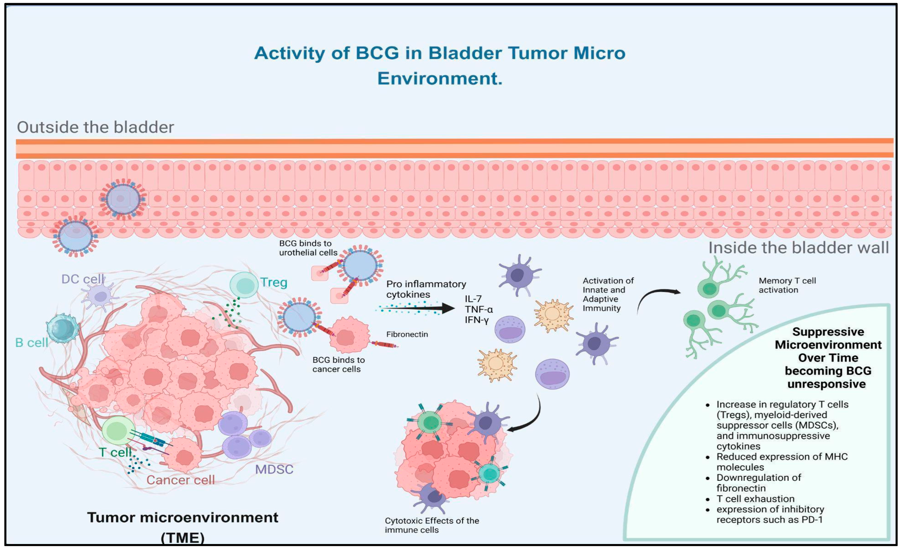
In the early 20th century, BCG was developed from the Mycobacterium bovis family, and its instillation acts as a cornerstone immunotherapy for NMIBC. BCG immunotherapy in bladder cancer works by activating the immune system. Initially, the urothelial cells are infected with BCG so that it is attached to the tumour cells via the extracellular matrix glycoprotein fibronectin, where it is subsequently internalised. BCG internalisation activates the innate immunity, which stimulates the activation of the reticuloendothelial system. The activation triggers the release of pro-inflammatory cytokines (interleukin-2, tumour necrosis factor-alpha, and interferon-gamma), which result in the activation of immune cells (T cells, macrophages, and natural killer cells). The activated immune cells attack and destroy bladder cancer cells through cytotoxic effects, creating an environment hostile to tumour recurrence. BCG therapy boosts adaptive immunity by activation of T cells and formation of memory T cells, thereby preventing recurrence [21,22]. Certain adverse events/complications are associated with the BCG immunotherapy, such as Reiter’s syndrome, parotid gland infection, enterocutaneous fistula, pleural effusion, and Poncet’s disease [23]. Despite the success rates, tumours adapt to evade the immune response, and hence a significant subset of patients become unresponsive to BCG therapy.
BCG unresponsive patients develop a suppressive microenvironment characterised by increased regulatory T cells (Tregs), myeloid-derived suppressor cells (MDSCs), immunosuppressive cytokines, reduced MHC expression, fibronectin downregulation, and PD-1-mediated T-cell exhaustion, which collectively impair effective anti-tumour immunity which is illustrated in Figure 1 [24,25,26].
Nearly 30% to 50% patients are BCG unresponsive, which follows standard therapy of radical cystectomy [9]. However, tumour grade plays a critical role in outcomes post-BCG. Patients with high-grade tumours, particularly T1 lesions and carcinoma in situ (CIS), have a significantly worse prognosis if recurrence occurs after BCG therapy compared to those with low-grade papillary tumours [27]. Although many patients remain recurrence-free at one year, up to 75% experience recurrence within five years, and a portion progresses to muscle-invasive disease, which dramatically affects survival [28]. While salvage intravesical chemotherapy, immunotherapy, and novel therapies are being studied, none have demonstrated durable responses in truly BCG-unresponsive high-grade cases [29]. Addressing BCG unresponsiveness requires alternative strategies, including combination therapies (e.g., PD-1/PD-L1 axis targeted by ICIs), novel intravesical agents, or systemic therapies. However, underlying mechanisms of resistance can guide personalised treatment approaches with other drug combinations to improve outcomes in BCG-unresponsive patients [25].

2.2. Co-Inhibitory and Co-Stimulating Signaling Therapies

Co-inhibitory molecules are key regulators of immune responses, which maintain immune tolerance by suppressing immune cell activation. The co-inhibitory signaling molecules are crucial for preventing autoimmunity, but tumours evade immune detection using the co-inhibitory signaling molecules. The immune balance is achieved by modulating these pathways through activation and inhibition, thereby promoting anti-tumour immunity while minimising adverse events. In bladder cancer, the upregulation of co-inhibitory molecules enables tumour cells to escape immune surveillance, making them a critical target for immunotherapy [30]. Its immune system evasion is mediated by pathways like CTLA-4, PD-1/PD-L1. The co-inhibitory receptor PD-1 is expressed on activated T cells. The ligands of the receptor [PD-L1, PD-L2] are overexpressed in tumour cells. The interaction of PD-1 and PD-L1 inhibits T-cell activation and induces immune tolerance [31]. CTLA-4 is a leukocyte differentiation antigen and a transmembrane receptor expressed on T cells. It binds with the antigen-presenting cells (APC), where it downregulates T-cell activation. CTLA-4 is another mechanism by which tumours inhibit immune responses [32].
Clinical trials of the PD-1/PD-L1 axis targeted by ICIs have proved efficacy in metastatic urothelial carcinoma therapy, cisplatin-ineligible locally advanced or metastatic cases, and neoadjuvant settings for MIBC. Approved ICIs for the treatment of bladder cancer include Atezolizumab, Avelumab, Durvalumab, Nivolumab, and Pembrolizumab [32]. However, in many cases, the patients develop resistance to the therapy. CTLA-4 inhibitors like ipilimumab, often studied in combination with other ICIs, have shown limited success in bladder cancer, potentially due to differing expression patterns and mechanisms of action [33]. Emerging Co-Inhibitory Targets like LAG-3, TIM-3, and VISTA have shown promise. Where LAG-3 co-expression with PD-1 correlates with poor prognosis, and dual blockade has demonstrated efficacy in early studies [34,35]. TIM-3 and VISTA also suppress T cell responses, and their inhibition, particularly in combination with PD-1 blockade, has shown potential in preclinical models [36,37].
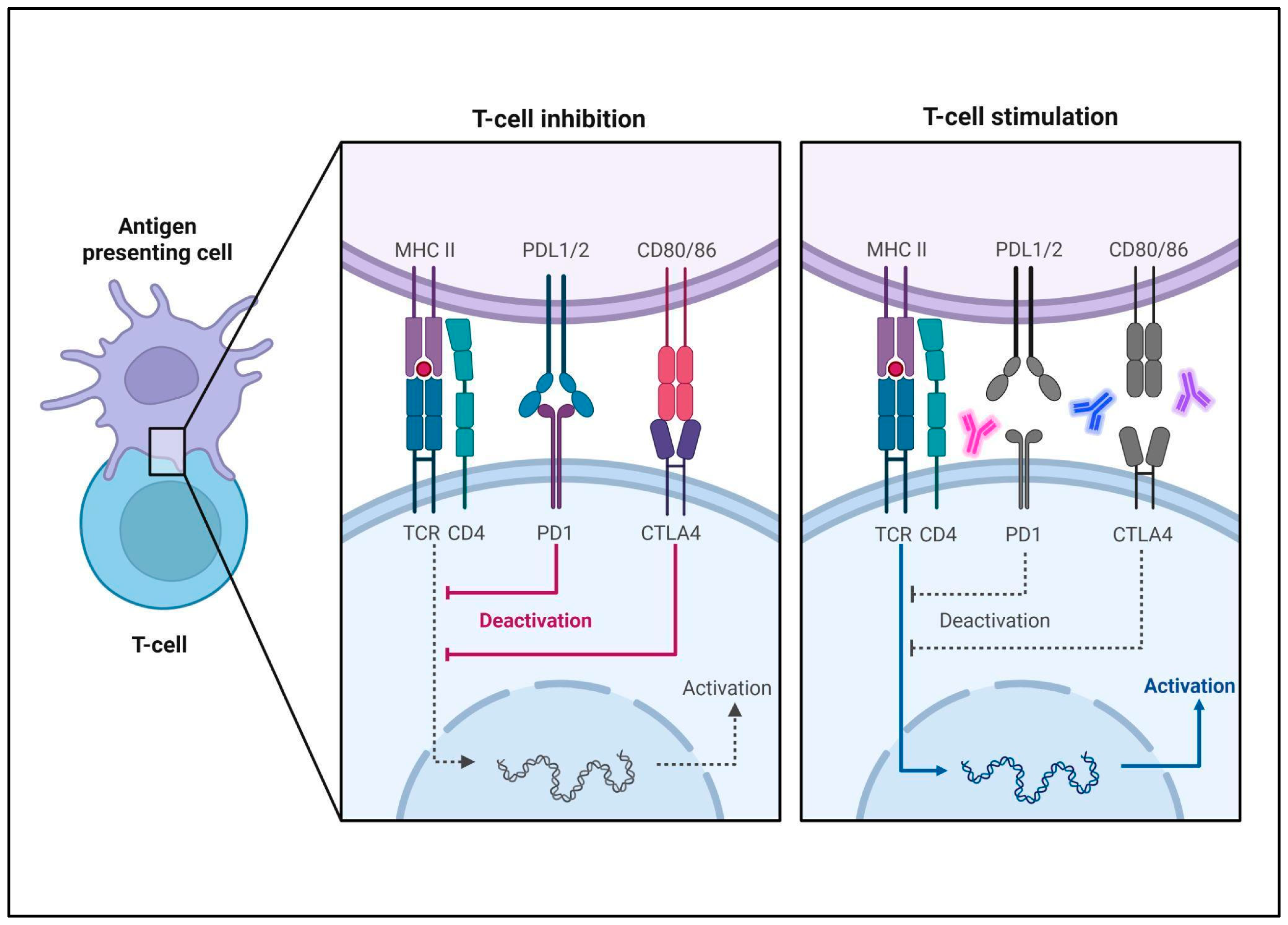
Figure 2 illustrates the molecular mechanisms that govern T-cell inhibition and stimulation through interactions between T cells and antigen-presenting cells (APCs). On the left side, under T-cell inhibition, binding of PD-1 on T cells with PD-L1/PD-L2 on APCs, along with CTLA-4 binding to CD80/CD86, leads to inhibitory signaling that deactivates the T cell, suppressing its immune response. This is often exploited by tumour cells to evade immune attack. On the right side, under T-cell stimulation, immune checkpoint inhibitors (represented by color highlighted antibodies) block the PD-1/PD-L1 and CTLA-4/CD80/86 interactions, thereby lifting the inhibitory signals. This allows the T-cell receptor and co-stimulatory molecules like CD28 to activate the T cell, enhancing its immune activity against targets such as cancer cells. The image gives a contrast on how immune checkpoints suppress T-cell activity and how their blockade can strengthen immune responses.
Co-stimulatory signaling molecules regulate and enhance the activation and proliferation of immune cells, particularly T cells, making them vital targets for immunotherapy as depicted in Figure 2 [38]. CD28 and 4-1BB (CD137) are the important co-stimulatory receptors present on T cells; they bind to their ligands CD80, CD37L respectively, present on APC. This binding activates the T cell and promotes its proliferation and cytokine production [39]. Activation of 4-1BB also improves immunotherapy outcomes by reinforcing T cell responses, especially in bladder cancer treatment [40,41]. Similar activity of co-stimulatory molecule OX40, also known as CD134, which is upregulated upon T-cell activation, binds to its ligand in APCs, enhancing T-cell survival, memory formation, and cytokine production, thus automatically improving the immune responses [42]. Newer targets like GITR and ICOS boost T cell activation and counteract tumour suppression [41]. FGFR3 (and by extension EGFR) in bladder cancer mainly upregulate co-inhibitory signals like PD-1 and CTLA-4 on T cells; sometimes PD-L1 on tumour cells in other contexts, while suppressing co-stimulatory conditions. Direct regulation of prototypical co-stimulatory receptors (e.g., CD28/B7, OX40/OX40L) has not been documented in these studies. However, by reducing antigen presentation (MHC I/II), chemokines and adhesion molecules, FGFR3/EGFR signaling prevents effective engagement of T-cell co-stimulatory pathways. The net effect is immune evasion: tumours harbouring FGFR3 or EGFR alterations show fewer CD8+ T cells and rely on exhausted T-cell phenotypes [43].

2.3. Combination Therapies

In immunotherapy, combining the co-stimulatory and co-inhibitory receptors will be a promising strategy for the treatment of bladder cancer. The combination approach, as tabulated in Table 1, ICIs based on co-inhibitory antagonists and co-stimulatory agonists, is to overcome the immune evasion mechanisms employed by tumours and to enhance the capacity of the immune system for effective identification of the cancer cells. In bladder cancer, these approaches aim to shift the balance from an immunosuppressive environment toward one that promotes immune activation and effective tumour eradication. PD-1/PD-L1 inhibitors, along with CTLA-4 inhibitors, such as nivolumab and ipilimumab, have shown effective responses in urothelial carcinoma, but toxicity remains a challenge [33]. Apart from ICIs that are listed in Table 1, other combinations are experimented for the different stages of bladder cancer treatments, Sasanlimab of phase 3 randomised clinical trial (CREST trial) on combination with BCG for BCG naïve/unresponsive for high risk NMIBC has shown efficacy in the cohort by high Event Free Survival (EFS) giving favorable prognosis in this condition [44].

2.4. Current Clinical Therapy Landscape

The current clinical landscape in bladder cancer involves chemotherapies and immunotherapies, which avoid cystectomy and ensure tumour stage-wise treatment in an effective, patient-friendly way. First-line treatment for NMIBC and intermediate, high-risk NMIBC generally involves BCG as adjuvant therapy besides resection of the tumour, which is the practising scenario, but when it proves to be ineffective, there are other treatment strategies that are followed [51]. According to Balar et al. 2021, the multicentric clinical trial has experimented and proved that the NMIBC category of bladder tumour has the immunotherapy choice of pembrolizumab, which is FDA-approved and treats high-risk BCG unresponsive patients (KEYNOTE-057) [52]. The patient cohort of Ta or T1/CIS stages of NMIBC high risk was confirmed to be BCG unresponsive and were ineligible for radical cystectomy, or did not want the procedure. The cohort responded well to pembrolizumab and showed anti-tumour and lesser toxicity for the drug [52].
Imfinzi, also known as durvalumab, is the FDA-approved combination drug with gemcitabine and cisplatin currently prescribed with these neoadjuvants for MIBC patients after radical cystectomy, with a follow-up of another dose of durvalumab standalone. This is the only FDA-approved preoperative immunotherapy for MIBC [53].
In the metastatic setting, the combination of enfortumab vedotin and pembrolizumab was approved by the FDA on 15 December 2023, for patients with locally advanced or metastatic urothelial carcinoma, based on results from the EV-302/KEYNOTE-A39 trial involving 886 patients without prior systemic therapy for advanced disease. Notably, this combination represents the first first-line regimen in decades to demonstrate superior efficacy compared to standard platinum-based chemotherapy [54].
FGFR-targeted therapy with erdafitinib (Balversa) has been approved by the FDA for adult patients with locally advanced or metastatic urothelial carcinoma harbouring FGFR3 alterations, following disease progression on one or more prior lines of systemic therapy, including prior PD-(L)1 inhibitor therapy where applicable. Erdafitinib remains the only approved targeted therapy for urothelial carcinoma to date. Currently, FGFR inhibitors are primarily used in the second-line setting, particularly in patients who have progressed after or are ineligible for immune checkpoint inhibitor therapy [55,56].

3. Mechanism of Resistance Formation

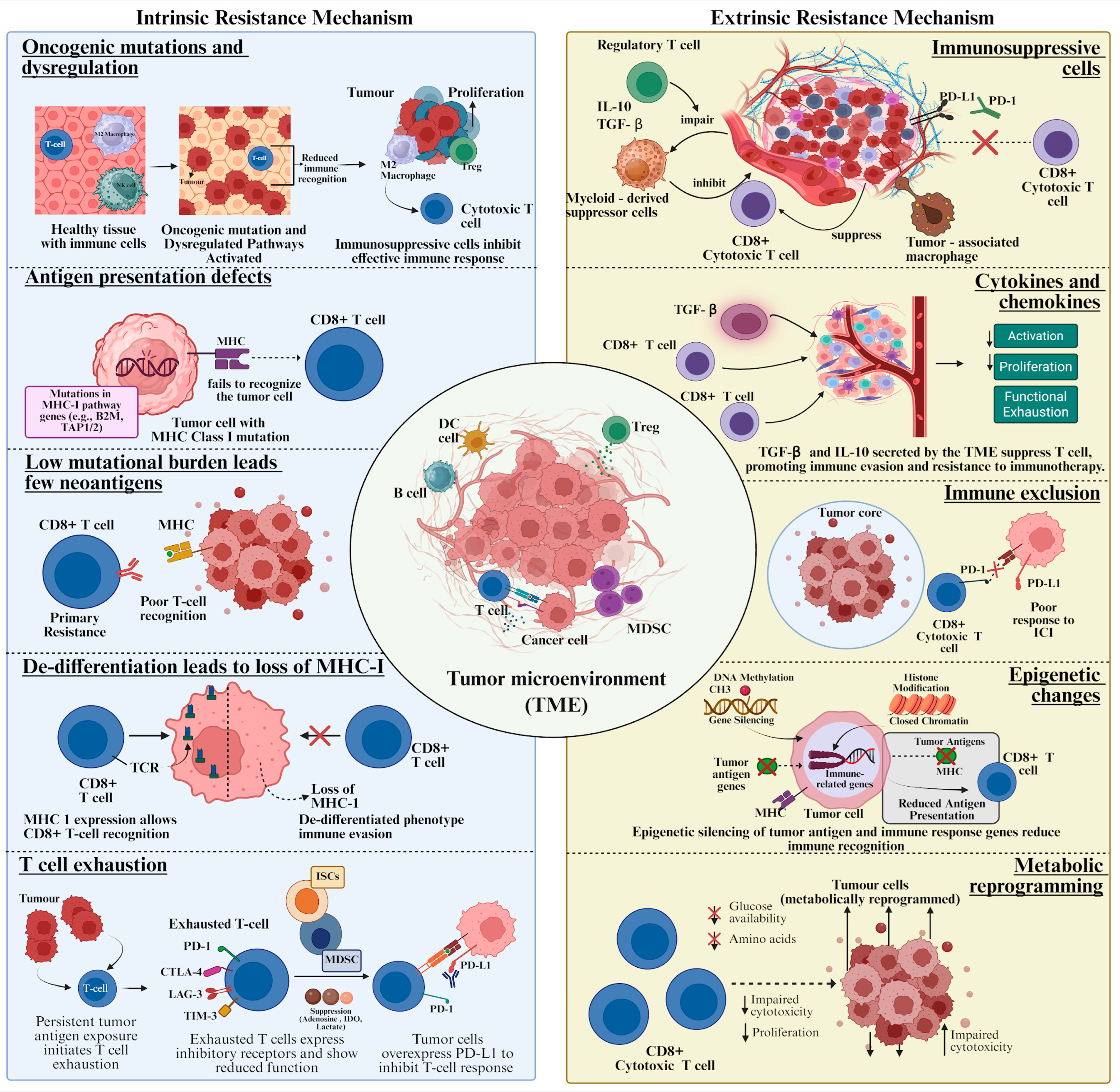
Figure 3 illustrates that resistance to ICIs can arise from both intrinsic and extrinsic factors within the tumour and its surrounding environment. Intrinsic factors are related to the tumour cells themselves and include oncogenic mutations and dysregulation that can alter signaling pathways, defects in antigen presentation that prevent immune recognition, a low mutational burden with few neoantigens to trigger an immune response, tumour cell de-differentiation leading to the loss of MHC-I expression, and T-cell exhaustion, where immune cells lose their ability to effectively attack the tumour. On the other hand, extrinsic factors involve the TME and include the presence of immunosuppressive cells that inhibit immune activity, the production of cytokines and chemokines that modulate immune responses, immune exclusion which prevents immune cells from infiltrating the tumour, epigenetic changes in gene expression alteration that do not change the DNA sequence, and metabolic reprogramming that supports tumour growth and suppresses immune cell function. Because of these multifaceted resistance mechanisms, combination therapies are often explored to overcome these barriers and enhance the efficacy of ICIs.
Development of resistance to CTLA-4 inhibitors and PD-1/PD-L1 inhibitors in bladder cancer patients involves intrinsic and extrinsic factors. Tumour cells employ various intrinsic mechanisms to evade immune detection and resist immunotherapy. Tumours with mutations in genes involved in antigen presentation, such as those in the major histocompatibility complex (MHC) class I pathway, are less likely to be recognised by T cells, leading to resistance to ICIs [57]. Tumours with low mutational burden or few neoantigens are less likely to elicit a robust T-cell response, contributing to primary resistance [58,59].
Intrinsically, bladder tumours exhibit high mutational burdens and frequent alterations in pathways such as p53, PI3K/AKT, DNA repair, and chromatin remodelling, which influence immune visibility and responsiveness (Cancer Genome Atlas, 2014) [60]. Additionally, tumours overproduce prostaglandin E2 (PGE2) via the COX2/mPGES1 pathway, which suppresses antigen presentation, impairs dendritic cell maturation, and promotes the recruitment of immunosuppressive myeloid cells [61]. Bladder cancer is also characterised by the aberrant production of hyaluronan, which contributes to tumour growth and immune evasion by interacting with CD44 on immune cells and modulating inflammatory signaling [62].
Extrinsically, the TME becomes highly immunosuppressive, populated by regulatory T cells (Tregs), MDSCs, tumour-associated macrophages (TAMs), and tolerogenic dendritic cells (tDCs). These immune cells express PD-L1 and secrete inhibitory cytokines such as IL-10, TGF-β, and IL-6, which blunt T cell responses and promote immune tolerance [63]. MDSCs, which accumulate in both peripheral blood and tumour sites of bladder cancer patients, suppress CD4+ and CD8+ T cells and can differentiate into immunosuppressive PD-L1+ macrophages or dendritic cells. Similarly, TAMs adopt an M2-like phenotype, supporting angiogenesis, tumour invasion, and directly suppressing cytotoxic T cells [64]. tDCs, influenced by tumour-secreted IL-10 and PGE2, fail to mature and instead promote Treg induction through PD-L1/PD-1 signaling. Tregs themselves are abundant in bladder tumours and contribute to immunosuppression via the expression of FOXP3, CTLA-4, and IL-10, effectively silencing effector T cell responses [65].
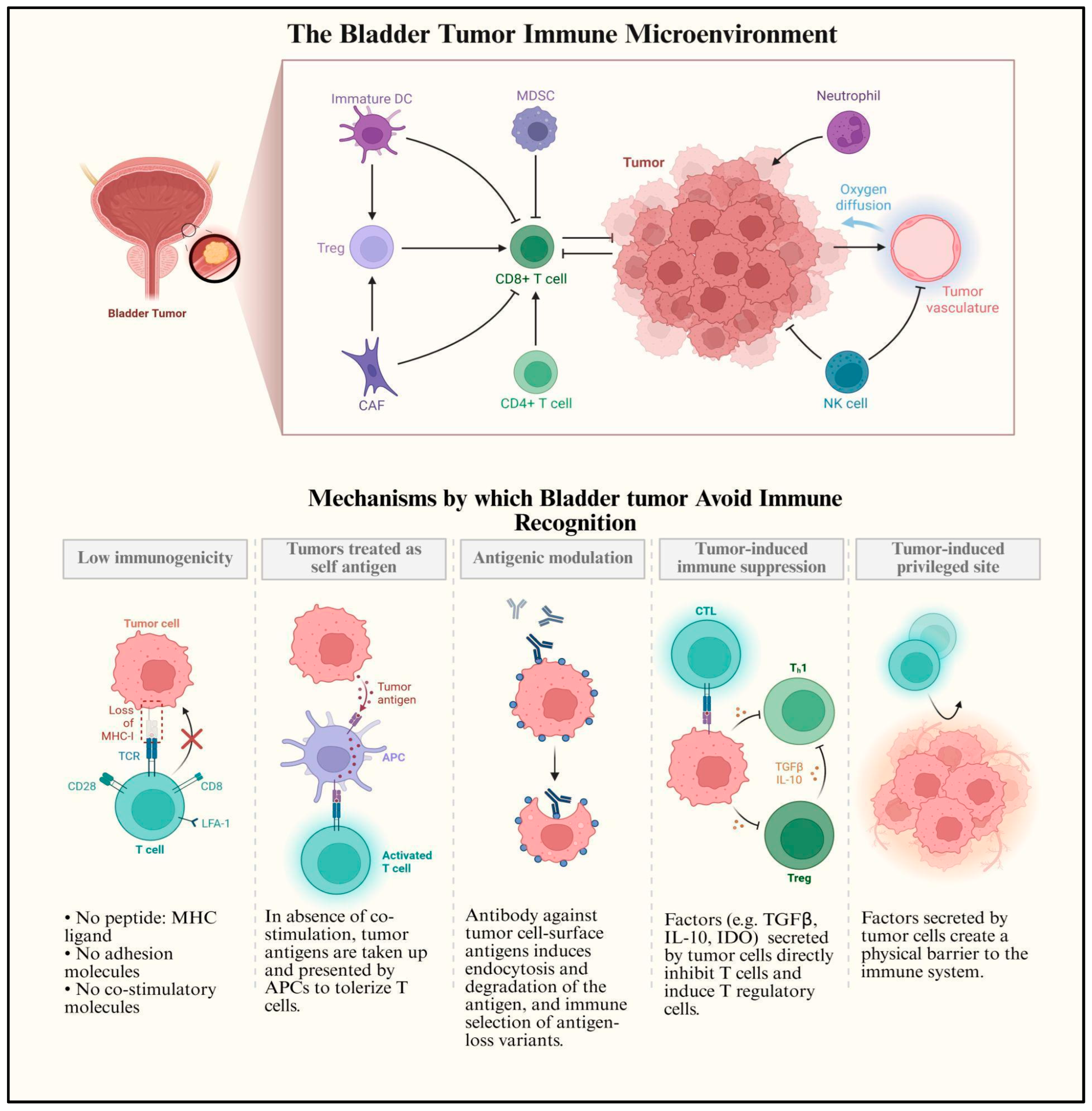
Figure 4 illustrates the complex immune microenvironment of bladder tumours and the diverse mechanisms they use to evade immune recognition. In the upper section, it shows how various immune and stromal cells, such as regulatory T cells (Tregs), MDSCs, cancer-associated fibroblasts (CAFs), immature dendritic cells (DCs), and neutrophils, interact with CD8+ and CD4+ T cells to suppress anti-tumour responses within the tumour milieu. The lower section outlines five distinct strategies by which bladder tumours avoid immune detection: (a) low immunogenicity due to loss of MHC, adhesion, and co-stimulatory molecules; (b) presentation of tumour antigens in the absence of co-stimulation, leading to T cell tolerance; (c) antigenic modulation through antibody-induced antigen degradation and selection of immune-resistant variants; (d) tumour-induced immune suppression via secretion of inhibitory cytokines like TGF-β and IL-10, which promote Treg activation; and (e) creation of an immune-privileged site through physical barriers. Together, these processes allow bladder tumours to escape immune surveillance and resist immunotherapy.
T-cell exhaustion is characterised by the expression of numerous inhibitory receptors and decreases T-cell efficacy, which can result from prolonged exposure to tumour antigens. The ISCs and metabolites further reduce the effective immune response. PD-1/PD-L1 inhibitors combined with other treatments, such as targeting TGFβ or employing epigenetic therapies, may enhance the efficacy of ICB by overcoming some of these resistance mechanisms [66]. Rosenberg et al. (2016) and Sharma et al. (2016) state that the evolution of ICI therapies in bladder cancer is due to the approval of several PD-1/PD-L1 inhibitors for metastatic/advanced bladder cancer stages, as demonstrated based on the clinical outcomes such as response, survival without progression, and overall survival [46,47]. However, a few patients do not respond well to these therapies and acquire resistance. Recent research studies are focused on the development of next-generation ICIs, combining ICIs with other treatments, and identifying biomarkers for personalised therapy [16].
The TME plays a critical role in modulating the immune response and promoting resistance to ICIs. The infiltration of immune suppressive cells, such as regulatory T cells (Tregs), macrophages, and MDSCs, creates an immunosuppressive TME that hampers the efficacy of ICIs. The TME can secrete cytokines and chemokines that inhibit T-cell activation and proliferation, such as TGF-β and IL-10, further promoting immune evasion. The spatial arrangement of immune cells within the TME can also influence treatment outcomes. For instance, the exclusion of CD8+ T cells from the tumour core is associated with poor response to ICIs [67,68,69].
Epigenetic changes, such as DNA methylation and histone modification, can downregulate the expression of tumour antigens and immune-related genes, reducing the efficacy of ICIs. Tumour cells can metabolically reprogram to deprive T cells of essential nutrients, such as glucose and amino acids, impairing their function and promoting resistance. CTLA-4 inhibitors, such as ipilimumab, work by blocking the interaction between CTLA-4 and its ligands, CD80 and CD86, enhancing T-cell activation. However, resistance to CTLA-4 inhibitors can occur by upregulating other immune checkpoints, such as PD-1, to compensate for CTLA-4 inhibition. The TME can remain immunosuppressive despite CTLA-4 inhibition, limiting the efficacy of the treatment [70,71,72].

4. FGFR3 Signaling Pathway

FGFRs are single-pass transmembrane proteins that consist of three domains—ECD (extracellular domain), TMD (transmembrane domain), and TKD (tyrosine kinase intracellular domain). The FGFR family comprise four transmembrane receptors, FGFR 1-4. These receptors are critical for the normal physiological functions, such as the development of embryos and the regulation of angiogenesis in adults. The transmembrane receptors are expressed in various types of cells, and they regulate cell proliferation, survival and migration [73]. FGFR3 signaling pathways are complex and involve multiple downstream effectors that regulate various cellular processes.
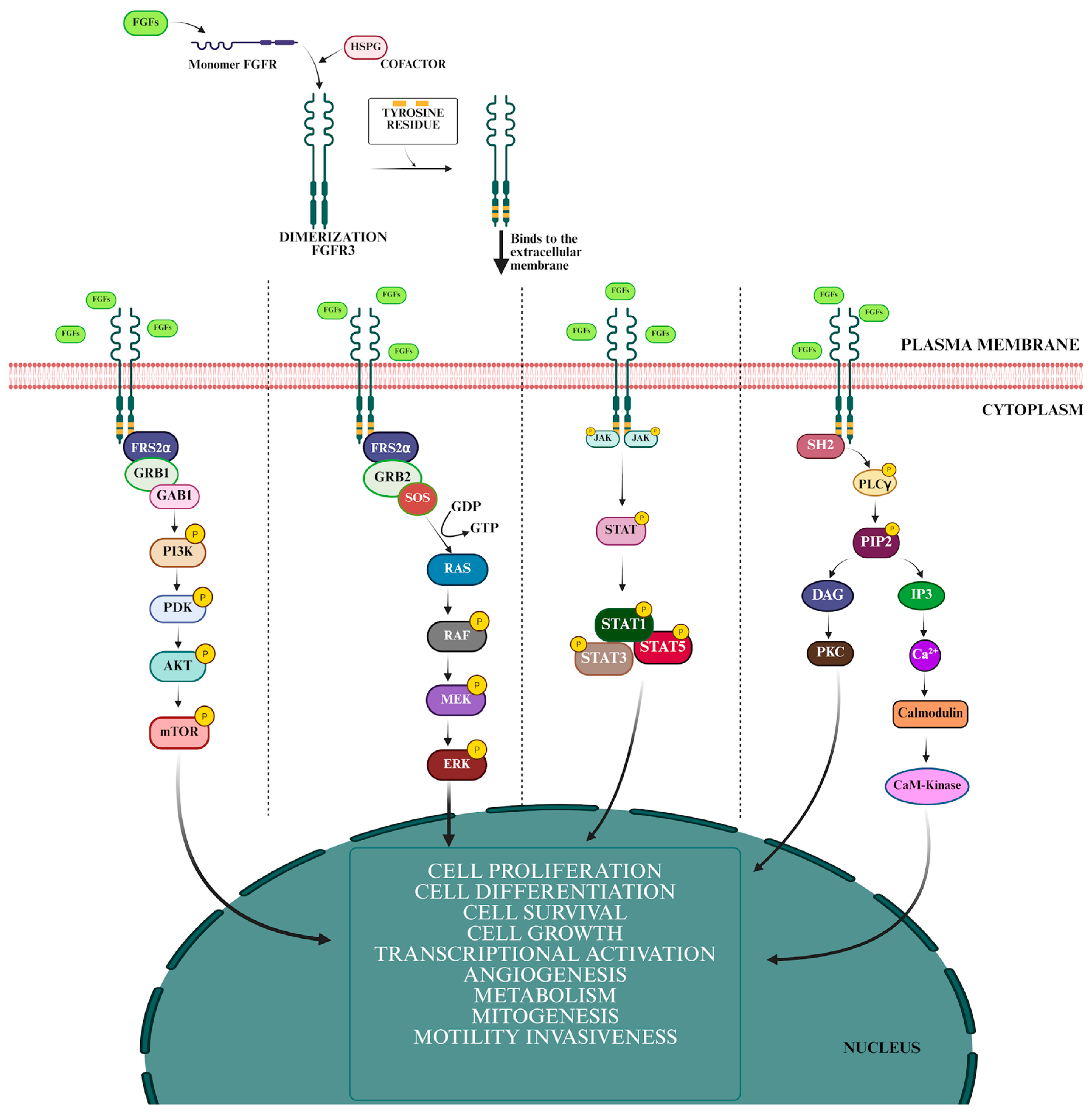
When FGFs (typically FGF1, FGF9, etc.) bind to FGFR3, often in the presence of heparin sulfate proteoglycans (co-factors), the receptor undergoes dimerisation (pairs with another FGFR3). The dimerised receptors activate each other’s intracellular tyrosine kinase domains through auto-phosphorylation, adding phosphate groups to specific tyrosine residues. Phosphorylated FGFR3 recruits and activates multiple downstream signaling pathways, promoting cell proliferation and differentiation. This cascade involves activation of RAS to RAF (including A-Raf, B-Raf, Raf-1) followed by MEK1/2 to ERK1/2, which then enters the nucleus to regulate gene expression, Supports cell survival and metabolism by phosphorylating and activating AKT.This involves activation of STAT transcription factors, especially STAT1 and STAT3, which modulate genes related to inflammation and immune evasion. The mechanism of FGFR3 activation illustrated in Figure 5 represents the fibroblast FGF ligands binding with the extracellular domain of the FGFR3 receptor. The binding leads to the dimerisation of FGFR3, which in turn undergoes auto-phosphorylation on specific tyrosine residues within its intracellular domain. The phosphorylation event resulted in the activation of downstream signaling pathways [74]. Upon activation, FGFR3 initiates cascades involving the mitogen-activated protein kinase (MAPK) pathway and phosphoinositide 3-kinase (PI3K)/Akt signaling [75], which are essential in mediating cell survival, differentiation and proliferation.

4.1. FGFR3 Aberrations in Bladder Cancer

FGFR3, a critical player in bladder cancer pathogenesis, has mutations of approximately 75% in low-grade NMIBC, including point mutations and gene fusions [75]. The most common mutation is S249C, which accounts for 63% of reported mutations, followed by Y375C. Both mutations account for more than 80% of all the detected mutations [76]. In addition to point mutations, FGFR3 gene fusions are critical in bladder cancer. The FGFR3-TACC3 fusion is one of the most studied, where the FGFR3 kinase domain fuses with the TACC3 gene, leading to continuous receptor activation. The FGFR3 fusion is linked with a poorer prognosis compared to low-grade tumours [77]. FGFR3 mutations frequently do not co exist with other carcinogenic mutations, including those in TP53 and RB1, suggesting a distinct tumour growth pathway. The main effect of the FGFR3 alterations is ligand-independent receptor dimerisation, which sets off subsequent signaling cascades that support cell survival and proliferation [78].
Mutations in FGFR3 signaling pathways have a multifaceted role in bladder cancer, involving tumourigenesis and modulation of the immune landscape, modifying the activity of infiltration by immune cells into the microenvironment of tumour cells. FGFR3 mutations alter the cytokine profiles, creating an immunosuppressive environment, and impede the effectiveness of anti-tumour immune responses [79,80]. Also, pERK and pERBB3, which are repeatedly activated in FGFR3-driven cells following anti-FGFR treatment, can be overcome by pan-ERBB receptor blockade. This also suggests that combination treatment with both inhibitors is more effective for treating FGFR3-fusion driven bladder cancers [80].

4.2. FGFR3 Inhibitors in Bladder Cancer

The FGFR3 dysregulation impacts the expression of ICM, potentially affecting the efficacy of ICIs. Understanding the interactions is critical for the novel treatment approaches by combining FGFR-targeted therapies with ICIs [81]. Moreover, the interaction between FGFR3 and other tyrosine kinase receptors (TKRs) can complicate treatment strategies. For example, resistance mechanisms have been identified where continuous exposure to FGFR inhibitors leads to upregulation of alternative TKRs, such as ERBB3 [80]. This highlights the necessity for combination therapies where multiple pathways can be targeted simultaneously for overcoming resistance and improving the treatment efficacy. Ascione et al. 2023 clearly state that the FGFR3 targeted therapy is based on TKIs, and they are categorised as selective and multi-targeting TKIs based on the mechanism of action and are tabulated in Table 2 [17]. Selective TKIs target only the FGFR kinase domain, whereas multi-targeting TKIs have non-specific inhibitors that exhibit affinity towards multiple additional receptors.

5. EGFR Signaling Pathway and Its Aberrations in Bladder Cancer

EGFR is a receptor tyrosine kinase (RTK) consisting of three domains—ligand binding ECD, TMD, and TKD. The EGFR family consists of four receptors, EGFR 1-4, also termed ErbB 1-4. Like FGFR3, EGFR, upon binding of its ligands, undergoes dimerisation and autophosphorylation, which results in the activation of the downstream signaling cascade that controls multiple cellular responses [18].
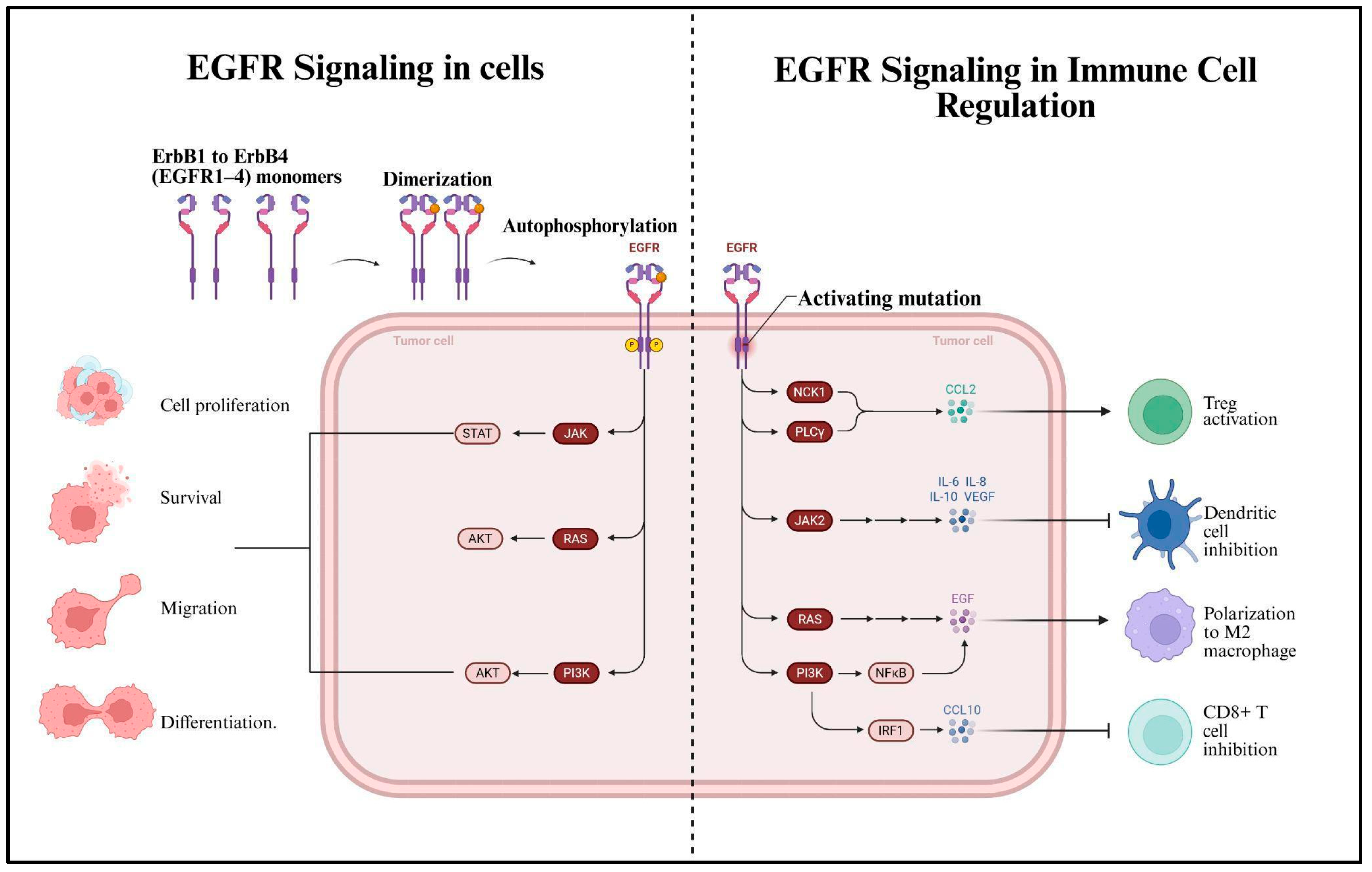
Figure 6 illustrates the dual role of EGFR signaling in cancer progression and immune regulation. On the left, it shows how EGFR (ErbB1-4) receptors dimerise and autophosphorylate upon ligand binding, activating downstream pathways such as JAK/STAT, RAS/AKT, and PI3K/AKT that promote tumour cell proliferation, survival, migration, and differentiation. On the right, it depicts how activating mutations in EGFR also contribute to immune evasion by altering the TME: NCK1 and PLCγ pathways stimulate CCL2 to activate regulatory T cells (Tregs), JAK2 signaling promotes secretion of immunosuppressive cytokines like IL-6, IL-8, IL-10, and VEGF, which inhibit dendritic cells, RAS signaling promotes M2 macrophage polarisation via EGF, and PI3K/NFκB/IRF1 signaling drives CCL10 expression, which inhibits CD8+ T cell activity. Together, these mechanisms enhance both tumour growth and suppression of anti-tumour immunity. EGFR, particularly in urothelial carcinoma, is overexpressed in MIBC and is frequently dysregulated in bladder cancer. Higher EGFR levels may act as a diagnostic indicator for advanced cancer stages [90]. It is also related to advanced tumour stages, correlating with poor patient prognosis [91]. It is reported that EGFR overexpression occurs in a significant percentage of MIBC cases, ranging from 27% to 74% [18]. Studies have shown that a subset of MIBC tumours, particularly those with a basal-like phenotype, exhibit high expression, which leads to EGFR pathway activation and are sensitive to anti-EGFR therapies. These basal cell markers can be used as a point in monitoring recurrence [92].
EGFR signaling participates in several biological functions, including cell migration, differentiation, survival, and proliferation for bladder cancer [93]. The activation of the EGFR is associated with enhanced tumour growth and metastasis, which can be a focus for therapeutic intervention [90]. One point where the expression of EGFR is influenced is by long noncoding RNAs (EGFR-AS1). These noncoding RNAs accelerate the growth of bladder cancer by accelerating EGFR mRNA, therefore increasing its expression, and facilitating the tumour invasion and metastasis. Like FGFR3, EGFR has a role in bladder cancer that is further complicated by its interaction with other TKRs. In a significant portion of bladder tumours, the co-expression of HER2 and EGFR has been observed, indicating potential synergistic effects on tumour progression [94].
Mutational effects in the EGFR gene are relatively rare in bladder cancer, though the overexpression of EGFR is observed in many patients. The oncogenic effects are mainly due to dysregulated signaling rather than genetic alterations [95]. The effectiveness of anti-EGFR treatments (small molecule inhibitors, monoclonal antibodies) has been investigated in clinical trials. The anti-EGFR treatments have shown varying degrees of success, particularly in MIBC. They may enhance the effects of chemotherapy, but resistance to the therapies poses the main challenge. However, this resistance to therapy can be controlled by combination strategies that target multiple pathways simultaneously [96].

Role of EGFR Inhibitors in Bladder Cancer’s Immune Landscape

EGFR signaling influences immune cell infiltration and activity within the TME by cytokines secreted by vascular endothelial cells or by tumour cells (in which EGFR is highly expressed, especially in bladder cancer) that can activate EGFR signaling pathways, promoting tumour cell proliferation and migration while simultaneously altering responses to the immune environment [97]. ICM expression is linked to EGFR activation, where this interaction brings about an immunosuppressive environment, making the tumour cell avoid immune identification. Targeting both pathways may improve treatment outcomes due to the reciprocation between immune checkpoints and EGFR signaling [98]. It is also crucial to remember that EGFR inhibition can boost T-cell infiltration and activity against tumours by blocking EGFR signaling. This can restore anti-tumour immunity by lowering immunosuppressive factors in the TME [99]. Therefore, combination therapies that incorporate both anti-EGFR and ICIs have a great deal of potential. The EGFR inhibitors that can be used in conjunction with ICIs are shown in Table 3. This can be started as research on how these combinations support current EGFR inhibitor treatments from the standpoint of comprehending immune landscapes in bladder cancer.

6. The Activity of Both FGFR3 and EGFR in the TME Affecting PD1 and PDL1 Axis of Bladder Cancer

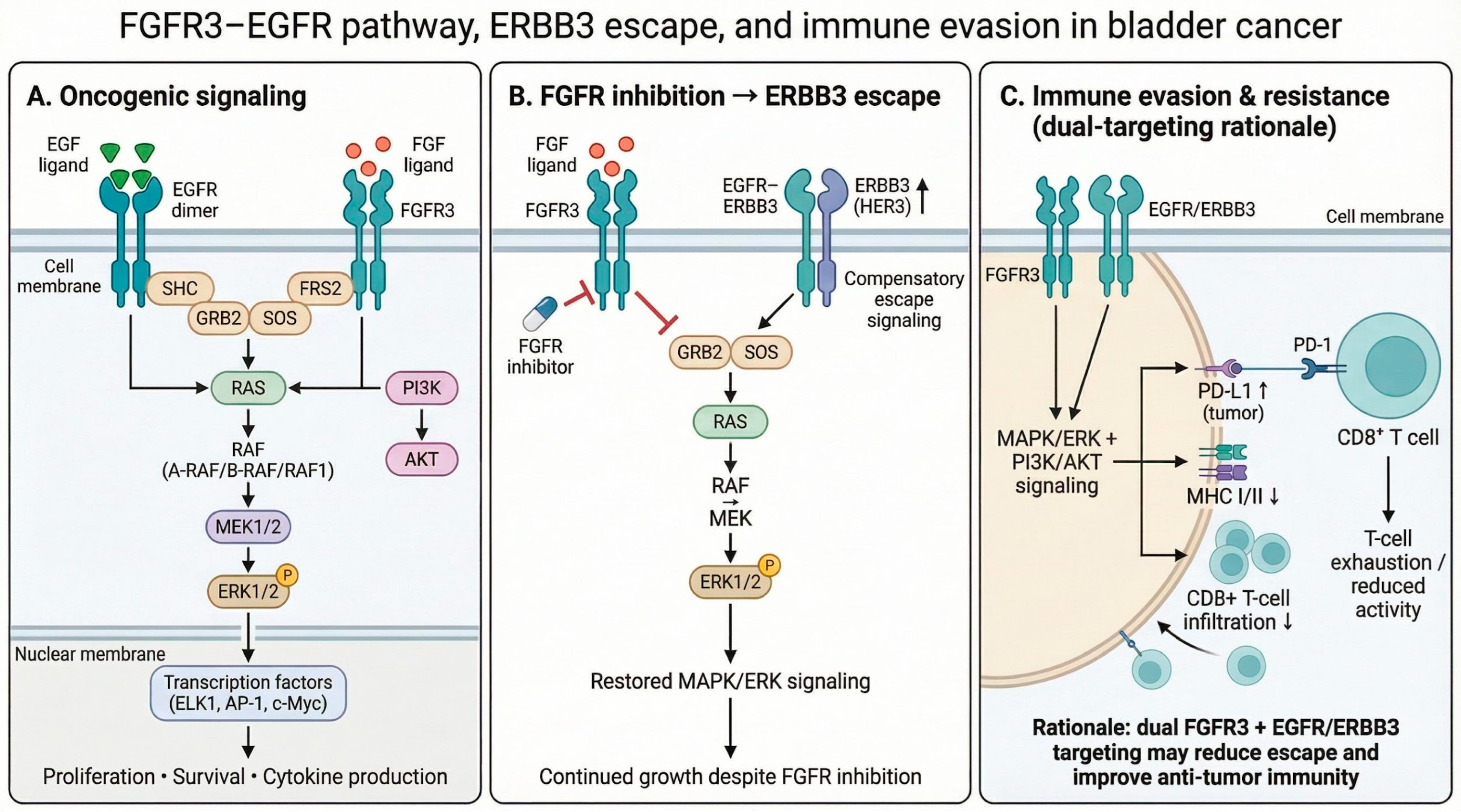
Figure 7 illustrates the convergence of EGFR and FGFR3 signaling pathways within the TME, emphasising their shared downstream effects on cellular behaviour. Upon ligand binding, EGFR and FGFR3 undergo dimerisation and activate a cascade involving adaptor proteins like SHC, GRB2, and SOS, which subsequently activate Ras. Ras signaling leads to the activation of TPL2 and the RAF family kinases (Raf1, B-Raf, A-Raf), followed by MEK1/2, and ultimately ERK1/2. Activated ERK1/2 translocates into the nucleus, where it phosphorylates a wide array of transcription factors, including ELK1, AP1, STAT1, c-Myc, and CREB. These transcription factors regulate the expression of genes involved in critical processes such as cytokine production, apoptosis, migration, proliferation, and differentiation. The diagram highlights how both FGFR3 and EGFR activate a shared MAPK/ERK pathway, underlining their overlapping roles in tumour progression and potential as combined therapeutic targets.
FGFR3 mutations have a higher prevalence in bladder cancer, altering the TME, suppressing the immune response, thereby hindering the activity of ICIs [106]. Immune evasion by bladder tumour cells occurs when FGFR3 activation can result in increased expression of PD-L1. On the contrary, EGFR signaling has been found to restrain the sensitivity of bladder cancer cells that are driven by FGFR3 to inhibitors of FGFR, thus establishing a cycle acting upon complicated immune responses [81]. Activation of EGFR promotes tumour proliferation and survival, leading to a repressed immune microenvironment that reduces T-cell activity and confers resistance to ICIs [18]. Dual targeting of FGFR3 and EGFR was presented in Figure 7 as a therapeutic strategy to bypass resistance mechanisms and ameliorate responses to immunotherapy, which will be a potential option for therapy.
FGFR3 activation is associated with T-cell exclusion, overexpression of co-inhibitory checkpoints including PD-1 and CTLA-4, and suppression of MHC expression and IFN-γ signaling, which leads to impaired antigen presentation and cytotoxic T-cell recruitment. There is a growing body of evidence suggesting that ERBB2 (HER2) may cooperate in this immune modulation since it forms heterodimers with ERBB3 and EGFR, potentiating downstream MAPK and PI3K signaling. The latter two are implicated in immune escape pathways through modulation of PD-L1 and suppression of IFN-mediated responses [107].

6.1. FGFR3 and EGFR Resistance Mechanisms

In bladder cancer, EGFR signaling is associated with low immune infiltration and resistance to immune checkpoint blockade. Although EGFR is highly expressed in the FGFR3-fusion lines SW780 and RT4, its role may become supportive by cooperating with ERBB2/ERBB3 to sustain the MAPK signaling that inhibits antigen presentation machinery and chemokine-mediated T-cell recruitment [106]. Thus, the interplay between FGFR3, EGFR, and ERBB2/3 not only promotes resistance to targeted therapy but may also contribute toward an immunologically “cold” TME-a notion supporting the rationale for the combination of tyrosine kinase inhibitors with immune checkpoint inhibitors in FGFR3/ERBB-driven bladder cancer [108]. By blocking both pathways, it might be possible to lower PD-L1 levels and make it easier for T cells to get into tumours, which would make ICIs work better [109,110]. Furthermore, comprehending the molecular mechanisms that govern this interaction is ideal for developing combination therapies capable of effectively altering the immune landscape in bladder cancer [111]. The synergistic activity of FGFR3 and EGFR profoundly affects the immune milieu in bladder cancer, modulating PD-1/PD-L1 interactions and creating avenues for innovative therapeutic approaches to augment immunotherapy effectiveness [112,113].

6.2. FGFR3 and EGFR as Therapy Targets

Erdafitinib, a FGFR3 inhibitor, has revolutionised treatment options of FGFR3-altered bladder tumours, particularly in patients with the stage of NMIBC, even though there is a response rate of suboptimal efficacy [114,115]. Combining FGFR inhibitors with ICIs may improve therapeutic outcomes by modulating the TME and enhancing anti-tumour immunity [116]. EGFR is a target in muscle-invasive bladder cancer; several anti-EGFR monoclonal antibodies and TKIs are currently under investigation.
FGFR3 and EGFR play central and interconnected roles in mediating therapeutic resistance through on-target mutations, compensatory signaling, and adaptive cellular reprogramming. FGFR3-driven tumours develop resistance to FGFR inhibitors via secondary gatekeeper mutations that impair drug binding while preserving kinase activity, as well as through bypass activation of alternative receptor tyrosine kinases, particularly EGFR and ERBB family members. Inhibition of FGFR3 disrupts negative feedback loops, leading to compensatory upregulation of EGFR signaling and reactivation of key downstream pathways such as MAPK/ERK and PI3K/AKT, thereby sustaining tumour proliferation despite targeted therapy [117]. Conversely, EGFR-targeted therapies are limited by both on-target resistance, including secondary kinase domain mutations such as T790M and C797S, which reduce inhibitor binding and restore receptor activity, and off-target mechanisms, including activation of parallel pathways, downstream signaling reactivation, and phenotypic transitions such as epithelial–mesenchymal transition (EMT) [118]. The drug trials that are approved by FDA for bladder cancer is mentioned below in Table 4, which are dependent on FGFR3 and EGFR mutations of bladder tumors.

7. Targeting the FGFR3-EGFR in the Immune Axis for Bladder Cancer

FGFR3 and EGFR significantly influence the TME and therapeutic resistance in bladder cancer, highlighting their roles in resistance to both targeted and immune therapies. FGFR3 mutations are highly prevalent in non-invasive low-grade tumours, promoting proliferation through ligand-independent receptor activation. These mutations often modulate the immune microenvironment, fostering immunosuppressive conditions that hinder anti-tumour responses.
Resistive FGFR3 alterations of NMIBC to FGFR inhibitors highlight the intervention of alternative pathways, such as upregulated ERBB2/3 signaling. The cooperation between FGFR3 and ICM has PD-L1 expression, which highlights the potential for combined FGFR-targeted and ICI therapies. Overexpression of EGFR is observed in high-grade bladder tumours with aggressive disease and recurrence. Here, anti-EGFR therapies combined with ICIs, when there is chemoresistance and immunosuppression, can also be compelling in moving forward to better joined drug strategies [124].
Similarly, EGFR overexpression has been associated with heightened PD-L1 expression, contributing to immune escape and resistance to ICIs. These findings highlight their utility in stratifying patients for personalised treatment strategies. FGFR3 mutations are associated with higher recurrence rates in NMIBC but lose predictive value in more advanced stages [125]. Conversely, EGFR overexpression is associated with higher recurrence rates and poor prognosis. In NMIBC, EGFR expression is often observed. While EGFR is expressed in normal urothelium, overexpression in NMIBC, particularly in advanced cases, is associated with biological aggressiveness and a poorer prognosis [92].
Likewise, co-activation of FGFR3 and EGFR complicates therapeutic responses, as their combined activity affects the PD-1/PD-L1 axis in a stronger sense. This interaction creates the opportunity for developing a dual-targeting point to overcome resistance formation and enhance immunotherapy outcomes. Blocking both pathways may reduce PD-L1 expression, increase T-cell infiltration, and improve ICI efficacy. FGFR3 and EGFR also serve as potential biomarkers for monitoring recurrence and predicting therapeutic responses [76].
Resistance to targeted therapies in bladder cancer is frequently driven by compensatory signaling mechanisms. FGFR3-driven tumours can acquire resistance to FGFR inhibitors through secondary kinase mutations and activation of alternative signaling pathways, including PI3K–mTOR alterations. Notably, activation of the EGFR/ERBB receptor family has been identified as a key bypass mechanism that restores downstream proliferative signaling. For instance, Facchinetti et al. 2023, demonstrated that resistance to the FGFR inhibitor erdafitinib can be mediated by EGFR activation, and that combined inhibition using erdafitinib and gefitinib can overcome this resistance [126]. In addition to targeted therapy resistance, FGFR3 alterations are also associated with reduced efficacy of immune checkpoint blockade (ICB). Also, Noeraparast et al. 2024, reported that FGFR3-mutant metastatic urothelial carcinoma exhibits a T-cell-depleted tumour microenvironment, contributing to diminished responses to immunotherapy [106]. Similarly, Weickhardt et al. 2022, showed that adaptive activation of ERBB3, a member of the EGFR family, limits FGFR inhibitor efficacy, while dual targeting of FGFR3 and ERBB3 enhances therapeutic response in preclinical models [80]. At the molecular level, FGFR3 and EGFR signaling pathways converge on shared downstream cascades, particularly the MAPK/ERK and PI3K/AKT pathways, providing a mechanistic basis for cross-resistance and supporting combination therapeutic strategies. Ouyang et al. 2023, demonstrated that mutant FGFR3 enhances serine synthesis, thereby activating PI3K/AKT signaling in tumour-associated macrophages, highlighting its role in modulating the tumour microenvironment [127]. In parallel, Wang et al. 2021, showed that EGFR activates the MEK/ERK/c-Jun axis, regulating downstream targets such as PD-L1 in bladder cancer cells [128]. Consistently, Komura et al. 2023, described FGFR signaling as engaging canonical RAS–MAPK and PI3K–AKT pathways, further supporting the convergence of FGFR3 and EGFR signaling in bladder cancer progression and therapeutic resistance [129].

8. Conclusions

From the review, an in-depth understanding of FGFR3 and EGFR functions in the TME can be gained in the clinical trial scenarios. Bladder cancer remains a major clinical challenge due to its high rates of recurrence, chemoresistance, and limited response to immunotherapy, largely driven by a dynamic tumour microenvironment. This review highlights the critical roles of FGFR3 and EGFR in regulating tumour progression, immune evasion, and therapeutic resistance. FGFR3 alterations and EGFR overexpression contribute not only to oncogenic signaling but also to the development of an immunosuppressive microenvironment, characterised by reduced antigen presentation, T-cell exclusion, and increased immune checkpoint activity.
Both genes exhibit significant signaling convergence, activating shared pathways such as MAPK/ERK and PI3K/AKT, which promote tumour survival and enable compensatory resistance mechanisms. Crosstalk between FGFR3 and EGFR further amplifies resistance to targeted therapies and immune checkpoint inhibitors. Collectively, these findings support a unified model of resistance centred on FGFR3–EGFR signaling within the immune axis. Targeting these pathways in combination with immunotherapy represents a promising strategy for a more personalised and effective therapeutic approach in bladder cancer.

Author Contributions

Conceptualisation and writing—original draft preparation, A.M.; writing, editing and supervision, C.E.J.W., A.B., N.K. and T.J.; writing—review and editing, A.M., S.D. and J.E.W.; image creation—A.M. and A.S. All authors have read and agreed to the published version of the manuscript.

Funding

This review received no specific grant from any funding agency in the public, commercial, or not-for-profit sector.

Institutional Review Board Statement

Not applicable.

Informed Consent Statement

Not applicable.

Data Availability Statement

No new data were created or analysed.

Conflicts of Interest

The authors declare no conflicts of interest.

References

  1. Global Cancer Observatory: Cancer Today. Available online: https://gco.iarc.who.int/media/globocan/factsheets/cancers/30-bladder-fact-sheet.pdf (accessed on 8 April 2026).
  2. Sung, H.; Ferlay, J.; Siegel, R.L.; Laversanne, M.; Soerjomataram, I.; Jemal, A.; Bray, F. Global cancer statistics 2020: GLOBOCAN estimates of incidence and mortality worldwide for 36 cancers in 185 countries. CA Cancer J. Clin. 2021, 71, 209–249. [Google Scholar] [CrossRef] [Scilit] [PubMed]
  3. Bringing Bladder Cancer to the Forefront: Our Bladder Cancer Awareness Month 2025 Report; World Bladder Cancer Patient Coalition: Brussels, Belgium, 2025; Available online: https://worldbladdercancer.org/news_events/bringing-bladder-cancer-to-the-forefront-our-bladder-cancer-awareness-month-2025-report/ (accessed on 8 April 2026).
  4. Burger, M.; Catto, J.W.F.; Dalbagni, G.; Grossman, H.B.; Herr, H.; Karakiewicz, P.; Kassouf, W.; Kiemeney, L.A.; La Vecchia, C.; Shariat, S.; et al. Epidemiology and risk factors of urothelial bladder cancer. Eur. Urol. 2013, 63, 234–241. [Google Scholar] [CrossRef] [Scilit]
  5. Yamaguchi, N.H. Smoking, immunity, and DNA damage. Transl. Lung Cancer Res. 2019, 8, S3–S6. [Google Scholar] [CrossRef] [Scilit]
  6. Zhao, X.; Wang, Y.; Liang, C. Cigarette smoking and risk of bladder cancer: A dose-response meta-analysis. Int. Urol. Nephrol. 2022, 54, 1169–1185. [Google Scholar] [CrossRef] [Scilit]
  7. Mossanen, M. The epidemiology of bladder cancer. Hematol. Oncol. Clin. N. Am. 2021, 35, 445–455. [Google Scholar] [CrossRef] [Scilit]
  8. Nieder, A.M.; Lee, D.J. Re: The causal role of cigarette smoking in bladder cancer initiation and progression, and the role of urologists in smoking cessation. J. Urol. 2008, 180, 2713. [Google Scholar] [CrossRef] [Scilit] [PubMed]
  9. Kamat, A.M.; Hahn, N.M.; Efstathiou, J.A.; Lerner, S.P.; Malmström, P.-U.; Choi, W.; Guo, C.C.; Lotan, Y.; Kassouf, W. Bladder cancer. Lancet 2016, 388, 2796–2810. [Google Scholar] [CrossRef] [Scilit]
  10. Babjuk, M.; Böhle, A.; Burger, M.; Capoun, O.; Cohen, D.; Compérat, E.M.; Hernández, V.; Kaasinen, E.; Palou, J.; Rouprêt, M.; et al. EAU guidelines on non–muscle-invasive urothelial carcinoma of the bladder: Update 2016. Eur. Urol. 2017, 71, 447–461. [Google Scholar] [CrossRef] [Scilit] [PubMed]
  11. Saginala, K.; Barsouk, A.; Aluru, J.S.; Rawla, P.; Padala, S.A.; Barsouk, A. Epidemiology of bladder cancer. Med. Sci. 2020, 8, 15. [Google Scholar] [CrossRef] [Scilit]
  12. Sanli, O.; Dobruch, J.; Knowles, M.A.; Burger, M.; Alemozaffar, M.; Nielsen, M.E.; Lotan, Y. Bladder cancer. Nat. Rev. Dis. Primers 2017, 3, 17022. [Google Scholar] [CrossRef] [Scilit]
  13. Sharma, P.; Allison, J.P. The future of immune checkpoint therapy. Science 2015, 348, 56–61. [Google Scholar] [CrossRef] [Scilit]
  14. Chen, D.S.; Mellman, I. Oncology meets immunology: The cancer-immunity cycle. Immunity 2013, 39, 1–10. [Google Scholar] [CrossRef] [Scilit]
  15. Pardoll, D.M. The blockade of immune checkpoints in cancer immunotherapy. Nat. Rev. Cancer 2012, 12, 252–264. [Google Scholar] [CrossRef] [Scilit]
  16. Roviello, G.; Catalano, M.; Santi, R.; Palmieri, V.E.; Vannini, G.; Galli, I.C.; Buttitta, E.; Villari, D.; Rossi, V.; Nesi, G. Immune checkpoint inhibitors in urothelial bladder cancer: State of the art and future perspectives. Cancers 2021, 13, 4411. [Google Scholar] [CrossRef] [Scilit]
  17. Ascione, C.M.; Napolitano, F.; Esposito, D.; Servetto, A.; Belli, S.; Santaniello, A.; Scagliarini, S.; Crocetto, F.; Bianco, R.; Formisano, L. Role of FGFR3 in bladder cancer: Treatment landscape and future challenges. Cancer Treat. Rev. 2023, 115, 102530. [Google Scholar] [CrossRef] [Scilit]
  18. Mooso, B.A.; Vinall, R.L.; Mudryj, M.; Yap, S.A.; deVere White, R.W.; Ghosh, P.M. The role of EGFR family inhibitors in muscle invasive bladder cancer: A review of clinical data and molecular evidence. J. Urol. 2015, 193, 19–29. [Google Scholar] [CrossRef] [Scilit]
  19. Dobruch, J.; Oszczudłowski, M. Bladder cancer: Current challenges and future directions. Medicina 2021, 57, 749. [Google Scholar] [CrossRef] [Scilit] [PubMed]
  20. Chamie, K.; Litwin, M.S.; Bassett, J.C.; Daskivich, T.J.; Lai, J.; Hanley, J.M.; Konety, B.R.; Saigal, C.S.; the Urologic Diseases in America Project. Recurrence of high-risk bladder cancer: A population-based analysis. Cancer 2013, 119, 3219–3227. [Google Scholar] [CrossRef] [Scilit] [PubMed]
  21. Redelman-Sidi, G.; Glickman, M.S.; Bochner, B.H. The mechanism of action of BCG therapy for bladder cancer--a current perspective. Nat. Rev. Urol. 2014, 11, 153–162. [Google Scholar] [CrossRef] [Scilit] [PubMed]
  22. Lidagoster, S.; Ben-David, R.; De Leon, B.; Sfakianos, J.P. BCG and alternative therapies to BCG therapy for non-muscle-invasive bladder cancer. Curr. Oncol. 2024, 31, 1063–1078. [Google Scholar] [CrossRef] [Scilit]
  23. Mukherjee, N.; Wheeler, K.M.; Svatek, R.S. Bacillus Calmette–Guérin treatment of bladder cancer. Curr. Opin. Urol. 2019, 29, 181–188. [Google Scholar] [CrossRef] [Scilit]
  24. Jiang, S.; Redelman-Sidi, G. BCG in bladder cancer immunotherapy. Cancers 2022, 14, 3073. [Google Scholar] [CrossRef] [Scilit]
  25. Kates, M.; Matoso, A.; Choi, W.; Baras, A.S.; Daniels, M.J.; Lombardo, K.; Brant, A.; Mikkilineni, N.; McConkey, D.J.; Kamat, A.M.; et al. Adaptive immune resistance to intravesical BCG in non-muscle invasive bladder cancer: Implications for prospective BCG-unresponsive trials. Clin. Cancer Res. 2020, 26, 882–891. [Google Scholar] [CrossRef] [Scilit]
  26. Strandgaard, T.; Lindskrog, S.V.; Nordentoft, I.; Christensen, E.; Birkenkamp-Demtröder, K.; Andreasen, T.G.; Lamy, P.; Kjær, A.; Ranti, D.; Wang, Y.A.; et al. Elevated T-cell exhaustion and urinary tumor DNA levels are associated with Bacillus calmette-Guérin failure in patients with non–muscle-invasive bladder cancer. Eur. Urol. 2022, 82, 646–656. [Google Scholar] [CrossRef] [Scilit]
  27. Mmeje, C.O.; Guo, C.C.; Shah, J.B.; Navai, N.; Grossman, H.B.; Dinney, C.P.; Kamat, A.M. Papillary recurrence of bladder cancer at first evaluation after induction Bacillus calmette-Guérin therapy: Implication for clinical trial design. Eur. Urol. 2016, 70, 778–785. [Google Scholar] [CrossRef] [Scilit] [PubMed]
  28. Sfakianos, J.P.; Kim, P.H.; Hakimi, A.A.; Herr, H.W. The effect of restaging transurethral resection on recurrence and progression rates in patients with nonmuscle invasive bladder cancer treated with intravesical Bacillus calmette-Guérin. J. Urol. 2014, 191, 341–345. [Google Scholar] [CrossRef] [Scilit] [PubMed]
  29. Hannouneh, Z.A.; Hijazi, A.; Alsaleem, A.A.; Hami, S.; Kheyrbek, N.; Tanous, F.; Khaddour, K.; Abbas, A.; Alshehabi, Z. Novel immunotherapeutic options for BCG-unresponsive high-risk non-muscle-invasive bladder cancer. Cancer Med. 2023, 12, 21944–21944–21968. [Google Scholar] [CrossRef] [Scilit] [PubMed]
  30. Yang, Z.; Xu, Y.; Bi, Y.; Zhang, N.; Wang, H.; Xing, T.; Bai, S.; Shen, Z.; Naz, F.; Zhang, Z.; et al. Immune escape mechanisms and immunotherapy of urothelial bladder cancer. J. Clin. Transl. Res. 2021, 7, 485–500. [Google Scholar]
  31. Liu, R.; Li, H.F.; Li, S. PD-1-mediated inhibition of T cell activation: Mechanisms and strategies for cancer combination immunotherapy. Cell Insight 2024, 3, 100146. [Google Scholar] [CrossRef] [Scilit]
  32. Carosella, E.D.; Ploussard, G.; LeMaoult, J.; Desgrandchamps, F. A systematic review of immunotherapy in urologic cancer: Evolving roles for targeting of CTLA-4, PD-1/PD-L1, and HLA-G. Eur. Urol. 2015, 68, 267–279. [Google Scholar] [CrossRef] [Scilit]
  33. McGregor, B.A.; Campbell, M.T.; Xie, W.; Farah, S.; Bilen, M.A.; Schmidt, A.L.; Sonpavde, G.P.; Kilbridge, K.L.; Choudhury, A.D.; Mortazavi, A.; et al. Results of a multicenter, phase 2 study of nivolumab and ipilimumab for patients with advanced rare genitourinary malignancies. Cancer 2021, 127, 840–849. [Google Scholar] [CrossRef] [Scilit]
  34. Matsuzaki, J.; Gnjatic, S.; Mhawech-Fauceglia, P.; Beck, A.; Miller, A.; Tsuji, T.; Eppolito, C.; Qian, F.; Lele, S.; Shrikant, P.; et al. Tumor-infiltrating NY-ESO-1-specific CD8+ T cells are negatively regulated by LAG-3 and PD-1 in human ovarian cancer. Proc. Natl. Acad. Sci. USA 2010, 107, 7875–7880. [Google Scholar] [CrossRef] [Scilit] [PubMed]
  35. Gestermann, N.; Saugy, D.; Martignier, C.; Tillé, L.; Marraco, S.A.F.; Zettl, M.; Tirapu, I.; Speiser, D.E.; Verdeil, G. LAG-3 and PD-1+LAG-3 inhibition promote anti-tumor immune responses in human autologous melanoma/T cell co-cultures. Oncoimmunology 2020, 9, 1736792. [Google Scholar] [CrossRef] [Scilit] [PubMed]
  36. Sakuishi, K.; Apetoh, L.; Sullivan, J.M.; Blazar, B.R.; Kuchroo, V.K.; Anderson, A.C. Targeting Tim-3 and PD-1 pathways to reverse T cell exhaustion and restore anti-tumor immunity. J. Exp. Med. 2010, 207, 2187–2194. [Google Scholar] [CrossRef] [Scilit] [PubMed]
  37. Wang, L.; Rubinstein, R.; Lines, J.L.; Wasiuk, A.; Ahonen, C.; Guo, Y.; Lu, L.-F.; Gondek, D.; Wang, Y.; Fava, R.A.; et al. VISTA, a novel mouse Ig superfamily ligand that negatively regulates T cell responses. J. Exp. Med. 2011, 208, 577–592. [Google Scholar] [CrossRef] [Scilit]
  38. Pourakbari, R.; Hajizadeh, F.; Parhizkar, F.; Aghebati-Maleki, A.; Mansouri, S.; Aghebati-Maleki, L. Co-stimulatory agonists: An insight into the immunotherapy of cancer. EXCLI J. 2021, 20, 1055–1085. [Google Scholar] [CrossRef] [Scilit]
  39. Leung, J.; Suh, W.K. The CD28-B7 family in anti-tumor immunity: Emerging concepts in cancer immunotherapy. Immune Netw. 2014, 14, 265–276. [Google Scholar] [CrossRef] [Scilit]
  40. Dawicki, W.; Watts, T.H. Expression and function of 4-1BB during CD4 versus CD8 T cell responses in vivo. Eur. J. Immunol. 2004, 34, 743–751. [Google Scholar] [CrossRef] [Scilit]
  41. O’Neill, R.E.; Cao, X. Co-stimulatory and co-inhibitory pathways in cancer immunotherapy. Adv. Cancer Res. 2019, 143, 145–194. [Google Scholar] [CrossRef] [Scilit]
  42. Deng, J.; Zhao, S.; Zhang, X.; Jia, K.; Wang, H.; Zhou, C.; He, Y. OX40 (CD134) and OX40 ligand, important immune checkpoints in cancer. OncoTargets Ther. 2019, 12, 7347–7353. [Google Scholar] [CrossRef] [Scilit]
  43. Ruan, R.; Li, L.; Li, X.; Huang, C.; Zhang, Z.; Zhong, H.; Zeng, S.; Shi, Q.; Xia, Y.; Zeng, Q.; et al. Unleashing the potential of combining FGFR inhibitor and immune checkpoint blockade for FGF/FGFR signaling in tumor microenvironment. Mol. Cancer 2023, 22, 60. [Google Scholar] [CrossRef] [Scilit]
  44. Shore, N.D.; Powles, T.B.; Bedke, J.; Galsky, M.D.; Redorta, J.P.; Ku, J.H.; Kretkowski, M.; Xylinas, E.; Alekseev, B.; Ye, D.; et al. Sasanlimab plus BCG in BCG-naive, high-risk non-muscle invasive bladder cancer: The randomized phase 3 CREST trial. Nat. Med. 2025, 31, 2806–2814. [Google Scholar] [CrossRef] [Scilit]
  45. Bellmunt, J.; De Wit, R.; Vaughn, D.J.; Fradet, Y.; Lee, J.-L.; Fong, L.; Vogelzang, N.J.; Climent, M.A.; Petrylak, D.P.; Choueiri, T.K.; et al. Pembrolizumab as second-line therapy for advanced urothelial carcinoma. N. Engl. J. Med. 2017, 376, 1015–1026. [Google Scholar] [CrossRef] [Scilit]
  46. Rosenberg, J.E.; Hoffman-Censits, J.; Powles, T.; van der Heijden, M.S.; Balar, A.V.; Necchi, A.; Dawson, N.; O’Donnell, P.H.; Balmanoukian, A.; Loriot, Y.; et al. Atezolizumab in patients with locally advanced and metastatic urothelial carcinoma who have progressed following treatment with platinum-based chemotherapy: A single-arm, multicentre, phase 2 trial. Lancet 2016, 387, 1909–1920. [Google Scholar] [CrossRef] [Scilit]
  47. Sharma, P.; Callahan, M.K.; Bono, P.; Kim, J.; Spiliopoulou, P.; Calvo, E.; Pillai, R.N.; A Ott, P.; de Braud, F.; Morse, M.; et al. Nivolumab monotherapy in recurrent metastatic urothelial carcinoma (CheckMate 032): A multicentre, open-label, two-stage, multi-arm, phase 1/2 trial. Lancet Oncol. 2016, 17, 1590–1598. [Google Scholar] [CrossRef] [Scilit]
  48. Faiena, I.; Cummings, A.L.; Crosetti, A.M.; Pantuck, A.J.; Chamie, K.; Drakaki, A. Durvalumab: An investigational anti-PD-L1 monoclonal antibody for the treatment of urothelial carcinoma. Drug Des. Dev. Ther. 2018, 12, 209–215. [Google Scholar] [CrossRef] [Scilit] [PubMed]
  49. Rao, A.; Patel, M.R. A review of avelumab in locally advanced and metastatic bladder cancer. Ther. Adv. Urol. 2019, 11, 1756287218823485. [Google Scholar] [CrossRef] [Scilit] [PubMed]
  50. Carthon, B.C.; Wolchok, J.D.; Yuan, J.; Kamat, A.; Tang, D.S.N.; Sun, J.; Ku, G.; Troncoso, P.; Logothetis, C.J.; Allison, J.P.; et al. Preoperative CTLA-4 blockade: Tolerability and immune monitoring in the setting of a presurgical clinical trial. Clin. Cancer Res. 2010, 16, 2861–2871. [Google Scholar] [CrossRef] [Scilit]
  51. Chang, S.S.; Boorjian, S.A.; Chou, R.; Clark, P.E.; Daneshmand, S.; Konety, B.R.; Pruthi, R.; Quale, D.Z.; Ritch, C.R.; Seigne, J.D.; et al. Diagnosis and treatment of non-muscle invasive bladder cancer: AUA/SUO guideline. J. Urol. 2016, 196, 1021–1029. [Google Scholar] [CrossRef] [Scilit] [PubMed]
  52. Balar, A.V.; Kamat, A.M.; Kulkarni, G.S.; Uchio, E.M.; Boormans, J.L.; Roumiguié, M.; Krieger, L.E.M.; Singer, E.A.; Bajorin, D.F.; Grivas, P.; et al. Pembrolizumab monotherapy for the treatment of high-risk non-muscle-invasive bladder cancer unresponsive to BCG (KEYNOTE-057): An open-label, single-arm, multicentre, phase 2 study. Lancet Oncol. 2021, 22, 919–930. [Google Scholar] [CrossRef] [Scilit]
  53. Mirkheshti, N.; Kpormegbey, D.E.; Weinstock, C.; Gao, X.; Pierce, W.F.; Fiero, M.H.; Amiri-Kordestani, L.; Pazdur, R.; Suzman, D.L. FDA Approval Summary: Durvalumab for the Treatment of Adult Patients with Muscle-Invasive Bladder Cancer. Clin. Cancer Res. 2026, 32, 1383–1388. [Google Scholar] [CrossRef] [Scilit]
  54. Vulsteke, C.; Adra, N.; Danchaivijitr, P.; Sabadash, M.; Rodriguez-Vida, A.; Zhang, Z.; Atduev, V.; Göger, Y.E.; Rausch, S.; Kang, S.-H.; et al. Perioperative enfortumab vedotin and pembrolizumab in bladder cancer. N. Engl. J. Med. 2026, 394, 1257–1269. [Google Scholar] [CrossRef] [Scilit] [PubMed]
  55. Franza, A.; Pirovano, M.; Giannatempo, P.; Cosmai, L. Erdafitinib in locally advanced/metastatic urothelial carcinoma with certain FGFR genetic alterations. Future Oncol. 2022, 18, 2455–2464. [Google Scholar] [CrossRef] [Scilit]
  56. Pant, S.; Schuler, M.; Iyer, G.; Witt, O.; Doi, T.; Qin, S.; Tabernero, J.; Reardon, D.A.; Massard, C.; Minchom, A.; et al. Erdafitinib in patients with advanced solid tumours with FGFR alterations (RAGNAR): An international, single-arm, phase 2 study. Lancet Oncol. 2023, 24, 925–935. [Google Scholar] [CrossRef] [Scilit]
  57. Lauss, M.; Phung, B.; Borch, T.H.; Harbst, K.; Kaminska, K.; Ebbesson, A.; Hedenfalk, I.; Yuan, J.; Nielsen, K.; Ingvar, C.; et al. Molecular patterns of resistance to immune checkpoint blockade in melanoma. Nat. Commun. 2024, 15, 3075. [Google Scholar] [CrossRef] [Scilit]
  58. Lee, J.; Kim, E.H. Mechanisms underlying response and resistance to immune checkpoint blockade in cancer immunotherapy. Front. Oncol. 2023, 13, 1233376. [Google Scholar] [CrossRef] [Scilit] [PubMed]
  59. Yang, M.; Cui, M.; Sun, Y.; Liu, S.; Jiang, W. Mechanisms, combination therapy, and biomarkers in cancer immunotherapy resistance. Cell Commun. Signal. 2024, 22, 338. [Google Scholar] [CrossRef] [Scilit]
  60. TCGA Bladder Cancer Study Reveals Potential Drug Targets, Similarities to Several Cancers. Genome.Gov. 6 March 2019. Available online: https://www.genome.gov/news/news-release/TCGA-bladder-cancer-study-reveals-potential-drug-targets-similarities-to-several-cancers (accessed on 9 April 2026).
  61. Prima, V.; Kaliberova, L.N.; Kaliberov, S.; Curiel, D.T.; Kusmartsev, S. COX2/mPGES1/PGE2 pathway regulates PD-L1 expression in tumor-associated macrophages and myeloid-derived suppressor cells. Proc. Natl. Acad. Sci. USA 2017, 114, 1117–1122. [Google Scholar] [CrossRef] [Scilit]
  62. Chen, C.; He, W.; Huang, J.; Wang, B.; Li, H.; Cai, Q.; Su, F.; Bi, J.; Liu, H.; Zhang, B.; et al. LNMAT1 promotes lymphatic metastasis of bladder cancer via CCL2 dependent macrophage recruitment. Nat. Commun. 2018, 9, 3826. [Google Scholar] [CrossRef]
  63. Pu, Y.; Ji, Q. Tumor-associated macrophages regulate PD-1/PD-L1 immunosuppression. Front. Immunol. 2022, 13, 874589. [Google Scholar] [CrossRef] [Scilit]
  64. Noy, R.; Pollard, J.W. Tumor-associated macrophages: From mechanisms to therapy. Immunity 2014, 41, 49–61. [Google Scholar] [CrossRef] [Scilit]
  65. Basurto-Olvera, P.; Serrano, H.; Maldonado-Bernal, C. Regulatory T cells in cancer: From immunosuppression to therapeutic targeting. Front. Immunol. 2025, 16, 1703211. [Google Scholar] [CrossRef] [Scilit]
  66. Bagchi, S.; Yuan, R.; Engleman, E.G. Immune checkpoint inhibitors for the treatment of cancer: Clinical impact and mechanisms of response and resistance. Annu. Rev. Pathol. 2021, 16, 223–249. [Google Scholar] [CrossRef] [Scilit] [PubMed]
  67. Wei, S.; Du, K.; Lan, H.; Yang, Z.; Deng, Y.; Wei, Z.; Frederick, D.T.; Lee, J.; Labrie, M.; Tian, T.; et al. A comprehensive proteogenomic and spatial analysis of innate and acquired resistance of metastatic melanoma to immune checkpoint blockade therapies. bioRxiv 2024. [Google Scholar] [CrossRef] [Scilit]
  68. Huang, S.; Chung, J.Y.-F.; Li, C.; Wu, Y.; Qiao, G.; To, K.-F.; Tang, P.M.-K. Corrigendum to “Cellular dynamics of tumor microenvironment driving immunotherapy resistance in non-small-cell lung carcinoma” [Cancer Lett. 604 (2024) 217272]. Cancer Lett. 2024, 611, 217425. [Google Scholar] [CrossRef] [Scilit] [PubMed]
  69. Kannabran, S.; Ajan, A.; Lui, L.; Yet, Z. Decoding the interplay: Exploring immunotherapy resistance in the tumor microenvironment. Berkeley Pharma Tech J. Med. 2024, 4, 113–142. [Google Scholar] [CrossRef] [Scilit]
  70. Cho, N.W.; Guldberg, S.M.; Nabet, B.Y.; Yu, J.Z.; Kim, E.J.; Hiam-Galvez, K.J.; Yee, J.L.; DeBarge, R.; Tenvooren, I.; Ashitey, N.A.; et al. T cells instruct immune checkpoint inhibitor therapy resistance in tumors responsive to IL1 and TNFα inflammation. Cancer Immunol. Res. 2025, 13, 229–244. [Google Scholar] [CrossRef] [Scilit]
  71. Khosravi, G.R.; Mostafavi, S.; Bastan, S.; Ebrahimi, N.; Gharibvand, R.S.; Eskandari, N. Immunologic tumor microenvironment modulators for turning cold tumors hot. Cancer Commun. 2024, 44, 521–553. [Google Scholar] [CrossRef] [Scilit]
  72. Wang, B.; Han, Y.; Zhang, Y.; Zhao, Q.; Wang, H.; Wei, J.; Meng, L.; Xin, Y.; Jiang, X. Overcoming acquired resistance to cancer immune checkpoint therapy: Potential strategies based on molecular mechanisms. Cell Biosci. 2023, 13, 120. [Google Scholar] [CrossRef] [Scilit]
  73. Xie, Y.; Su, N.; Yang, J.; Tan, Q.; Huang, S.; Jin, M.; Ni, Z.; Zhang, B.; Zhang, D.; Luo, F.; et al. FGF/FGFR signaling in health and disease. Signal Transduct. Target. Ther. 2020, 5, 181. [Google Scholar] [CrossRef] [Scilit]
  74. Ferguson, H.R.; Smith, M.P.; Francavilla, C. Fibroblast Growth Factor receptors (FGFRs) and noncanonical partners in cancer signaling. Cells 2021, 10, 1201. [Google Scholar] [CrossRef] [Scilit]
  75. Knowles, M.A. FGFR3—A Central Player in Bladder Cancer Pathogenesis? Bladder Cancer 2020, 6, 403–423. [Google Scholar] [CrossRef] [Scilit]
  76. Bogale, D.E. The roles of FGFR3 and c-MYC in urothelial bladder cancer. Discov. Oncol. 2024, 15, 295. [Google Scholar] [CrossRef] [Scilit] [PubMed]
  77. Costa, R.; Carneiro, B.A.; Taxter, T.; Tavora, F.A.; Kalyan, A.; Pai, S.A.; Chae, Y.K.; Giles, F.J. FGFR3-TACC3 fusion in solid tumors: Mini review. Oncotarget 2016, 7, 55924–55938. [Google Scholar] [CrossRef] [Scilit] [PubMed]
  78. Liu, Q.; Huang, J.; Yan, W.; Liu, Z.; Liu, S.; Fang, W. FGFR families: Biological functions and therapeutic interventions in tumors. MedComm 2023, 4, e367. [Google Scholar] [CrossRef] [Scilit]
  79. Wang, Z.; Muthusamy, V.; Petrylak, D.P.; Anderson, K.S. Tackling FGFR3-driven bladder cancer with a promising synergistic FGFR/HDAC targeted therapy. npj Precis. Oncol. 2023, 7, 70. [Google Scholar] [CrossRef] [Scilit]
  80. Weickhardt, A.J.; Lau, D.K.; Hodgson-Garms, M.; Lavis, A.; Jenkins, L.J.; Vukelic, N.; Ioannidis, P.; Luk, I.Y.; Mariadason, J.M. Dual targeting of FGFR3 and ERBB3 enhances the efficacy of FGFR inhibitors in FGFR3 fusion-driven bladder cancer. BMC Cancer 2022, 22, 478. [Google Scholar] [CrossRef] [Scilit]
  81. Zhang, P.; Yue, L.; Leng, Q.; Chang, C.; Gan, C.; Ye, T.; Cao, D. Targeting FGFR for cancer therapy. J. Hematol. Oncol. 2024, 17, 39. [Google Scholar] [CrossRef] [Scilit]
  82. Milowsky, M.I.; Dittrich, C.; Durán, I.; Jagdev, S.; Millard, F.E.; Sweeney, C.J.; Bajorin, D.; Cerbone, L.; Quinn, D.I.; Stadler, W.M.; et al. Phase 2 trial of dovitinib in patients with progressive FGFR3-mutated or FGFR3 wild-type advanced urothelial carcinoma. Eur. J. Cancer 2014, 50, 3145–3152. [Google Scholar] [CrossRef] [Scilit]
  83. Hahn, N.M.; Bivalacqua, T.J.; Ross, A.E.; Netto, G.J.; Baras, A.; Park, J.C.; Chapman, C.; Masterson, T.A.; Koch, M.O.; Bihrle, R.; et al. A phase II trial of dovitinib in BCG-unresponsive urothelial carcinoma with FGFR3 mutations or Overexpression: Hoosier cancer research network trial HCRN 12-157. Clin. Cancer Res. 2017, 23, 3003–3011. [Google Scholar] [CrossRef] [Scilit]
  84. Necchi, A.; Ramlau, R.; González, A.F.; Chaudhry, A.; Todenhöfer, T.; Tahbaz, R.; Fontana, E.; Giannatempo, P.; Deville, J.-L.; Pouessel, D.; et al. Derazantinib alone and with atezolizumab in metastatic urothelial carcinoma with activating FGFR aberrations. JNCI Cancer Spectr. 2024, 8, pkae030. [Google Scholar] [CrossRef] [Scilit]
  85. Marandino, L.; Raggi, D.; Giannatempo, P.; Farè, E.; Necchi, A. Erdafitinib for the treatment of urothelial cancer. Expert Rev. Anticancer. Ther. 2019, 19, 835–846. [Google Scholar] [CrossRef] [Scilit]
  86. Grünewald, S.; Politz, O.; Bender, S.; Héroult, M.; Lustig, K.; Thuss, U.; Kneip, C.; Kopitz, C.; Zopf, D.; Collin, M.; et al. Rogaratinib: A potent and selective pan-FGFR inhibitor with broad antitumor activity in FGFR-overexpressing preclinical cancer models. Int. J. Cancer 2019, 145, 1346–1357. [Google Scholar] [CrossRef] [Scilit] [PubMed]
  87. Liu, P.C.C.; Koblish, H.; Wu, L.; Bowman, K.; Diamond, S.; DiMatteo, D.; Zhang, Y.; Hansbury, M.; Rupar, M.; Wen, X.; et al. INCB054828 (pemigatinib), a potent and selective inhibitor of fibroblast growth factor receptors 1, 2, and 3, displays activity against genetically defined tumor models. PLoS ONE 2020, 15, e0231877. [Google Scholar] [CrossRef] [Scilit]
  88. Guagnano, V.; Furet, P.; Spanka, C.; Bordas, V.; Le Douget, M.; Stamm, C.; Brueggen, J.; Jensen, M.R.; Schnell, C.; Schmid, H.; et al. Discovery of 3-(2,6-Dichloro-3,5-dimethoxy-phenyl)-1-6-[4-(4-ethyl-piperazin-1-yl)-phenylamino]-pyrimidin-4-yl-1-methyl-urea (NVP-BGJ398), A Potent and Selective Inhibitor of the Fibroblast Growth Factor Receptor Family of Receptor Tyrosine Kinase. J. Med. Chem. 2011, 54, 7066–7083. [Google Scholar] [CrossRef] [Scilit]
  89. Sootome, H.; Fujita, H.; Ito, K.; Ochiiwa, H.; Fujioka, Y.; Ito, K.; Miura, A.; Sagara, T.; Ito, S.; Ohsawa, H.; et al. Futibatinib Is a Novel Irreversible FGFR 1-4 Inhibitor That Shows Selective Antitumor Activity against FGFR-Deregulated Tumors. Cancer Res. 2020, 80, 4986–4997. [Google Scholar] [CrossRef] [Scilit] [PubMed]
  90. Huang, Z.; Zhang, M.; Chen, G.; Wang, W.; Zhang, P.; Yue, Y.; Guan, Z.; Wang, X.; Fan, J. Bladder cancer cells interact with vascular endothelial cells triggering EGFR signals to promote tumor progression. Int. J. Oncol. 2019, 54, 1555–1566. [Google Scholar] [CrossRef] [Scilit]
  91. Izumi, K.; Zheng, Y.; Li, Y.I.; Zaengle, J.; Miyamoto, H. Epidermal growth factor induces bladder cancer cell proliferation through activation of the androgen receptor. Int. J. Oncol. 2012, 41, 1587–1592. [Google Scholar] [CrossRef] [Scilit]
  92. Rebouissou, S.; Bernard-Pierrot, I.; de Reyniès, A.; Lepage, M.-L.; Krucker, C.; Chapeaublanc, E.; Hérault, A.; Kamoun, A.; Caillault, A.; Letouzé, E.; et al. EGFR as a potential therapeutic target for a subset of muscle-invasive bladder cancers presenting a basal-like phenotype. Sci. Transl. Med. 2014, 6, 244ra91. [Google Scholar] [CrossRef] [Scilit] [PubMed]
  93. Wang, A.; Jiang, A.; Gan, X.; Wang, Z.; Huang, J.; Dong, K.; Liu, B.; Wang, L.; Chen, M. EGFR-AS1 promotes bladder cancer progression by upregulating EGFR. BioMed Res. Int. 2020, 2020, 6665974. [Google Scholar] [CrossRef] [Scilit]
  94. Rose, M.; Maurer, A.; Wirtz, J.; Bleilevens, A.; Waldmann, T.; Wenz, M.; Eyll, M.; Geelvink, M.; Gereitzig, M.; Rüchel, N.; et al. EGFR activity addiction facilitates anti-ERBB based combination treatment of squamous bladder cancer. Oncogene 2020, 39, 6856–6870. [Google Scholar] [CrossRef] [Scilit]
  95. Halder, S.; Basu, S.; Lall, S.P.; Ganti, A.K.; Batra, S.K.; Seshacharyulu, P. Targeting the EGFR signaling pathway in cancer therapy: What’s new in 2023? Expert Opin. Ther. Targets 2023, 27, 305–324. [Google Scholar] [CrossRef] [Scilit]
  96. Li, P.; Li, L.; Li, Z.; Wang, S.; Li, R.; Zhao, W.; Feng, Y.; Huang, S.; Li, L.; Qiu, H.; et al. Annexin A1 promotes the progression of bladder cancer via regulating EGFR signaling pathway. Cancer Cell Int. 2022, 22, 7. [Google Scholar] [CrossRef] [Scilit]
  97. Tripathi, A.; MacDougall, K.; Sonpavde, G.P. Therapeutic landscape beyond immunotherapy in advanced urothelial carcinoma: Moving past the checkpoint. Drugs 2022, 82, 1649–1662. [Google Scholar] [CrossRef] [Scilit] [PubMed]
  98. Rodriguez, S.M.B.; Kamel, A.; Ciubotaru, G.V.; Onose, G.; Sevastre, A.-S.; Sfredel, V.; Danoiu, S.; Dricu, A.; Tataranu, L.G. An overview of EGFR mechanisms and their implications in targeted therapies for glioblastoma. Int. J. Mol. Sci. 2023, 24, 11110. [Google Scholar] [CrossRef] [Scilit]
  99. Garousi, S.; Godehkahriz, S.J.; Esfahani, K.; Lohrasebi, T.; Mousavi, A.; Salmanian, A.H.; Rezvani, M.; Moein, M. Meta-analysis of EGF-stimulated normal and cancer cell lines to discover EGF-associated oncogenic signaling pathways and prognostic biomarkers. Iran. J. Biotechnol. 2022, 20, e3245. [Google Scholar] [CrossRef] [Scilit] [PubMed]
  100. Wong, Y.-N.; Litwin, S.; Vaughn, D.; Cohen, S.; Plimack, E.R.; Lee, J.; Song, W.; Dabrow, M.; Brody, M.; Tuttle, H.; et al. Phase II trial of cetuximab with or without paclitaxel in patients with advanced urothelial tract carcinoma. J. Clin. Oncol. 2012, 30, 3545–3551. [Google Scholar] [CrossRef] [Scilit] [PubMed]
  101. Pruthi, R.S.; Nielsen, M.; Heathcote, S.; Wallen, E.M.; Rathmell, W.K.; Godley, P.; Whang, Y.; Fielding, J.; Schultz, H.; Grigson, G.; et al. A phase II trial of neoadjuvant erlotinib in patients with muscle-invasive bladder cancer undergoing radical cystectomy: Clinical and pathological results. BJU Int. 2010, 106, 349–354. [Google Scholar] [CrossRef] [Scilit]
  102. Peng, M.; Huang, Y.; Tao, T.; Peng, C.-Y.; Su, Q.; Xu, W.; Darko, K.O.; Tao, X.; Yang, X. Metformin and gefitinib cooperate to inhibit bladder cancer growth via both AMPK and EGFR pathways joining at Akt and Erk. Sci. Rep. 2016, 6, 28611. [Google Scholar] [CrossRef] [Scilit]
  103. Ke, K.; Gu, P.; Chen, Y.; Li, H.; Shen, Z.; Lin, M.C.; Zhang, R.; Gao, Z.; Shi, X. The antibiotic and antiprotozoal agent dibromopropamidine dihydrochloride is a new EGFR inhibitor and potential anticancer drug for bladder cancer. ChemistrySelect 2022, 7, e202200424. [Google Scholar] [CrossRef] [Scilit]
  104. Powles, T.; Bhatia, A.; Burtness, B.; Kogawa, T.; Nishina, T.; Nakayama, I.; Fountzilas, C.; Castillo, D.R.; McKean, M.; Meric-Bernstam, F.; et al. HERTHENA-PanTumor01: A phase II study of patritumab deruxtecan (HER3-DXd) in previously treated advanced solid tumors. Future Oncol. 2025, 21, 3283–3292. [Google Scholar] [CrossRef] [Scilit]
  105. Kim, M.; Lee, J.L.; Shin, S.J. Phase II study of a trastuzumab biosimilar in combination with paclitaxel for HER2-positive recurrent or metastatic urothelial carcinoma: KCSG GU18-18. ESMO Open 2023, 8, 101588. [Google Scholar] [CrossRef] [Scilit]
  106. Noeraparast, M.; Krajina, K.; Pichler, R.; Niedersüß-Beke, D.; Shariat, S.F.; Grünwald, V.; Ahyai, S.; Pichler, M. FGFR3 alterations in bladder cancer: Sensitivity and resistance to targeted therapies. Cancer Commun. 2024, 44, 1189–1208. [Google Scholar] [CrossRef] [Scilit]
  107. Arienti, C.; Pignatta, S.; Tesei, A. Epidermal growth factor receptor family and its role in gastric cancer. Front. Oncol. 2019, 9, 1308. [Google Scholar] [CrossRef] [Scilit] [PubMed]
  108. Wu, B.; Zhang, B.; Li, B.; Wu, H.; Jiang, M. Cold and hot tumors: From molecular mechanisms to targeted therapy. Signal Transduct. Target. Ther. 2024, 9, 274. [Google Scholar] [CrossRef] [Scilit] [PubMed]
  109. Song, Y.; Jiang, S.; Peng, Y.; Qin, C.; Du, Y.; Xu, T. Effect of FGFR alteration on prognosis in 1963 urothelial carcinoma patients with immune checkpoint inhibitors: Implying combination of FGFR inhibitor and immunotherapy for FGFR-altered urothelial carcinoma. Pharmacol. Res. 2024, 205, 107230. [Google Scholar] [CrossRef] [Scilit] [PubMed]
  110. Cheng, W.-L.; Feng, P.-H.; Lee, K.-Y.; Chen, K.-Y.; Sun, W.-L.; Van Hiep, N.; Luo, C.-S.; Wu, S.-M. The role of EREG/EGFR pathway in tumor progression. Int. J. Mol. Sci. 2021, 22, 12828. [Google Scholar] [CrossRef] [Scilit]
  111. Liu, H.; Lin, J.; Phan, Q.T.; Bruno, V.M.; Filler, S.G. Epidermal growth factor receptor signaling governs the host inflammatory response to invasive aspergillosis. mBio 2024, 15, e0267124. [Google Scholar] [CrossRef] [Scilit]
  112. Zhao, B.; Wang, L.; Qiu, H.; Zhang, M.; Sun, L.; Peng, P.; Yu, Q.; Yuan, X. Mechanisms of resistance to anti-EGFR therapy in colorectal cancer. Oncotarget 2017, 8, 3980–4000. [Google Scholar] [CrossRef] [Scilit]
  113. Li, G.-C.; Jia, X.-C.; Zhao, Q.-C.; Zhang, H.-W.; Yang, P.; Xu, L.-L.; Pang, F.-N.; Sun, J.-B. The expression of epidermal growth factor receptor 1 and human epidermal growth factor receptor 2 based on tumor location affect survival in gastric cancer. Medicine 2020, 99, e20460. [Google Scholar] [CrossRef] [Scilit]
  114. Siddiqui, M.R.; Railkar, R.; Sanford, T.; Crooks, D.R.; Eckhaus, M.A.; Haines, D.; Choyke, P.L.; Kobayashi, H.; Agarwal, P.K. Targeting epidermal growth factor receptor (EGFR) and human epidermal growth factor receptor 2 (HER2) expressing bladder cancer using combination photoimmunotherapy (PIT). Sci. Rep. 2019, 9, 2084. [Google Scholar] [CrossRef] [Scilit]
  115. van der Fels, C.A.M.; Leliveld, A.; Buikema, H.; van den Heuvel, M.C.; de Jong, I.J. VEGF, EGFR and PSMA as possible imaging targets of lymph node metastases of urothelial carcinoma of the bladder. BMC Urol. 2022, 22, 213. [Google Scholar] [CrossRef] [Scilit] [PubMed]
  116. Xiao, D.; Hu, X.; Peng, M.; Deng, J.; Zhou, S.; Xu, S.; Wu, J.; Yang, X. Inhibitory role of proguanil on the growth of bladder cancer via enhancing EGFR degradation and inhibiting its downstream signaling pathway to induce autophagy. Cell Death Dis. 2022, 13, 499. [Google Scholar] [CrossRef] [Scilit]
  117. Lau, D.K.; Jenkins, L.; Weickhardt, A. Mechanisms of acquired resistance to fibroblast growth factor receptor targeted therapy. Cancer Drug Resist. 2019, 2, 568–579. [Google Scholar] [CrossRef] [Scilit] [PubMed]
  118. Chhouri, H.; Alexandre, D.; Grumolato, L. Mechanisms of acquired resistance and tolerance to EGFR targeted therapy in non-small cell lung cancer. Cancers 2023, 15, 504. [Google Scholar] [CrossRef] [Scilit]
  119. Necchi, A.; Pouessel, D.; Leibowitz, R.; Gupta, S.; Fléchon, A.; García-Donas, J.; Bilen, M.; Debruyne, P.; Milowsky, M.; Friedlander, T.; et al. Pemigatinib for metastatic or surgically unresectable urothelial carcinoma with FGF/FGFR genomic alterations: Final results from FIGHT-201. Ann. Oncol. 2024, 35, 200–210. [Google Scholar] [CrossRef] [Scilit]
  120. Guercio, B.J.; Sarfaty, M.; Teo, M.Y.; Ratna, N.; Duzgol, C.; Funt, S.A.; Lee, C.-H.; Aggen, D.H.; Regazzi, A.M.; Chen, Z.; et al. Clinical and genomic landscape of FGFR3-altered urothelial carcinoma and treatment outcomes with erdafitinib: A real-world experience. Clin. Cancer Res. 2023, 29, 4586–4595. [Google Scholar] [CrossRef] [Scilit]
  121. Javle, M.; Roychowdhury, S.; Kelley, R.K.; Sadeghi, S.; Macarulla, T.; Weiss, K.H.; Waldschmidt, D.-T.; Goyal, L.; Borbath, I.; El-Khoueiry, A.; et al. Infigratinib (BGJ398) in previously treated patients with advanced or metastatic cholangiocarcinoma with FGFR2 fusions or rearrangements: Mature results from a multicentre, open-label, single-arm, phase 2 study. Lancet Gastroenterol. Hepatol. 2021, 6, 803–815. [Google Scholar] [CrossRef] [Scilit]
  122. Mazzola, C.R.; Siddiqui, K.M.; Billia, M.; Chin, J. Dovitinib: Rationale, preclinical and early clinical data in urothelial carcinoma of the bladder. Expert Opin. Investig. Drugs 2014, 23, 1553–1562. [Google Scholar] [CrossRef] [Scilit]
  123. Clinicaltrials.Gov. Available online: https://clinicaltrials.gov/study/NCT05521698 (accessed on 9 April 2026).
  124. Benjamin, D.J.; Hsu, R. Treatment approaches for FGFR-altered urothelial carcinoma: Targeted therapies and immunotherapy. Front. Immunol. 2023, 14, 1258388. [Google Scholar] [CrossRef] [Scilit] [PubMed]
  125. Herrera-Abreu, M.T.; Pearson, A.; Campbell, J.; Shnyder, S.D.; Knowles, M.A.; Ashworth, A.; Turner, N.C. Parallel RNA interference screens identify EGFR activation as an escape mechanism in FGFR3-mutant cancer. Cancer Discov. 2013, 3, 1058–1071. [Google Scholar] [CrossRef] [Scilit] [PubMed]
  126. Facchinetti, F.; Hollebecque, A.; Braye, F.; Vasseur, D.; Pradat, Y.; Bahleda, R.; Pobel, C.; Bigot, L.; Déas, O.; Arango, J.D.F.; et al. Resistance to selective FGFR inhibitors in FGFR-driven urothelial cancer. Cancer Discov. 2023, 13, 1998–2011. [Google Scholar] [CrossRef] [Scilit]
  127. Ouyang, Y.; Ou, Z.; Zhong, W.; Yang, J.; Fu, S.; Ouyang, N.; Chen, J.; Xu, L.; Wu, D.; Qian, J.; et al. FGFR3 alterations in bladder cancer stimulate Serine synthesis to induce immune-inert macrophages that suppress T-cell recruitment and activation. Cancer Res. 2023, 83, 4030–4046. [Google Scholar] [CrossRef] [Scilit]
  128. Wang, L.; Xu, T.; Yang, X.; Liang, Z.; Zhang, J.; Li, D.; Chen, Y.; Ma, G.; Wang, Y.; Liang, Y.; et al. Immunosuppression induced by glutamine deprivation occurs via activating PD-L1 transcription in bladder cancer. Front. Mol. Biosci. 2021, 8, 687305. [Google Scholar] [CrossRef] [Scilit] [PubMed]
  129. Komura, K.; Hirosuna, K.; Tokushige, S.; Tsujino, T.; Nishimura, K.; Ishida, M.; Hayashi, T.; Ura, A.; Ohno, T.; Yamazaki, S.; et al. The impact of FGFR3 alterations on the tumor microenvironment and the efficacy of immune checkpoint inhibitors in bladder cancer. Mol. Cancer 2023, 22, 185. [Google Scholar] [CrossRef] [Scilit] [PubMed]
Figure 1. Mechanism of BCG inside the bladder after instillation (created in BioRender. M, A. (2025) https://BioRender.com/joph89f, accessed on 9 April 2026).
Immuno 06 00036 g001
Figure 2. Co-inhibitory signaling and co-stimulatory signaling pathways in TME (created in BioRender. M, A. (2025) https://BioRender.com/qzdfdlb, accessed on 9 April 2026).
Immuno 06 00036 g002
Figure 3. The resistance mechanism by immune checkpoint inhibitors in the TME of the immune axis (created in BioRender. M, A. (2026) https://BioRender.com/ukzv2m0, accessed on 9 April 2026).
Immuno 06 00036 g003
Figure 4. Mechanism of immune evasion by bladder cancer immune microenvironment (created in BioRender. M, A. (2025) https://BioRender.com/joph89f, accessed on 9 April 2026).
Immuno 06 00036 g004
Figure 5. FGFR3 signaling pathway (created in BioRender. M, A. (2025) https://BioRender.com/yxpnw22, accessed on 9 April 2026).
Immuno 06 00036 g005
Figure 6. EGFR signaling pathways (created in BioRender. M, A. (2025) https://BioRender.com/t3r4lza, accessed on 9 April 2026).
Immuno 06 00036 g006
Figure 7. Role of FGFR3 and EGFR signaling pathways in bladder TME affecting immune axis (created in BioRender. M, A. (2025) https://BioRender.com/8bxbruy, accessed on 9 April 2026).
Immuno 06 00036 g007
Table 1. Immune checkpoint inhibitors involved in co-inhibitory, co-stimulatory combination therapies.
S. NoICIsTarget SiteMechanism of ActionReferences
1PembrolizumabPD-1Pembrolizumab is a highly selective monoclonal antibody against PD-1. The drug can disrupt the engagement of PD-1 with its ligands and hinder inhibitory signals in T cells.[45]
2NivolumabPD-1Nivolumab is a fully human monoclonal antibody blocking PD-1 and thereby enhancing anti-tumour immune mechanisms.[46]
3AtezolizumabPD-L1By blocking the PD-L1/PD-1 immune checkpoint, atezolizumab reduces immunosuppressive signals found within the TME and, consequently, increases T-cell-mediated immunity against the tumour.[47]
4DurvalumabPD-L1Durvalumab is a high-affinity human immunoglobulin G1 kappa monoclonal antibody that blocks the interaction of PD-L1 with PD-1 and CD80.[48]
5AvelumabPD-L1Avelumab is a fully human IgG1 monoclonal antibody ICI that binds to PD-L1 on tumour cells, blocking its interaction with PD-1 and B7-1 receptors on T-lymphocytes.[49]
6IpilimumabCTLA-4Ipilimumab is a recombinant, fully human monoclonal antibody that binds to and blocks human cytotoxic T lymphocyte-associated antigen 4 (CTLA-4). Blocking CTLA-4 results in T-cell activation, proliferation, and lymphocyte infiltration into organ tissues and tumours, which leads to tumour cell death.[50]
7Nivolumab
and Ipilimumab
PD-1/PD-L1and CTLA-4 inhibitorsPD-1/PD-L1 inhibitors in combination with CTLA-4 inhibitors, such as nivolumab and ipilimumab, have shown durable responses in metastatic urothelial carcinoma.[33]
Table 2. FGFR3 inhibitors in bladder cancer treatment.
S. NoInhibitor MoleculesTypeMechanism of ActionReferences
1Dovitinib Multi-targeting TKIsDovitinib is a multi-targeted TKI targeting vascular endothelial growth factor receptor (VEGFR), platelet-derived growth factor receptor (PDGFR), and FGFR1-3.[82,83]
2DerazantinibMulti-targeting TKIsDerazantinib is a multi-kinase inhibitor, active against FGFR1-3, colony-stimulating factor receptor 1 (CSF1R) and VEGFR2.[84]
3ErdafitinibSelective TKIsErdafitinib is selective for the FGFR kinase. It is currently used for treating patients with locally advanced or metastatic urothelial carcinoma, with FGFR2 or FGFR3 genetic aberrations.[85]
4RogaratinibSelective TKIsRogaratinib is an FGFR inhibitor that reversibly occupies the ATP-binding pocket. The compound inhibits FGFR1-4.[86]
5PemigatinibSelective TKIsPemigatinib is a selective and reversible ATP-competitive FGFR1-3 inhibitor.[87]
6InfigratinibSelective TKIsInfigratinib is a selective FGFR1-3 inhibitor. In bladder cancer, infigratinib suppressed cell proliferation in cell lines overexpressing FGFR3.[88]
7FutibatinibSelective TKIsFutibatinib is an irreversible FGFR1-4 inhibitor, which demonstrated anti-tumour activity in bladder cancer with FGFR3 fusions.[89]
Table 3. EGFR inhibitors in bladder cancer treatment.
S. NoInhibitor MoleculesTypeMechanism of ActionReferences
1Cetuximab Anti-EGFR monoclonal antibodyCetuximab inhibits EGFR activation by binding to its extracellular domain, preventing ligand binding.[100]
2ErlotinibEGFR TKIErlotinib blocks the intracellular kinase domain of EGFR, inhibiting downstream signaling pathways.[101]
3GefitinibEGFR TKIGefitinib suppressed EGFR signaling and inhibited phosphorylation of ERK and Akt.[102]
4Afatinib and DacomitinibSecond-generation EGFR TKIswhich irreversibly inhibit EGFR and related receptors (HER2, HER4), have shown anti-tumour effects and synergism with radiation in preclinical models.[28]
5Dibromopropamidine DihydrochlorideEGFR TKIIdentified as a new EGFR inhibitor, it showed high cytotoxicity and effectively inhibited EGFR kinase activity, promoting apoptosis in bladder cancer cells. This compound also demonstrated significant tumour growth suppression in vivo.[103]
6Patritumab deruxtecan (HER3-DXd)HER3-targeted antibody-drug conjugateBinds with high specificity to HER3 on the surface of cancer cells. Once bound to HER3, the antibody–receptor complex is internalised into the cancer cell via receptor-mediated endocytosis.[104]
7TrastuzumabMonoclonal antibodyIt targets the HER2/neu receptor (ERBB2).
It binds to the extracellular domain IV of HER2, inhibiting dimerisation and downstream signaling, resulting in reduced cell proliferation and promoting antibody-dependent cellular cytotoxicity (ADCC).
[105]
Table 4. Clinical trials that have succeeded and are ongoing with FGFR3 and EGFR targeting drugs for Urothelial carcinoma.
Drug/TrialPopulationStudy TypeResponse Rate (ORR)Survival OutcomesBiomarkers/SelectionKey Toxicities/NotesRef.
Pemigatinib (FIGHT-201)Metastatic UC (FGFR3 altered)Phase II17.8–23.3%PFS: 4.0–4.3 months; OS: 6.8–8.9 monthsFGFR3 mutations/fusions; resistance mutations (V555M, N540K)Diarrhoea, hyperphosphatemia, stomatitis[119]
Erdafitinib (Real-world)FGFR2/3-altered UCReal-world cohort~40%PFS: 2.8 months; OS: 6.6 monthsFGFR3 alterations; heterogeneity (~26% discordance)Dose reduction (38%), interruptions (50%)[120]
Infigratinib (BGJ398)FGFR3-altered advanced UCClinical trial25.4% (DCR 64.2%)Not specifiedFGFR3 altered tumoursHyperphosphatemia, fatigue, and higher creatinine [121]
DovitinibBCG-unresponsive NMIBCPhase IICR: 8% (33% subgroup)Limited efficacyFGFR3 mutation/overexpressionHigh grade 3–4 toxicity[122]
Apalutamide (NCT05521698)NMIBCPhase I randomisedNot reportedNot reportedEGFR mRNA expression; AR status; FGFR3 exploratory analysisEvaluates EGFR modulation; includes immune TME profiling and CD8+ T-cell analysis[123]
Disclaimer/Publisher’s Note: The statements, opinions and data contained in all publications are solely those of the individual author(s) and contributor(s) and not of MDPI and/or the editor(s). MDPI and/or the editor(s) disclaim responsibility for any injury to people or property resulting from any ideas, methods, instructions or products referred to in the content.