
Potential of Small-Molecule Natural Products Against Autophagy Dysfunction in Kidney Diseases

 ,
,  , , ,
, , ,  ,
, 
Abstract
1. Introduction
2. Materials and Methods
3. Role of Autophagy Dysfunction in the Pathophysiology of Kidney Diseases
3.1. Autophagy and Oxidative Stress
3.2. Autophagy and Inflammation
3.3. Autophagy and Fibrosis
3.4. Autophagy and ER Stress
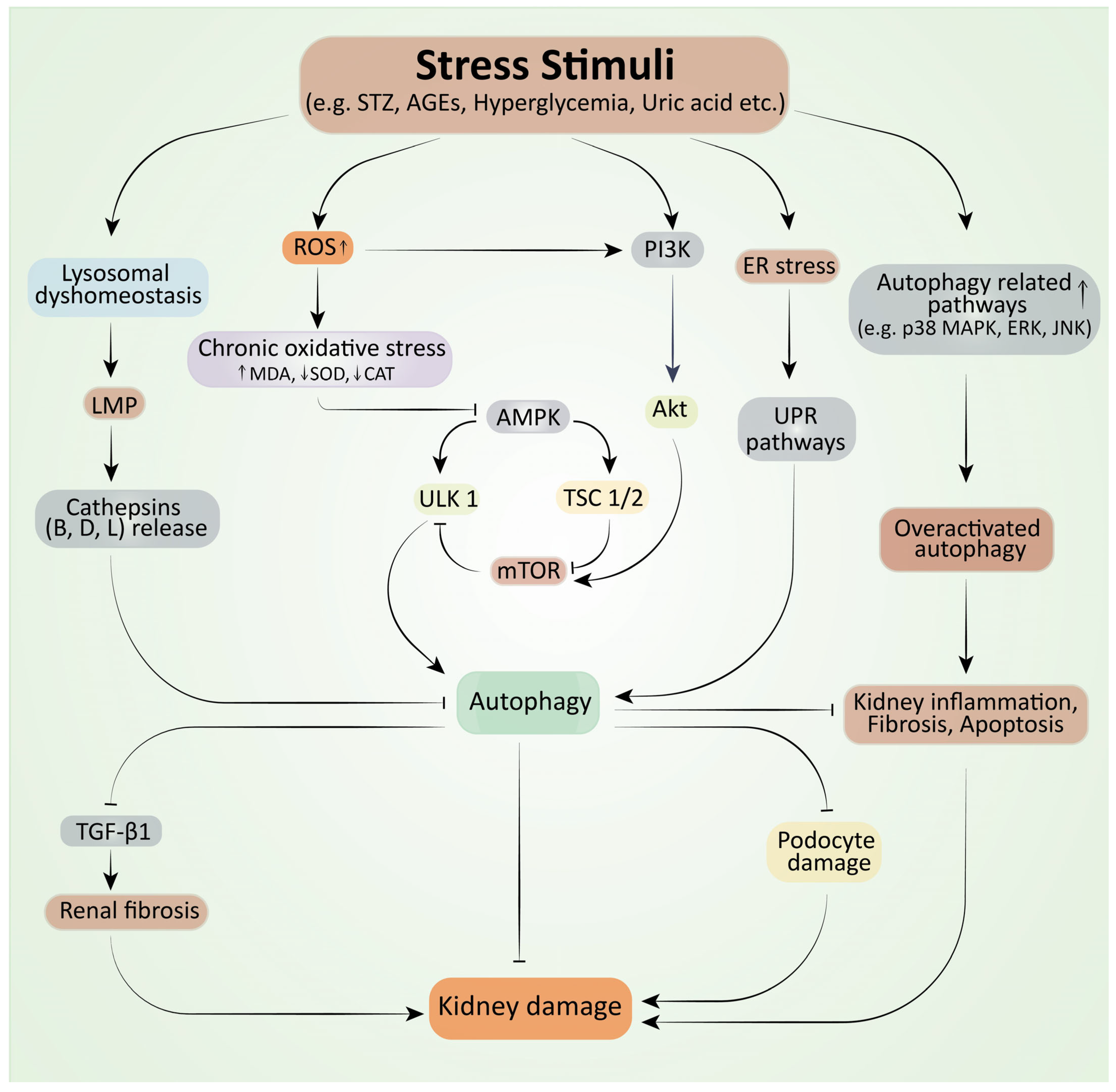
3.5. Molecular Mechanisms Involved in Autophagy Dysfunction in Kidney Disease
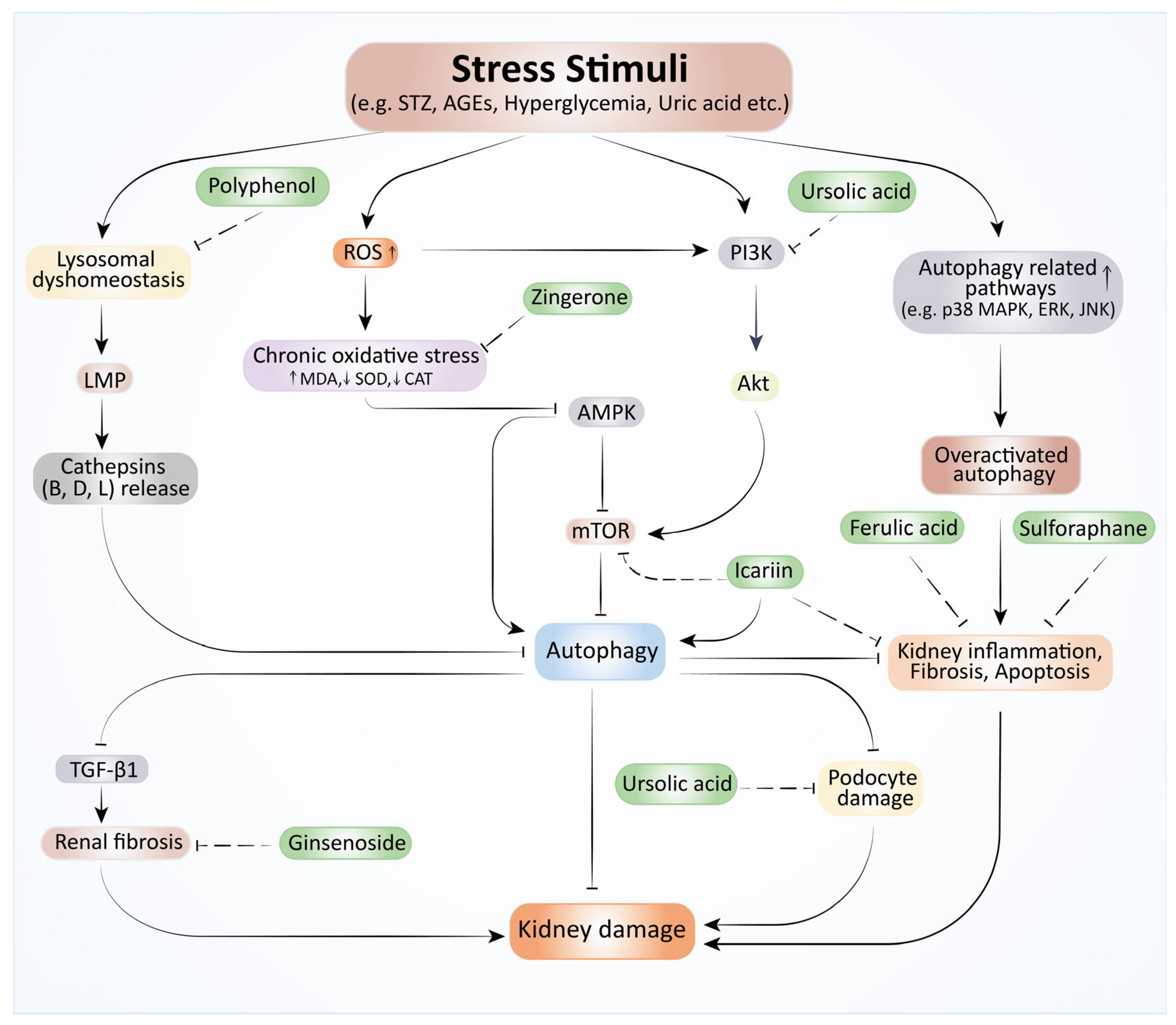
4. Therapeutic Effects of Phytochemicals Against Autophagy Dysfunction in Kidney Diseases
4.1. Acute Kidney Injury
4.2. Chronic Kidney Disease
4.3. Obesity-Related Nephropathy
4.4. Diabetic Nephropathy
4.5. Hypertensive Nephropathy
4.6. Obstructive Nephropathy
5. Combination Therapy of Natural Molecules with Drugs Used in CKD Treatment
6. Clinical Trials of Natural Products Against Autophagy Dysfunction in CKD
7. Recent Updates on Bioactive Compounds for Autophagy in Kidney Diseases
8. Prospects and Limitations
9. Conclusions
Author Contributions
Funding
Data Availability Statement
Conflicts of Interest
References
- Bikbov, B.; Purcell, C.A.; Levey, A.S.; Smith, M.; Abdoli, A.; Abebe, M.; Adebayo, O.M.; Afarideh, M.; Agarwal, S.K.; Agudelo-Botero, M.; et al. Global, regional, and national burden of chronic kidney disease, 1990–2017: A systematic analysis for the Global Burden of Disease Study 2017. Lancet 2020, 395, 709–733. [Google Scholar] [CrossRef] [Scilit]
- Jager, K.J.; Kovesdy, C.; Langham, R.; Rosenberg, M.; Jha, V.; Zoccali, C. A single number for advocacy and communication-worldwide more than 850 million individuals have kidney diseases. Nephrol. Dial. Transpl. 2019, 34, 1803–1805. [Google Scholar] [CrossRef] [Scilit] [PubMed]
- Francis, A.; Harhay, M.N.; Ong, A.C.M.; Tummalapalli, S.L.; Ortiz, A.; Fogo, A.B.; Fliser, D.; Roy-Chaudhury, P.; Fontana, M.; Nangaku, M.; et al. Chronic kidney disease and the global public health agenda: An international consensus. Nat. Rev. Nephrol. 2024, 20, 473–485. [Google Scholar] [CrossRef] [Scilit] [PubMed]
- Sureshbabu, A.; Ryter, S.W.; Choi, M.E. Oxidative stress and autophagy: Crucial modulators of kidney injury. Redox Biol. 2015, 4, 208–214. [Google Scholar] [CrossRef] [Scilit] [PubMed]
- Mizushima, N. A brief history of autophagy from cell biology to physiology and disease. Nat. Cell Biol. 2018, 20, 521–527. [Google Scholar] [CrossRef] [Scilit]
- Levine, B.; Klionsky, D.J. Autophagy wins the 2016 Nobel Prize in Physiology or Medicine: Breakthroughs in baker’s yeast fuel advances in biomedical research. Proc. Natl. Acad. Sci. USA 2017, 114, 201–205. [Google Scholar] [CrossRef] [Scilit]
- Wang, Z.; Choi, M.E. Autophagy in kidney health and disease. Antioxid. Redox Signal 2014, 20, 519–537. [Google Scholar] [CrossRef] [Scilit]
- Pandiri, I.; Moni, A. Ocimum herb species: A potential treatment strategy for diabetic kidney disease. J. Adv. Biotechnol. Exp. Ther. 2018, 1, 88–91. [Google Scholar] [CrossRef] [Scilit]
- Rahman, M.A.; Hannan, M.A.; Dash, R.; Rahman, M.H.; Islam, R.; Uddin, M.J.; Sohag, A.A.M.; Rahman, M.H.; Rhim, H. Phytochemicals as a Complement to Cancer Chemotherapy: Pharmacological Modulation of the Autophagy-Apoptosis Pathway. Front. Pharmacol. 2021, 12, 639628. [Google Scholar] [CrossRef] [Scilit]
- Chowdhury, S.; Ghosh, S.; Das, A.K.; Sil, P.C. Ferulic Acid Protects Hyperglycemia-Induced Kidney Damage by Regulating Oxidative Insult, Inflammation and Autophagy. Front. Pharmacol. 2019, 10, 27. [Google Scholar] [CrossRef] [Scilit]
- Liu, P.; Zhu, W.; Wang, Y.; Ma, G.; Zhao, H.; Li, P. Chinese herbal medicine and its active compounds in attenuating renal injury via regulating autophagy in diabetic kidney disease. Front. Endocrinol. 2023, 14, 1142805. [Google Scholar] [CrossRef] [Scilit]
- Putra, I.; Fakhrudin, N.; Nurrochmad, A.; Wahyuono, S. A Review of Medicinal Plants with Renoprotective Activity in Diabetic Nephropathy Animal Models. Life 2023, 13, 560. [Google Scholar] [CrossRef] [Scilit]
- Rahman, M.A.; Akter, S.; Dorotea, D.; Mazumder, A.; Uddin, M.N.; Hannan, M.A.; Hossen, M.J.; Ahmed, M.S.; Kim, W.; Kim, B.; et al. Renoprotective potentials of small molecule natural products targeting mitochondrial dysfunction. Front. Pharmacol. 2022, 13, 925993. [Google Scholar] [CrossRef] [Scilit]
- Uddin, M.J.; Kim, E.H.; Hannan, M.A.; Ha, H. Pharmacotherapy against oxidative stress in chronic kidney disease: Promising small molecule natural products targeting Nrf2-HO-1 signaling. Antioxidants 2021, 10, 258. [Google Scholar] [CrossRef] [Scilit] [PubMed]
- Mao, S.; Zhang, J. Role of autophagy in chronic kidney diseases. Int. J. Clin. Exp. Med. 2015, 8, 22022–22029. [Google Scholar]
- Stanigut, A.M.; Tuta, L.; Pana, C.; Alexandrescu, L.; Suceveanu, A.; Blebea, N.-M.; Vacaroiu, I.A. Autophagy and Mitophagy in Diabetic Kidney Disease—A Literature Review. Int. J. Mol. Sci. 2025, 26, 806. [Google Scholar] [CrossRef] [Scilit]
- Andrade, L.; Rodrigues, C.E.; Gomes, S.A.; Noronha, I.L. Acute Kidney Injury as a Condition of Renal Senescence. Cell Transplant. 2018, 27, 739–753. [Google Scholar] [CrossRef] [Scilit] [PubMed]
- Cinà, D.P.; Onay, T.; Paltoo, A.; Li, C.; Maezawa, Y.; De Arteaga, J.; Jurisicova, A.; Quaggin, S.E. Inhibition of MTOR Disrupts Autophagic Flux in Podocytes. J. Am. Soc. Nephrol. 2012, 23, 412–420. [Google Scholar] [CrossRef] [Scilit]
- Distefano, G.; Boca, M.; Rowe, I.; Wodarczyk, C.; Ma, L.; Piontek, K.B.; Germino, G.G.; Pandolfi, P.P.; Boletta, A. Polycystin-1 Regulates Extracellular Signal-Regulated Kinase-Dependent Phosphorylation of Tuberin To Control Cell Size through mTOR and Its Downstream Effectors S6K and 4EBP1. Mol. Cell. Biol. 2009, 29, 2359–2371. [Google Scholar] [CrossRef] [Scilit] [PubMed]
- Ravichandran, K.; Edelstein, C.L. Polycystic Kidney Disease: A Case of Suppressed Autophagy? Semin. Nephrol. 2014, 34, 27–33. [Google Scholar] [CrossRef] [Scilit]
- Kim, S.I.; Na, H.-J.; Ding, Y.; Wang, Z.; Lee, S.J.; Choi, M.E. Autophagy Promotes Intracellular Degradation of Type I Collagen Induced by Transforming Growth Factor (TGF)-β1*. J. Biol. Chem. 2012, 287, 11677–11688. [Google Scholar] [CrossRef] [Scilit]
- Angoorani, P.; Ejtahed, H.-S.; Mirmiran, P.; Mirzaei, S.; Azizi, F. Dietary consumption of advanced glycation end products and risk of metabolic syndrome. Int. J. Food Sci. Nutr. 2016, 67, 170–176. [Google Scholar] [CrossRef] [Scilit]
- Lin, T.-A.; Wu, V.C.-C.; Wang, C.-Y. Autophagy in Chronic Kidney Diseases. Cells 2019, 8, 61. [Google Scholar] [CrossRef] [Scilit]
- Wharram, B.L.; Goyal, M.; Wiggins, J.E.; Sanden, S.K.; Hussain, S.; Filipiak, W.E.; Saunders, T.L.; Dysko, R.C.; Kohno, K.; Holzman, L.B.; et al. Podocyte Depletion Causes Glomerulosclerosis: Diphtheria Toxin–Induced Podocyte Depletion in Rats Expressing Human Diphtheria Toxin Receptor Transgene. J. Am. Soc. Nephrol. 2005, 16, 2941–2952. [Google Scholar] [CrossRef] [Scilit] [PubMed]
- Kim, J.W.; Nam, S.A.; Koh, E.-S.; Kim, H.W.; Kim, S.; Woo, J.J.; Kim, Y.K. The Impairment of Endothelial Autophagy Accelerates Renal Senescence by Ferroptosis and NLRP3 Inflammasome Signaling Pathways with the Disruption of Endothelial Barrier. Antioxidants 2024, 13, 886. [Google Scholar] [CrossRef] [Scilit] [PubMed]
- Tavernier, Q.; Mami, I.; Rabant, M.; Karras, A.; Laurent-Puig, P.; Chevet, E.; Thervet, E.; Anglicheau, D.; Pallet, N. Urinary Angiogenin Reflects the Magnitude of Kidney Injury at the Infrahistologic Level. J. Am. Soc. Nephrol. 2017, 28, 678–690. [Google Scholar] [CrossRef] [Scilit]
- Zhang, Z.; Sun, Y.; Xue, J.; Jin, D.; Li, X.; Zhao, D.; Lian, F.; Qi, W.; Tong, X. The critical role of dysregulated autophagy in the progression of diabetic kidney disease. Front. Pharmacol. 2022, 13, 977410. [Google Scholar] [CrossRef] [Scilit] [PubMed]
- Wang, X.; Gao, Y.; Tian, N.; Zhu, Z.; Wang, T.; Xu, J.; Wu, B.; Zhang, N. Astragaloside IV represses high glucose-induced mesangial cells activation by enhancing autophagy via SIRT1 deacetylation of NF-κB p65 subunit. Drug Des. Dev. Ther. 2018, 12, 2971–2980. [Google Scholar] [CrossRef] [Scilit]
- Kaushal, G.P.; Chandrashekar, K.; Juncos, L.A. Molecular Interactions Between Reactive Oxygen Species and Autophagy in Kidney Disease. Int. J. Mol. Sci. 2019, 20, 3791. [Google Scholar] [CrossRef] [Scilit]
- Sridhar, S.; Botbol, Y.; Macian, F.; Cuervo, A.M. Autophagy and disease: Always two sides to a problem. J. Pathol. 2012, 226, 255–273. [Google Scholar] [CrossRef] [Scilit]
- Wible, D.J.; Bratton, S.B. Reciprocity in ROS and autophagic signaling. Curr. Opin. Toxicol. 2018, 7, 28–36. [Google Scholar] [CrossRef] [Scilit]
- Seo, S.U.; Woo, S.M.; Lee, H.-S.; Kim, S.H.; Min, K.-J.; Kwon, T.K. mTORC1/2 inhibitor and curcumin induce apoptosis through lysosomal membrane permeabilization-mediated autophagy. Oncogene 2018, 37, 5205–5220. [Google Scholar] [CrossRef] [Scilit]
- Birben, E.; Sahiner, U.M.; Sackesen, C.; Erzurum, S.; Kalayci, O. Oxidative stress and antioxidant defense. World Allergy Organ. J. 2012, 5, 9–19. [Google Scholar] [CrossRef] [Scilit]
- Melk, A.; Baisantry, A.; Schmitt, R. The yin and yang of autophagy in acute kidney injury. Autophagy 2016, 12, 596–597. [Google Scholar] [CrossRef] [Scilit] [PubMed]
- Jiang, M.; Liu, K.; Luo, J.; Dong, Z. Autophagy is a renoprotective mechanism during in vitro hypoxia and in vivo ischemia-reperfusion injury. Am. J. Pathol. 2010, 176, 1181–1192. [Google Scholar] [CrossRef] [Scilit] [PubMed]
- Lin, F. Autophagy in renal tubular injury and repair. Acta Physiol. 2017, 220, 229–237. [Google Scholar] [CrossRef] [Scilit] [PubMed]
- Kimura, T.; Takabatake, Y.; Takahashi, A.; Kaimori, J.Y.; Matsui, I.; Namba, T.; Kitamura, H.; Niimura, F.; Matsusaka, T.; Soga, T.; et al. Autophagy protects the proximal tubule from degeneration and acute ischemic injury. J. Am. Soc. Nephrol. 2011, 22, 902–913. [Google Scholar] [CrossRef] [Scilit] [PubMed]
- Kimura, T.; Takahashi, A.; Takabatake, Y.; Namba, T.; Yamamoto, T.; Kaimori, J.Y.; Matsui, I.; Kitamura, H.; Niimura, F.; Matsusaka, T.; et al. Autophagy protects kidney proximal tubule epithelial cells from mitochondrial metabolic stress. Autophagy 2013, 9, 1876–1886. [Google Scholar] [CrossRef] [Scilit]
- Jiang, M.; Wei, Q.; Dong, G.; Komatsu, M.; Su, Y.; Dong, Z. Autophagy in proximal tubules protects against acute kidney injury. Kidney Int. 2012, 82, 1271–1283. [Google Scholar] [CrossRef] [Scilit]
- Chen, W.T.; Hung, K.C.; Wen, M.S.; Hsu, P.Y.; Chen, T.H.; Wang, H.D.; Fang, J.T.; Shie, S.S.; Wang, C.Y. Impaired leukocytes autophagy in chronic kidney disease patients. Cardiorenal Med. 2013, 3, 254–264. [Google Scholar] [CrossRef] [Scilit]
- Kosztelnik, M.; Kurucz, A.; Papp, D.; Jones, E.; Sigmond, T.; Barna, J.; Traka, M.H.; Lorincz, T.; Szarka, A.; Banhegyi, G.; et al. Suppression of AMPK/aak-2 by NRF2/SKN-1 down-regulates autophagy during prolonged oxidative stress. FASEB J. Off. Publ. Fed. Am. Soc. Exp. Biol. 2019, 33, 2372–2387. [Google Scholar] [CrossRef] [Scilit] [PubMed]
- Ai, S.; Li, Y.; Zheng, H.; Wang, Z.; Liu, W.; Tao, J.; Li, Y.; Wang, Y. Global research trends and hot spots on autophagy and kidney diseases: A bibliometric analysis from 2000 to 2022. Front. Pharmacol. 2023, 14, 1275792. [Google Scholar] [CrossRef] [Scilit]
- Anders, H.J.; Schaefer, L. Beyond tissue injury-damage-associated molecular patterns, toll-like receptors, and inflammasomes also drive regeneration and fibrosis. J. Am. Soc. Nephrol. 2014, 25, 1387–1400. [Google Scholar] [CrossRef] [Scilit]
- Kinsey, G.R.; Li, L.; Okusa, M.D. Inflammation in acute kidney injury. Nephron Exp. Nephrol. 2008, 109, e102–e107. [Google Scholar] [CrossRef] [Scilit]
- Akchurin, O.M.; Kaskel, F. Update on inflammation in chronic kidney disease. Blood Purif. 2015, 39, 84–92. [Google Scholar] [CrossRef] [Scilit]
- Pahwa, R.; Goyal, A.; Jialal, I. Chronic Inflammation. In StatPearls; StatPearls Publishing Copyright© 2025; StatPearls Publishing LLC.: Treasure Island, FL, USA, 2025. [Google Scholar]
- Banerjee, P.; Basu, A.; Wegiel, B.; Otterbein, L.E.; Mizumura, K.; Gasser, M.; Waaga-Gasser, A.M.; Choi, A.M.; Pal, S. Heme oxygenase-1 promotes survival of renal cancer cells through modulation of apoptosis- and autophagy-regulating molecules. J. Biol. Chem. 2012, 287, 32113–32123. [Google Scholar] [CrossRef] [Scilit]
- Ding, Y.; Fu, X.; Wang, Q.; Liu, H.; Wang, H.; Wu, D. The Complex Interplay between Autophagy and NLRP3 Inflammasome in Renal Diseases. Int. J. Mol. Sci. 2021, 22, 12766. [Google Scholar] [CrossRef] [Scilit]
- Jianbing, H.; Xiaotian, L.; Jie, T.; Xueying, C.; Honge, J.; Bo, Z.; Lirong, H.; Lei, Z. The Effect of Allograft Inflammatory Factor-1 on Inflammation, Oxidative Stress, and Autophagy via miR-34a/ATG4B Pathway in Diabetic Kidney Disease. Oxid. Med. Cell. Longev. 2022, 2022, 1668000. [Google Scholar] [CrossRef] [Scilit] [PubMed]
- Kato, H.; Gruenwald, A.; Suh, J.H.; Miner, J.H.; Barisoni-Thomas, L.; Taketo, M.M.; Faul, C.; Millar, S.E.; Holzman, L.B.; Susztak, K. Wnt/β-catenin pathway in podocytes integrates cell adhesion, differentiation, and survival. J. Biol. Chem. 2011, 286, 26003–26015. [Google Scholar] [CrossRef] [Scilit] [PubMed]
- Fabian, S.L.; Penchev, R.R.; St-Jacques, B.; Rao, A.N.; Sipilä, P.; West, K.A.; McMahon, A.P.; Humphreys, B.D. Hedgehog-Gli pathway activation during kidney fibrosis. Am. J. Pathol. 2012, 180, 1441–1453. [Google Scholar] [CrossRef] [Scilit]
- White, E.S.; Mantovani, A.R. Inflammation, wound repair, and fibrosis: Reassessing the spectrum of tissue injury and resolution. J. Pathol. 2013, 229, 141–144. [Google Scholar] [CrossRef] [Scilit]
- Kuppe, C.; Ibrahim, M.M.; Kranz, J.; Zhang, X.; Ziegler, S.; Perales-Patón, J.; Jansen, J.; Reimer, K.C.; Smith, J.R.; Dobie, R.; et al. Decoding myofibroblast origins in human kidney fibrosis. Nature 2021, 589, 281–286. [Google Scholar] [CrossRef] [Scilit]
- Sato, Y.; Yanagita, M. Resident fibroblasts in the kidney: A major driver of fibrosis and inflammation. Inflamm. Regen. 2017, 37, 17. [Google Scholar] [CrossRef] [Scilit]
- Ferenbach, D.A.; Bonventre, J.V. Mechanisms of maladaptive repair after AKI leading to accelerated kidney ageing and CKD. Nat. Rev. Nephrol. 2015, 11, 264–276. [Google Scholar] [CrossRef] [Scilit] [PubMed]
- Kumar, S. Cellular and molecular pathways of renal repair after acute kidney injury. Kidney Int. 2018, 93, 27–40. [Google Scholar] [CrossRef] [Scilit]
- Humphreys, B.D. Mechanisms of renal fibrosis. Annu. Rev. Physiol. 2018, 80, 309–326. [Google Scholar] [CrossRef] [Scilit]
- Podestà, M.A.; Faravelli, I.; Ponticelli, C. Autophagy in lupus nephritis: A delicate balance between regulation and disease. Autoimmun. Rev. 2022, 21, 103132. [Google Scholar] [CrossRef] [Scilit]
- Gui, Z.; Suo, C.; Wang, Z.; Zheng, M.; Fei, S.; Chen, H.; Sun, L.; Han, Z.; Tao, J.; Ju, X.; et al. Impaired ATG16L-dependent autophagy promotes renal interstitial fibrosis in chronic renal graft dysfunction through inducing EndMT by NF-κB signal pathway. Front. Immunol. 2021, 12, 650424. [Google Scholar] [CrossRef] [Scilit] [PubMed]
- Kim, W.Y.; Nam, S.A.; Song, H.C.; Ko, J.S.; Park, S.H.; Kim, H.L.; Choi, E.J.; Kim, Y.S.; Kim, J.; Kim, Y.K. The role of autophagy in unilateral ureteral obstruction rat model. Nephrology 2012, 17, 148–159. [Google Scholar] [CrossRef] [Scilit] [PubMed]
- Ma, Z.; Li, L.; Livingston, M.J.; Zhang, D.; Mi, Q.; Zhang, M.; Ding, H.-F.; Huo, Y.; Mei, C.; Dong, Z. p53/microRNA-214/ULK1 axis impairs renal tubular autophagy in diabetic kidney disease. J. Clin. Investig. 2020, 130, 5011–5026. [Google Scholar] [CrossRef] [Scilit]
- Yan, Q.; Song, Y.; Zhang, L.; Chen, Z.; Yang, C.; Liu, S.; Yuan, X.; Gao, H.; Ding, G.; Wang, H. Autophagy activation contributes to lipid accumulation in tubular epithelial cells during kidney fibrosis. Cell Death Discov. 2018, 4, 39. [Google Scholar] [CrossRef] [Scilit] [PubMed]
- Fu, Y.; Xiang, Y.; Wu, W.; Cai, J.; Tang, C.; Dong, Z. Persistent activation of autophagy after cisplatin nephrotoxicity promotes renal fibrosis and chronic kidney disease. Front. Pharmacol. 2022, 13, 918732. [Google Scholar] [CrossRef] [Scilit]
- Livingston, M.J.; Ding, H.-F.; Huang, S.; Hill, J.A.; Yin, X.-M.; Dong, Z. Persistent activation of autophagy in kidney tubular cells promotes renal interstitial fibrosis during unilateral ureteral obstruction. Autophagy 2016, 12, 976–998. [Google Scholar] [CrossRef] [Scilit]
- Hetz, C.; Zhang, K.; Kaufman, R.J. Mechanisms, regulation and functions of the unfolded protein response. Nat. Rev. Mol. Cell Biol. 2020, 21, 421–438. [Google Scholar] [CrossRef] [Scilit]
- Wang, M.; Chen, Z.; Tang, Z.; Tang, S. Natural products derived from traditional Chinese medicines targeting ER stress for the treatment of kidney diseases. Ren. Fail. 2024, 46, 2396446. [Google Scholar] [CrossRef] [Scilit]
- Hasegawa, S.; Inagi, R. Organelle stress and crosstalk in kidney disease. Kidney360 2020, 1, 1157–1164. [Google Scholar] [CrossRef] [Scilit] [PubMed]
- Fan, Y.; Xiao, W.; Lee, K.; Salem, F.; Wen, J.; He, L.; Zhang, J.; Fei, Y.; Cheng, D.; Bao, H.; et al. Inhibition of reticulon-1a–mediated endoplasmic reticulum stress in early AKI attenuates renal fibrosis development. J. Am. Soc. Nephrol. 2017, 28, 2007–2021. [Google Scholar] [CrossRef] [Scilit]
- Fan, Y.; Xiao, W.; Li, Z.; Li, X.; Chuang, P.Y.; Jim, B.; Zhang, W.; Wei, C.; Wang, N.; Jia, W.; et al. RTN1 mediates progression of kidney disease by inducing ER stress. Nat. Commun. 2015, 6, 7841. [Google Scholar] [CrossRef] [Scilit]
- Xiao, W.; Fan, Y.; Wang, N.; Chuang, P.Y.; Lee, K.; He, J.C. Knockdown of RTN1A attenuates ER stress and kidney injury in albumin overload-induced nephropathy. Am. J. Physiol. Ren. Physiol. 2016, 310, F409–F415. [Google Scholar] [CrossRef] [Scilit]
- Yan, M.; Shu, S.; Guo, C.; Tang, C.; Dong, Z. Endoplasmic reticulum stress in ischemic and nephrotoxic acute kidney injury. Ann. Med. 2018, 50, 381–390. [Google Scholar] [CrossRef] [Scilit] [PubMed]
- Yamamoto, H.; Matsui, T. Molecular mechanisms of macroautophagy, microautophagy, and chaperone-mediated autophagy. J. Nippon. Med. Sch. 2024, 91, 2–9. [Google Scholar]
- Aman, Y.; Schmauck-Medina, T.; Hansen, M.; Morimoto, R.I.; Simon, A.K.; Bjedov, I.; Palikaras, K.; Simonsen, A.; Johansen, T.; Tavernarakis, N. Autophagy in healthy aging and disease. Nat. Aging 2021, 1, 634–650. [Google Scholar] [CrossRef] [Scilit]
- Sohn, M.; Kim, K.; Uddin, M.J.; Lee, G.; Hwang, I.; Kang, H.; Kim, H.; Lee, J.H.; Ha, H. Delayed treatment with fenofibrate protects against high-fat diet-induced kidney injury in mice: The possible role of AMPK autophagy. Am. J. Physiol. Ren. Physiol. 2017, 312, F323–F334. [Google Scholar] [CrossRef] [Scilit]
- Liu, F.; Zhao, L.; Wu, T.; Yu, W.; Li, J.; Wang, W.; Huang, C.; Diao, Z.; Xu, Y. Targeting autophagy with natural products as a potential therapeutic approach for diabetic microangiopathy. Front. Pharmacol. 2024, 15, 1364616. [Google Scholar] [CrossRef] [Scilit]
- Liu, J.; Livingston, M.J.; Dong, G.; Tang, C.; Su, Y.; Wu, G.; Yin, X.-M.; Dong, Z. Histone deacetylase inhibitors protect against cisplatin-induced acute kidney injury by activating autophagy in proximal tubular cells. Cell Death Dis. 2018, 9, 322. [Google Scholar] [CrossRef] [Scilit]
- Tang, C.; Han, H.; Yan, M.; Zhu, S.; Liu, J.; Liu, Z.; He, L.; Tan, J.; Liu, Y.; Liu, H.; et al. PINK1-PRKN/PARK2 pathway of mitophagy is activated to protect against renal ischemia-reperfusion injury. Autophagy 2018, 14, 880–897. [Google Scholar] [CrossRef] [Scilit] [PubMed]
- Kaufman, D.R.; Papillon, J.; Larose, L.; Iwawaki, T.; Cybulsky, A.V. Deletion of inositol-requiring enzyme-1α in podocytes disrupts glomerular capillary integrity and autophagy. Mol. Biol. Cell 2017, 28, 1636–1651. [Google Scholar] [CrossRef] [Scilit] [PubMed]
- Rashid, H.-O.; Yadav, R.K.; Kim, H.-R.; Chae, H.-J. ER stress: Autophagy induction, inhibition and selection. Autophagy 2015, 11, 1956–1977. [Google Scholar] [CrossRef] [Scilit]
- Hartleben, B.; Gödel, M.; Meyer-Schwesinger, C.; Liu, S.; Ulrich, T.; Köbler, S.; Wiech, T.; Grahammer, F.; Arnold, S.J.; Lindenmeyer, M.T.; et al. Autophagy influences glomerular disease susceptibility and maintains podocyte homeostasis in aging mice. J. Clin. Investig. 2010, 120, 1084–1096. [Google Scholar] [CrossRef] [Scilit] [PubMed]
- Cybulsky, A.V. The intersecting roles of endoplasmic reticulum stress, ubiquitin–proteasome system, and autophagy in the pathogenesis of proteinuric kidney disease. Kidney Int. 2013, 84, 25–33. [Google Scholar] [CrossRef] [Scilit]
- Tang, C.; Livingston, M.J.; Liu, Z.; Dong, Z. Autophagy in kidney homeostasis and disease. Nat. Rev. Nephrol. 2020, 16, 489–508. [Google Scholar] [CrossRef] [Scilit] [PubMed]
- Ruby, M.; Gifford, C.C.; Pandey, R.; Raj, V.S.; Sabbisetti, V.S.; Ajay, A.K. Autophagy as a Therapeutic Target for Chronic Kidney Disease and the Roles of TGF-β1 in Autophagy and Kidney Fibrosis. Cells 2023, 12, 412. [Google Scholar] [CrossRef] [Scilit]
- Wu, M.; Ma, Y.; Chen, X.; Liang, N.; Qu, S.; Chen, H. Hyperuricemia causes kidney damage by promoting autophagy and NLRP3-mediated inflammation in rats with urate oxidase deficiency. Dis. Model. Mech. 2021, 14, dmm048041. [Google Scholar] [CrossRef] [Scilit]
- Wu, M.; Zhang, M.; Zhang, Y.; Li, Z.; Li, X.; Liu, Z.; Liu, H.; Li, X. Relationship between lysosomal dyshomeostasis and progression of diabetic kidney disease. Cell Death Dis. 2021, 12, 958. [Google Scholar] [CrossRef] [Scilit]
- Yuan, Q.; Tang, B.; Zhang, C. Signaling pathways of chronic kidney diseases, implications for therapeutics. Signal Transduct. Target. Ther. 2022, 7, 182. [Google Scholar] [CrossRef] [Scilit]
- Wang, H.; Gao, L.; Zhao, C.; Fang, F.; Liu, J.; Wang, Z.; Zhong, Y.; Wang, X. The role of PI3K/Akt signaling pathway in chronic kidney disease. Int. Urol. Nephrol. 2024, 56, 2623–2633. [Google Scholar] [CrossRef] [Scilit] [PubMed]
- Miricescu, D.; Balan, D.G.; Tulin, A.; Stiru, O.; Vacaroiu, I.A.; Mihai, D.A.; Popa, C.C.; Papacocea, R.I.; Enyedi, M.; Sorin, N.A.; et al. PI3K/AKT/mTOR signalling pathway involvement in renal cell carcinoma pathogenesis (Review). Exp. Ther. Med. 2021, 21, 540. [Google Scholar] [CrossRef] [Scilit]
- Muratsubaki, S.; Kuno, A.; Tanno, M.; Miki, T.; Yano, T.; Sugawara, H.; Shibata, S.; Abe, K.; Ishikawa, S.; Ohno, K.; et al. Suppressed autophagic response underlies augmentation of renal ischemia/reperfusion injury by type 2 diabetes. Sci. Rep. 2017, 7, 5311. [Google Scholar] [CrossRef] [Scilit]
- Huang, R.; Xu, Y.; Wan, W.; Shou, X.; Qian, J.; You, Z.; Liu, B.; Chang, C.; Zhou, T.; Lippincott-Schwartz, J.; et al. Deacetylation of nuclear LC3 drives autophagy initiation under starvation. Mol. Cell 2015, 57, 456–466. [Google Scholar] [CrossRef] [Scilit]
- Sun, M.; Li, J.; Mao, L.; Wu, J.; Deng, Z.; He, M.; An, S.; Zeng, Z.; Huang, Q.; Chen, Z. p53 Deacetylation Alleviates Sepsis-Induced Acute Kidney Injury by Promoting Autophagy. Front. Immunol. 2021, 12, 685523. [Google Scholar] [CrossRef] [Scilit] [PubMed]
- Deng, J.K.; Zhang, X.; Wu, H.L.; Gan, Y.; Ye, L.; Zheng, H.; Zhu, Z.; Liu, W.J.; Liu, H.F. ROS-ERK Pathway as Dual Mediators of Cellular Injury and Autophagy-Associated Adaptive Response in Urinary Protein-Irritated Renal Tubular Epithelial Cells. J. Diabetes Res. 2021, 2021, 6614848. [Google Scholar] [CrossRef] [Scilit]
- Yang, J.; Yuan, L.; Liu, F.; Li, L.; Liu, J.; Chen, Y.; Lu, Y.; Yuan, Y. Molecular mechanisms and physiological functions of autophagy in kidney diseases. Front. Pharmacol. 2022, 13, 974829. [Google Scholar] [CrossRef] [Scilit]
- Sanz, A.B.; Sanchez-Niño, M.D.; Ramos, A.M.; Ortiz, A. Regulated cell death pathways in kidney disease. Nat. Rev. Nephrol. 2023, 19, 281–299. [Google Scholar] [CrossRef] [Scilit]
- Jia, Z.; Wang, K.; Zhang, Y.; Duan, Y.; Xiao, K.; Liu, S.; Ding, X. Icariin Ameliorates Diabetic Renal Tubulointerstitial Fibrosis by Restoring Autophagy via Regulation of the miR-192-5p/GLP-1R Pathway. Front. Pharmacol. 2021, 12, 720387. [Google Scholar] [CrossRef] [Scilit]
- Matboli, M.; Ibrahim, D.; Hasanin, A.H.; Hassan, M.K.; Habib, E.K.; Bekhet, M.M.; Afifi, A.M.; Eissa, S. Epigenetic modulation of autophagy genes linked to diabetic nephropathy by administration of isorhamnetin in Type 2 diabetes mellitus rats. Epigenomics 2021, 13, 187–202. [Google Scholar] [CrossRef] [Scilit]
- Zhang, X.X.; Jiang, C.H.; Liu, Y.; Lou, D.X.; Huang, Y.P.; Gao, M.; Zhang, J.; Yin, Z.Q.; Pan, K. Cyclocarya paliurus triterpenic acids fraction attenuates kidney injury via AMPK-mTOR-regulated autophagy pathway in diabetic rats. Phytomedicine 2019, 64, 153060. [Google Scholar] [CrossRef] [Scilit] [PubMed]
- Akaras, N.; Gur, C.; Kucukler, S.; Kandemir, F.M. Zingerone reduces sodium arsenite-induced nephrotoxicity by regulating oxidative stress, inflammation, apoptosis and histopathological changes. Chem. Biol. Interact. 2023, 374, 110410. [Google Scholar] [CrossRef] [Scilit]
- Kho, M.C.; Park, J.H.; Han, B.H.; Tan, R.; Yoon, J.J.; Kim, H.Y.; Ahn, Y.M.; Lee, Y.J.; Kang, D.G.; Lee, H.S. Plantago asiatica L. Ameliorates Puromycin Aminonucleoside-Induced Nephrotic Syndrome by Suppressing Inflammation and Apoptosis. Nutrients 2017, 9, 386. [Google Scholar] [CrossRef] [Scilit] [PubMed]
- Zhang, D.; Zhang, H.; Lv, S.; Zhu, C.; Gong, S.; Yu, X.; Wang, Y.; Huang, X.; Yuan, S.; Ding, X.; et al. Sulforaphane alleviates renal fibrosis through dual regulation on mTOR-mediated autophagy pathway. Int. Urol. Nephrol. 2025, 57, 1277–1287. [Google Scholar] [CrossRef] [Scilit] [PubMed]
- Xu, L.; Fan, Q.; Wang, X.; Li, L.; Lu, X.; Yue, Y.; Cao, X.; Liu, J.; Zhao, X.; Wang, L. Ursolic acid improves podocyte injury caused by high glucose. Nephrol. Dial. Transpl. 2017, 32, 1285–1293. [Google Scholar] [CrossRef] [Scilit]
- Ou, X.; Yu, Z.; Pan, C.; Zheng, X.; Li, D.; Qiao, Z.; Zheng, X. Paeoniflorin: A review of its pharmacology, pharmacokinetics and toxicity in diabetes. Front. Pharmacol. 2025, 16, 1551368. [Google Scholar] [CrossRef] [Scilit]
- Tian, H.; Zheng, X.; Wang, H. Isorhapontigenin ameliorates high glucose-induced podocyte and vascular endothelial cell injuries via mitigating oxidative stress and autophagy through the AMPK/Nrf2 pathway. Int. Urol. Nephrol. 2023, 55, 423–436. [Google Scholar] [CrossRef] [Scilit] [PubMed]
- Turgut, F.; Awad, A.S.; Abdel-Rahman, E.M. Acute Kidney Injury: Medical Causes and Pathogenesis. J. Clin. Med. 2023, 12, 375. [Google Scholar] [CrossRef] [Scilit] [PubMed]
- Zeng, Y.; Li, S.; Wu, J.; Chen, W.; Sun, H.; Peng, W.; Yu, X.; Yang, X. Autophagy inhibitors promoted aristolochic acid I induced renal tubular epithelial cell apoptosis via mitochondrial pathway but alleviated nonapoptotic cell death in mouse acute aritolochic acid nephropathy model. Apoptosis 2014, 19, 1215–1224. [Google Scholar] [CrossRef] [Scilit]
- Tan, X.; Zhu, H.; Tao, Q.; Guo, L.; Jiang, T.; Xu, L.; Yang, R.; Wei, X.; Wu, J.; Li, X.; et al. FGF10 Protects Against Renal Ischemia/Reperfusion Injury by Regulating Autophagy and Inflammatory Signaling. Front. Genet. 2018, 9, 556. [Google Scholar] [CrossRef] [Scilit]
- Kaushal, G.P.; Shah, S.V. Autophagy in acute kidney injury. Kidney Int. 2016, 89, 779–791. [Google Scholar] [CrossRef] [Scilit]
- Djavaheri-Mergny, M.; Amelotti, M.; Mathieu, J.; Besançon, F.; Bauvy, C.; Codogno, P. Regulation of autophagy by NFkappaB transcription factor and reactives oxygen species. Autophagy 2007, 3, 390–392. [Google Scholar] [CrossRef] [Scilit]
- Vijayan, A. Tackling AKI: Prevention, timing of dialysis and follow-up. Nat. Rev. Nephrol. 2020, 17, 87–88. [Google Scholar] [CrossRef] [Scilit]
- Chen, L.; Wang, Q.; Li, T.; Li, L.; Wang, C.; Xu, B.; Gong, X. Exploring therapeutic mechanisms of Chuan Huang Fang-II in the treatment of acute kidney injury on chronic kidney disease patients from the perspective of lipidomics. Ren. Fail. 2024, 46, 2356021. [Google Scholar] [CrossRef] [Scilit] [PubMed]
- Shi, Y.; Shi, X.; Zhao, M.; Chang, M.; Ma, S.; Zhang, Y. Ferroptosis: A new mechanism of traditional Chinese medicine compounds for treating acute kidney injury. Biomed. Pharmacother. 2023, 163, 114849. [Google Scholar] [CrossRef] [Scilit]
- Qi, J.; Xue, Q.; Kuang, L.; Xie, L.; Luo, R.; Nie, X. Berberine alleviates cisplatin-induced acute kidney injury by regulating mitophagy via PINK 1/Parkin pathway. Transl. Androl. Urol. 2020, 9, 1712–1724. [Google Scholar] [CrossRef] [Scilit]
- Gong, X.; Duan, Y.; Zheng, J.; Ye, Z.; Hei, T.K. Tetramethylpyrazine Prevents Contrast-Induced Nephropathy via Modulating Tubular Cell Mitophagy and Suppressing Mitochondrial Fragmentation, CCL2/CCR2-Mediated Inflammation, and Intestinal Injury. Oxid. Med. Cell. Longev. 2019, 2019, 7096912. [Google Scholar] [CrossRef] [Scilit]
- Wen, J.; Xu, B.; Sun, Y.; Lian, M.; Li, Y.; Lin, Y.; Chen, D.; Diao, Y.; Almoiliqy, M.; Wang, L. Paeoniflorin protects against intestinal ischemia/reperfusion by activating LKB1/AMPK and promoting autophagy. Pharmacol. Res. 2019, 146, 104308. [Google Scholar] [CrossRef] [Scilit] [PubMed]
- Li, H.; Tang, Y.; Wen, L.; Kong, X.; Chen, X.; Liu, P.; Zhou, Z.; Chen, W.; Xiao, C.; Xiao, P.; et al. Neferine reduces cisplatin-induced nephrotoxicity by enhancing autophagy via the AMPK/mTOR signaling pathway. Biochem. Biophys. Res. Commun. 2017, 484, 694–701. [Google Scholar] [CrossRef] [Scilit] [PubMed]
- Zuo, Z.; Li, Q.; Zhou, S.; Yu, R.; Wu, C.; Chen, J.; Xiao, Y.; Chen, H.; Song, J.; Pan, Y.; et al. Berberine ameliorates contrast-induced acute kidney injury by regulating HDAC4-FoxO3a axis-induced autophagy: In vivo and in vitro. Phytother. Res. 2024, 38, 1761–1780. [Google Scholar] [CrossRef] [Scilit]
- Kalantar-Zadeh, K.; Jafar, T.H.; Nitsch, D.; Neuen, B.L.; Perkovic, V. Chronic kidney disease. Lancet 2021, 398, 786–802. [Google Scholar] [CrossRef] [Scilit] [PubMed]
- Levin, A.; Okpechi, I.G.; Caskey, F.J.; Yang, C.-W.; Tonelli, M.; Jha, V. Perspectives on early detection of chronic kidney disease: The facts, the questions, and a proposed framework for 2023 and beyond. Kidney Int. 2023, 103, 1004–1008. [Google Scholar] [CrossRef] [Scilit]
- Ding, Y.; Choi, M.E. Autophagy in diabetic nephropathy. J. Endocrinol. 2014, 224, R15. [Google Scholar] [CrossRef] [Scilit]
- Periyasamy-Thandavan, S.; Jiang, M.; Schoenlein, P.; Dong, Z. Autophagy: Molecular machinery, regulation, and implications for renal pathophysiology. Am. J. Physiol. Ren. Physiol. 2009, 297, F244–F256. [Google Scholar] [CrossRef] [Scilit]
- Di Tu, Q.; Jin, J.; Hu, X.; Ren, Y.; Zhao, L.; He, Q. Curcumin improves the renal autophagy in rat experimental membranous nephropathy via regulating the PI3K/AKT/mTOR and Nrf2/HO-1 signaling pathways. BioMed Res. Int. 2020, 2020, 7069052. [Google Scholar] [CrossRef] [Scilit]
- Liu, X.; Chen, J.; Sun, N.; Li, N.; Zhang, Z.; Zheng, T.; Li, Z. Ginsenoside Rb1 ameliorates autophagy via the AMPK/mTOR pathway in renal tubular epithelial cells in vitro and in vivo. Int. J. Biol. Macromol. 2020, 163, 996–1009. [Google Scholar] [CrossRef] [Scilit]
- Lu, R.; Chen, J.; Liu, B.; Lin, H.; Bai, L.; Zhang, P.; Chen, D.; Li, H.; Li, J.; Pang, Y.; et al. Protective role of Astragaloside IV in chronic glomerulonephritis by activating autophagy through PI3K/AKT/AS160 pathway. Phytother. Res. 2020, 34, 3236–3248. [Google Scholar] [CrossRef] [Scilit]
- Tseng, C.-Y.; Yu, P.-R.; Hsu, C.-C.; Lin, H.-H.; Chen, J.-H. The effect of isovitexin on lipopolysaccharide-induced renal injury and inflammation by induction of protective autophagy. Food Chem. Toxicol. 2023, 172, 113581. [Google Scholar] [CrossRef] [Scilit]
- Yang, F.; Gao, Y.; Xie, S.; Yang, W.; Wang, Q.; Ye, W.; Sun, L.; Zhou, J.; Feng, X. Dietary phytosterol supplementation mitigates renal fibrosis via activating mitophagy and modulating the gut microbiota. Food Funct. 2025, 16, 2316–2334. [Google Scholar] [CrossRef] [Scilit]
- Fan, L.; Chen, H.-H.; Liu, H.-J.; Chen, H.-J.; Zhu, L.-L.; Zhang, T. Experimental study on danggui shaoyao san improving renal fibrosis by promoting autophagy. Evid. Based Complement. Altern. Med. 2022, 2022, 6761453. [Google Scholar] [CrossRef] [Scilit]
- Chen, R.-J.; Lee, Y.-H.; Yeh, Y.-L.; Wu, W.-S.; Ho, C.-T.; Li, C.-Y.; Wang, B.-J.; Wang, Y.-J. Autophagy-inducing effect of pterostilbene: A prospective therapeutic/preventive option for skin diseases. J. Food Drug Anal. 2017, 25, 125–133. [Google Scholar] [CrossRef] [Scilit]
- Tu, Y.; Gu, L.; Chen, D.; Wu, W.; Liu, H.; Hu, H.; Wan, Y.; Sun, W. Rhein inhibits autophagy in rat renal tubular cells by regulation of AMPK/mTOR signaling. Sci. Rep. 2017, 7, 43790. [Google Scholar] [CrossRef] [Scilit] [PubMed]
- Li, J.; Qin, X.; Xu, W.; Zhang, H.; Huang, S.; Yang, Y.; Qin, M.; Mi, Z.; Zhong, X. Herb pair of Rhubarb-Astragalus mitigates renal interstitial fibrosis through downregulation of autophagy via p38-MAPK/TGF-β1 and p38-MAPK/smad2/3 pathways. Int. J. Biochem. Cell Biol. 2024, 169, 106549. [Google Scholar] [CrossRef] [Scilit]
- Kounatidis, D.; Vallianou, N.G.; Stratigou, T.; Voukali, M.; Karampela, I.; Dalamaga, M. The Kidney in Obesity: Current Evidence, Perspectives and Controversies. Curr. Obes. Rep. 2024, 13, 680–702. [Google Scholar] [CrossRef] [Scilit] [PubMed]
- Martínez-Montoro, J.I.; Morales, E.; Cornejo-Pareja, I.; Tinahones, F.J.; Fernández-García, J.C. Obesity-related glomerulopathy: Current approaches and future perspectives. Obes. Rev. 2022, 23, e13450. [Google Scholar] [CrossRef] [Scilit] [PubMed]
- Hao, M.; Lv, Y.; Liu, S.; Guo, W. The New Challenge of Obesity—Obesity-Associated Nephropathy. Diabetes Metab. Syndr. Obes. 2024, 17, 1957–1971. [Google Scholar] [CrossRef] [Scilit]
- Yau, K.; Kuah, R.; Cherney, D.Z.I.; Lam, T.K.T. Obesity and the kidney: Mechanistic links and therapeutic advances. Nat. Rev. Endocrinol. 2024, 20, 321–335. [Google Scholar] [CrossRef] [Scilit]
- Yao, E.; Yang, X.; Huang, X.; Mi, Y.; Wu, X.; Fang, M.; Huang, J.; Qiu, Y.; Hong, X.; Peng, L.; et al. Phytochemical wedelolactone reverses obesity by prompting adipose browning through SIRT1/AMPK/PPARα pathway via targeting nicotinamide N-methyltransferase. Phytomedicine 2022, 94, 153843. [Google Scholar] [CrossRef] [Scilit]
- Axelsson, A.S.; Tubbs, E.; Mecham, B.; Chacko, S.; Nenonen, H.A.; Tang, Y.; Fahey, J.W.; Derry, J.M.J.; Wollheim, C.B.; Wierup, N.; et al. Sulforaphane reduces hepatic glucose production and improves glucose control in patients with type 2 diabetes. Sci. Transl. Med. 2017, 9, eaah4477. [Google Scholar] [CrossRef] [Scilit]
- Bai, X.; Bian, Z.; Zhang, M. Targeting the Nrf2 signaling pathway using phytochemical ingredients: A novel therapeutic road map to combat neurodegenerative diseases. Phytomedicine 2023, 109, 154582. [Google Scholar] [CrossRef] [Scilit] [PubMed]
- Mafra, D.; Borges, N.A.; Lindholm, B.; Shiels, P.G.; Evenepoel, P.; Stenvinkel, P. Food as medicine: Targeting the uraemic phenotype in chronic kidney disease. Nat. Rev. Nephrol. 2021, 17, 153–171. [Google Scholar] [CrossRef] [Scilit] [PubMed]
- Mykkänen, O.T.; Huotari, A.; Herzig, K.H.; Dunlop, T.W.; Mykkänen, H.; Kirjavainen, P.V. Wild blueberries (Vaccinium myrtillus) alleviate inflammation and hypertension associated with developing obesity in mice fed with a high-fat diet. PLoS ONE 2014, 9, e114790. [Google Scholar] [CrossRef] [Scilit] [PubMed]
- Du, J.; Shen, L.; Tan, Z.; Zhang, P.; Zhao, X.; Xu, Y.; Gan, M.; Yang, Q.; Ma, J.; Jiang, A.; et al. Betaine Supplementation Enhances Lipid Metabolism and Improves Insulin Resistance in Mice Fed a High-Fat Diet. Nutrients 2018, 10, 131. [Google Scholar] [CrossRef] [Scilit] [PubMed]
- Mafra, D.; Esgalhado, M.; Borges, N.A.; Cardozo, L.; Stockler-Pinto, M.B.; Craven, H.; Buchanan, S.J.; Lindholm, B.; Stenvinkel, P.; Shiels, P.G. Methyl Donor Nutrients in Chronic Kidney Disease: Impact on the Epigenetic Landscape. J. Nutr. 2019, 149, 372–380. [Google Scholar] [CrossRef] [Scilit]
- Bahrampour, N.; Mirzababaei, A.; Hosseininasab, D.; Abaj, F.; Clark, C.C.T.; Mirzaei, K. High intake of dietary phytochemical index may be related to reducing risk of diabetic nephropathy: A case-control study. BMC Nutr. 2023, 9, 14. [Google Scholar] [CrossRef] [Scilit]
- Moreira, L.S.G.; Brum, I.; de Vargas Reis, D.C.M.; Trugilho, L.; Chermut, T.R.; Esgalhado, M.; Cardozo, L.; Stenvinkel, P.; Shiels, P.G.; Mafra, D. Cinnamon: An aromatic condiment applicable to chronic kidney disease. Kidney Res. Clin. Pr. 2023, 42, 4–26. [Google Scholar] [CrossRef] [Scilit]
- Dinda, B.; Chakraborty, A. Pharmacokinetics and Metabolism of Phytochemicals Having Anti-Obesity and Antidiabetic Activity; Springer: Cham, Switzerland, 2022; pp. 469–505. [Google Scholar]
- Wang, W.; Sun, W.; Cheng, Y.; Xu, Z.; Cai, L. Role of sirtuin-1 in diabetic nephropathy. J. Mol. Med. 2019, 97, 291–309. [Google Scholar] [CrossRef] [Scilit]
- Kim, H.; Dusabimana, T.; Kim, S.R.; Je, J.; Jeong, K.; Kang, M.C.; Cho, K.M.; Kim, H.J.; Park, S.W. Supplementation of Abelmoschus manihot Ameliorates Diabetic Nephropathy and Hepatic Steatosis by Activating Autophagy in Mice. Nutrients 2018, 10, 1703. [Google Scholar] [CrossRef] [Scilit] [PubMed]
- Sagoo, M.K.; Gnudi, L. Diabetic Nephropathy: An Overview. Methods Mol. Biol. 2020, 2067, 3–7. [Google Scholar] [CrossRef] [Scilit] [PubMed]
- Jefferson, J.A.; Shankland, S.J.; Pichler, R.H. Proteinuria in diabetic kidney disease: A mechanistic viewpoint. Kidney Int. 2008, 74, 22–36. [Google Scholar] [CrossRef] [Scilit] [PubMed]
- Kume, S.; Koya, D. Autophagy: A Novel Therapeutic Target for Diabetic Nephropathy. Diabetes Metab. J. 2015, 39, 451–460. [Google Scholar] [CrossRef] [Scilit]
- Kume, S.; Koya, D.; Uzu, T.; Maegawa, H. Role of nutrient-sensing signals in the pathogenesis of diabetic nephropathy. Biomed. Res. Int. 2014, 2014, 315494. [Google Scholar] [CrossRef] [Scilit]
- Ding, Y.; Kim, S.; Lee, S.Y.; Koo, J.K.; Wang, Z.; Choi, M.E. Autophagy regulates TGF-β expression and suppresses kidney fibrosis induced by unilateral ureteral obstruction. J. Am. Soc. Nephrol. 2014, 25, 2835–2846. [Google Scholar] [CrossRef] [Scilit]
- Kume, S.; Yamahara, K.; Yasuda, M.; Maegawa, H.; Koya, D. Autophagy: Emerging therapeutic target for diabetic nephropathy. Semin. Nephrol. 2014, 34, 9–16. [Google Scholar] [CrossRef] [Scilit]
- Jin, Y.; Liu, S.; Ma, Q.; Xiao, D.; Chen, L. Berberine enhances the AMPK activation and autophagy and mitigates high glucose-induced apoptosis of mouse podocytes. Eur. J. Pharmacol. 2017, 794, 106–114. [Google Scholar] [CrossRef] [Scilit]
- Wei, Y.; Gao, J.; Qin, L.; Xu, Y.; Shi, H.; Qu, L.; Liu, Y.; Xu, T.; Liu, T. Curcumin suppresses AGEs induced apoptosis in tubular epithelial cells via protective autophagy. Exp. Ther. Med. 2017, 14, 6052–6058. [Google Scholar] [CrossRef] [Scilit] [PubMed]
- Ezzat, S.M.; Abdallah, H.M.I.; Yassen, N.N.; Radwan, R.A.; Mostafa, E.S.; Salama, M.M.; Salem, M.A. Phenolics from Physalis peruviana fruits ameliorate streptozotocin-induced diabetes and diabetic nephropathy in rats via induction of autophagy and apoptosis regression. Biomed. Pharmacother. 2021, 142, 111948. [Google Scholar] [CrossRef] [Scilit]
- Yang, X.; Han, X.; Wen, Q.; Qiu, X.; Deng, H.; Chen, Q. Protective Effect of Keluoxin against Diabetic Nephropathy in Type 2 Diabetic Mellitus Models. Evid. Based Complement. Altern. Med. 2021, 2021, 8455709. [Google Scholar] [CrossRef] [Scilit]
- Chen, Y.; Zheng, Y.F.; Lin, X.H.; Zhang, J.P.; Lin, F.; Shi, H. Dendrobium mixture attenuates renal damage in rats with diabetic nephropathy by inhibiting the PI3K/Akt/mTOR pathway. Mol. Med. Rep. 2021, 24, 590. [Google Scholar] [CrossRef] [Scilit]
- Liu, Y.; Liu, W.; Zhang, Z.; Hu, Y.; Zhang, X.; Sun, Y.; Lei, Q.; Sun, D.; Liu, T.; Fan, Y.; et al. Yishen capsule promotes podocyte autophagy through regulating SIRT1/NF-κB signaling pathway to improve diabetic nephropathy. Ren. Fail. 2021, 43, 128–140. [Google Scholar] [CrossRef] [Scilit]
- Amanat, M.; Lal, K.; Singh, T.G.; Singh, R. Molecular insights of diabetic nephropathy and chemical constituents-based treatment approach. Phytochem. Rev. 2025, 24, 5163–5206. [Google Scholar] [CrossRef] [Scilit]
- Zhan, X.; Yan, C.; Chen, Y.; Wei, X.; Xiao, J.; Deng, L.; Yang, Y.; Qiu, P.; Chen, Q. Celastrol antagonizes high glucose-evoked podocyte injury, inflammation and insulin resistance by restoring the HO-1-mediated autophagy pathway. Mol. Immunol. 2018, 104, 61–68. [Google Scholar] [CrossRef] [Scilit] [PubMed]
- Wang, Y.; Zhao, H.; Wang, Q.; Zhou, X.; Lu, X.; Liu, T.; Zhan, Y.; Li, P. Chinese Herbal Medicine in Ameliorating Diabetic Kidney Disease via Activating Autophagy. J. Diabetes Res. 2019, 2019, 9030893. [Google Scholar] [CrossRef] [Scilit]
- Xu, X.H.; Ding, D.F.; Yong, H.J.; Dong, C.L.; You, N.; Ye, X.L.; Pan, M.L.; Ma, J.H.; You, Q.; Lu, Y.B. Resveratrol transcriptionally regulates miRNA-18a-5p expression ameliorating diabetic nephropathy via increasing autophagy. Eur. Rev. Med. Pharmacol. Sci. 2017, 21, 4952–4965. [Google Scholar]
- Huang, S.S.; Ding, D.F.; Chen, S.; Dong, C.L.; Ye, X.L.; Yuan, Y.G.; Feng, Y.M.; You, N.; Xu, J.R.; Miao, H.; et al. Resveratrol protects podocytes against apoptosis via stimulation of autophagy in a mouse model of diabetic nephropathy. Sci. Rep. 2017, 7, 45692. [Google Scholar] [CrossRef] [Scilit]
- Al Shahrani, M.; Chandramoorthy, H.C.; Alshahrani, M.; Abohassan, M.; Eid, R.A.; Ravichandran, K.; Rajagopalan, P. Cassia auriculata leaf extract ameliorates diabetic nephropathy by attenuating autophagic necroptosis via RIP-1/RIP-3-p-p38MAPK signaling. J. Food Biochem. 2021, 45, e13810. [Google Scholar] [CrossRef] [Scilit]
- Yang, F.; Qu, Q.; Zhao, C.; Liu, X.; Yang, P.; Li, Z.; Han, L.; Shi, X. Paecilomyces cicadae-fermented Radix astragali activates podocyte autophagy by attenuating PI3K/AKT/mTOR pathways to protect against diabetic nephropathy in mice. Biomed. Pharmacother. 2020, 129, 110479. [Google Scholar] [CrossRef] [Scilit]
- Han, J.; Zhang, Y.; Shi, X.; Peng, Z.; Xing, Y.; Pang, X. Tongluo Digui decoction treats renal injury in diabetic rats by promoting autophagy of podocytes. J. Tradit. Chin. Med. 2021, 41, 125–132. [Google Scholar] [CrossRef] [Scilit] [PubMed]
- Zhang, Y.; Arzaghi, H.; Ma, Z.; Roye, Y.; Musah, S. Epigenetics of Hypertensive Nephropathy. Biomedicines 2024, 12, 2622. [Google Scholar] [CrossRef] [Scilit]
- Pitzer, A.; Elijovich, F.; Laffer, C.L.; Ertuglu, L.A.; Sahinoz, M.; Saleem, M.; Krishnan, J.; Dola, T.; Aden, L.A.; Sheng, Q.; et al. DC ENaC-Dependent Inflammasome Activation Contributes to Salt-Sensitive Hypertension. Circ. Res. 2022, 131, 328–344. [Google Scholar] [CrossRef] [Scilit] [PubMed]
- Bidani, A.K.; Griffin, K.A. Pathophysiology of hypertensive renal damage: Implications for therapy. Hypertension 2004, 44, 595–601. [Google Scholar] [CrossRef] [Scilit] [PubMed]
- Laffer, C.L.; Elijovich, F.; Sahinoz, M.; Pitzer, A.; Kirabo, A. New Insights Into the Renin-Angiotensin System in Chronic Kidney Disease. Circ. Res. 2020, 127, 607–609. [Google Scholar] [CrossRef] [Scilit]
- Ngowi, E.E.; Sarfraz, M.; Afzal, A.; Khan, N.H.; Khattak, S.; Zhang, X.; Li, T.; Duan, S.F.; Ji, X.Y.; Wu, D.D. Roles of Hydrogen Sulfide Donors in Common Kidney Diseases. Front. Pharmacol. 2020, 11, 564281. [Google Scholar] [CrossRef] [Scilit]
- Hackfort, B.T.; Mishra, P.K. Emerging role of hydrogen sulfide-microRNA crosstalk in cardiovascular diseases. Am. J. Physiol. Heart Circ. Physiol. 2016, 310, H802–H812. [Google Scholar] [CrossRef] [Scilit]
- Weber, G.J.; Pushpakumar, S.B.; Sen, U. Hydrogen sulfide alleviates hypertensive kidney dysfunction through an epigenetic mechanism. Am. J. Physiol. Heart Circ. Physiol. 2017, 312, H874–H885. [Google Scholar] [CrossRef] [Scilit]
- Ye, Q.; Ren, M.; Fan, D.; Mao, Y.; Zhu, Y.Z. Identification and Validation of the miR/RAS/RUNX2 Autophagy Regulatory Network in AngII-Induced Hypertensive Nephropathy in MPC5 Cells Treated with Hydrogen Sulfide Donors. Antioxidants 2024, 13, 958. [Google Scholar] [CrossRef] [Scilit]
- Liu, P.; Hu, T.; Gao, B.; Xia, R.; Dai, X.; Wang, J. Network pharmacology and in vitro experiments reveal the autophagy mechanism of Yanggan-Yishui granules in improving hypertensive renal injury. Arch. Med. Sci. 2024, 22, 387–407. [Google Scholar] [CrossRef] [Scilit]
- Klahr, S. Obstructive nephropathy. Intern. Med. 2000, 39, 355–361. [Google Scholar] [CrossRef] [Scilit]
- Chevalier, R.L.; Cachat, F. Role of Angiotensin II in Chronic Ureteral Obstruction. Contrib. Nephrol. 2001, 135, 250–260. [Google Scholar] [CrossRef] [Scilit]
- Ichikawa, I.; Kuwayama, F.; Pope, J.C.t.; Stephens, F.D.; Miyazaki, Y. Paradigm shift from classic anatomic theories to contemporary cell biological views of CAKUT. Kidney Int. 2002, 61, 889–898. [Google Scholar] [CrossRef] [Scilit]
- Bander, S.J.; Buerkert, J.E.; Martin, D.; Klahr, S. Long-term effects of 24-hr unilateral ureteral obstruction on renal function in the rat. Kidney Int. 1985, 28, 614–620. [Google Scholar] [CrossRef] [Scilit] [PubMed]
- Chevalier, R.L.; Forbes, M.S.; Thornhill, B.A. Ureteral obstruction as a model of renal interstitial fibrosis and obstructive nephropathy. Kidney Int. 2009, 75, 1145–1152. [Google Scholar] [CrossRef] [Scilit]
- Li, L.; Zepeda-Orozco, D.; Black, R.; Lin, F. Autophagy is a component of epithelial cell fate in obstructive uropathy. Am. J. Pathol. 2010, 176, 1767–1778. [Google Scholar] [CrossRef] [Scilit]
- Zhang, B.; Ru, F.; Chen, X.; Chen, Z. Autophagy attenuates renal fibrosis in obstructive nephropathy through inhibiting epithelial-to-mesenchymal transition. Zhong Nan Da Xue Xue Bao Yi Xue Ban 2021, 46, 601–608. [Google Scholar] [CrossRef] [Scilit]
- Wang, Z.; Chen, Z.; Li, B.; Zhang, B.; Du, Y.; Liu, Y.; He, Y.; Chen, X. Curcumin attenuates renal interstitial fibrosis of obstructive nephropathy by suppressing epithelial-mesenchymal transition through inhibition of the TLR4/NF-кB and PI3K/AKT signalling pathways. Pharm. Biol. 2020, 58, 828–837. [Google Scholar] [CrossRef] [Scilit]
- Zhao, J.; Tostivint, I.; Xu, L.; Huang, J.; Gambotti, L.; Boffa, J.-J.; Yang, M.; Wang, L.; Sun, Z.; Chen, X.; et al. Efficacy of combined abelmoschus manihot and irbesartan for reduction of albuminuria in patients with type 2 diabetes and diabetic kidney disease: A multicenter randomized double-blind parallel controlled clinical trial. Diabetes Care 2022, 45, e113–e115. [Google Scholar] [CrossRef] [Scilit]
- Eskandrani, A.A.; Khater, S.I.; El-Emam, M.M.A.; Altalhi, R.; Metwally, M.M.M.; Khamis, T.; Mostafa, M.; Soliman, M.M.; Albattal, S.B.; Althobaiti, S.A.; et al. Nano-liposomal berberine and vildagliptin combination: A novel therapeutic approach against oxidative stress, inflammation, autophagy dysregulation, and fibrosis in diabetic nephropathy. Tissue Cell 2026, 100, 103355. [Google Scholar] [CrossRef] [Scilit]
- Sattarinezhad, A.; Roozbeh, J.; Shirazi Yeganeh, B.; Omrani, G.R.; Shams, M. Resveratrol reduces albuminuria in diabetic nephropathy: A randomized double-blind placebo-controlled clinical trial. Diabetes Metab. 2019, 45, 53–59. [Google Scholar] [CrossRef] [Scilit] [PubMed]
- Khajehdehi, P.; Pakfetrat, M.; Javidnia, K.; Azad, F.; Malekmakan, L.; Nasab, M.H.; Dehghanzadeh, G. Oral supplementation of turmeric attenuates proteinuria, transforming growth factor-β and interleukin-8 levels in patients with overt type 2 diabetic nephropathy: A randomized, double-blind and placebo-controlled study. Scand. J. Urol. Nephrol. 2011, 45, 365–370. [Google Scholar] [CrossRef] [Scilit] [PubMed]
- Liu, L.J.; Yang, Y.Z.; Shi, S.F.; Bao, Y.F.; Yang, C.; Zhu, S.N.; Sui, G.L.; Chen, Y.Q.; Lv, J.C.; Zhang, H. Effects of Hydroxychloroquine on Proteinuria in IgA Nephropathy: A Randomized Controlled Trial. Am. J. Kidney Dis. 2019, 74, 15–22. [Google Scholar] [CrossRef] [Scilit]
- Perico, N.; Antiga, L.; Caroli, A.; Ruggenenti, P.; Fasolini, G.; Cafaro, M.; Ondei, P.; Rubis, N.; Diadei, O.; Gherardi, G.; et al. Sirolimus therapy to halt the progression of ADPKD. J. Am. Soc. Nephrol. 2010, 21, 1031–1040. [Google Scholar] [CrossRef] [Scilit]
- Levine, B.; Packer, M.; Codogno, P. Development of autophagy inducers in clinical medicine. J. Clin. Investig. 2015, 125, 14–24. [Google Scholar] [CrossRef] [Scilit] [PubMed]
- Liu, Y.-F.; Wang, H.-H.; Geng, Y.-H.; Han, L.; Tu, S.-H.; Wang, H. Advances of berberine against metabolic syndrome-associated kidney disease: Regarding effect and mechanism. Front. Pharmacol. 2023, 14, 1112088. [Google Scholar] [CrossRef] [Scilit]
- Malhotra, B.; Kulkarni, G.T.; Dhiman, N.; Joshi, D.D.; Chander, S.; Kharkwal, A.; Sharma, A.K.; Kharkwal, H. Recent advances on Berberis aristata emphasizing berberine alkaloid including phytochemistry, pharmacology and drug delivery system. J. Herb. Med. 2021, 27, 100433. [Google Scholar] [CrossRef] [Scilit]
- Wang, Z.; Wang, Y.-S.; Chang, Z.-m.; Li, L.; Zhang, Y.; Lu, M.-m.; Zheng, X.; Li, M.; Shao, D.; Li, J.; et al. Berberine-loaded Janus nanocarriers for magnetic field-enhanced therapy against hepatocellular carcinoma. Chem. Biol. Drug Des. 2017, 89, 464–469. [Google Scholar] [CrossRef] [Scilit]
- Lu, W.-B.; Chen, W.-W.; Li, R.-L. Determination of berberine chloride in mouse plasma after intragastric administration of Lianxiang microemulsion. Zhong Yao Cai Zhongyaocai J. Chin. Med. Mater. 2010, 33, 957–960. [Google Scholar]
- Mei, D.; Mao, S.; Sun, W.; Wang, Y.; Kissel, T. Effect of chitosan structure properties and molecular weight on the intranasal absorption of tetramethylpyrazine phosphate in rats. Eur. J. Pharm. Biopharm. 2008, 70, 874–881. [Google Scholar] [CrossRef] [Scilit] [PubMed]
- Liu, Y.; Yang, G.; Cui, W.; Zhang, Y.; Liang, X. Regulatory mechanisms of tetramethylpyrazine on central nervous system diseases: A review. Front. Pharmacol. 2022, 13, 948600. [Google Scholar] [CrossRef] [Scilit] [PubMed]
- Xia, H.; Cheng, Z.; Cheng, Y.; Xu, Y. Investigating the passage of tetramethylpyrazine-loaded liposomes across blood-brain barrier models in vitro and ex vivo. Mater. Sci. Eng. C 2016, 69, 1010–1017. [Google Scholar] [CrossRef] [Scilit]
- Li, J.; Wei, J.; Wan, Y.; Du, X.; Bai, X.; Li, C.; Lin, Y.; Liu, Z.; Zhou, M.; Zhong, Z. TAT-modified tetramethylpyrazine-loaded nanoparticles for targeted treatment of spinal cord injury. J. Control. Release 2021, 335, 103–116. [Google Scholar] [CrossRef] [Scilit]
- Tan, G.; He, W. Effects of neferine nanoliposomes on cerebral anoxia and cerebral embolism in mice. J. Guangdong Coll. Pharm. 2010, 26, 377–379. [Google Scholar]
- Chen, Y.-Q.; Chen, H.-Y.; Tang, Q.-Q.; Li, Y.-F.; Liu, X.-S.; Lu, F.-H.; Gu, Y.-Y. Protective effect of quercetin on kidney diseases: From chemistry to herbal medicines. Front. Pharmacol. 2022, 13, 968226. [Google Scholar] [CrossRef] [Scilit]
- Cai, T.; Wang, X.; Li, B.; Xiong, F.; Wu, H.; Yang, X. Deciphering the synergistic network regulation of active components from SiNiSan against irritable bowel syndrome via a comprehensive strategy: Combined effects of synephrine, paeoniflorin and naringin. Phytomedicine 2021, 86, 153527. [Google Scholar] [CrossRef] [Scilit]
- Xu, W.; Fang, C.; Jiang, M.J. The therapeutic effect of Berberine on rodent models of kidney injury: A systematic review and meta–analysis. BMC Nephrol. 2025, 26, 315. [Google Scholar] [CrossRef] [Scilit] [PubMed]
- Yan, L.; Yan, Y. Therapeutic potential of sulforaphane in liver diseases: A review. Front. Pharmacol. 2023, 14, 1256029. [Google Scholar] [CrossRef] [Scilit]



| Kidney Cell Type | Disease Stage/Models | Activator/ Inactivator | Mechanisms/Pathway Involved | Markers | Role of Autophagy | Ref. |
|---|---|---|---|---|---|---|
| Podocytes | CKD | mTOR inhibition (rapamycin, starvation) | mTOR | ↑ LC3, Lysotracker | Protective, ↓ Podocytes apoptosis | [17] |
| Podocytes | ESRD | mTOR deletion | mTOR–autophagy signaling pathway | LC3, autophagosomes, damaged mitochondria | Harmful, ↑ Proteinuria and ESRD | [18] |
| Tubular epithelial cells | ADPKD | Mutation of pkd1 and pkd2 | mTOR signaling pathway, cAMP signaling pathway, growth factor signaling | ↑ LC3-II, Beclin-1, autophagosomes | Harmful, ↑ cyst formation and enlargement | [19,20] |
| Primary mouse mesangial cells | Renal Fibrosis model | TGF-β1, TFP, CO (low dose) | TAK1-MKK3-p38 signaling | ↑ Beclin-1, ↑ LC3, LAMP-1 colocalization | Protective, ↓ Col-I protein and collagen accumulation | [21] |
| Mesangial cells | DN | AGEs | RAGE-PI3K—AKT-mTOR signaling | ↑ LC3-II/LC3-I, Atg5 ↓ p62 | Protective, ↓ AGEs-induced injury and apoptosis | [22] |
| Mesangial cells | Toxic kidney injury | Cadmium exposure | ROS, GSK-3β activation; Ca2+ signaling, mitochondrial depolarization | ↑ ROS, LC3 | Dual role, ↑ cell death but also acts as protective housekeeping and toxicity biomarker | [23] |
| Mesangial cells | Stress condition | Stress | ↑ LC3, Atg | Dual role, ↑ Type II programmed cell death, TGF-β1 | [7] | |
| Podocytes | Aging-associated CKD | Atg5 gene knockout (autophagy inhibition) | Atg5-dependent autophagy and proteasome pathway dysfunction | Ubiquitinated proteins, oxidative proteins | Harmful, ↑ Damaged proteins, podocyte loss, proteinuria, and glomerulosclerosis | [24] |
| Distal tubular epithelial cells | Renal fibrosis (UUO model) | Atg7 gene deletion | TGF-β/Smad4 signaling, NLRP3 inflammasome activation, EMT | LC3-II/LC3-I, PAI-1, E-cadherin, α-SMA, vimentin, FSP-1 | Protective, ↓ NLRP3 activation, apoptosis, EMT, and tubulointerstitial fibrosis | [25] |
| Tubular epithelial cells | AKI | ER Stress/XBP1 | XBP1 activation, ULK1, PI3K-Beclin1 initiation | Angiogenin, CRELD2, LC3-II | Protective, ↑ Homeostasis ↓ Misfolded proteins. | [26] |
| Proximal tubular cells | DKD | High Glucose | Lysosomal dyshomeostasis; mTOR overactivation | LC3, p62 accumulation | Harmful, ↑ Cellular damage and end-stage failure. | [27] |
| Mesangial cells | DKD | High Glucose | SIRT1 deacetylation, NF-ĸB inhibition | p65 acetylation | Protective, ↑ Renal function ↓ Fibrosis | [28] |
| Disease Models (Animals and Cells) | Phytochemicals | Doses and Times | Alterations in Autophagy and Renal Outcome | Alterations in Mechanism/Pathway Involved | Ref. |
|---|---|---|---|---|---|
| Sepsis-induced AKI in rats | Resveratrol | 30 mg/kg; i. p. for 5 days | ↓ Renal tubular damage | ↑ p53 deacetylation, deacetylase Sirt1 | [91] |
| DKD in experimental rats | Ferulic acid | 50 mg/kg, orally, daily for 8 weeks | ↓ Kidney injury, apoptosis, inflammation, and defective autophagy | ↑ AGEs, MAPKs, NF-κB, modulation Autophagy, ↓ mTOR | [10] |
| STZ-induced diabetic rats | Icariin | 20, 40, and 80 mg/kg orally, daily for 12 weeks | ↑ Autophagy ↓ Tubulointerstitial fibrosis | ↑ GLP-1R ↓ mTOR phosphorylation | [95] |
| STZ/HFD-induced T2DM rats | Isorhamnetin | 50 mg/kg, orally and daily for 4 or 8 weeks | ↑ Renal function, glucose/lipid metabolism, autophagy | ↑ FYCO1, ULK-1, TECPR1, WIPI | [96] |
| STZ-treated Male Sprague Dawley rats | Cyclocarya paliurus triterpenic acids | 40 and 160 mg/kg CPT intragastric, daily for 10 weeks | ↑ Autophagy; ↓ kidney injury and apoptosis | ↑ p-AMPK; ↓ p-mTOR | [97] |
| Arsenic-induced nephrotoxicity in rats | Zingerone | 50 mg/kg, orally for 14 days | ↓ Oxidative stress, inflammation, apoptosis, and kidney damage; excessive autophagy | ↑ AKT2 and FOXO1, ↓ NF-κB and IL-1β, TNF, IL-6, iNOS, COX-2, MAPK14, MAPK15, JNK | [98] |
| Sprague-Dawley rats with puromycin aminonucleoside | Plantago asiatica/major (Hispidulin) | 200 mg/kg oral gavage, daily for 7 days | ↑Autophagy; ↓ Podocyte apoptosis, Proteinuria | ↑ MAPK pathway (animal models) | [99] |
| Male C57BL/6 UUO mice model | Sulforaphane | 25–75 mg/kg; i. p. daily for 7 days | ↓ Renal fibrosis | ↑ mTOR pathway | [100] |
| High-glucose-induced podocyte injury | Ursolic acid | In vitro: 5 μmol/L for 24 h (in vitro) | ↑ Autophagy, ↓ Podocyte injury | ↓ miR-21, ↑ PTEN, ↓PI3K/Akt/mTOR pathway | [101] |
| Serum of DKD mice | Tripterygium glycoside | In vitro: 1.25 μg/mL, 72 h | ↑ Autophagy; ↓ EMT and podocyte apoptosis | ↓mTOR/Twist1 pathway | [102] |
| High-glucose-induced podocyte | Isorhapontigenin (polyphenol) | In vitro: 10 µM and 20 µM for 10 to 14 days | ↑ Autophagy, ↓ Oxidative stress, podocyte/endothelial cell damage | ↑ AMPK/Nrf2 pathway, Beclin-1, Atg5, ↓ P62 | [103] |
Disclaimer/Publisher’s Note: The statements, opinions and data contained in all publications are solely those of the individual author(s) and contributor(s) and not of MDPI and/or the editor(s). MDPI and/or the editor(s) disclaim responsibility for any injury to people or property resulting from any ideas, methods, instructions or products referred to in the content. |
© 2026 by the authors. Licensee MDPI, Basel, Switzerland. This article is an open access article distributed under the terms and conditions of the Creative Commons Attribution (CC BY) license.
Share and Cite
Shoyshob, T.Z.; Afrin, N.; Islam, M.M.; Farjana, M.; Roni, M.T.A.; Sen, M.; Azam, M.S.; Hridoy, A.I.; Moni, A.; Uddin, M.J. Potential of Small-Molecule Natural Products Against Autophagy Dysfunction in Kidney Diseases. Immuno 2026, 6, 22. https://doi.org/10.3390/immuno6020022
Shoyshob TZ, Afrin N, Islam MM, Farjana M, Roni MTA, Sen M, Azam MS, Hridoy AI, Moni A, Uddin MJ. Potential of Small-Molecule Natural Products Against Autophagy Dysfunction in Kidney Diseases. Immuno. 2026; 6(2):22. https://doi.org/10.3390/immuno6020022
Chicago/Turabian StyleShoyshob, Tanvir Zaman, Nusrat Afrin, Md Minhajul Islam, Mithila Farjana, Md. Tarek Ahmed Roni, Mithila Sen, Mohammad Shafiul Azam, Ajahar Islam Hridoy, Akhi Moni, and Md Jamal Uddin. 2026. "Potential of Small-Molecule Natural Products Against Autophagy Dysfunction in Kidney Diseases" Immuno 6, no. 2: 22. https://doi.org/10.3390/immuno6020022
APA StyleShoyshob, T. Z., Afrin, N., Islam, M. M., Farjana, M., Roni, M. T. A., Sen, M., Azam, M. S., Hridoy, A. I., Moni, A., & Uddin, M. J. (2026). Potential of Small-Molecule Natural Products Against Autophagy Dysfunction in Kidney Diseases. Immuno, 6(2), 22. https://doi.org/10.3390/immuno6020022