2. Detailed Case Description
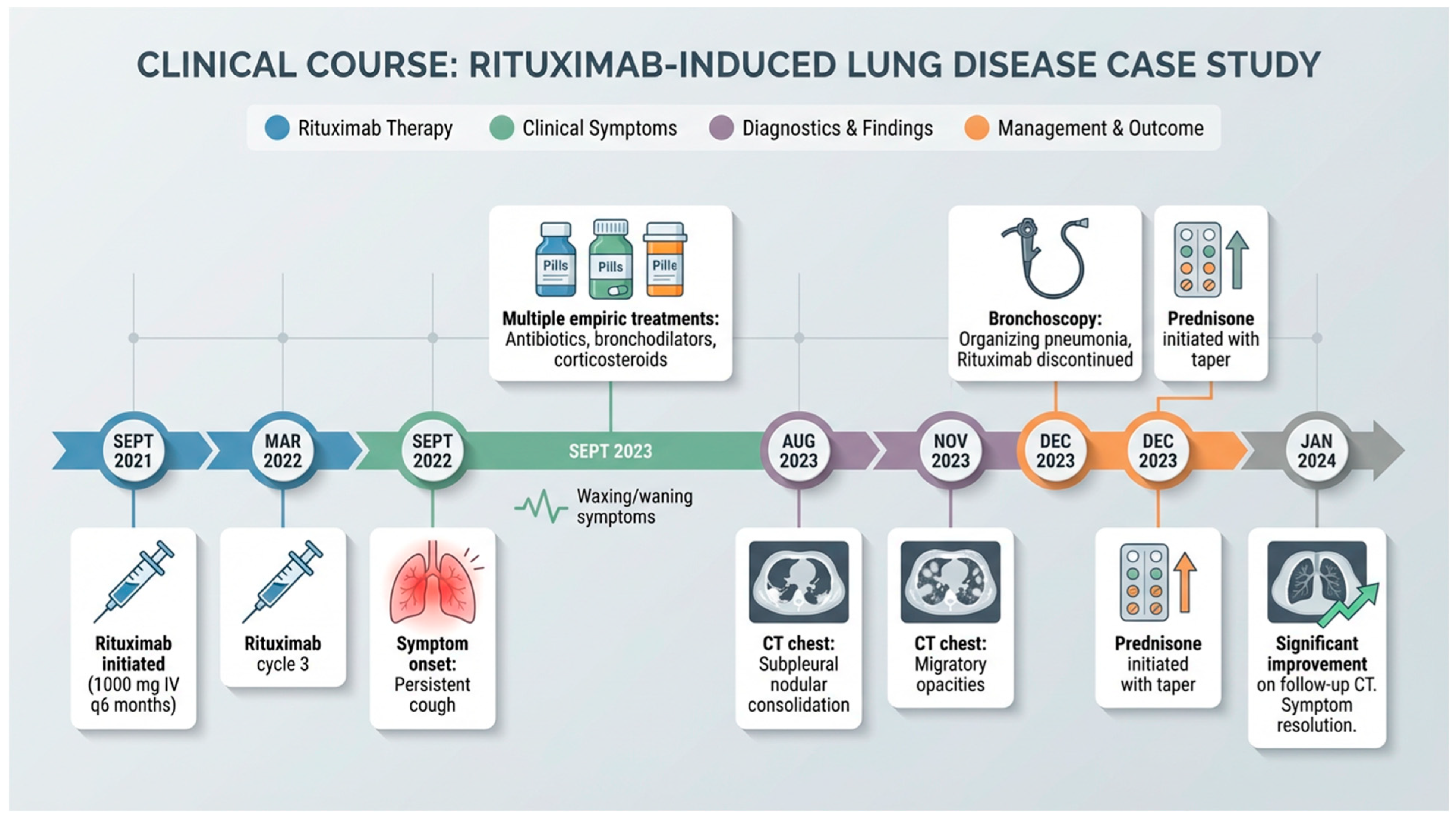
A 72-year-old woman with a history of seropositive rheumatoid arthritis diagnosed in 2008 and treated with rituximab presented to the pulmonary clinic in July 2023 with an eight-month history of persistent cough. The cough was intermittently productive with green sputum but frequently dry and was associated with mild chest wall discomfort and occasional palpitations during coughing episodes. Symptoms waxed and waned without complete resolution. She had been treated multiple times with antibiotics, bronchodilators, and short courses of corticosteroids for presumed bronchitis, with only transient improvement. She denied dyspnea at rest, hemoptysis, weight loss, or night sweats. Intermittent subjective fevers were reported at home. She was a never-smoker with no history of chronic lung disease, asthma, or environmental exposures and was a retired school teacher.
The patient had a 17-year history of rheumatoid arthritis. After intolerance to methotrexate with continued disease activity, the patient had been treated with rituximab 1000 mg IV infusions for several years. Her last rituximab infusion prior to this period was in April 2020, following which she achieved sustained remission. In January 2021, rituximab was formally placed on hold given continued disease control. In August 2021, she developed a clinical flare with joint stiffness, swelling, and synovitis, prompting re-initiation of rituximab in September 2021. Rituximab was initially administered every 6 months and later adjusted to every 4 months. She was also maintained on hydroxychloroquine 200 mg twice daily and meloxicam 15 mg daily. Respiratory symptoms began approximately 14 months after rituximab was restarted.
On initial evaluation in the pulmonology clinic, she was afebrile, normotensive without tachycardia, and saturating 99% on room air. Physical exam demonstrated faint bilateral basal crackles, otherwise diffusely normal. There was no joint swelling or deformities. Autoimmune workup revealed positive Anti-Nuclear Antibody (ANA) 1:40 in a nuclear homogenous pattern. Current rheumatoid factor (RF) and anti-cyclic citrullinated peptide (Anti-CCP) were negative, consistent with seroreversion following rituximab therapy. Further testing including immunoglobulins, anti-smooth muscle antibody, and anti-smith antibody were negative. Pulmonary function testing demonstrated mild restrictive physiology without evidence of obstruction (FVC 1.78 L, 71% predicted; FEV1 1.48 L, 75% predicted; FEV1/FVC 0.83; TLC 3.75 L, 79% predicted) with mildly reduced diffusion capacity (DLCO 75% predicted).
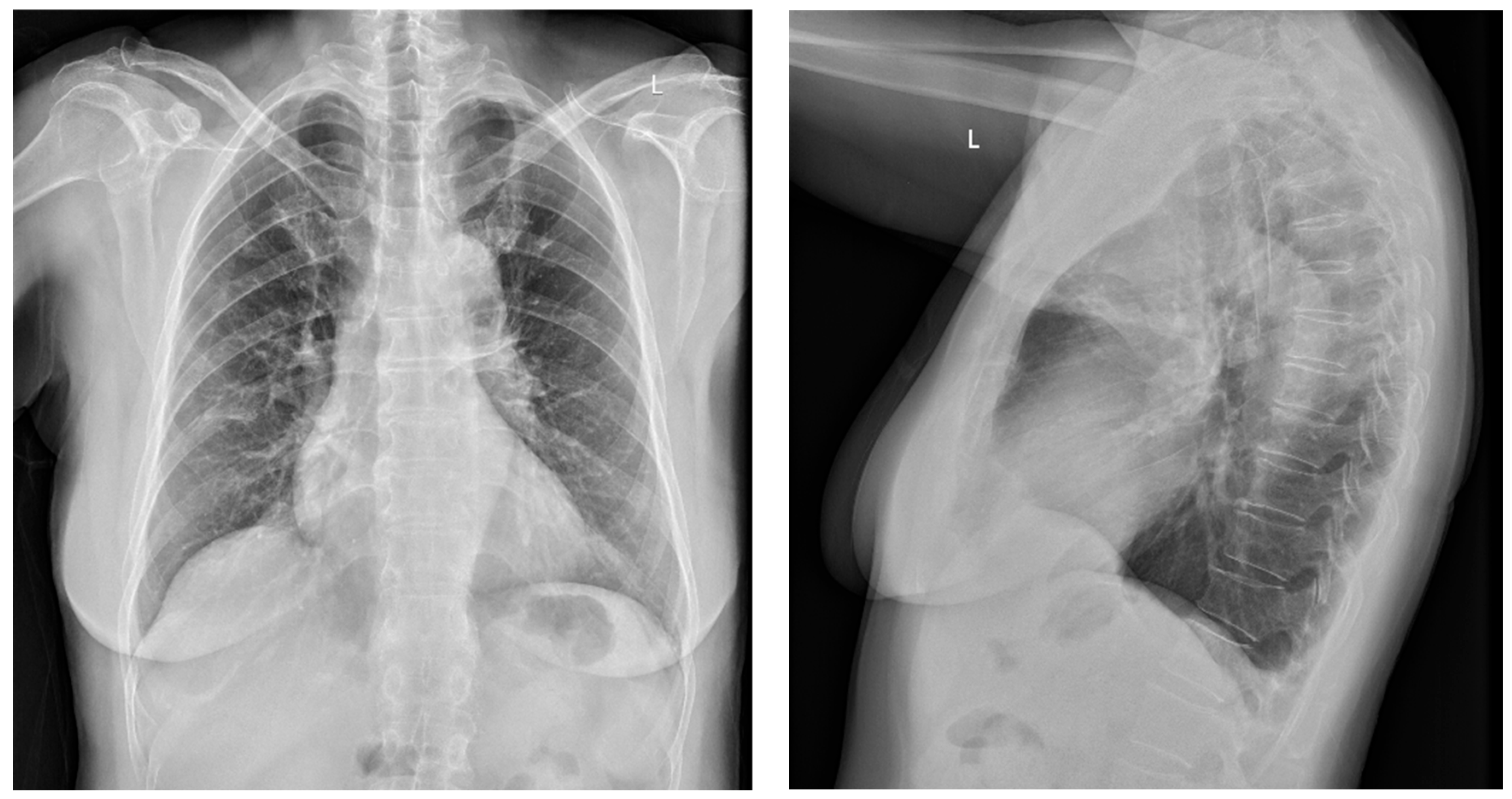
A chest radiograph ordered by her primary care physician on 29 December 2022 and obtained on 5 January 2023, demonstrated a faint right upper lobe anterior segment opacity (
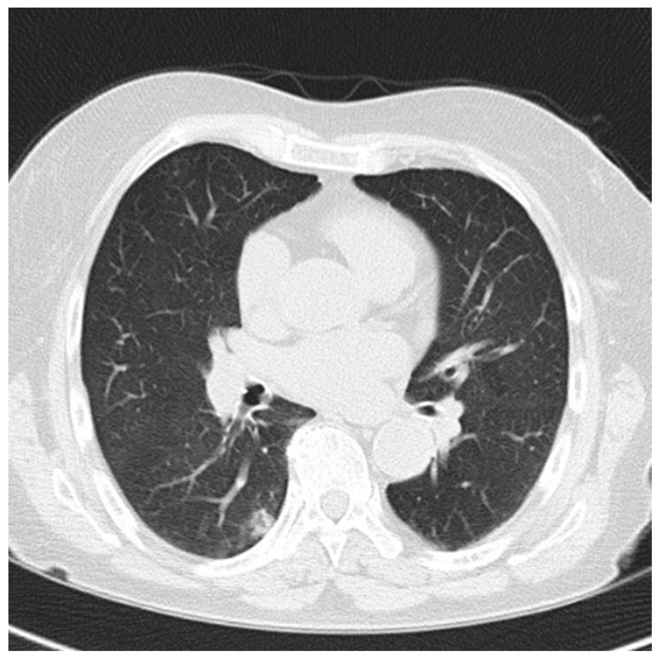
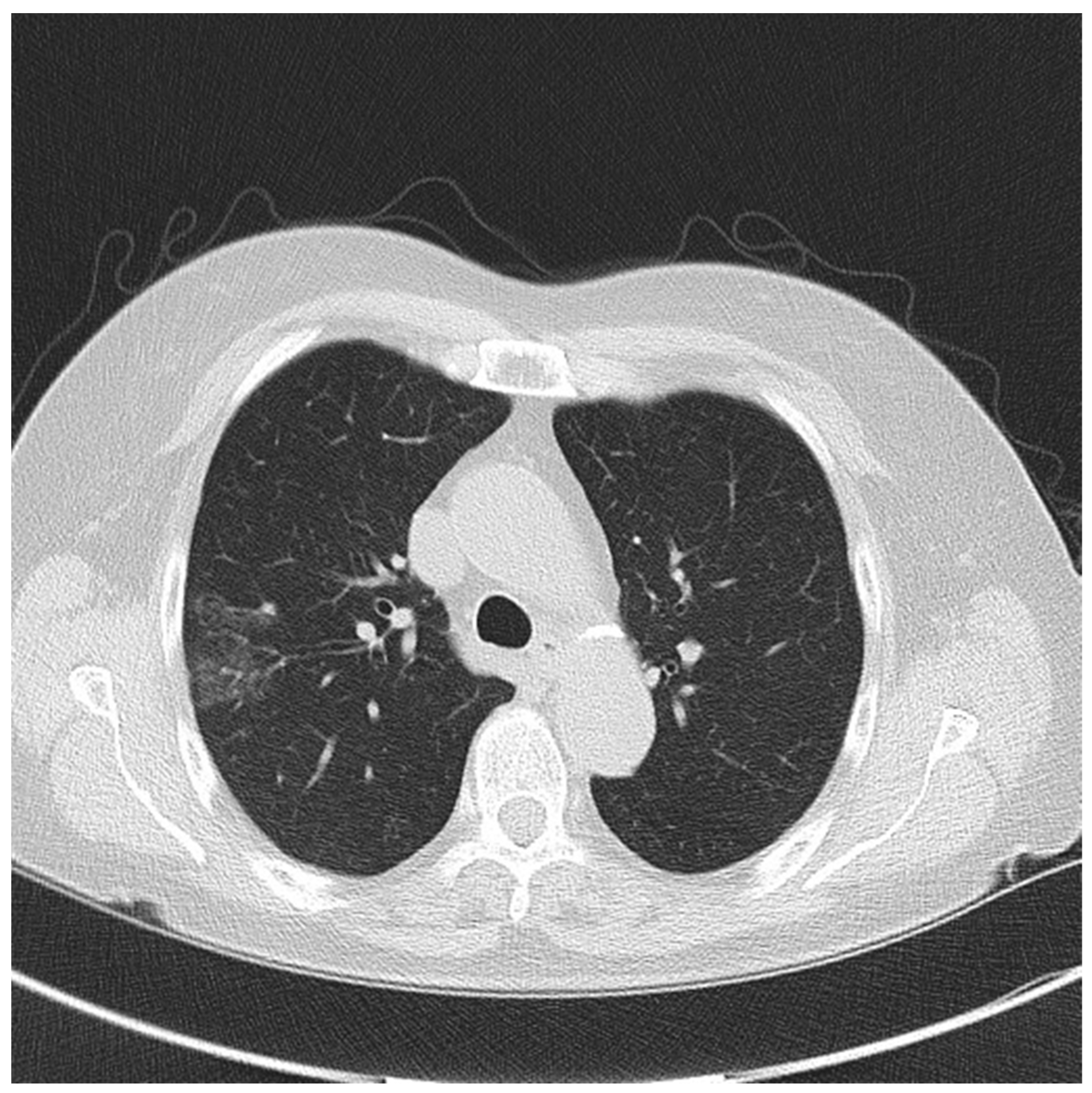
Figure 1). Follow-up on January 18th revealed similar subtle findings. Serial chest CT imaging performed in August 2023, November 2023, and January 2024 demonstrated evolving findings. Initial imaging on 9 August 2023 showed subtle interstitial changes and a small subpleural nodular consolidation, initially interpreted as inflammatory or infectious in nature (
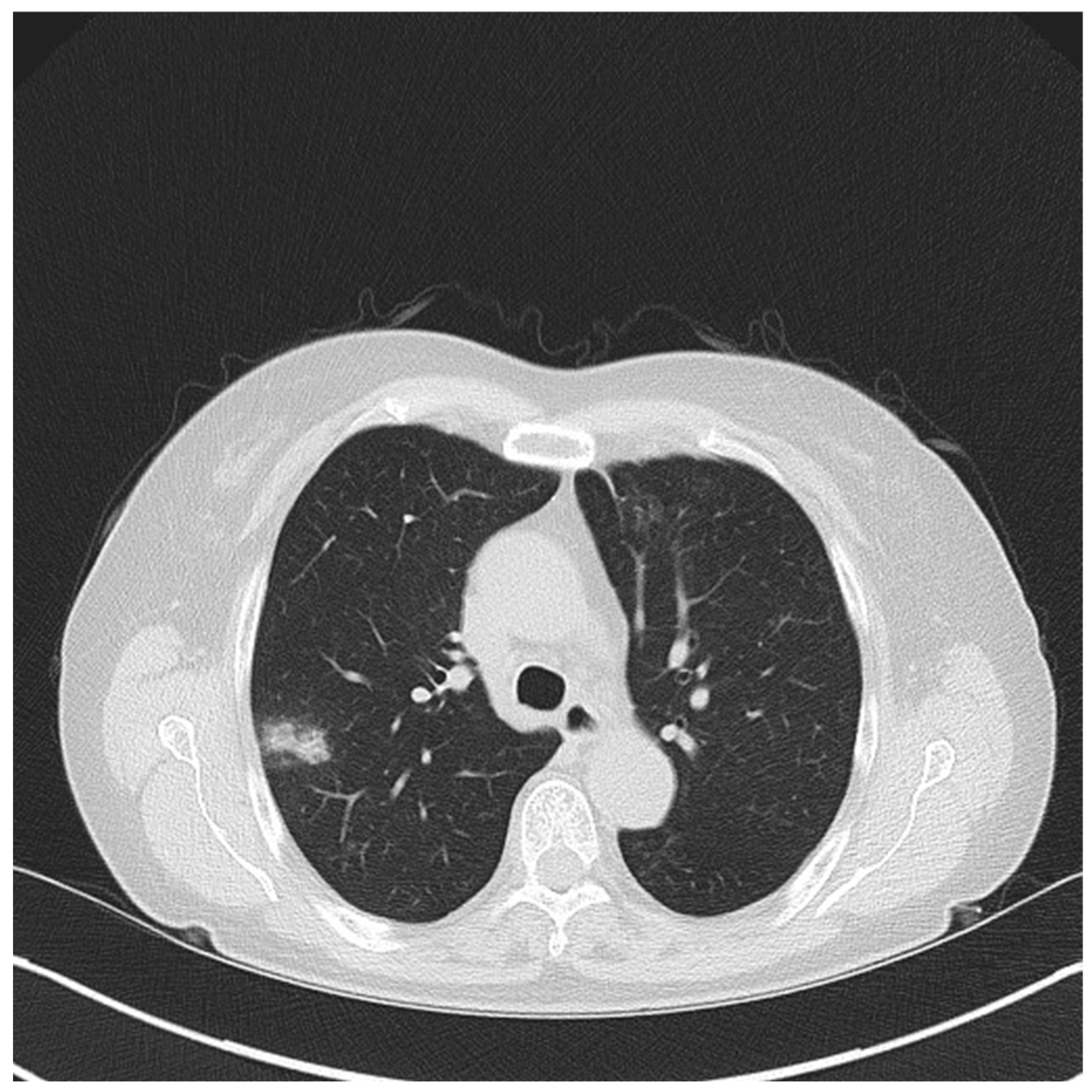
Figure 2). Repeat imaging on 15 November 2023 revealed waxing and waning patchy nodular opacities involving different lung regions, suggesting a migratory inflammatory process rather than progressive fibrotic disease (
Figure 3). Follow-up imaging on 22 January 2024 demonstrated radiographic improvement following treatment (
Figure 4).
Given persistent symptoms, inability to obtain sputum for microbiologic evaluation, and evolving radiographic abnormalities, bronchoscopy was performed on 13 December 2023 using robotic-assisted navigation with radial endobronchial ultrasound guidance. Transbronchial biopsy demonstrated lymphoplasmacytic inflammation with early organization consistent with organizing pneumonia. Bronchoalveolar lavage demonstrated a neutrophil-predominant profile. Microbiologic studies were unrevealing. Bronchoalveolar bacterial cultures demonstrated normal respiratory flora. Fungal cultures, acid-fast bacilli stains and cultures, Aspergillus galactomannan testing, and an expanded respiratory pathogen PCR panel including SARS-CoV-2, influenza, Respiratory Syncytial Virus, Adenovirus, Mycoplasma pneumoniae, Chlamydia pneumoniae, and Legionella species were negative. Cytology was negative for malignant cells.
The case was reviewed in a multidisciplinary interstitial lung disease conference. The patient’s rheumatoid arthritis remained clinically quiescent with negative inflammatory markers and absence of extrapulmonary disease activity, making rheumatoid arthritis–associated interstitial lung disease unlikely. Given the temporal association with rituximab exposure, exclusion of infection and malignancy, and histopathologic confirmation of organizing pneumonia, rituximab-induced lung disease was considered the most likely diagnosis. Although organizing pneumonia may occur idiopathically or in association with connective tissue disease, the patient’s rheumatoid arthritis remained clinically quiescent, and the overall clinical course favored a drug-induced etiology.
Following bronchoscopy confirmation of organizing pneumonia and multidisciplinary review, rituximab was discontinued in December 2023 and a prednisone 40 mg daily was initiated, followed by a gradual taper over approximately six months. Follow-up CT imaging on 22 January 2024, approximately one month after rituximab discontinuation and corticosteroid initiation, demonstrated radiographic improvement, and the patient reported resolution of cough and respiratory symptoms (
Figure 5). The patient remained clinically asymptomatic without recurrence at both 6-month and 12-month follow-up evaluations. No additional follow-up information was available thereafter.
3. Discussion
Rituximab-induced lung disease (R-ILD) is an uncommon but increasingly recognized complication of anti-CD20 therapy. Although rituximab is generally well tolerated, pulmonary toxicity has been reported across a broad clinical spectrum ranging from mild respiratory symptoms to severe acute respiratory distress syndrome [
1,
2,
3,
4]. The true incidence remains uncertain and is likely underestimated due to overlap with infection, autoimmune lung disease, and attribution bias [
2,
3]. Most reported cases occur in patients receiving rituximab as part of combination chemotherapy regimens for hematologic malignancies, whereas pulmonary toxicity associated with rituximab monotherapy in autoimmune disease is less frequently described and may therefore be underrecognized [
2,
5]. In a 2007 case series by Wagner et al., 16 known cases of interstitial lung disease (ILD) were evaluated in patients undergoing treatment with rituximab for non-Hodgkins lymphoma, of which only 4 received rituximab monotherapy [
2]. Interstitial pneumonitis and bilateral infiltrates were the predominant imaging findings.
Clinical manifestations of R-ILD are heterogeneous but most commonly include cough, dyspnea, and fever, symptoms that frequently mimic infectious pneumonia and contribute to delayed diagnosis [
2,
3]. Radiographic findings in R-ILD are variable and may include ground-glass opacities, patchy consolidations, nodular infiltrates, or diffuse interstitial changes [
1]. In a 2010 systematic literature review by Liote et al. [
3], 45 cases of lung disease were evaluated in patients receiving rituximab as part of combination chemotherapy regimens. Symptoms of lung disease often occurred around the 4th cycle. Ground glass opacities were the second most common presentation on high-resolution CT, and the predominant histopathologic pattern was organizing pneumonia [
3]. The present case demonstrated a particularly prolonged and fluctuating clinical course characterized by waxing and waning respiratory symptoms and transient improvement with empiric therapy for presumed bronchitis. Such an indolent and migratory presentation has been described in delayed forms of R-ILD, particularly those associated with organizing pneumonia [
2,
3,
6].
The migratory and waxing-and-waning nodular opacities observed in this patient favored an inflammatory rather than fibrotic process and are characteristic of organizing pneumonia and certain forms of drug-induced lung injury [
3,
6]. Serial imaging demonstrating evolving inflammatory lesions rather than progressive fibrosis provided an important diagnostic clue and supported further tissue evaluation.
In a study evaluating patients receiving rituximab-containing chemotherapy by Liu et al., interstitial pneumonitis was observed following multiple treatment cycles, suggesting a potential cumulative or immune-mediated mechanism of lung injury [
7]. The present case supports a cumulative toxicity mechanism, as the patient received 7 infusion cycles over more than 2 years before definitive diagnosis.
Organizing pneumonia is one of the most often reported histopathologic manifestations of delayed R-ILD [
2,
3,
6]. The pathogenesis remains incompletely understood but is believed to involve immune dysregulation following rituximab-mediated B-cell depletion. This process may result in altered cytokine signaling, imbalance between B- and T-cell immune responses, and downstream inflammatory lung injury [
3,
4]. Experimental and clinical observations suggest that T-cell-mediated immune activation and cytokine dysregulation may play a key role in organizing pneumonia and other inflammatory lung injury patterns [
4]. Cytokines such as tumor necrosis factor-α and interleukin-6 have been implicated in interstitial lung disease pathogenesis and may contribute to the inflammatory cascade observed in R-ILD [
3,
4].
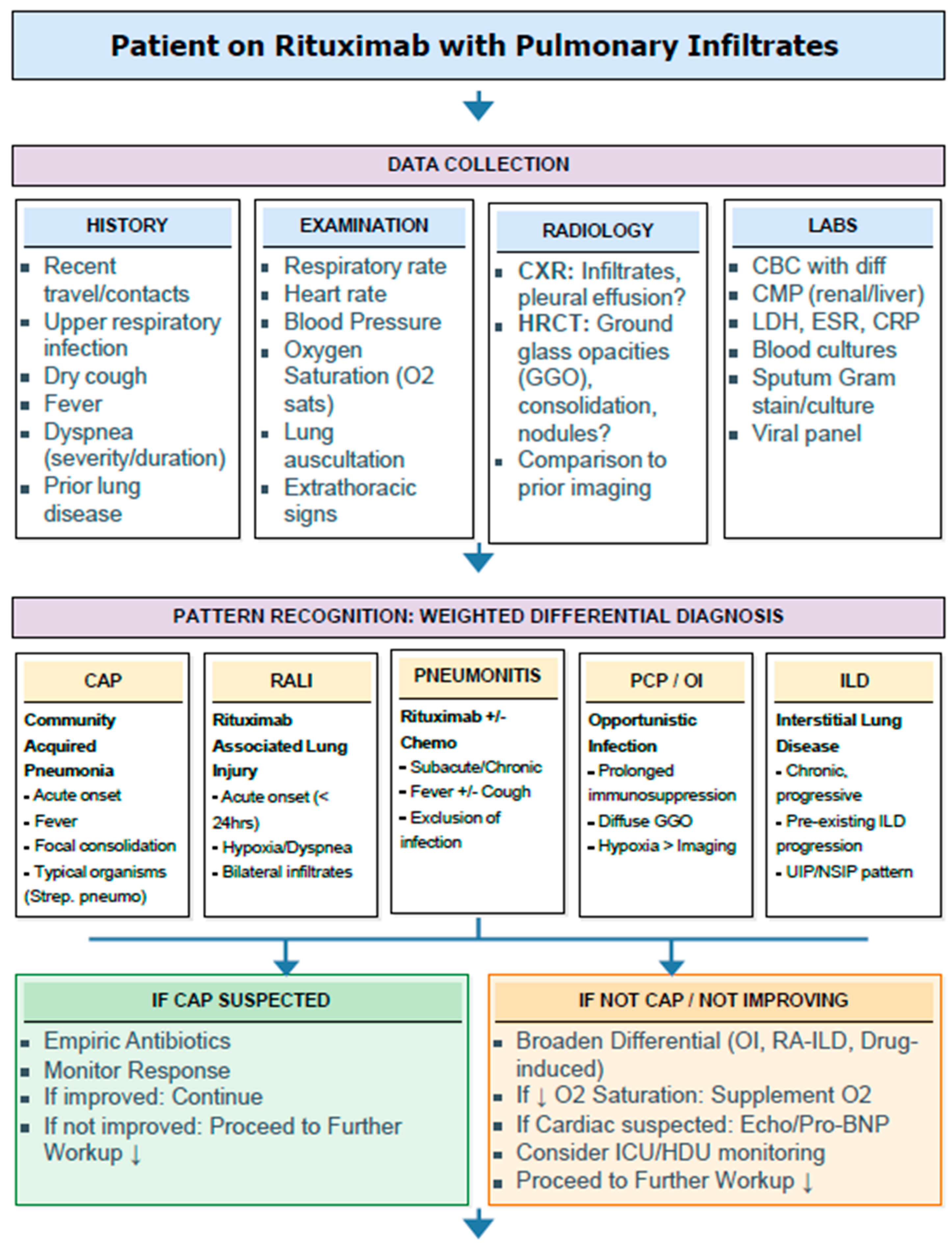
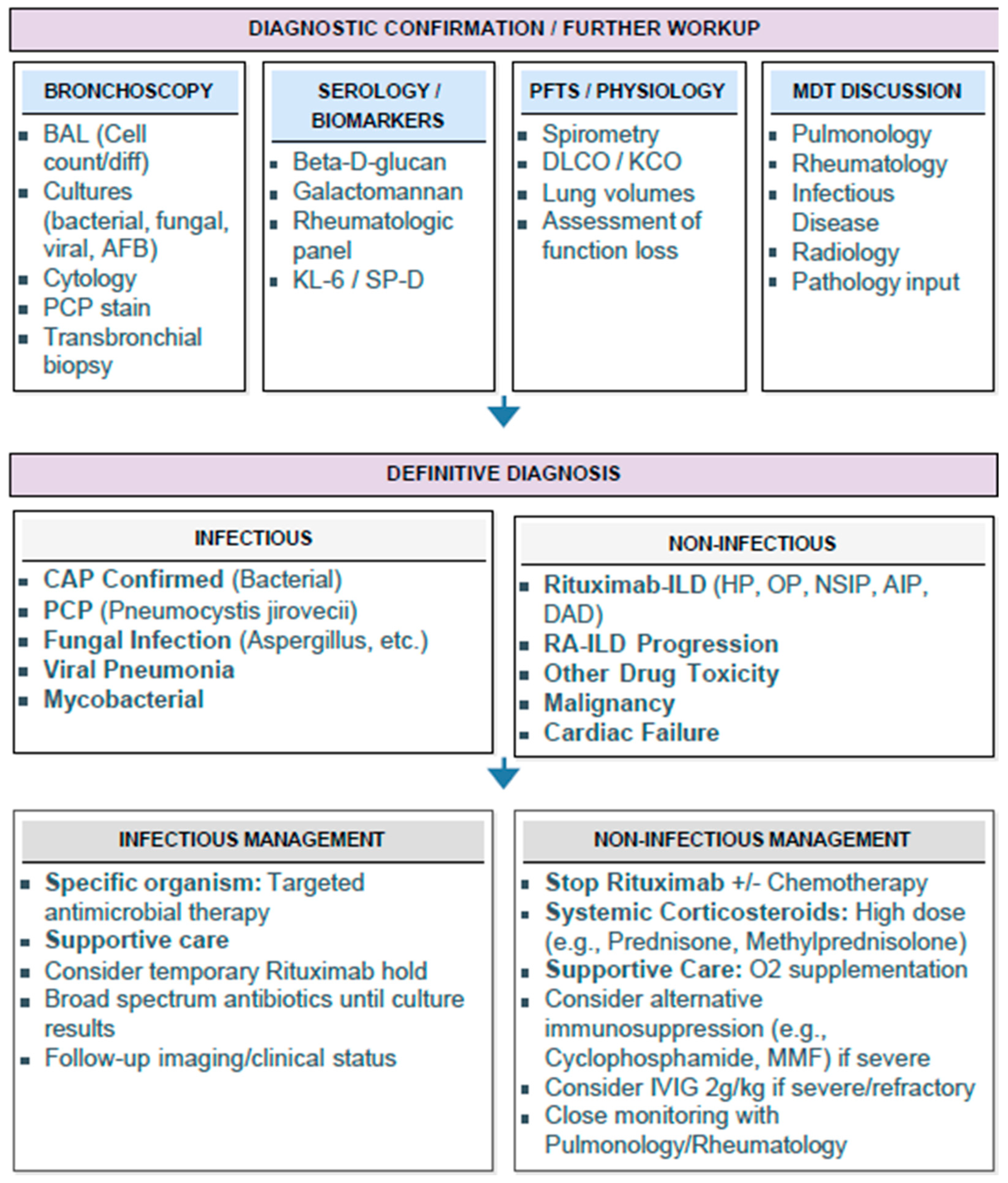
The diagnosis of R-ILD is primarily one of exclusion and requires careful evaluation to rule out infection, malignancy, and autoimmune-associated interstitial lung disease [
1,
2,
3]. Distinguishing R-ILD from rheumatoid arthritis-associated interstitial lung disease can be particularly challenging, as both may present with overlapping clinical and radiographic features. Alternative explanations merit consideration. Organizing pneumonia may occur as a manifestation of rheumatoid arthritis-associated lung disease, even in the absence of extensive systemic symptoms, and may also develop following respiratory infection. In the present case, these possibilities could not be completely excluded. However, the patient’s rheumatoid arthritis remained clinically quiescent without evidence of active systemic disease, microbiologic studies were unrevealing, and multidisciplinary review favored a drug-induced process. Therefore, while causality cannot be definitively established, the overall clinical, radiographic, and histopathologic findings were considered most consistent with rituximab-associated organizing pneumonia. In this case, several findings supported a drug-induced etiology, including clinically stable rheumatoid arthritis, negative microbiologic studies, seroreversion of rheumatoid factor following rituximab therapy, and the temporal association between rituximab exposure and pulmonary manifestations. Histopathologic confirmation demonstrating organizing pneumonia further strengthened the diagnosis in the appropriate clinical context (
Figure 6) [
2,
6].
Advanced bronchoscopic techniques played a key role in establishing the diagnosis. Robotic-assisted bronchoscopy with radial endobronchial ultrasound enabled tissue acquisition from peripheral lesions and reduced the need for surgical lung biopsy. Although bronchoalveolar lavage findings in R-ILD are often nonspecific, histopathologic confirmation can be critical when imaging and clinical findings are inconclusive [
2,
3].
Management of R-ILD centers on discontinuation of rituximab and initiation of systemic corticosteroid therapy [
2,
3,
6]. While standardized treatment regimens have not been established, most reported cases demonstrate favorable clinical and radiographic outcomes when therapy is initiated early [
2,
3,
6]. Re-exposure to rituximab is generally discouraged due to the potential risk of recurrence. While most R-ILD cases respond favorably to early intervention, mortality rates of 15–29% have been reported in systematic reviews, emphasizing the importance of early recognition. In Hadjinicolaou et al.’s review of 121 cases, R-ILD was fatal in 18 cases (15%), and in Lioté et al.’s series of 45 cases, 8 patients died (18%) [
1,
2,
4,
5]. In the present case, cessation of rituximab and corticosteroid treatment resulted in resolution of symptoms and radiographic improvement, consistent with prior reports describing reversible inflammatory lung injury [
6].
This case highlights several important clinical considerations. First, R-ILD should be suspected in patients receiving rituximab monotherapy who develop unexplained respiratory symptoms, even in the absence of combination chemotherapy. Second, the delayed onset of symptoms following rituximab re-exposure and the presence of migratory or waxing-and-waning pulmonary opacities should prompt consideration of organizing pneumonia or drug-induced lung disease. Third, multidisciplinary evaluation, advanced bronchoscopic tissue acquisition, and histopathologic confirmation may be required to distinguish R-ILD from infection, malignancy, or autoimmune lung disease. Finally, the favorable clinical course, including sustained symptom resolution without recurrence at 6- and 12-month follow-up, highlights the importance of early recognition and intervention. Early recognition and intervention remain critical, as timely discontinuation of the offending agent and corticosteroid therapy may result in favorable outcomes.