Guidelines

S2k Guideline: Breathing, Respiratory Support and Ventilation in Acute and Chronic Spinal Cord Injury


1
School of Health Professions, Bern University of Applied Sciences, 3008 Bern, Switzerland
2
BG Trauma Hospital Frankfurt am Main gGmbh, 60389 Frankfurt, Germany
3
BG Hospital Hamburg, 21033 Hamburg, Germany
4
BG Trauma Hospital Murnau, 82418 Murnau, Germany
5
BDH Clinic Vallendar, 56179 Vallendar, Germany
6
Representation of the Perspective of People Affected, 70192 Stuttgart, Germany
7
Swiss Paraplegic Centre, 6207 Nottwil, Switzerland
*
Author to whom correspondence should be addressed.
J. Respir. 2026, 6(3), 13; https://doi.org/10.3390/jor6030013
Submission received: 15 December 2025 / Revised: 24 May 2026 / Accepted: 12 June 2026 / Published: 7 July 2026

Abstract

Background/Objectives: Spinal cord injury (SCI) frequently leads to impaired respiratory function and increased pneumonia risk due to reduced respiratory muscle strength, altered thoracic compliance, autonomic dysregulation, and diaphragm displacement. This guideline aims to ensure appropriate, high-quality, and quality-assured care for individuals requiring respiratory support. Methods: This work represents a consensus-based S2k guideline developed by the “BeAtmung” working group of the German-speaking Medical Society for Paraplegiology (DMGP), in collaboration with experts from Germany and Switzerland, and endorsed by eight professional societies. Development followed the German Association of the Scientific Medical Societies (AWMF) guidance manual. Topics were defined in editorial meetings, supported by targeted literature searches and existing guidelines. Drafts were revised regularly with written feedback and final approval was granted after a consensus conference on 18 January 2022. Results: The guideline includes 11 specific recommendations addressing ventilation, monitoring, care structures, and discharge planning. Recommendations were graded using a three-level scheme (“need,” “should,” and “can be considered”) and consensus strength was classified as strong (>95%), consensus (>75–95%), majority (>50–75%), or no consensus (<50%). These recommendations support clinical decision-making and promote standardised, high-quality respiratory care for people with SCI. Conclusions: This guideline provides consensus-based recommendations for the comprehensive respiratory management of individuals with acute and chronic SCI, integrating diagnostic, therapeutic, and long-term care strategies to support high-quality, coordinated care across settings and healthcare systems.

1. Introduction

Spinal cord injury (SCI) is associated with a wide range of respiratory complications that evolve over time [1]. Beyond the immediate effects of muscle paralysis, long-term changes in lung mechanics, thoracic compliance, and central respiratory control contribute to a progressive decline in pulmonary function [2,3,4,5,6]. Additional factors such as autonomic dysregulation, altered thoraco-abdominal interactions, and secondary conditions like obesity or gastrointestinal dysfunction further complicate respiratory management [7,8,9,10,11,12]. These complex and multifactorial changes underscore the need for structured assessment and individualized respiratory care in people living with SCI.
This guideline aims to provide essential theoretical knowledge for the treatment and care of individuals with SCI who require respiratory support. It pursues six core objectives: First (1), to deepen the understanding of respiratory complications specific to SCI, including the impact of impaired gastrointestinal function on respiratory mechanics. Second (2), to outline diagnostic and therapeutic approaches for both temporary and long-term ventilation needs. Third (3), to present strategies for managing cough effectiveness and airway secretion clearance. Fourth (4), to describe procedures and requirements for transitioning patients to home-based or other out-of-hospital care settings. Fifth (5), to define technical and professional standards for treatment centres and for the care of ventilated or respiratory-assisted individuals outside of hospital environments. Sixth (6), to establish principles of quality assurance and lifelong follow-up in long-term care, regardless of the care setting.

2. Methods

This consensus-based S2k guideline was developed by the “BeAtmung” working group of the German-speaking Medical Society for Paraplegiology (DMGP), in collaboration with experts from Germany and Switzerland and with consensus from eight professional societies (Table 1). The development followed the German Association of the Scientific Medical Societies (AWMF) guidance manual.
The structure and key topics of the guideline were defined during several editorial meetings. Each expert was assigned a specific topic and tasked with preparing a draft for their section. The editorial team consisted of eight experts from the fields of medicine, nursing, physiotherapy, and respiratory therapy, including one person living with high-level SCI and dependent on ventilation, ensuring that lived experience was directly integrated into the development process.
Each contributor conducted topic-focused literature searches for their assigned area in Pub med/Medline, Cochrane Library, and Embase, with the overall search approach discussed and refined collaboratively. Relevant international guidelines and recommendations from professional societies were reviewed and, where appropriate, taken into consideration. The individual drafts were compiled into a preliminary version, which underwent continuous revision in documented meetings and through repeated opportunities for written feedback. The final draft was circulated among all working group members and submitted to the DMGP guideline commission.

2.1. Consensus Conference

The consensus conference was held on 18 January 2022, as a pandemic-related hybrid meeting, with in-person participation at BG-Klinik Frankfurt and additional participation via videoconference (Zoom). A total of ten voting participants took part in the formal consensus process, comprising members of the author group and representatives of the participating professional societies. All professional societies had submitted written comments on the draft guideline in advance. These comments were systematically reviewed prior to the consensus conference and incorporated into a revised draft. In addition, representatives of the professional societies were given the opportunity to actively participate in the discussion during the Zoom conference.
During the consensus conference, the previously formulated recommendations were discussed stepwise and transformed into recommendations. Voting was conducted in accordance with the AWMF methodological framework, and the level of consensus was defined for each recommendation (>75% consensus, >95% strong consensus).
Consensus was achieved for all recommendations, with the exception of the originally proposed recommendation 7 (ventilator-associated lower respiratory tract infections). This statement was removed without replacement. One dissenting vote was recorded for this decision, while all remaining participants agreed with the removal. For all other recommendations, strong consensus (100% agreement) was achieved.
Following the consensus conference, the revised version of the guideline was circulated again to all participants to allow verification that their comments and suggestions had been appropriately addressed.

2.2. Recommendations

The guideline includes 11 specific recommendations addressing ventilation, monitoring, care structures, and discharge planning. Recommendations were graded using a three-level scheme (“need,” “should,” and “can be considered”) (Table 2), and consensus strength was classified as strong (>95%), consensus (>75–95%), majority (>50–75%), or no consensus (<50%) (Table 3). According to the AWMF classification system, this is an S2k guideline.
To improve reporting transparency for this publication, the manuscript was prepared in accordance with the Reporting Items for Practice Guidelines in Healthcare (RIGHT) checklist, which is provided as Supplementary Materials.

3. Results

3.1. Respiratory Consequences of Spinal Cord Injury
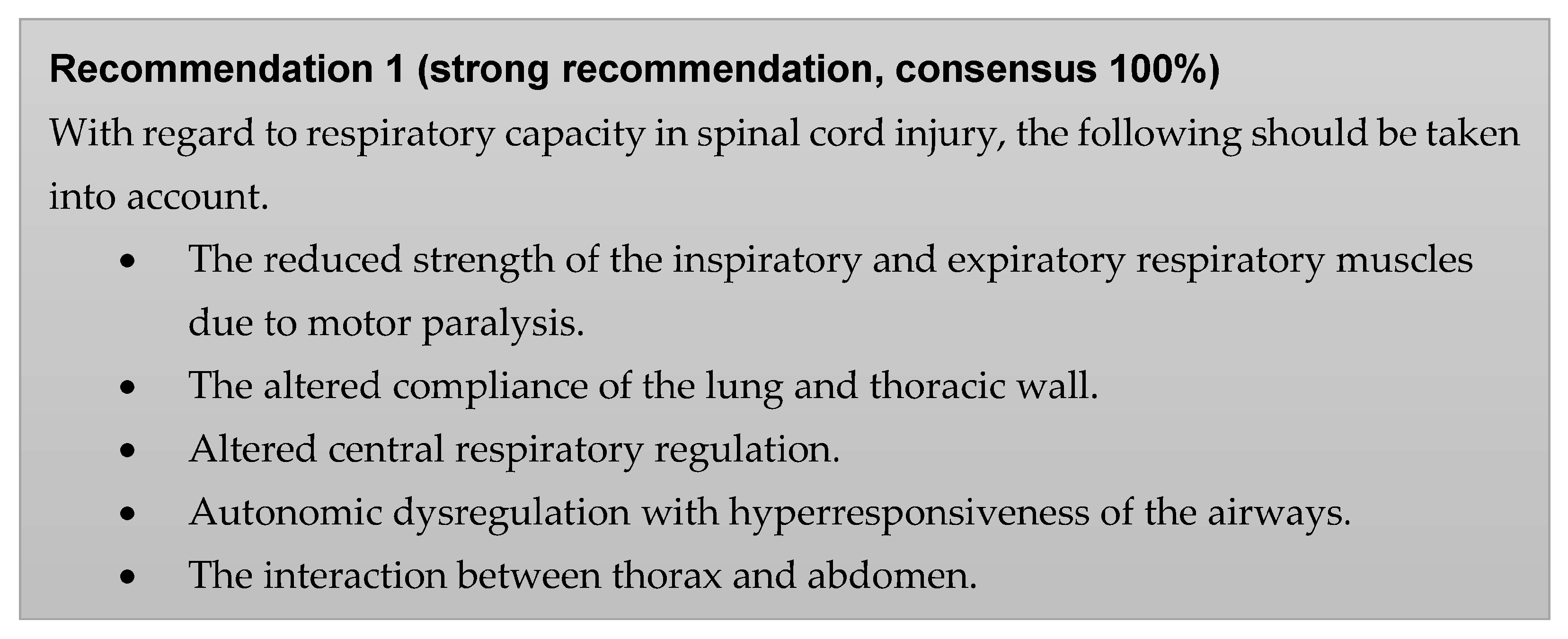
SCI often leads to reduced respiratory function and consequently to an increased risk of pneumonia [1]. Contributing factors include weakened inspiratory and expiratory muscles due to paralysis, including auxiliary muscles, altered lung and thoracic wall compliance, especially over time, impaired central respiratory control, reduced airway diameter and responsiveness caused by autonomic dysregulation, and disrupted thoraco-abdominal interaction due to diaphragm displacement and pressure imbalances [13].
As a result of the decrease in the strength of the inspiratory muscles, the vital capacity (inspiratory (VC) and forced VC (FVC)), the respiratory volume (AZV), the one-second capacity (FEV1), and the inspiratory capacity decrease depending on the level of SCI [14]. Respiratory minute volume (AMV) is maintained through compensatory increases in respiratory rate. Additionally, the lack of rib cage stabilization by the intercostal muscles leads to paradoxical inward movement during inspiration [15,16]. The decrease in expiratory muscle strength leads to a reduction in expiratory reserve volume (ERV), depending on the level of paralysis, and thus to an increase in residual volume (RV), and additionally reduces VC [17,18]. These changes are collectively described as “complex restrictive patterns” [19]. Maximum achievable airflow during coughing is reduced, making effective clearance of bronchial secretions difficult. When peak cough flow (PCF) falls below 270 L/min, secretion expectoration is no longer reliably effective, and becomes nearly impossible below 160 L/min [20,21]. PCF can be easily measured with a peak flow meter and should be assessed in all patients with potentially impaired respiratory muscle function and increased risk of secretion retention.
Lung and thoracic compliance is reduced in people with tetraplegia due to a decrease in lung volumes and changes in surfactant when breathing with low lung volumes, even one year after the onset of SCI [2,3]. The stiffening of the rib thorax due to ankylosis of the rib joints and the spasticity of the intercostal muscles further reduce thoracic compliance in the course of time [4,5,6].
Centrally controlled breathing is impaired in patients with tetraplegia. Respiratory drive in response to hypercapnia is diminished [22,23,24]. Central respiratory regulation disorders occur more frequently at night, partly caused by drugs with respiratory depressive side effects that are used for complications of paraplegia (e.g., pain, spasticity) [25].
In cases of SCI above Th6, the sympathetic innervation of the airways is disrupted. The absence of sympathetic activity, combined with unopposed vagal cholinergic stimulation, results in bronchoconstriction, increased secretion production, and bronchial hyperreactivity [26,27].
The interaction between the abdomen and thorax becomes particularly relevant in tetraplegia, influenced by changes in position, increased intra-abdominal pressure, and trunk spasticity [28,29]. Due to increased abdominal compliance following loss of abdominal muscle innervation, the diaphragm is passively displaced caudally and flattened in the sitting position. Simultaneously, intra-abdominal organs shift ventrally and caudally, reducing the diaphragm’s lateral apposition zone and vertical contraction force. This leads to decreased AZV, increased work of breathing, and more rapid onset of dyspnoea. Unlike classic orthopnoea, AZV may be lower in the sitting than in the lying position; however, evidence regarding position-dependent changes in lung volumes in SCI is inconsistent [30,31,32]. Additionally, increased intra-abdominal pressure caused by coprostasis, flatulence, or abdominal muscle spasticity further elevates the work of breathing and contributes to dyspnoea. These phenomena are frequently observed in individuals with SCI due to delayed gastrointestinal transit [28,29].
In the long-term, lung function may decline beyond normal age-related changes [7,8]. Factors such as obesity, reduced inspiratory muscle strength, persistent bronchial obstruction, and lesion level influence this progression [7,8,9,10,11,12]. The incidence of lung disease correlates with the degree of lung function impairment, smoking, signs of obstructive pulmonary disease, and previous pneumonia, but not with lesion size, classification, or duration [33].
Jor 06 00013 i001

3.2. Cough and Secretion Management

Non-invasive mechanical ventilatory support (NIV) or invasive ventilation, as well as electrical stimulation of the diaphragm, can only be successful if the airway is open and free of secretions. Obstruction of airways by secretions leads to decreased ventilation and worsening of the ventilation-perfusion ratio and increases the risk of shunts, atelectasis and bronchopneumonia. Monitoring and interventions regarding cough and secretion management must therefore take place before initiating mechanical respiratory support or electrical stimulation [34,35]. Cough support should be considered in all patients with a PCF below 270 L/min [20,21,36]. Given the central role of effective cough and secretion management, a range of specific techniques may be required, including breathing exercises, mechanical insufflation–exsufflation [37,38], high-frequency chest wall oscillation, manual chest physiotherapy, autogenic drainage, oscillating positive expiratory pressure (OPEP), positive expiratory pressure (PEP), active cycle of breathing techniques, and intrapulmonary percussive ventilation [39].
The goals of effective cough and secretion management are to ensure efficient expectoration of secretions, prevent secretion retention and atelectasis, and improve ventilation while reducing the risk of bronchopneumonia [40].
These goals are achieved as follows:
  • Ventilate the lungs to the maximum possible inspiratory capacity (MIC) before coughing up [41]. This can be achieved by mobilisation, change of position, air stacking, inflation with the resuscitator or by means of glossopharyngeal breathing.
  • A strengthening of the in- and expiratory musculature to improve inspiratory capacity and maximum expiratory flow [42,43].
  • Specific positioning techniques (e.g., head-down and/or 135° positioning) to facilitate and improve expectoration [44].
  • Improving maximum airflow during cough thrust by compressing the abdomen using a three-part abdominal belt [45].
  • Assisted coughing techniques with a target maximum cough thrust of >270 L/min [20]: Deeper inspiration by air stacking, glossopharyngeal breathing, insufflation with a resuscitator, intermittent positive pressure breathing (IPPB device) or in-exsufflators should be used to achieve the highest possible inspiratory volume in order to then maximally increase the air flow during expiration by means of manual compression on the upper abdomen in a dorso-cranial direction [46,47].
  • Inhalation with hyperosmolar saline, preferably as part of IPPB therapy.
  • Extra- or intrathoracic vibration for secretion mobilization.
Cough and secretion management is an integral part of the long-term success of efficient ventilation. For any form of long-term ventilation, the patient and the patient’s care team must be trained in the efficient coughing up of secretions.

3.2.1. Mechanical Coughing Techniques

These techniques can be used if, with correctly performed manual-assisted techniques, the maximum cough output remains low (<270 L/min) or the secretions cannot be coughed up effectively. Correct patient and carer attitude and training is critical for success.

3.2.2. Special Aspects in Tracheotomised and Invasively Ventilated Patients

Assisted coughing and mechanical coughing aids (in-exsufflators) are recommended to reduce invasive suctioning as much as possible [48]. Mechanically assisted expectoration using an in-exsufflator can also be used for invasively ventilated patients [49,50]. It should be noted that the lumen of the tracheal cannula has an influence on the pressure applied intrathoracically [51]. Patients usually prefer mechanical expectoration to invasive suction [52].
In addition, invasively ventilated patients should be ventilated with a cannula without a cuff as soon as possible, taking into account adequate volumes, or for as long as possible with an unblocked cannula [53]. The aim here is, in addition to preventing tracheal damage, to improve secretion management and swallowing function, as well as to enable speech [54].
Endotracheal suctioning: Minimally invasive suctioning is preferable to deep suctioning reaching into the main bronchi in tracheotomised patients, as this is associated with fewer side effects [48]. Particularly in the case of a high degree of recently occurred SCI, it should be noted that the autonomic dysregulation can provoke a drop in blood pressure or bradycardia up to asystole [55,56]. Depending on the situation, this may still be necessary. Attention is drawn to atraumatic procedures.
Two principles are available for respiratory gas heating and humidification—controlled heating and humidification (active respiratory gas conditioning) or a heat exchanger (passive respiratory gas conditioning, e.g., a heat and moisture exchanger (HME) filter—and these should be used in invasively ventilated patients depending on the nature of the secretion [57].
An algorithm for cough and secretion management can be used to structure a consistent approach [58] (Figure 1). Reference is made to the algorithm for secretion management during non-invasive ventilation in neuromuscular diseases [58].
If coughing is reduced without signs of secretion stasis, consider training the patient and caregiver to be prepared in case of increased secretion.
In case of decreased cough thrust with signs of secretion stasis, characterised by episodes of respiratory distress, thoracic tightness, drop in saturation of >3% from baseline or a drop in saturation < 95%, prompt manually assisted expectoration is necessary.
If expectoration of secretions is not assured despite manually assisted coughing techniques, and the maximum cough thrust under assisted coughing techniques is <270 L/min, then the use of an in/exsufflator is justified.

3.3. Atelectasis Prophylaxis

Atelectasis is a common complication in people with SCI [59]. The development in people with SCI is usually due to a combination of reduced ventilation as a result of bronchoconstriction or obstruction due to retention of secretions, reduced inspiratory capacity with a decrease in the strength of the inspiratory muscles and, particularly in the acute phase of SCI, pleural effusions [59]. Although studies with a high level of evidence for the prevention of these complications are lacking, the following recommendations can nevertheless be made [60]:
  • Efficient mobilisation of secretions through nursing, physiotherapeutic and/or equipment-assisted measures to keep the airways open.
  • The use of an abdominal belt improves diaphragmatic function by increasing the apposition zone (ideal in a sitting or 70° standing table position) and thus leads to improved inspiration and more efficient coughing in the wheelchair [45,61].
Jor 06 00013 i002

3.4. Sleep-Related Respiratory Disorders

Breathing disorders during sleep, such as obstructive and central sleep apnoea or alveolar hypoventilation, are more common in people with spinal paralysis than in the normal population [62,63]. Especially in people with tetraplegia, breathing disorders during sleep are present in approx. 60% within the first year after paralysis [64]. Several interacting mechanisms are thought to underlie the increased prevalence of sleep-related breathing disorders in this population. These include increased upper airway obstruction due to spasticity of the respiratory support muscles and, in some cases, hypertrophy of the cervical and neck musculature, as well as increased intra-abdominal pressure and reduced lung volumes. In addition, elevated nasal resistance has been demonstrated in people with tetraplegia and may further contribute to upper airway obstruction during sleep [65]. Moreover, a fluid shift from the lower extremities to the upper trunk when changing from a wheelchair to the supine position—particularly in individuals with para- or tetraplegia—has been proposed to increase upper airway resistance and may partly explain the higher prevalence of OSA in this group. However, this mechanism remains hypothetical and requires further investigation [66]. An increased neck circumference has also been described as a potential risk factor for sleep apnoea in tetraplegic patients, although its relevance remains controversial in the literature [64]. Additional factors such as increased intra-abdominal pressure, reduced lung volumes, the use of centrally acting analgesic and/or antispastic medications, and obesity related to physical inactivity appear to exert additive negative effects on the occurrence of OSA [67]. In addition to acute hypercapnia due to a lack of voluntary use of the still existing auxiliary respiratory muscles, chronic hypercapnia can develop, as in people with neuromuscular diseases, which then also causes a reduction in the hypercapnic respiratory drive [68]. The symptoms in people with tetraplegia with obstructive sleep apnoea syndrome include, in addition to the typical symptoms such as daytime sleepiness, sleepiness [69] and blood pressure changes [70], neurocognitive impairments such as reduced attention, concentration and learning ability as well as memory deficits [71].

3.4.1. Therapy

Continuous positive airway pressure (CPAP) and auto-CPAP therapy, bilevel positive airway pressure (PAP) therapy and adaptive servo-ventilation have been established for the therapy of sleep-associated respiratory disorders, depending on the underlying respiratory disorder. In people with tetraplegia, it has been shown that lower CPAP pressure settings are needed for successful therapy when compared with non-paraplegic people (in 69% of people with tetraplegia < 10 cmH2O) [72]. However, it is important to note that adherence to CPAP therapy is reduced in this patient population [67], a finding that has also been confirmed in a more recent study [73]. In particular, in individuals with tetraplegia, safety aspects related to mask selection and handling become highly relevant and may restrict the range of feasible therapeutic options. A randomized controlled trial further demonstrated that, in patients with acute tetraplegia and nocturnal breathing disorders, (A)CPAP therapy improved daytime sleepiness but did not improve neurocognitive dysfunction [74].
The choice of respiratory support mode (CPAP; ASV; bilevel PAP) is basically identical to that for non-paraplegic people. Experience shows that CPAP therapy is sometimes not tolerated in people with severe SCI, especially when higher pressure levels are required, because of the increased work of breathing associated with reduced strength of the respiratory muscles [75]. If necessary, a switch to bilevel PAP therapy may be considered [75], provided that an appropriate backup rate is established. In this context, careful monitoring is essential, as inadequate settings may increase the risk of inducing or exacerbating central sleep apnoea. To date, there are no studies demonstrating proof of concept for the feasibility or effectiveness of this approach specifically in the SCI population.
Independent handling of the mask must be ensured, in particular safe removal of the mask, if necessary by means of individual adjustments and, if hand and finger function are insufficient, securing of the alarm option or implementation and monitoring by an assistant.
In addition to a ramp function (a slow pressure build-up over a longer period of time until the therapy pressure is reached), people with tetraplegia in particular find it pleasant to switch on the CPAP device by triggering it through the nose. Furthermore, the breathing air should be humidified in order to optimally humidify the airways and to avoid an increased formation of viscous secretions.

3.4.2. Sleep Quality Under Ventilation

A carefully adapted form of ventilation during initial treatment, regardless of whether it is pressure- or volume-controlled or diaphragm-stimulated, usually leads to sufficient sleep quality. Only patients who have been continuously treated with a phrenic nerve stimulator (PNS) show significant sleep quality deficits when they are returned to external positive pressure ventilation [76].

3.4.3. Screening

Despite frequent daytime symptoms as a result of breathing disorders during sleep, sleep apnoea syndrome and sleep-related hypoventilation are often not diagnosed and remain untreated [77]. Observed apnoea, pronounced snoring, daytime sleepiness (determined with the Epworth sleepiness scale or the Berlin questionnaire), arterial hypertension, or lack of blood pressure reduction at night are reasons for further clarification in a centre with a sleep laboratory and experience in long-term ventilation (stepwise procedure analogous to the recommendations of the DGP guideline) [58], polygraphy, polysomnography, continuous SaO2 and PtcCO2 measurement). In people with tetraplegia, a clarification should be considered within the first year after the onset of SCI, even if there are few or no daytime symptoms [78].
In high SCI, central nocturnal respiratory disorders are also found more frequently. Polysomnography is recommended to correctly classify and determine the extent of the nocturnal breathing disorder. It is useful to perform a capnography at the same time. Polysomnography is also recommended to assess the stages of sleep. The sleep-related breathing disorders first occur in REM sleep [79].
Jor 06 00013 i003

3.5. Acute Respiratory Insufficiency

The restriction of the respiratory pump in acute SCI often leads to protracted hypoxaemic or ventilatory insufficiency, depending on the lesion level and in combination with additional thoracoabdominal injuries. For these reasons, rapid weaning from ventilation is often not possible. In the case of higher SCI, a narrower indication for invasive ventilation results from the above-mentioned SCI-specific features, such as, and in particular, the management of secretions, whereas in the case of lower SCI levels, the route via a non-invasive form of ventilation can be considered as an alternative.

3.5.1. Tracheotomy

Tracheostomy for acute tetraplegia is recommended for the following cases:
  • In case of motoric complete tetraplegia according to American Spinal Injury Association Impairment Scale (AIS) A and B [80] (Supplement S1) and existing respiratory insufficiency.
  • Where VC ≤ 500 mL.
  • Where there is an Injury Severity Score (ISS) > 32.
Early tracheotomy (<10 days after onset of a SCI) shortens both ICU length of stay and total ventilation time (assuming successful weaning). Unaffected by this are both the pneumonia rate and the total duration of cross-section-specific treatment [81]. The tracheotomy can be performed dilatatively or with plastic surgery. Advantages are the withdrawal of analgosedation and thus the earlier integration of the awake, ventilated patient into the first phases of the cross-section-specific rehabilitative therapies (e.g., mobilisation into the wheelchair, speech training, oral feeding and, if necessary, weaning).
People with high tetraplegia can occasionally be NIV-breathed even in the acute phase [49] or extubated early and then NIV-breathed [82]. However, this procedure is reserved for experienced centres, as otherwise the incidence of re-intubations and re-tracheotomies is increased [83].
For permanently invasively ventilated patients, the tracheostoma should be stable according to the current S2k guideline “Non-invasive and invasive ventilation” of the DGP [58,84]. As a rule, these are surgically created, epithelialized tracheostomata. In exceptional cases, a stable, non-epithelialized tracheostoma may also be sufficient [85].
After a tracheostomy, the possibility of decannulation should be evaluated early and regularly. In this context, ‘early’ is defined as the point when stable ventilation is achieved, effective secretion management is established, and pulmonary infection is absent. For further details, please refer to the flowchart in the Supplementary Materials. The timing for initiating the decannulation procedure varies for each patient; here, ‘early’ emphasizes the importance of promptly considering the transition away from tracheostomy.

3.5.2. Decannulation

Whenever decannulation is sought in people with SCI, a structured procedure is useful. For this, reference can be made to the detailed decannulation scheme of the DMGP (Supplement S2) or to the NEURODECANN Clinical Score [86].
Jor 06 00013 i004

3.5.3. Early Mobilisation

Additional risk factors in acute and long-term ventilation of people with SCI are pneumonia [87], pressure ulcers [88], and thromboembolism [89]. Therefore, early mobilisation of these patients is already recommended in ICU and intermediate care units (IMCs) [88], taking into account the necessary equipment and personnel requirements.

3.6. Invasive Ventilation

Special features must be emphasised for people with SCI who require invasive ventilation because of prolonged ventilatory insufficiency. See also chapter 14 SCI of the S2k guideline of the DGP, “Non-Invasive and Invasive Ventilation as a Therapy for Chronic Respiratory Insufficiency”, Revision 2017 [58].

3.6.1. Ventilation Settings

The goals in choosing the ventilation mode for people with SCI are as follows:
  • Adequate oxygenation and CO2 elimination with subjective comfort.
  • Prevention of atelectasis.
  • Possibility of phonation under ventilation.

3.6.2. Pressure and Volume-Controlled Modes

There are no reliable studies to answer the question of whether volume-controlled or pressure-controlled ventilation methods should preferably be chosen [84]. Both methods can therefore be chosen with the appropriate expertise and clinical circumstances. If pressure-controlled ventilation is used, a minimum/backup volume should be stored, as spasticity and positioning can cause the tidal volume to drop immediately.
Based on biomechanical considerations, pressure-controlled forms of ventilation are advantageous over volume-controlled ventilation for the prevention of atelectasis and for possible compensation in case of leakage ventilation (to allow the patient to speak).

3.6.3. Target Volume and Hypocapnia

The target volume to be aimed at with the set inspiratory pressure level is judged differently. According to the current recommendations, the target volume should be within the limits of 6–8 mL/kg ideal body weight [90]. In the past, 10–15 mL/kg was recommended in the acute phase, based on the ideal body weight [91]. These high tidal volumes can still be found in people with tetraplegia who have been permanently ventilated for decades in some cases. In the current practice of German-speaking centres for SCI, the target volume is 8 to a maximum of 10 mL/kg ideal body weight. A PEEP of 5–6 cmH2O can be selected for atelectasis prophylaxis with a blocked tracheal cannula.
Normocapnia is the goal. However, hypocapnia is often observed, to which the ventilated person with tetraplegia becomes accustomed. This can result in subjective dyspnoea under normocapnia.
Although hyperventilation is frequently observed in long-term tracheostomized ventilated patients, correction of this condition is often difficult, as reductions in ventilatory volume are commonly associated with patient discomfort, particularly sensations of dyspnoea. Nevertheless, when ventilatory volumes can be safely reduced, several advantages may be observed [92,93]:
  • Improvement of the ability to speak.
  • Prevention of atelectasis.
  • Enabling fluctuating minute volumes without suffering hypoxaemia.
  • Preventing a decrease in static compliance.
  • Preventing subjective breathlessness under ventilation.
The potential risk of volutrauma should be carefully considered. In parallel, the formation of atelectasis must be anticipated and prevented as far as possible.
Ventilatory management in isolated ventilatory pump failure, as seen in high SCI, differs fundamentally from ventilation strategies required in combined ventilatory pump and lung parenchymal failure, such as chronic obstructive pulmonary disease (COPD) or acute respiratory distress syndrome (ARDS). Consequently, direct comparisons between these conditions are limited.
Lung parenchyma in patients with acute SCI is generally preserved, apart from in cases of complications such as atelectasis or pneumonia. Mechanical ventilation strategies in this population have therefore traditionally focused on correcting hypoventilation and preventing atelectasis, including the cautious use of increased tidal volumes. Low tidal volumes may promote atelectasis in this population, whereas carefully increased tidal volumes may reduce its incidence.
This approach contrasts with lung-protective ventilation strategies recommended for patients with diffuse lung injury such as ARDS, which should be applied if ARDS develops.

3.6.4. Summary

Thus, the literature provides the following recommendations on ventilation modes for invasively long-term ventilated and lung-healthy people with tetraplegia:
  • Use of pressure-controlled ventilation with relatively high tidal volumes (starting with 8–10 mL/kg ideal body weight).
  • Use of a backup/minimum volume under pressure-controlled ventilation, as spasticity and positioning can immediately cause the tidal volume to drop.
  • Due to vegetative dysregulation (temperature dysregulation, circulatory dysregulation, etc.) it is often necessary to adjust parameters (e.g., inspiratory pressure or ventilation frequency) to the situation.
  • In ventilated persons with high tetraplegia, normoventilation is the goal, although patients often prefer hypocapnia due to a sensation of breathlessness despite normal blood gas values. For better phonation and avoidance of tracheal lesions, the longest possible unblocking time of the tracheal cannula under ventilation should be made possible for each individual patient.
Jor 06 00013 i005

3.7. Communication and Phonation

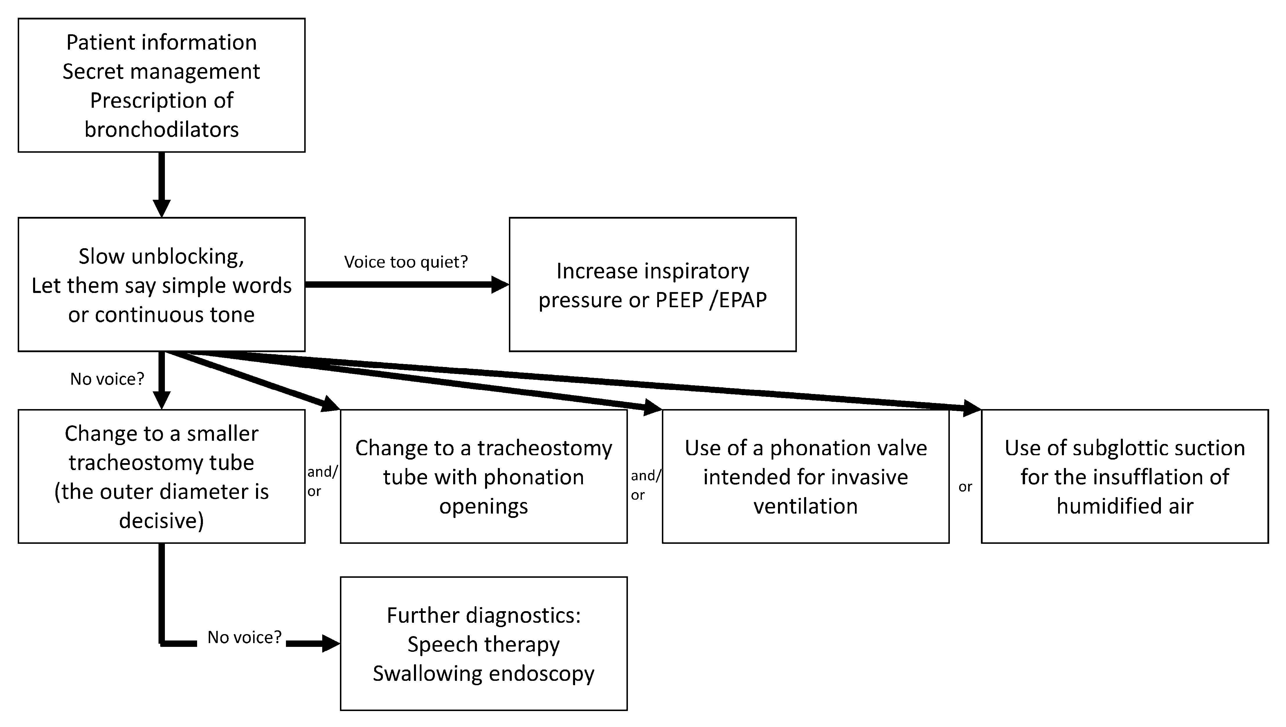
In particular, invasively ventilated people with SCI should have the possibility to speak under ventilation, even on ICU and IMC wards (Figure 2). In addition to the advantages of better communication and higher quality of life, this increase in laryngeal awareness can improve the act of swallowing and thus also prevent aspiration [94,95].

3.7.1. Phonation Under Ventilation

The inability to communicate via speech represents a significant restriction of participation in everyday life for the invasively ventilated or partially ventilated patient. Loud and clear phonation under ventilation with moderate or non-existent dysphagia is basically possible and can be learned [96,97,98]. There are three ways to do this:
  • Leakage ventilation.
  • Leakage ventilation using speaking valves in ventilation systems (so-called phonation valves).
  • Use of speaking cannulae, as follows:
    • Fenestrated tracheal cannulae with inner cannula (“core”).
    • Non-fenestrated tracheal cannula with subglottic air introduction.

3.7.2. Leakage Ventilation

When ventilating with a tracheal cannula and blocked cuff, the airflow is directed unilaterally towards the tracheo-bronchial tree. If the cuff is vented (leakage) under controlled ventilation, air also flows retrogradely next to the cannula shaft through the glottis, so that phonation is possible depending on the flow strength of the air stream—even in the exhalation phase [99,100]. It is important to note that the machine-set PEEP is not maintained, but alveolar ventilation can still be ensured by the increased inspiratory pressure with appropriate adjustment of pressure or volume. An increased PEEP level (between 10 and 12 cmH2O) leads to a higher airflow in expiration, so that a clearer and louder phonation can be achieved [101,102,103,104]. The continuous airflow generated by the increased PEEP is perceived as disturbing or not tolerated by some patients. A bilevel ventilation mode has advantages over an assisted controlled ventilation mode for speech in this situation [102].

3.7.3. Leakage Ventilation Using a Phonation Valve

If a phonation valve is interposed (the cuff must be unblocked), the phonation possibility is improved because no air escapes through the cannula during exhalation and thus the entire exhalation volume flows through the glottis. Care must be taken to ensure that there is sufficient space for the escaping exhaled air next to the cannula, as the exhaled air can only escape through the upper airways (e.g., cannula with smaller outer diameter, less volume of the unblocked cuff, windowing in the cannula). Assessing exhalation pressure using a pressure gauge can be helpful in these situations. In this way, there is the additional advantage that phonation is possible with almost equal force in the inhalation and exhalation phases [91,92,93,94,95].

3.7.4. Speaking Tubes

(a)
Fenestrated tracheal cannulae with inner cannula (“soul”)
There are many tracheal cannulae that are designed to improve the airflow to the vocal folds by fenestration or screening in the bend. However, care must be taken to ensure that the fenestration is correctly positioned and, if necessary, this must also be checked endoscopically. For effective ventilation with a blocked cuff without the possibility of phonation, the fenestration can be removed by means of a closed inner cannula.
(b)
Non-fenestrated tracheal cannula with subglottic air supply
If unblocking or the use of a speaking tube is not possible, it is possible to use a blockable tube with subglottic suction capability to introduce air through this channel above the cuff instead of suctioning above it. In this way, phonation can be trained continuously, independent of the breathing cycle. If unblocking of the cannula is not possible, the use of this type of cannula, depending on the proximity of the air introduction channel to the upper cuff pole, often results in the problem of saliva lying in front of the channel entrance, causing a “bubbling” speech. The air flow that must necessarily be introduced is also perceived as very annoying by some patients, although during pauses in speech the air flow could be prevented by a fingertip—however, this is not usable for people with high tetraplegia without outside help. Overall, this type of cannula is only used by a few patients and is tolerated over a longer period of time.
Jor 06 00013 i006

3.8. Ventilator-Associated Lower Respiratory Tract Infections

People with SCI under invasive ventilation with failure of the respiratory pump and simultaneously reduced coughing ability are more frequently carriers of multi-resistant pathogens in the airways. In the case of gram-negative pathogens, an increase in the degree of resistance is particularly evident due to the escalation of intravenous antibiotic therapies, especially in patients receiving long-term treatment. In this regard, we refer to the publications currently published by the Robert Koch Institute on the use of antibiotics [105].
Ventilator-associated tracheobronchitis (VAT) is characterised by fever, increased secretion volume, positive culture of bronchial secretions without evidence of radiologically detectable infiltrates. This can also be described as a condition between lower airway colonisation and ventilator-associated pneumonia (VAP) [106]. The incidence is between 1.4–11% [107]. Frequently detected pathogens are Pseudomonas aeruginosa, Staphylococcus aureus, and Acinetobacter baumannii [108]. This also includes multi-resistant germs [109]. VAT is associated with longer mechanical ventilation, longer ICU length of stay and higher mortality [107]. In addition to intravenous administration, antibiotics can also be successful in treating VAT by inhalation [110,111].
Inhaled antibiotic therapy can be considered for recurrent respiratory tract infections and bronchopneumonia, as well as for recurrent secretions due to exacerbations of tracheobronchitis in analogy to the therapy recommendations for “non-CF bronchiectasis” [112] and the S3 guideline “Epidemiology, diagnosis and therapy of adult patients with nosocomial pneumonia” [112,113].

3.9. Weaning

Weaning people with tetraplegia is a challenge for treatment centres for many reasons. It is mostly a case of prolonged weaning. The contents of the S2k guideline “Prolonged Weaning,” Revision 2020 of the DGP [114], as well as the contents of the DGNR guideline “Prolonged Weaning in Neurological–Neurosurgical Early Rehabilitation” [115] are considered in the following [85].
As this is usually a case of prolonged, discontinuous weaning in tracheotomised patients [116,117], the length of stay in specialised wards reported in the literature is between 40 and 292 days [118]. Prolonged weaning is therefore the rule, as recurrent pulmonary infections, among other things, delay the weaning process [117,119]. The failure rate after long-term weaning is about 30% [116,118]. In parallel, vegetative dysregulations such as hypotonia, bradycardia, temperature regulation disorders, and autonomic dysreflexia can complicate the course of treatment [120,121]. Transfer to NIV should be considered early in the weaning process.

3.9.1. Pathophysiological Aspects

All people with high SCI are restricted in their breathing [119]. This is rarely due to (pre-)existing lung diseases, but mainly to the impairment of the muscular respiratory pump. Often the diaphragm remains, which alone has to do the work of breathing [122]. It should be borne in mind that even short-term ventilation under analgosedation can additionally impair diaphragmatic activity [123,124,125]. Training of this muscle and its remaining function should be performed carefully and systematically, while strictly avoiding fatigue. This training has the effect that certain types of muscle fibres, which initially fatigue quickly, can now provide an endurance performance [126,127]. This phenomenon has been studied in detail for the first time in people with SCI after stimulation of the phrenic nerve [128]. In this way, respirator-free times can be extended continuously and gradually. Reaching diaphragmatic fatigue must be avoided, as it may delay the weaning process or render successful weaning impossible [122,124,129].

3.9.2. Characteristics of People with Spinal Cord Injury

The majority of patients with impaired respiratory function are people with tetraplegia or -paresis with a level of injury C0–C7 or AIS A, B or C [130]. More rarely, however, people with SCI are found with additional concomitant injuries and/or diseases that impair respiratory performance.

3.9.3. Supplementary Exclusion Criteria for the Start of the Weaning Process

In the case of SCI, the following situations should, in particular, be noted, in which weaning cannot be started or continued:
  • Persistent complete diaphragmatic paralysis.
  • Untreated, highly pronounced autonomic dysreflexia.
  • Non-compensated trunk spasticity significantly affecting breathing.
  • Any form of sepsis.

3.9.4. Practical Procedure of the Weaning Process

Our daily clinical experience with patients with SCI consistently shows different weaning patterns compared with other patient populations [131]. Owing to their neurological condition, patients with SCI rely predominantly on diaphragmatic breathing during the weaning process. This breathing pattern is known to generate higher tidal volumes in the supine position, where the effects of gravity on abdominal content and diaphragm excursion are minimized. Although additional references cited in this context [28,29] date from 1987 and 2001, we consider them fully applicable to patients with SCI, as the underlying pathophysiological mechanisms remain unchanged. Our approach does not reject the awakening and breathing coordination (ABC) principles but modifies them into a progressive, long-term strategy. A key element of this modification is the transition from ‘lying to sitting.’ Weaning in SCI is not just a pulmonary task but a verticalization process. We perform weaning during a gradual elevation of the upper body, with the final successful steps often occurring while the patient is seated in a wheelchair. This ‘modified ABC approach’ allows for successful weaning (or partial daytime independence) even in cases where standard protocols would have failed.
Strict adherence to conventional weaning windows frequently leads to SCI patients being prematurely classified as ‘permanently ventilator dependent,’ which significantly diminishes their quality of life. Our approach does not reject the ABC principles but modifies them into a progressive, long-term strategy. A key element of this modification is the transition from ‘lying to sitting.’ Weaning in SCI is not just a pulmonary task but a verticalization process. We perform weaning during a gradual elevation of the upper body, with the final successful steps often occurring while the patient is seated in a wheelchair. This ‘modified ABC approach’ allows for successful weaning (or partial daytime independence) even in cases where standard protocols would have failed.
Our clinical experience is supported by recent meta-analytical data [132], which demonstrate that weaning success significantly increases in specialized rehabilitation settings (82%) compared with acute ICUs (63%), often after prolonged periods of ventilation. Furthermore, specialized guidelines, such as those from the RISCI group (2023), advocate for a progressive ‘ventilator-free breathing’ approach rather than conventional rapid weaning trials, acknowledging the unique pathophysiological needs of SCI patients. Therefore, we would like to present a concept that “works in daily business,” which, even if it is not backed up by a study protocol, is nevertheless guided by the current literature. Whatever protocol you utilize while performing weaning, your SCI patient clinical focus must remain on preventing the ‘overloading’ of accessory respiratory and spared genuine respiratory muscles. Failure to monitor for signs of exhaustion can jeopardize the entire weaning process and delay the transition to independent breathing. From a spontaneous breathing time of 20 min/h, weaning can usually be carried out in a sitting position (e.g., in a wheelchair) due to the trained diaphragm [133]. Monitoring of the AZV and capnometry must be ensured.
The weaning concept is carried out during the day as follows: A weaning cycle is planned over 12 h (e.g., 8–20 h). Ideally, for each hour there is a share of spontaneous breathing and a share of relief time on the ventilator (adapted to the needs of the patient and the possibilities of the weaning station), this is a discontinuous weaning. This results in up to 12 training units per day. To determine the increase in the proportion of spontaneous breathing, experience tells us that the measurement of the average AZV (spirometrically measured average AZV over 20 spontaneous breaths) is helpful.
Other influencing variables are VC, respiratory rate, extent of diaphragmatic mobility (sonography), and cough thrust testing. Important discontinuation criteria for spontaneous breathing are a decrease in AZV (mean AZV minus 30%) and the development of vegetative symptoms (spasticity, dysreflexia). The increase units are to be checked every day by means of these parameters and on the basis of the clinical experience of the practitioners and should be corrected if necessary. If weaning is successfully completed during the day, i.e., the patient is respiratory stable for about a week during the day, weaning is started at night as follows: The spontaneous breathing time is extended by one hour per day. The patient has the choice of extending the times into the night or disconnecting from the ventilator earlier in the early morning hours. Once the patient has been weaned for 24 h, they should be observed for at least three days to see if spontaneous breathing is stable. If there is evidence of respiratory insufficiency during night-time weaning, or in the days following weaning, transfer to an NIV should be made. Standardised protocols have proven helpful in the weaning process and should be used as part of the documentation process [131]. For further detailed information see SCIRE 2022 [133].
In cases of patients with long ventilation times, even NIV can be an option to gap ventilatory insufficiency and is worth exploring in contrast to tracheostomy to improve quality of life. Such a procedure will be reserved for exceptional cases. While performing NIV, mouthpiece ventilation may be applied in addition to the use of nasal masks.
Jor 06 00013 i007

3.10. Electrostimulation Diaphragm

This is a form of negative-pressure ventilation that is comparable to positive-pressure ventilation in terms of control parameters, as it too is controlled by a control unit—just like an external ventilator—in terms of tidal volume and respiratory rate. However, since electrical impulses trigger diaphragmatic contraction, this controlled form of ventilation is comparable to natural diaphragmatic breathing. In the case of a spinal cord lesion and/or congenital or acquired central respiratory regulation disorders, the following systems can be implanted to stimulate the diaphragm. There are two different systems, direct and indirect diaphragmatic stimulation. Both systems are suitable for long-term stimulation and for use over 24 h. Positive effects regarding infection rate, lung ventilation, especially in the posterior-basal lung segments, and an improvement in secretion management have been proven. In addition, an improvement in phonation, swallowing, and the sense of smell and taste has been demonstrated [128,134]. The indirect systems can also be used in patients with residual respiratory function and can be used unilaterally [128,135].
Exclusion criteria are severe cognitive impairment, a severely damaged heart or lung parenchyma, as well as prefinally diseased patients. Prerequisites for implantation are intact alpha-motoneurons (2nd motoneuron) of both undamaged phrenic nerves, as well as a functioning diaphragm muscle. Lower pulmonary complication rates and improvement in speech have been described [128,134,136]. Long-term studies have showed a lower mortality rate compared with conventionally ventilated patients [128,134].
Due to the savings in consumables, these systems are more cost-effective than invasive ventilation after approx. 3–5 years [128].
In addition, other negative aspects of invasive positive pressure ventilation are avoided, primarily diaphragmatic muscle atrophy and also the consequences of the non-physiological change in thoracic pressure conditions with reduced venous return.

3.10.1. Diaphragmatic Stimulation—Indirect Stimulation (PNS)

Model types (at the time of writing the guideline):
  • Atrostim Yukka®/Atrotech Oy, Tampere, Finland
  • Avery System®/Avery Biomedical Devices Inc., Commack, NY, USA.
  • Thoma System®/Medimplant GmbH, Vienna, Austria (did not have CE approval at the time of publication).
PNS has been an option for negative pressure ventilation of patients with respiratory insufficiency since the mid-1960s [137]. Electrodes are implanted on both phrenic nerves at the level of the 3rd–4th intercostal space mediastinally or in the scalenus gap and all receivers and cables are inserted intracorporeally. Stimulation is performed transcutaneously by induction with closed skin conditions. Thoracic bleeding and pneumothoraces are possible peri- and postoperative complications. Injuries to the phrenic nerve have been described.
The threshold and stimulation current values do not change in long-term studies, so that the above-mentioned systems are suitable for long-term ventilation [138]. Ventilation times are internationally reported as 24 h/day in more than 50% of cases due to the special type of stimulation.

3.10.2. Diaphragmatic Stimulation—Direct Stimulation (DPS)

Model types (at the time of writing the guideline):
  • NeurX®/Synapse Biomedical Inc., Oberlin, OH, USA
  • TransAeris®/Synapse Biomedical Inc., Oberlin, OH, USA
In this procedure, electrodes are inserted laparoscopically directly into the diaphragmatic muscle near the entrance of the phrenic nerve and the cables are passed out of the abdomen [139]. Peri- and postoperative capno- and pneumothorax, cable breaks, and pain during stimulation are possible complications [140,141].
MRI examinations can be carried out in spite of the inserted electrodes. Continuous stimulation over 24 h is possible, as is use in partially ventilated and partially spontaneously breathing patients. Support during weaning or to avoid mechanical ventilation in the case of increasing respiratory exhaustion or in phases of acute respiratory insufficiency can be carried out with passive systems (TransAeris®) [142].
Jor 06 00013 i008

3.11. Non-Invasive Ventilation (NIV)

NIV can avoid intubation and tracheotomy with the associated complications. The body’s own filtering and immune defence as well as the optimal warming and humidification of the respiratory air are thus largely preserved. NIV is used both for long-term therapy and, increasingly, for acute treatment of respiratory insufficiency to support the respiratory pump [143].
NIV is intended to be used in the acute situation of exhaustion of many years of adequate spontaneous breathing to bridge the acute situation and return to spontaneous breathing.
NIV can provide support in the course of intensive therapy for a first-time acquired SCI after necessary invasive ventilation following extubation for transition to spontaneous breathing. It is inferior to tracheostomy and invasive ventilation when the contextual factors of intensive care indicate that longer-term ventilation is required. In these cases, early (<10 days) tracheotomy shortens the time of intensive care treatment [144]. NIV is used for night-time relief during spontaneous breathing during the day. It can also be used in combination with mask and mouthpiece ventilation for long ventilation times per day [145].

3.11.1. NIV in the Acute Situation

Patient-related restrictions in the use of NIV, and in particular the limited possibility of cooperation, must be taken into account in the acute situation after SCI. Experience and training in the use of NIV, both for the medical staff and the nursing staff, are decisive for the success and duration of the application. The mandatory prerequisites for successful NIV, such as functional preservation of the facial and pharyngeal muscles, remain. The same applies to the contraindications of an uncontrollable secretion as well as abdominal complications of possible aerophagia. Discontinuation criteria are subject to the criteria of intensive care ventilation therapy and are based on the blood gas analyses and the necessary ventilation pressures.

3.11.2. NIV in the Chronic Situation of SCI

Acute respiratory failure is more common in people with SCI. Reasons for this are pneumonia, atelectasis, pleural effusion, non-pulmonary complications such as sepsis, long periods of inactivity when lying down due to decubitus ulcer, abdominal complications, and a slowly progressive exhaustion of the respiratory pump with increasing age [146].
Respiratory insufficiency often manifests first during sleep. The reduced active AZV with low tidal volumes not only leads to hypoventilation (increased in REM sleep as a result of the reduced activity of the respiratory support muscles), but also to the promotion of obstructive apnoea as a result of the reduced dilation of the upper airways [147].
Disturbed sleep, headaches, fatigue, sleepiness during the day, blood pressure dysregulation, intellectual performance loss with impaired concentration and memory are the consequences.
To date, no reliable predictors of progressive ventilatory insufficiency have been established. Analogous to patients with neuromuscular disorders, the 20–30–40 rule (VC less than 20 mL/kg ideal body weight; maximal inspiratory pressure (Pimax) less than 30 cmH2O; maximal expiratory pressure (Pemax) less than 40 cmH2O [148] or FVC is <40%) may help anticipate impending respiratory failure and support timely decision-making regarding airway protection before emergency intubation and invasive ventilation become necessary, thereby preventing life-threatening complications. Importantly, these patients may be at substantial risk of ventilatory failure despite the absence of CO2 retention. Consequently, blood gas analysis alone is not a reliable parameter for determining the need for airway-securing interventions [149,150,151,152].
Prerequisites for the implementation of the NIV are as follows [153]:
  • Functional preservation of the facial and pharyngeal musculature.
  • Stable cardiac and circulatory function.
  • Absence of severe disturbance of consciousness.
  • Absence of facial trauma or facial surgery.
  • Experience of the care team.
  • Arterial PaCO2 > 45 mmHg during the day, taking into account symptoms of discomfort.
  • In the presence of nocturnal hypoventilation under the following conditions [154]:
    If there is an increase in arterial PaCO2 or tcCO2 > 55 mmHg for >10 min during sleep.
    If there is an increase in PaCO2 (tcCO2) during sleep of >10 mmHg compared with wakefulness to a value of >50 mmHg for a minimum of 10 min.
Contraindications are as follows:
  • Lack of cooperation of the patient.
  • Increased risk of aspiration in the absence of protective reflexes.
  • Obstruction of the upper airway.
  • Secretions that cannot be controlled by non-invasive means.
  • Decubital ulcers in the area of the mask surfaces.
  • Ileus due to possible aerophagy.
Initially, 24 h/day NIV is possible and can often be gradually reduced in the further course. In order to perform long-term NIV in paraplegia over 24 h/day, a high level of patient adherence and expertise of the treatment team is required. This applies to both clinical and out-of-hospital care. When initiating NIV in people with SCI, the following aspects must be considered, depending on the level of the lesion [49,155]:
  • If hand/arm function is limited or absent, the mask can only be put on and removed by an assistant or special adaptations. Particular attention should be paid to the possibility of rapid independent removal of the mask in emergency situations.
  • When using mouth/nose and full-face masks, there is, among other things, a risk of suffocation (e.g., in case of vomiting or malfunction of the ventilator) and/or aspiration, especially during sleep. Therefore, if removal by the patient is not possible, a qualified nurse must continuously monitor ventilation as part of treatment care. Additional technical monitoring (pulse oximetry) is necessary.
  • The possibility for the patient to raise the alarm can be restricted by the NIV (e.g., loud calling or operating the patient call) and must be technically ensured.
  • Assistance with expectoration must also be ensured during the time phases of NIV application, depending on the needs of the patient.
  • The operation of the device by the patient or caregiver must be checked and ensured.
  • Fears of the patient, especially unpleasant hyperaesthesia in the facial area (often the last innervated areas), and a lack of communication with the help of the mimic musculature during ventilation should be taken into account.
  • Aerophagy can occasionally increase intra-abdominal pressure with the risk of malaise, vomiting or ileus. Whether nasal masks have an advantage over full-face masks in this context is not documented in the literature. Papers dealing with the differences between the two types of masks assess the effectiveness of the masks based on the criteria of sleep quality and the outcome of alveolar ventilation. Patients assessed the advantages and disadvantages of the other mask system differently and more often preferred the other system to the one they were used to. Objectively, there was no significant difference in effectiveness, so that the individual adaptation of the interface and the adjustment of the ventilation parameters under blood gas control are decisive [156,157]. In a study on the long-term follow-up of NIV, aerophagia is described as a frequent occurrence that rarely leads to serious complications and is essentially dependent on the inspiratory pressure [158].

3.11.3. Ventilation Settings

There are many suitable ventilators available for NIV, often offering a variety of ventilation modes. However, there is no standardised nomenclature for these modes.
The settings of the ventilation modes have to be selected individually for people with SCI. Settings that include controlled ventilation parameters are used more often than they would be in non-paralysed patients. This also applies to people with SCI with a distended abdomen. It may be useful to store a backup/minimum volume.
Simultaneously present pulmonary skeletal diseases must be taken into account. The optimal setting of ventilation parameters in chronic respiratory insufficiency due to high SCI is not known. Therefore, it makes sense to follow the guidelines for NIV and adjust the ventilation parameters to the level of paralysis, the lung disease, the thoracic deformities (kyphoscoliosis typical of paralysis), the abdominal pressure increase (coprostasis and meteorism), and the extent of dyspnoea as well as the blood gas analysis [159,160].
Furthermore, a respiratory drive disorder is observed at night, especially in people with high tetraplegia. Therefore, a sufficiently high background (so-called backup) frequency must be selected because of the possible lack of triggering.
It is advantageous to titrate NIV adequately under monitored supervision, with the aim of normoventilation. To improve tolerance, this can be done in a protracted or a gradual manner.
Initiation of NIV in SCI should initially be with a nasal or nasal pillow mask with active humidification during the day [155]. If an oral–nasal mask is necessary for people with tetraplegia, permanent monitoring by a qualified nurse who is permanently present and by pulse oximetry must be carried out and the possibility of alarm must be ensured. To improve patient tolerance, NIV should be initiated with low pressure settings, guided by the degree of hypercapnia and the target ventilation required to achieve normoventilation. In addition, the use of auto-EPAP may facilitate optimal pressure titration to maintain upper airway patency.
In addition, polygraphic or polysomnographic monitoring may be recommended if appropriate [161].
After the initial adjustment, short-term control examinations are necessary to monitor success. In addition to recording symptoms using structured questionnaires (e.g., Epworth sleepiness scale; Berlin questionnaire), compliance data should be recorded (e.g., average nocturnal duration of use; documentation of nights with use of the therapy).
Subsequently, a combined technical and medical check-up is recommended after three and six months and at 6–12 monthly intervals if the course is stable.
The goals of nocturnal NIV are normalisation of arterial PaCO2 during the day, normoventilation under NIV at night (AHI < 5/h), stable O2 saturation, and improvement of daytime symptoms.
In the long-term course, the aim is to achieve a survival advantage, an improvement or ideally a normalisation of blood gases during the day, and an advantage in terms of quality of life [162].
Jor 06 00013 i009

3.12. Transition

In Germany, the framework agreement on discharge management came into force on 1 October 2017 under the precondition of the Act to Strengthen Health Care Provision of 23 July 2015 [163]. As a result, the contents of the contract were defined to implement the insured person’s entitlement to discharge management vis-à-vis the hospital, as well as to support discharge management by the health or long-term care insurance provider. The specific aspects of transitioning highly paralysed, ventilated patients are outlined below and aim to support standardisation in patient care within the framework of discharge management. The SCI, as the primary condition, leads to a lifelong need for medical treatment, which is derived from the long-term therapeutic goals.
The general treatment goals are as follows:
  • To ensure survival.
  • To improve the somatic condition, but at least to prevent deterioration, including possible complications, and to treat it if necessary.
  • To improve the mental state, but at least to prevent deterioration.
  • To minimise the complications of SCI.
  • The specific treatment goals are as follows:
  • Daily mobilisation to the wheelchair.
  • Restoration/maintenance of the ability to speak (also under ventilation).
  • Ensuring communication.
  • Restoration of the greatest possible independence in activities of daily living, occupational and social integration.

3.12.1. Nursing Care

Continuous personal observation of the patient by appropriately qualified nursing staff, along with the reliable implementation of all disease-specific nursing interventions, must be ensured by a nursing service that adheres to quality standards for structural, process, and outcome quality in accordance with § 80 SGB XI and SGB V (applicable in the Federal Republic of Germany) [164,165]. A high medical standard must be set for the specialist nursing qualifications of outpatient services, as ventilated individuals with SCI present diverse and complex care challenges.
In individual cases, it must also be ensured that a second person is available during the provision of basic care (e.g., major personal hygiene, transfers) when ventilation is required and additional factors complicate mobilisation (e.g., obesity, contractures, spasticity, autonomic dysregulation).
To ensure the quality of long-term care for ventilated people with SCI, the following aspects are particularly important [166] for the evaluation of the patient’s overall situation before transfer home:
  • The structured and controlled training of staff in all treatment groups.
  • The mastery of risk management in case of emergency.
  • Interventions to be managed after discharge.
  • The designation of a structured follow-up programme to monitor quality standards, especially in specific respiratory care.
  • To minimise the risk of incidents, the following factors should be considered and behaviour trained accordingly [166]:
  • Ensuring a contact person in treatment and respiration centres.
  • Alarm management.
  • Measures to prevent disconnections.
  • Power supply and its backup even in the event of failures.
  • Seamless and structured exchange of information by means of common or shared documentation.
  • Creation of individualised checklists and their regular review.
Relatives who wish to provide basic or treatment-related care may be suitable for doing so. However, their professional competence should be carefully assessed. The theoretical and practical training required for caring for individuals with this condition is demanding. It is particularly important to consider that, in emergency situations, relatives may be emotionally affected, which could impair their ability to respond appropriately.

3.12.2. Provision in the Employer Model/Assistance Model

Individuals who have chosen the employer or personal assistance model of care independently and responsibly manage the selection of their personal assistants, as well as assume responsibility for their competence. It is strongly recommended to involve external experts with experience in the clinical condition and therapeutic procedures, at least for training purposes, to ensure safe and effective care [84]. It must also be ensured that, in cases of mandatory ventilation, a sufficient number of qualified nursing staff are always available to provide assistance, even in challenging situations [167].

3.13. Technical Equipment

Individuals with SCI who are permanently ventilated are continuously dependent on external assistance for all activities of daily living. Therefore, in addition to clinical observation, comprehensive technical monitoring is essential to ensure the maintenance of vital functions. Moreover, a frequent and SCI-specific complication—autonomic dysreflexia—can be detected through such monitoring. This condition typically presents with a shift from tachycardia to bradycardia accompanied by persistent hypertension [168,169], as well as an initial rise in CO2 followed by a drop in peripheral oxygen saturation [170]. Respiratory disturbances such as hypoxia or hypercapnia may themselves trigger dysreflexive episodes [171], further underscoring the need for continuous monitoring of these parameters.

3.13.1. Pulse Oximetry and Capnography/Capnometry

Providing a pulse oximeter for individuals with SCI who are partially or fully ventilated is essential, as they may not perceive life-threatening oxygen desaturation, nor are they able to communicate or independently correct it.
The following patient groups should be equipped with a possibility for non-invasive PCO2 measurement:
  • Unstable ventilation situation in autonomic dysreflexia [168,169], spasticity, increased intra-abdominal pressure in meteorism and coprostasis.
  • With implanted PNS or DPS (due to missing volumetry).
  • In the case of spontaneous breathing performance dependent on the form of the day with the risk of exhaustion of the respiratory pump.

3.13.2. Respirometer

A respirometer is indicated for PNS/DPS ventilation due to the absence of volumetric monitoring. It is also essential for assessing spontaneous breathing performance during intermittent ventilation.

3.13.3. Supply with a Second Ventilator

The clinical practice of using second ventilators has been well established over many years and is reflected in recommendations and guidelines issued by experts, expert panels, and professional societies [84,172,173,174,175]. Of particular note is the DGP S2k Guideline “Non-Invasive and Invasive Ventilation as Therapy for Chronic Respiratory Insufficiency,” Revision 2017, which explicitly addresses this aspect [58].
This results in the following recommendations:
A second ventilator (so-called “back-up”) must be available if daily ventilation exceeds 16 h. In addition, the indication for a second ventilator and an external battery is always warranted, regardless of daily ventilation duration, if there is a risk of sudden, complete ventilator dependency due to SCI-specific complications and individual patient characteristics.
Furthermore, the daily mobilisation of the patient into the wheelchair, enabling participation in everyday life, is a key therapeutic goal. Therefore, a second device, specifically adapted to the wheelchair setting and equipped with an appropriate power supply (e.g., an additional battery), must be available. Frequent changes throughout the day, including switching the tubing and humidification system from nocturnal to daytime wheelchair ventilation, compromise both the safety and continuity of respiratory support.
If ventilation is carried out via a PNS or DPS (=first device), an additional ventilator is absolutely necessary as a second device in case of stimulation failure or during pauses in stimulation.
Jor 06 00013 i010

3.14. Preventive Care/Aftercare

Lifelong follow-up care is important to prevent medical problems, diagnose them at an early stage and regularly review the rehabilitation process in the outpatient setting. After discharge from the initial rehabilitation, regular outpatient follow-up checks/site assessments are ideally carried out. In this way, the transition from inpatient treatment to the situation at home should be accompanied holistically, emerging medical problems should be recognised in time and the rehabilitation process should be continued with therapies and with social and occupational integration. Knowledge of the specific problems in the long-term course and complications after SCI have led to the development of comprehensive paraplegiological and structured annual check-ups. The updated concept of preventive and follow-up care for people with SCI takes into account the specific problems of these patients [176]. For detailed recommendations on lifelong follow-up care, we refer to the current AWMF guideline “Lifelong Follow-Up Care 179-014” of the DMGP.

3.14.1. Long-Term Complications

The complications in people with SCI reported in the literature show clear differences between ventilated and non-ventilated people with tetraplegia. While pulmonary complications are typical for people with ventilation, decubital ulceration, pain, and constipation are more common in non-ventilated people with tetraplegia [117].

3.14.2. Mortality

It is worth noting that understanding which complications most commonly lead to death in individuals with long-term SCI provides valuable insights for the optimization of follow-up care. Differences and commonalities between findings from various populations and healthcare systems must be taken into account when interpreting these data. According to a German survey, post-discharge mortality in SCI is largely (90%) caused by paralysis-related complications, the majority of which are of pulmonary origin [177].
This finding underlines the importance of specialized respiratory management during long-term follow up.
Notably, the relatively low mortality observed during the initial treatment phase is followed by a critical two-year period after discharge, during which patients remain particularly vulnerable. After this phase is successfully overcome, further complications appear to occur less frequently. A plausible explanation may be the combination of closely monitored inpatient treatment at the beginning and a subsequent learning process in the patient’s home environment—shared by the patient, family members, primary care physicians, and caregivers [178]. More recent data are available for the Italian population [179].

3.15. Recommendations for Prevention

3.15.1. Therapy

Even at home, basic breathing exercises may be helpful to reduce the risk of pulmonary complications. As we know the difficulty of maintaining a routine, and because we consider them to be meaningful, some of these exercises are again listed below.
  • At least 10 min of intensive training are required daily [180].
  • Training with inspiratory resistance supported by respiratory muscle training devices is most effective [181,182,183]. A frequency of 4–5 times a week and between 60–80% of maximum inspiratory resistance (Pimax) is advisable [184].
Follow-up examinations should comprise a check of pulmonary function by spirometry and measurement of in- and expiratory muscle strength. Capacity to cough up secretions is monitored by measurement of peak cough flow and maximum inspiratory capacity. During the examination, patient education may help in keeping to the routine protocols by reviewing manually assisted and device-assisted coughing techniques.
In contrast to the effectiveness of vaccinations and the risk of pulmonary infections even in patients with SCI, the vaccination rate is still considered low [185]. Therefore, we have listed vaccinations that are supported by extensive literature and public healthcare recommendations. For the sake of completeness, potential post-vaccination complications must be kept in mind and discussed with every patient to ensure informed consent.

3.15.2. Vaccinations

  • Annual flu vaccination.
  • Pneumococcal polysaccharide vaccination [186,187,188,189].
  • Pertussis [190].
  • COVID-19 booster vaccination [191,192].

3.15.3. Progress Controls

As part of lifelong follow-up, respiratory function should be checked, especially in people with tetraplegia, by spirometry and measurement of in- and expiratory muscle strength. A symptom-based search (e.g., polygraphy/polysomnography) for respiratory disorders during sleep is recommended because of the frequency and increase with age. As part of lifelong follow-up, the capacity to cough up secretions efficiently should be checked regularly, especially in people with higher levels of SCI. This is done by measuring peak cough flow, maximum inspiratory capacity and reviewing assisted and device-assisted coughing techniques. For detailed recommendations on lifelong follow-up care, please refer to the current AWMF guideline “Lifelong Follow-Up Care 179–014” of the DMGP.
Jor 06 00013 i011

4. Methodological Limitations

While a formal systematic literature search is not mandatory for S2k guidelines, the topic-focused approach to the literature review without detailed documentation represents a methodological limitation with respect to transparency recommended by the AWMF.
Given the time interval between the consensus conference and publication, and in preparation for the upcoming 2027 revision, no post-conference literature update is currently being conducted. This may represent a limitation; however, a comprehensive literature review will be performed for the 2027 guideline revision.

5. Conclusions

This guideline provides a comprehensive framework for the respiratory management of individuals with acute and chronic SCI. It offers consensus-based recommendations for diagnostic and therapeutic interventions, ventilation strategies, and long-term care planning. Developed by experts from Germany and Switzerland, it is intended to support individuals affected by SCI as well as the healthcare professionals involved in their treatment, care, and support. Although this guideline was developed within the German-speaking healthcare context, it may serve as a transferable framework for other healthcare systems, providing guidance that can be adapted to country-specific legal, organizational, and reimbursement structures to support respiratory care in SCI. By addressing both clinical and organizational aspects of respiratory care, the guideline aims to promote appropriate, high-quality, economical, and quality-assured respiratory support across all care settings. Successful implementation requires practical expertise, which should be fostered through targeted hands-on training and specialized education.

Supplementary Materials

The following supporting information can be downloaded at https://www.mdpi.com/article/10.3390/jor6030013/s1, Supplement S1: American Spinal Injury Association Impairment Scale, Supplement S2: Decanulation Scheme.

Author Contributions

Conceptualization: F.M., O.M., M.L., S.T., S.H., G.K. and A.M.R.; formal analysis: A.M.R.; methodology: F.M., O.M., M.L., S.T., S.H., G.K. and A.M.R.; project administration: F.M. and A.M.R.; visualization: A.M.R.; writing—original draft for AWMF: F.M., O.M., M.L., S.T., S.H., G.K. and A.M.R.; writing—review and editing: F.M., O.M., M.L., S.T., S.H., G.K., M.-C.H. and A.M.R.; writing—English draft: A.M.R.; writing—review and editing English draft: F.M., O.M., M.L., S.T., S.H., G.K., M.-C.H. and A.M.R. All authors have read and agreed to the published version of the manuscript.

Funding

This study received no external funding.

Institutional Review Board Statement

Not applicable.

Informed Consent Statement

Not applicable.

Data Availability Statement

No new data were created or analyzed in this study. Data sharing is not applicable to this article.

Acknowledgments

All members of the DMGP working group “BeAtmung” deserve great thanks for their cooperation and support during the preparation of the guideline.

Conflicts of Interest

The authors declare no conflicts of interest.

Abbreviations

AHIApnoea-hypopnea index
AISAmerican Spinal Injury Association Impairment Scale
ALSAmyotrophic lateral sclerosis
AMVRespiratory minute volume
ARDSAcute respiratory distress syndrome
ARFAcute respiratory failure
ASBAssisted spontaneous breathing
ASSPCVAssisted pressure-controlled ventilation
ASVAdaptive servo ventilation
AVAPSAverage volume-assured pressure support
AWMFGerman Association of the Scientific Medical Societies
AZVBreathing volume
BMIBody mass index
COPDChronic obstructive lung disease
CPAPContinuous positive airway pressure
DPSDiaphragm stimulation
EPAPExpiratory positive airway pressure
ERVExpiratory reserve volume
FEV1Forced expiratory volume in 1 s
FiO2Inspiratory oxygen fraction
FRCFunctional residual capacity
FVCForced vital capacity
ICUIntensive care unit
IPAPInspiratory positive airway pressure
IPPBIntermittent positive pressure breathing
IVAPSIntelligent volume-assured pressure support
MICMaximal inspiratory capacity
NIVNon-invasive ventilation
NPVNegative pressure ventilation
OSA(S)Obstructive sleep apnoea (syndrome)
PemaxPeak expiratory pressure
PimaxPeak inspiratory pressure
PAVProportional assist ventilation
PCFPeak cough flow
PCVPressure-controlled ventilation
PEEPPositive end-expiratory pressure
PEFPeak expiratory flow
PNSPhrenic nerve stimulation
PPVPneumococcal polysaccharide vaccination
PSVPressure support ventilation
RVResidual volume
TLCTotal lung capacity
VAPVentilator associated pneumonia
VATVentilator associated tracheobronchitis
VCVVolume controlled ventilation
VtTidal volume

References

  1. Raab, A.M.; Mueller, G.; Elsig, S.; Gandevia, S.C.; Zwahlen, M.; Hopman, M.T.E.; Hilfiker, R. Systematic Review of Incidence Studies of Pneumonia in Persons with Spinal Cord Injury. J. Clin. Med. 2021, 11, 211. [Google Scholar] [CrossRef] [Scilit] [PubMed]
  2. Brown, R.; DiMarco, A.F.; Hoit, J.D.; Garshick, E. Respiratory Dysfunction and Management in Spinal Cord Injury. Respir. Care 2006, 51, 853–868; discussion 869–870. [Google Scholar] [CrossRef] [Scilit] [PubMed]
  3. Tow, A.M.; Graves, D.E.; Carter, R.E. Vital Capacity in Tetraplegics Twenty Years and Beyond. Spinal Cord 2001, 39, 139–144. [Google Scholar] [CrossRef] [Scilit] [PubMed]
  4. Scanlon, P.D.; Loring, S.H.; Pichurko, B.M.; McCool, F.D.; Slutsky, A.S.; Sarkarati, M.; Brown, R. Respiratory Mechanics in Acute Quadriplegia. Lung and Chest Wall Compliance and Dimensional Changes during Respiratory Maneuvers. Am. Rev. Respir. Dis. 1989, 139, 615–620. [Google Scholar] [CrossRef] [Scilit] [PubMed]
  5. Goldman, J.M.; Williams, S.J.; Denison, D.M. The Rib Cage and Abdominal Components of Respiratory System Compliance in Tetraplegic Patients. Eur. Respir. J. 1988, 1, 242–247. [Google Scholar] [CrossRef] [Scilit] [PubMed]
  6. Estenne, M.; De Troyer, A. The Effects of Tetraplegia on Chest Wall Statics. Am. Rev. Respir. Dis. 1986, 134, 121–124. [Google Scholar] [CrossRef] [Scilit] [PubMed]
  7. Postma, K.; Haisma, J.A.; de Groot, S.; Hopman, M.T.; Bergen, M.P.; Stam, H.J.; Bussmann, J.B. Changes in Pulmonary Function during the Early Years after Inpatient Rehabilitation in Persons with Spinal Cord Injury: A Prospective Cohort Study. Arch. Phys. Med. Rehabil. 2013, 94, 1540–1546. [Google Scholar] [CrossRef] [Scilit] [PubMed]
  8. Mueller, G.; de Groot, S.; van der Woude, L.; Hopman, M.T. Time-Courses of Lung Function and Respiratory Muscle Pressure Generating Capacity after Spinal Cord Injury: A Prospective Cohort Study. J. Rehabil. Med. 2008, 40, 269–276. [Google Scholar] [CrossRef] [Scilit] [PubMed]
  9. Almenoff, P.L.; Spungen, A.M.; Lesser, M.; Bauman, W.A. Pulmonary Function Survey in Spinal Cord Injury: Influences of Smoking and Level and Completeness of Injury. Lung 1995, 173, 297–306. [Google Scholar] [CrossRef] [Scilit] [PubMed]
  10. Linn, W.S.; Spungen, A.M.; Gong, H.; Bauman, W.A.; Adkins, R.H.; Waters, R.L. Smoking and Obstructive Lung Dysfunction in Persons with Chronic Spinal Cord Injury. J. Spinal Cord Med. 2003, 26, 28–35. [Google Scholar] [CrossRef] [Scilit] [PubMed]
  11. Stepp, E.L.; Brown, R.; Tun, C.G.; Gagnon, D.R.; Jain, N.B.; Garshick, E. Determinants of Lung Volumes in Chronic Spinal Cord Injury. Arch. Phys. Med. Rehabil. 2008, 89, 1499–1506. [Google Scholar] [CrossRef] [Scilit] [PubMed]
  12. Berlowitz, D.J.; Wadsworth, B.; Ross, J. Respiratory Problems and Management in People with Spinal Cord Injury. Breathe 2016, 12, 328–340. [Google Scholar] [CrossRef] [Scilit] [PubMed]
  13. Schilero, G.J.; Spungen, A.M.; Bauman, W.A.; Radulovic, M.; Lesser, M. Pulmonary Function and Spinal Cord Injury. Respir. Physiol. Neurobiol. 2009, 166, 129–141. [Google Scholar] [CrossRef] [Scilit] [PubMed]
  14. Mueller, G.; de Groot, S.; van der Woude, L.H.; Perret, C.; Michel, F.; Hopman, M.T. Prediction Models and Development of an Easy to Use Open-Access Tool for Measuring Lung Function of Individuals with Motor Complete Spinal Cord Injury. J. Rehabil. Med. 2012, 44, 642–647. [Google Scholar] [CrossRef] [Scilit] [PubMed]
  15. Linn, W.S.; Adkins, R.H.; Gong, H.; Waters, R.L. Pulmonary Function in Chronic Spinal Cord Injury: A Cross-Sectional Survey of 222 Southern California Adult Outpatients. Arch. Phys. Med. Rehabil. 2000, 81, 757–763. [Google Scholar] [CrossRef] [Scilit] [PubMed]
  16. Linn, W.S.; Spungen, A.M.; Gong, H.; Adkins, R.H.; Bauman, W.A.; Waters, R.L. Forced Vital Capacity in Two Large Outpatient Populations with Chronic Spinal Cord Injury. Spinal Cord 2001, 39, 263–268. [Google Scholar] [CrossRef] [Scilit] [PubMed]
  17. De Troyer, A.; Estenne, M. The Expiratory Muscles in Tetraplegia. Paraplegia 1991, 29, 359–363. [Google Scholar] [CrossRef] [Scilit] [PubMed]
  18. Fujiwara, T.; Hara, Y.; Chino, N. Expiratory Function in Complete Tetraplegics: Study of Spirometry, Maximal Expiratory Pressure, and Muscle Activity of Pectoralis Major and Latissimus Dorsi Muscles. Am. J. Phys. Med. Rehabil. 1999, 78, 464–469. [Google Scholar] [CrossRef] [Scilit] [PubMed]
  19. Clay, R.D.; Iyer, V.N.; Reddy, D.R.; Siontis, B.; Scanlon, P.D. The “Complex Restrictive” Pulmonary Function Pattern: Clinical and Radiologic Analysis of a Common but Previously Undescribed Restrictive Pattern. Chest 2017, 152, 1258–1265. [Google Scholar] [CrossRef] [Scilit] [PubMed]
  20. Bach, J.R.; Saporito, L.R. Criteria for Extubation and Tracheostomy Tube Removal for Patients with Ventilatory Failure. A Different Approach to Weaning. Chest 1996, 110, 1566–1571. [Google Scholar] [CrossRef] [Scilit] [PubMed]
  21. Sancho, J.; Servera, E.; Diaz, J.; Marin, J. Predictors of Ineffective Cough during a Chest Infection in Patients with Stable Amyotrophic Lateral Sclerosis. Am. J. Respir. Crit. Care Med. 2007, 175, 1266–1271. [Google Scholar] [CrossRef] [Scilit] [PubMed]
  22. Bergofsky, E.H. Mechanism for Respiratory Insufficiency after Cervical Cord Injury; a Source of Alveolar Hypoventilation. Ann. Intern. Med. 1964, 61, 435–447. [Google Scholar] [PubMed]
  23. Manning, H.L.; Brown, R.; Scharf, S.M.; Leith, D.E.; Weiss, J.W.; Weinberger, S.E.; Schwartzstein, R.M. Ventilatory and Po.1 Response to Hypercapnia in Quadriplegia. Respir. Physiol. 1992, 89, 97–112. [Google Scholar] [CrossRef] [Scilit] [PubMed]
  24. Ben-Dov, I.; Zlobinski, R.; Segel, M.J.; Gaides, M.; Shulimzon, T.; Zeilig, G. Ventilatory Response to Hypercapnia in C(5-8) Chronic Tetraplegia: The Effect of Posture. Arch. Phys. Med. Rehabil. 2009, 90, 1414–1417. [Google Scholar] [CrossRef] [Scilit] [PubMed]
  25. Berlowitz, D.J.; Graco, M. An Update on Sleep Disordered Breathing in Spinal Cord Injury. Curr. Opin. Pulm. Med. 2025, 31, 584–590. [Google Scholar] [CrossRef] [Scilit] [PubMed]
  26. Grimm, D.R.; Chandy, D.; Almenoff, P.L.; Schilero, G.; Lesser, M. Airway Hyperreactivity in Subjects with Tetraplegia Is Associated with Reduced Baseline Airway Caliber. Chest 2000, 118, 1397–1404. [Google Scholar] [CrossRef] [Scilit] [PubMed]
  27. Schilero, G.J.; Grimm, D.R.; Bauman, W.A.; Lenner, R.; Lesser, M. Assessment of Airway Caliber and Bronchodilator Responsiveness in Subjects with Spinal Cord Injury. Chest 2005, 127, 149–155. [Google Scholar] [CrossRef] [Scilit] [PubMed]
  28. Estenne, M.; De Troyer, A. Mechanism of the Postural Dependence of Vital Capacity in Tetraplegic Subjects. Am. Rev. Respir. Dis. 1987, 135, 367–371. [Google Scholar] [CrossRef] [Scilit] [PubMed]
  29. Baydur, A.; Adkins, R.H.; Milic-Emili, J. Lung Mechanics in Individuals with Spinal Cord Injury: Effects of Injury Level and Posture. J. Appl. Physiol. 2001, 90, 405–411. [Google Scholar] [CrossRef] [Scilit] [PubMed]
  30. Chen, C.F.; Lien, I.N.; Wu, M.C. Respiratory Function in Patients with Spinal Cord Injuries: Effects of Posture. Paraplegia 1990, 28, 81–86. [Google Scholar] [CrossRef] [Scilit] [PubMed]
  31. Galeiras Vazquez, R.; Rascado Sedes, P.; Mourelo Farina, M.; Montoto Marques, A.; Ferreiro Velasco, M.E. Respiratory Management in the Patient with Spinal Cord Injury. Biomed. Res. Int. 2013, 2013, 168757. [Google Scholar] [CrossRef] [Scilit] [PubMed]
  32. Miccinilli, S.; Morrone, M.; Bastianini, F.; Molinari, M.; Scivoletto, G.; Silvestri, S.; Ranieri, F.; Sterzi, S. Optoelectronic Plethysmography to Evaluate the Effect of Posture on Breathing Kinematics in Spinal Cord Injury: A Cross Sectional Study. Eur. J. Phys. Rehabil. Med. 2016, 52, 36–47. [Google Scholar] [PubMed]
  33. Stolzmann, K.L.; Gagnon, D.R.; Brown, R.; Tun, C.G.; Garshick, E. Risk Factors for Chest Illness in Chronic Spinal Cord Injury: A Prospective Study. Am. J. Phys. Med. Rehabil. 2010, 89, 576–583. [Google Scholar] [CrossRef] [Scilit] [PubMed]
  34. Bach, J.R.; Ishikawa, Y.; Kim, H. Prevention of Pulmonary Morbidity for Patients with Duchenne Muscular Dystrophy. Chest 1997, 112, 1024–1028. [Google Scholar] [CrossRef] [Scilit] [PubMed]
  35. Tzeng, A.C.; Bach, J.R. Prevention of Pulmonary Morbidity for Patients with Neuromuscular Disease. Chest 2000, 118, 1390–1396. [Google Scholar] [CrossRef] [Scilit] [PubMed]
  36. Pillastrini, P.; Bordini, S.; Bazzocchi, G.; Belloni, G.; Menarini, M. Study of the Effectiveness of Bronchial Clearance in Subjects with Upper Spinal Cord Injuries: Examination of a Rehabilitation Programme Involving Mechanical Insufflation and Exsufflation. Spinal Cord 2006, 44, 614–616. [Google Scholar] [CrossRef] [Scilit] [PubMed]
  37. Hyun, S.E.; Hwang, W.; Ji, H.M.; Shin, H.-I. Effect of Body Position on Peak Expiratory Flow during Mechanical Insufflation–Exsufflation in People with Cervical Spinal Cord Injury: A Pilot Study. Sci. Rep. 2023, 13, 16548. [Google Scholar] [CrossRef] [Scilit] [PubMed]
  38. Shah, N.M.; Apps, C.; Kaltsakas, G.; Madden-Scott, S.; Suh, E.-S.; D’Cruz, R.F.; Arbane, G.; Patout, M.; Lhuillier, E.; Hart, N.; et al. The Effect of Pressure Changes During Mechanical Insufflation-Exsufflation on Respiratory and Airway Physiology. Chest 2024, 165, 929–941. [Google Scholar] [CrossRef] [Scilit] [PubMed]
  39. Rose, L.; McKim, D.; Leasa, D.; Nonoyama, M.; Tandon, A.; Kaminska, M.; O’Connell, C.; Loewen, A.; Connolly, B.; Murphy, P.; et al. Monitoring Cough Effectiveness and Use of Airway Clearance Strategies: A Canadian and UK Survey. Respir. Care 2018, 63, 1506–1513. [Google Scholar] [CrossRef] [Scilit] [PubMed]
  40. Hess, D.R. The Evidence for Secretion Clearance Techniques. Respir. Care 2001, 46, 1276–1293. [Google Scholar] [CrossRef] [Scilit] [PubMed]
  41. Kang, S.W.; Bach, J.R. Maximum Insufflation Capacity: Vital Capacity and Cough Flows in Neuromuscular Disease. Am. J. Phys. Med. Rehabil. 2000, 79, 222–227. [Google Scholar] [CrossRef] [Scilit] [PubMed]
  42. Berlowitz, D.J.; Tamplin, J. Respiratory Muscle Training for Cervical Spinal Cord Injury. Cochrane Database Syst. Rev. 2013, 7, CD008507. [Google Scholar] [CrossRef] [Scilit] [PubMed]
  43. Wong, S.L.; Shem, K.; Crew, J. Specialized Respiratory Management for Acute Cervical Spinal Cord Injury: A Retrospective Analysis. Top. Spinal Cord Inj. Rehabil. 2012, 18, 283–290. [Google Scholar] [CrossRef] [Scilit] [PubMed]
  44. Fink, J.B. Positioning versus Postural Drainage. Respir. Care 2002, 47, 769–777. [Google Scholar] [CrossRef] [Scilit] [PubMed]
  45. Julia, P.E.; Sa’ari, M.Y.; Hasnan, N. Benefit of Triple-Strap Abdominal Binder on Voluntary Cough in Patients with Spinal Cord Injury. Spinal Cord 2011, 49, 1138–1142. [Google Scholar] [CrossRef] [Scilit] [PubMed]
  46. Senent, C.; Golmard, J.L.; Salachas, F.; Chiner, E.; Morelot-Panzini, C.; Meninger, V.; Lamouroux, C.; Similowski, T.; Gonzalez-Bermejo, J. A Comparison of Assisted Cough Techniques in Stable Patients with Severe Respiratory Insufficiency Due to Amyotrophic Lateral Sclerosis. Amyotroph. Lateral Scler. 2011, 12, 26–32. [Google Scholar] [CrossRef] [Scilit] [PubMed]
  47. Ishikawa, Y.; Bach, J.R.; Komaroff, E.; Miura, T.; Jackson-Parekh, R. Cough Augmentation in Duchenne Muscular Dystrophy. Am. J. Phys. Med. Rehabil. 2008, 87, 726–730. [Google Scholar] [CrossRef] [Scilit] [PubMed]
  48. Van de Leur, J.P.; Zwaveling, J.H.; Loef, B.G.; Van der Schans, C.P. Endotracheal Suctioning versus Minimally Invasive Airway Suctioning in Intubated Patients: A Prospective Randomised Controlled Trial. Intensive Care Med. 2003, 29, 426–432. [Google Scholar] [CrossRef] [Scilit] [PubMed]
  49. Bach, J.R. Noninvasive Respiratory Management of High Level Spinal Cord Injury. J. Spinal Cord Med. 2012, 35, 72–80. [Google Scholar] [CrossRef] [Scilit] [PubMed]
  50. Branson, R.D. Secretion Management in the Mechanically Ventilated Patient. Respir. Care 2007, 52, 1328–1342; discussion 1342–1347. [Google Scholar] [CrossRef] [Scilit] [PubMed]
  51. Guerin, C.; Bourdin, G.; Leray, V.; Delannoy, B.; Bayle, F.; Germain, M.; Richard, J.C. Performance of the Coughassist Insufflation-Exsufflation Device in the Presence of an Endotracheal Tube or Tracheostomy Tube: A Bench Study. Respir. Care 2011, 56, 1108–1114. [Google Scholar] [CrossRef] [Scilit] [PubMed]
  52. Garstang, S.V.; Kirshblum, S.C.; Wood, K.E. Patient Preference for In-Exsufflation for Secretion Management with Spinal Cord Injury. J. Spinal Cord Med. 2000, 23, 80–85. [Google Scholar] [CrossRef] [Scilit] [PubMed]
  53. Elliott, M.W. Stepping up and down from NIV to Tracheostomy Ventilation. In ERS Practical Handbook Noninvasive Ventilation; Simonds, A.K., Ed.; ERS-European Respiratory Society: Brussels, Belgium, 2015; pp. 155–162. [Google Scholar]
  54. Suiter, D.M.; McCullough, G.H.; Powell, P.W. Effects of Cuff Deflation and One-Way Tracheostomy Speaking Valve Placement on Swallow Physiology. Dysphagia 2003, 18, 284–292. [Google Scholar] [CrossRef] [Scilit] [PubMed]
  55. Mathias, C.J. Bradycardia and Cardiac Arrest during Tracheal Suction--Mechanisms in Tetraplegic Patients. Eur. J. Intensive Care Med. 1976, 2, 147–156. [Google Scholar] [CrossRef] [Scilit] [PubMed]
  56. Hector, S.M.; Biering-Sorensen, T.; Krassioukov, A.; Biering-Sorensen, F. Cardiac Arrhythmias Associated with Spinal Cord Injury. J. Spinal Cord Med. 2013, 36, 591–599. [Google Scholar] [CrossRef] [Scilit] [PubMed]
  57. Simonds, A.K. (Ed.) ERS Practical Handbook of Noninvasive Ventilation; European Respiratory Society: Brussels, Belgium, 2015; ISBN 978-1-84984-076-7. [Google Scholar]
  58. Windisch, W.; Dreher, M.; Geiseler, J.; Siemon, K.; Brambring, J.; Dellweg, D.; Grolle, B.; Hirschfeld, S.; Kohnlein, T.; Mellies, U.; et al. Guidelines for Non-Invasive and Invasive Home Mechanical Ventilation for Treatment of Chronic Respiratory Failure—Update 2017. Pneumologie 2017, 71, 722–795. [Google Scholar] [CrossRef] [PubMed]
  59. Jackson, A.B.; Groomes, T.E. Incidence of Respiratory Complications Following Spinal Cord Injury. Arch. Phys. Med. Rehabil. 1994, 75, 270–275. [Google Scholar] [CrossRef] [Scilit] [PubMed]
  60. Casha, S.; Christie, S. A Systematic Review of Intensive Cardiopulmonary Management after Spinal Cord Injury. J. Neurotrauma 2011, 28, 1479–1495. [Google Scholar] [CrossRef] [Scilit] [PubMed]
  61. Reid, W.D.; Brown, J.A.; Konnyu, K.J.; Rurak, J.M.; Sakakibara, B.M. Physiotherapy Secretion Removal Techniques in People with Spinal Cord Injury: A Systematic Review. J. Spinal Cord Med. 2010, 33, 353–370. [Google Scholar] [CrossRef] [Scilit] [PubMed]
  62. Tran, K.; Hukins, C.; Geraghty, T.; Eckert, B.; Fraser, L. Sleep-Disordered Breathing in Spinal Cord-Injured Patients: A Short-Term Longitudinal Study. Respirology 2010, 15, 272–276. [Google Scholar] [CrossRef] [Scilit] [PubMed]
  63. Berlowitz, D.J.; Brown, D.J.; Campbell, D.A.; Pierce, R.J. A Longitudinal Evaluation of Sleep and Breathing in the First Year after Cervical Spinal Cord Injury. Arch. Phys. Med. Rehabil. 2005, 86, 1193–1199. [Google Scholar] [CrossRef] [Scilit] [PubMed]
  64. Stockhammer, E.; Tobon, A.; Michel, F.; Eser, P.; Scheuler, W.; Bauer, W.; Baumberger, M.; Muller, W.; Kakebeeke, T.H.; Knecht, H.; et al. Characteristics of Sleep Apnea Syndrome in Tetraplegic Patients. Spinal Cord 2002, 40, 286–294. [Google Scholar] [CrossRef] [Scilit] [PubMed]
  65. Gainche, L.; Berlowitz, D.J.; LeGuen, M.; Ruehland, W.R.; O’Donoghue, F.J.; Trinder, J.; Graco, M.; Schembri, R.; Eckert, D.J.; Rochford, P.D.; et al. Nasal Resistance Is Elevated in People with Tetraplegia and Is Reduced by Topical Sympathomimetic Administration. J. Clin. Sleep Med. JCSM 2016, 12, 1487–1492. [Google Scholar] [CrossRef] [Scilit] [PubMed]
  66. Sankari, A.; Vaughan, S.; Bascom, A.; Martin, J.L.; Badr, M.S. Sleep-Disordered Breathing and Spinal Cord Injury: A State-of-the-Art Review. Chest 2019, 155, 438–445. [Google Scholar] [CrossRef] [Scilit] [PubMed]
  67. Burns, S.P.; Rad, M.Y.; Bryant, S.; Kapur, V. Long-Term Treatment of Sleep Apnea in Persons with Spinal Cord Injury. Am. J. Phys. Med. Rehabil. 2005, 84, 620–626. [Google Scholar] [CrossRef] [Scilit] [PubMed]
  68. Rialp, G.; Raurich, J.M.; Llompart-Pou, J.A.; Ayestaran, I.; Ibanez, J. Central Respiratory Drive in Patients with Neuromuscular Diseases. Respir. Care 2013, 58, 450–457. [Google Scholar] [CrossRef] [Scilit] [PubMed]
  69. Sankari, A.; Bascom, A.; Oomman, S.; Badr, M.S. Sleep Disordered Breathing in Chronic Spinal Cord Injury. J. Clin. Sleep Med. 2014, 10, 65–72. [Google Scholar] [CrossRef] [Scilit] [PubMed]
  70. McEvoy, R.D.; Mykytyn, I.; Sajkov, D.; Flavell, H.; Marshall, R.; Antic, R.; Thornton, A.T. Sleep Apnoea in Patients with Quadriplegia. Thorax 1995, 50, 613–619. [Google Scholar] [CrossRef] [Scilit] [PubMed]
  71. Sajkov, D.; Marshall, R.; Walker, P.; Mykytyn, I.; McEvoy, R.D.; Wale, J.; Flavell, H.; Thornton, A.T.; Antic, R. Sleep Apnoea Related Hypoxia Is Associated with Cognitive Disturbances in Patients with Tetraplegia. Spinal Cord 1998, 36, 231–239. [Google Scholar] [CrossRef] [Scilit] [PubMed]
  72. Le Guen, M.C.; Cistulli, P.A.; Berlowitz, D.J. Continuous Positive Airway Pressure Requirements in Patients with Tetraplegia and Obstructive Sleep Apnoea. Spinal Cord 2012, 50, 832–835. [Google Scholar] [CrossRef] [Scilit] [PubMed]
  73. Sankari, A.; Aldwaikat, A.; Habra, M.; Salloum, A.; Zeineddine, S.; Pandya, N.; Martin, J.L.; Badr, M.S. Sleep Apnea in Individuals with Spinal Cord Injury. J. Clin. Sleep Med. JCSM 2025, 21, 1529–1537. [Google Scholar] [CrossRef] [Scilit] [PubMed]
  74. Berlowitz, D.J.; Schembri, R.; Graco, M.; Ross, J.M.; Ayas, N.; Gordon, I.; Lee, B.; Graham, A.; Cross, S.V.; McClelland, M.; et al. Positive Airway Pressure for Sleep-Disordered Breathing in Acute Quadriplegia: A Randomised Controlled Trial. Thorax 2019, 74, 282–290. [Google Scholar] [CrossRef] [Scilit] [PubMed]
  75. Berlowitz, D.J.; Spong, J.; Pierce, R.J.; Ross, J.; Barnes, M.; Brown, D.J. The Feasibility of Using Auto-Titrating Continuous Positive Airway Pressure to Treat Obstructive Sleep Apnoea after Acute Tetraplegia. Spinal Cord 2009, 47, 868–873. [Google Scholar] [CrossRef] [Scilit] [PubMed]
  76. Hirschfeld, S.; Thietje, R. Schlafqualität Bei Querschnittgelähmten. Somnologie Schlafforschung Schlafmed. 2014, 18, 34. [Google Scholar]
  77. Sankari, A.; Martin, J.L.; Badr, M. A Retrospective Review of Sleep-Disordered Breathing, Hypertenstion and Cardiovascular Diseases in Spinal Cord Injury Patients. Spinal Cord 2015, 53, 496–497. [Google Scholar] [CrossRef] [Scilit] [PubMed]
  78. Graco, M.; McDonald, L.; Green, S.E.; Jackson, M.L.; Berlowitz, D.J. Prevalence of Sleep-Disordered Breathing in People with Tetraplegia-a Systematic Review and Meta-Analysis. Spinal Cord 2021, 59, 474–484. [Google Scholar] [CrossRef] [Scilit] [PubMed]
  79. Sowho, M.; Amatoury, J.; Kirkness, J.P.; Patil, S.P. Sleep and Respiratory Physiology in Adults. Clin. Chest Med. 2014, 35, 469–481. [Google Scholar] [CrossRef] [Scilit] [PubMed]
  80. Menaker, J.; Kufera, J.A.; Glaser, J.; Stein, D.M.; Scalea, T.M. Admission ASIA Motor Score Predicting the Need for Tracheostomy after Cervical Spinal Cord Injury. J. Trauma Acute Care Surg. 2013, 75, 629–634. [Google Scholar] [CrossRef] [Scilit] [PubMed]
  81. Choi, H.J.; Paeng, S.H.; Kim, S.T.; Lee, K.S.; Kim, M.S.; Jung, Y.T. The Effectiveness of Early Tracheostomy (within at Least 10 Days) in Cervical Spinal Cord Injury Patients. J. Korean Neurosurg. Soc. 2013, 54, 220–224. [Google Scholar] [CrossRef] [Scilit] [PubMed]
  82. Berney, S.; Bragge, P.; Granger, C.; Opdam, H.; Denehy, L. The Acute Respiratory Management of Cervical Spinal Cord Injury in the First 6 Weeks after Injury: A Systematic Review. Spinal Cord 2011, 49, 17–29. [Google Scholar] [CrossRef] [Scilit] [PubMed]
  83. Hassid, V.J.; Schinco, M.A.; Tepas, J.J.; Griffen, M.M.; Murphy, T.L.; Frykberg, E.R.; Kerwin, A.J. Definitive Establishment of Airway Control Is Critical for Optimal Outcome in Lower Cervical Spinal Cord Injury. J. Trauma Acute Care Surg. 2008, 65, 1328–1332. [Google Scholar] [CrossRef] [Scilit] [PubMed]
  84. Windisch, W.; Brambring, J.; Budweiser, S.; Dellweg, D.; Geiseler, J.; Gerhard, F.; Kohnlein, T.; Mellies, U.; Schonhofer, B.; Schucher, B.; et al. Non-invasive and invasive mechanical ventilation for treatment of chronic respiratory failure. S2-Guidelines published by the German Medical Association of Pneumology and Ventilatory Support. Pneumologie 2010, 64, 207–240. [Google Scholar] [CrossRef] [Scilit] [PubMed]
  85. Schonhofer, B.; Geiseler, J.; Dellweg, D.; Moerer, O.; Barchfeld, T.; Fuchs, H.; Karg, O.; Rosseau, S.; Sitter, H.; Weber-Carstens, S.; et al. Prolonged weaning: S2k-guideline published by the German Respiratory Society. Pneumologie 2014, 68, 19–75. [Google Scholar] [CrossRef] [Scilit] [PubMed]
  86. Otto-Yáñez, M.; Monge, G.; Muñoz, T.; Segovia, E.; Villalobos, P.; Vera-Uribe, R.; Resqueti, V.; Fregonezi, G.; Torres-Castro, R. Predicting Decannulation Success in Patients with Neurological Conditions: Development and Validation of the NEURODECANN Clinical Score. Nurs. Health Sci. 2025, 27, e70200. [Google Scholar] [CrossRef] [Scilit] [PubMed]
  87. Chen, Y.; Shao, J.; Zhu, W.; Jia, L.S.; Chen, X.S. Identification of Risk Factors for Respiratory Complications in Upper Cervical Spinal Injured Patients with Neurological Impairment. Acta Orthop. Traumatol. Turc. 2013, 47, 111–117. [Google Scholar] [CrossRef] [Scilit] [PubMed]
  88. Leyk, G.; Hirschfeld, S.; Bothig, R.; Willenbrock, U.; Thietje, R.; Lonnecker, S.; Stuhr, M. Spinal cord injury (SCI)—Aspects of intensive medical care. Anasthesiol. Intensivmed. Notfallmed. Schmerzther. 2014, 49, 506–512; quiz 513. [Google Scholar] [CrossRef] [Scilit] [PubMed]
  89. Do, J.G.; Kim, D.H.; Sung, D.H. Incidence of Deep Vein Thrombosis after Spinal Cord Injury in Korean Patients at Acute Rehabilitation Unit. J. Korean Med. Sci. 2013, 28, 1382–1387. [Google Scholar] [CrossRef] [Scilit] [PubMed]
  90. Brower, R.G.; Matthay, M.A.; Morris, A.; Schoenfeld, D.; Thompson, B.T.; Wheeler, A. Ventilation with Lower Tidal Volumes as Compared with Traditional Tidal Volumes for Acute Lung Injury and the Acute Respiratory Distress Syndrome. N. Engl. J. Med. 2000, 342, 1301–1308. [Google Scholar] [CrossRef] [Scilit] [PubMed]
  91. Arora, S.; Flower, O.; Murray, N.P.; Lee, B.B. Respiratory Care of Patients with Cervical Spinal Cord Injury: A Review. Crit. Care Resusc. 2012, 14, 64–73. [Google Scholar] [CrossRef] [Scilit] [PubMed]
  92. Watt, J.W.; Devine, A. Does Dead Space Ventilation Always Alleviate Hypocapnia? Long-Term Ventilation with Plain Tracheostomy Tubes. Anaesthesia 1995, 50, 688–691. [Google Scholar] [CrossRef] [Scilit] [PubMed]
  93. Watt, J.W.; Fraser, M.H. The Effect of Insufflation Leaks in Long-Term Ventilation. Waking and Sleeping Transcutaneous Gas Tensions in Ventilator-Dependent Patients with an Uncuffed Tracheostomy Tube. Anaesthesia 1994, 49, 328–330. [Google Scholar] [PubMed]
  94. Price, G.J.; Jones, C.J.; Charlton, R.A.; Allen, C.M. A Combined Approach to the Assessment of Neurological Dysphagia. Clin. Otolaryngol. Allied Sci. 1987, 12, 197–201. [Google Scholar] [CrossRef] [Scilit] [PubMed]
  95. Shem, K.; Castillo, K.; Wong, S.L.; Chang, J.; Kolakowsky-Hayner, S. Dysphagia and Respiratory Care in Individuals with Tetraplegia: Incidence, Associated Factors, and Preventable Complications. Top. Spinal Cord Inj. Rehabil. 2012, 18, 15–22. [Google Scholar] [CrossRef] [Scilit] [PubMed]
  96. Hess, D.R. Facilitating Speech in the Patient with a Tracheostomy. Respir. Care 2005, 50, 519–525. [Google Scholar] [CrossRef] [Scilit] [PubMed]
  97. Diesener, P. Kommunikation Und Kostaufbau Unter Intensivmedizinischen Bedingungen. Available online: https://akademie-gesundheitsberufe.glkn.de/media/dokumente/aktuelles/publikationen/Schriftenreihe/SR24-d.pdf (accessed on 11 May 2026).
  98. Schwegler, H. Sprechen und Schlucken bei invasiver Beatmung. Available online: https://www.dysphagie.ch/men%C3%BC-deutsch/infos-f%C3%BCr-fachleute/sprechen-und-schlucken-bei-invasiver-beatmung (accessed on 11 May 2026).
  99. Prigent, H.; Lejaille, M.; Terzi, N.; Annane, D.; Figere, M.; Orlikowski, D.; Lofaso, F. Effect of a Tracheostomy Speaking Valve on Breathing-Swallowing Interaction. Intensive Care Med. 2012, 38, 85–90. [Google Scholar] [CrossRef] [Scilit] [PubMed]
  100. Garguilo, M.; Leroux, K.; Lejaille, M.; Pascal, S.; Orlikowski, D.; Lofaso, F.; Prigent, H. Patient-Controlled Positive End-Expiratory Pressure with Neuromuscular Disease: Effect on Speech in Patients with Tracheostomy and Mechanical Ventilation Support. Chest 2013, 143, 1243–1251. [Google Scholar] [CrossRef] [Scilit] [PubMed]
  101. Prigent, H.; Garguilo, M.; Pascal, S.; Pouplin, S.; Bouteille, J.; Lejaille, M.; Orlikowski, D.; Lofaso, F. Speech Effects of a Speaking Valve versus External PEEP in Tracheostomized Ventilator-Dependent Neuromuscular Patients. Intensive Care Med. 2010, 36, 1681–1687. [Google Scholar] [CrossRef] [Scilit] [PubMed]
  102. Prigent, H.; Samuel, C.; Louis, B.; Abinun, M.F.; Zerah-Lancner, F.; Lejaille, M.; Raphael, J.C.; Lofaso, F. Comparative Effects of Two Ventilatory Modes on Speech in Tracheostomized Patients with Neuromuscular Disease. Am. J. Respir. Crit. Care Med. 2003, 167, 114–119. [Google Scholar] [CrossRef] [Scilit] [PubMed]
  103. Manzano, J.L.; Lubillo, S.; Henriquez, D.; Martin, J.C.; Perez, M.C.; Wilson, D.J. Verbal Communication of Ventilator-Dependent Patients. Crit. Care Med. 1993, 21, 512–517. [Google Scholar] [CrossRef] [Scilit] [PubMed]
  104. Byrick, R.J. Improved Communication with the Passy-Muir Valve: The Aim of Technology and the Result of Training. Crit. Care Med. 1993, 21, 483–484. [Google Scholar] [PubMed]
  105. Empfehlung der Kommission für Krankenhaushygiene und Infektionsprävention (KRINKO) beim Robert Koch-Institut (RKI). Hygienemaßnahmen Bei Infektionen Oder Besiedlung Mit Multiresistenten Gramnegativen Stäbchen; Springer: Berlin/Heidelberg, Germany, 2012; pp. 1311–1354. [Google Scholar]
  106. Horan, T.C.; Andrus, M.; Dudeck, M.A. CDC/NHSN Surveillance Definition of Health Care-Associated Infection and Criteria for Specific Types of Infections in the Acute Care Setting. Am. J. Infect. Control 2008, 36, 309–332. [Google Scholar] [CrossRef] [Scilit] [PubMed]
  107. Nseir, S.; Di Pompeo, C.; Pronnier, P.; Beague, S.; Onimus, T.; Saulnier, F.; Grandbastien, B.; Mathieu, D.; Delvallez-Roussel, M.; Durocher, A. Nosocomial Tracheobronchitis in Mechanically Ventilated Patients: Incidence, Aetiology and Outcome. Eur. Respir. J. 2002, 20, 1483–1489. [Google Scholar] [CrossRef] [Scilit] [PubMed]
  108. Arnold, H.M.; Sawyer, A.M.; Kollef, M.H. Use of Adjunctive Aerosolized Antimicrobial Therapy in the Treatment of Pseudomonas Aeruginosa and Acinetobacter Baumannii Ventilator-Associated Pneumonia. Respir. Care 2012, 57, 1226–1233. [Google Scholar] [CrossRef] [Scilit] [PubMed]
  109. Dallas, J.; Skrupky, L.; Abebe, N.; Boyle, W.A.; Kollef, M.H. Ventilator-Associated Tracheobronchitis in a Mixed Surgical and Medical ICU Population. Chest 2011, 139, 513–518. [Google Scholar] [CrossRef] [Scilit] [PubMed]
  110. Palmer, L.B.; Smaldone, G.C.; Chen, J.J.; Baram, D.; Duan, T.; Monteforte, M.; Varela, M.; Tempone, A.K.; O’Riordan, T.; Daroowalla, F.; et al. Aerosolized Antibiotics and Ventilator-Associated Tracheobronchitis in the Intensive Care Unit. Crit. Care Med. 2008, 36, 2008–2013. [Google Scholar] [CrossRef] [Scilit] [PubMed]
  111. Abu-Salah, T.; Dhand, R. Inhaled Antibiotic Therapy for Ventilator-Associated Tracheobronchitis and Ventilator-Associated Pneumonia: An Update. Adv. Ther. 2011, 28, 728–747. [Google Scholar] [CrossRef] [Scilit] [PubMed]
  112. Polverino, E.; Goeminne, P.C.; McDonnell, M.J.; Aliberti, S.; Marshall, S.E.; Loebinger, M.R.; Murris, M.; Canton, R.; Torres, A.; Dimakou, K.; et al. European Respiratory Society Guidelines for the Management of Adult Bronchiectasis. Eur. Respir. J. 2017, 50, 1700629. [Google Scholar] [CrossRef] [Scilit] [PubMed]
  113. Dalhoff, K.; Abele-Horn, M.; Andreas, S.; Deja, M.; Ewig, S.; Gastmeier, P.; Gatermann, S.; Gerlach, H.; Grabein, B.; Heußel, C.P.; et al. Epidemiology, Diagnosis and Treatment of Adult Patients with Nosocomial Pneumonia—Update 2017—S3 Guideline of the German Society for Anaesthesiology and Intensive Care Medicine, the German Society for Infectious Diseases, the German Society for Hygiene and Microbiology, the German Respiratory Society and the Paul-Ehrlich-Society for Chemotherapy, the German Radiological Society and the Society for Virology. Pneumologie 2018, 72, 15–63. [Google Scholar] [CrossRef] [Scilit] [PubMed]
  114. Schonhofer, B.; Geiseler, J.; Dellweg, D.; Fuchs, H.; Moerer, O.; Weber-Carstens, S.; Westhoff, M.; Windisch, W. Prolonged Weaning: S2k Guideline Published by the German Respiratory Society. Respiration 2020, 99, 982–1084. [Google Scholar] [CrossRef] [Scilit] [PubMed]
  115. Rollnik, J.D.; Adolphsen, J.; Bauer, J.; Bertram, M.; Brocke, J.; Dohmen, C.; Donauer, E.; Hartwich, M.; Heidler, M.D.; Huge, V.; et al. Prolonged Weaning during Early Neurological and Neurosurgical Rehabilitation: S2k Guideline Published by the Weaning Committee of the German Neurorehabilitation Society (DGNR). Nervenarzt 2017, 88, 652–674. [Google Scholar] [CrossRef] [Scilit] [PubMed]
  116. Harrop, J.S.; Sharan, A.D.; Scheid, E.H.; Vaccaro, A.R.; Przybylski, G.J. Tracheostomy Placement in Patients with Complete Cervical Spinal Cord Injuries: American Spinal Injury Association Grade A. J. Neurosurg. 2004, 100, 20–23. [Google Scholar] [CrossRef] [Scilit] [PubMed]
  117. Hirschfeld, S.; Exner, G.; Tiedemann, S.; Thietje, R. Langzeitbeatmung Querschnittgelähmter Patienten. Trauma Berufskrankh. 2010, 12, 177–181. [Google Scholar] [CrossRef] [Scilit]
  118. Chiodo, A.E.; Scelza, W.; Forchheimer, M. Predictors of Ventilator Weaning in Individuals with High Cervical Spinal Cord Injury. J. Spinal Cord Med. 2008, 31, 72–77. [Google Scholar] [CrossRef] [Scilit] [PubMed]
  119. Fromm, B.; Hundt, G.; Gerner, H.J.; Baer, G.A.; Exner, G.; Botel, U.; Naumann, C.P.; Baumberger, M.E.; Zach, G. Management of Respiratory Problems Unique to High Tetraplegia. Spinal Cord 1999, 37, 239–244. [Google Scholar] [CrossRef] [Scilit] [PubMed]
  120. Popa, C.; Popa, F.; Grigorean, V.T.; Onose, G.; Sandu, A.M.; Popescu, M.; Burnei, G.; Strambu, V.; Sinescu, C. Vascular Dysfunctions Following Spinal Cord Injury. J. Med. Life 2010, 3, 275–285. [Google Scholar] [PubMed]
  121. Gondim, F.A.; Lopes, A.C.; Oliveira, G.R.; Rodrigues, C.L.; Leal, P.R.; Santos, A.A.; Rola, F.H. Cardiovascular Control after Spinal Cord Injury. Curr. Vasc. Pharmacol. 2004, 2, 71–79. [Google Scholar] [CrossRef] [Scilit] [PubMed]
  122. Edwards, R.H. The Diaphragm as a Muscle. Mechanisms Underlying Fatigue. Am. Rev. Respir. Dis. 1979, 119, 81–84. [Google Scholar] [CrossRef] [Scilit] [PubMed]
  123. Levine, S.; Nguyen, T.; Taylor, N.; Friscia, M.E.; Budak, M.T.; Rothenberg, P.; Zhu, J.; Sachdeva, R.; Sonnad, S.; Kaiser, L.R.; et al. Rapid Disuse Atrophy of Diaphragm Fibers in Mechanically Ventilated Humans. N. Engl. J. Med. 2008, 358, 1327–1335. [Google Scholar] [CrossRef] [Scilit] [PubMed]
  124. Roussos, C.S.; Macklem, P.T. Diaphragmatic Fatigue in Man. J. Appl. Physiol. 1977, 43, 189–197. [Google Scholar] [CrossRef] [Scilit] [PubMed]
  125. Schepens, T.; Verbrugghe, W.; Dams, K.; Corthouts, B.; Parizel, P.M.; Jorens, P.G. The Course of Diaphragm Atrophy in Ventilated Patients Assessed with Ultrasound: A Longitudinal Cohort Study. Crit. Care 2015, 19, 422. [Google Scholar] [CrossRef] [Scilit] [PubMed]
  126. Faulkner, J.A.; Maxwell, L.C.; Ruff, G.L.; White, T.P. The Diaphragm as a Muscle. Contractile Properties. Am. Rev. Respir. Dis. 1979, 119, 89–92. [Google Scholar] [CrossRef] [Scilit] [PubMed]
  127. Mantilla, C.B.; Seven, Y.B.; Zhan, W.Z.; Sieck, G.C. Diaphragm Motor Unit Recruitment in Rats. Respir. Physiol. Neurobiol. 2010, 173, 101–106. [Google Scholar] [CrossRef] [Scilit] [PubMed]
  128. Hirschfeld, S.; Exner, G.; Luukkaala, T.; Baer, G.A. Mechanical Ventilation or Phrenic Nerve Stimulation for Treatment of Spinal Cord Injury-Induced Respiratory Insufficiency. Spinal Cord 2008, 46, 738–742. [Google Scholar] [CrossRef] [Scilit] [PubMed]
  129. Walker, D.J.; Walterspacher, S.; Schlager, D.; Ertl, T.; Roecker, K.; Windisch, W.; Kabitz, H.J. Characteristics of Diaphragmatic Fatigue during Exhaustive Exercise until Task Failure. Respir. Physiol. Neurobiol. 2011, 176, 14–20. [Google Scholar] [CrossRef] [Scilit] [PubMed]
  130. American Spinal Injury Association. Reference Manual of the International Standards for Neurological Classification of Spinal Cord Injury; American Spinal Injury Association: Chicago, IL, USA, 2003. [Google Scholar]
  131. Fussenich, W.; Hirschfeld Araujo, S.; Kowald, B.; Hosman, A.; Auerswald, M.; Thietje, R. Discontinuous Ventilator Weaning of Patients with Acute SCI. Spinal Cord 2018, 56, 461–468. [Google Scholar] [CrossRef] [Scilit] [PubMed]
  132. Schreiber, A.F.; Garlasco, J.; Vieira, F.; Lau, Y.H.; Stavi, D.; Lightfoot, D.; Rigamonti, A.; Burns, K.; Friedrich, J.O.; Singh, J.M.; et al. Separation from Mechanical Ventilation and Survival after Spinal Cord Injury: A Systematic Review and Meta-Analysis. Ann. Intensive Care 2021, 11, 149. [Google Scholar] [CrossRef] [Scilit] [PubMed]
  133. Vu, V.; McIntyre, A.; Herrera, C.C.; Querée, M.; Teasell, R. Acute Respiratory Management Following Spinal Cord Injury; Spinal Cord Injury Research Evidence: London, UK, 2022. [Google Scholar]
  134. Romero, F.J.; Gambarrutta, C.; Garcia-Forcada, A.; Marin, M.A.; de la Lastra, E.D.; Paz, F.; Fernandez-Dorado, M.T.; Mazaira, J. Long-Term Evaluation of Phrenic Nerve Pacing for Respiratory Failure Due to High Cervical Spinal Cord Injury. Spinal Cord 2012, 50, 895–898. [Google Scholar] [CrossRef] [Scilit] [PubMed]
  135. Tokunaga, S.; Miyakoshi, A.; Sato, S.; Hirata, Y.; Adachi, H.; Arai, D.; Sato, T.; Kawanabe, Y. Complete Liberation from Mechanical Ventilation Using Diaphragm Pacing in a Patient with Traumatic Spinal Cord Injury Despite Persistent Unilateral Diaphragmatic Paralysis. NMC Case Rep. J. 2026, 13, 55–61. [Google Scholar] [CrossRef] [Scilit] [PubMed]
  136. Esclarin, A.; Bravo, P.; Arroyo, O.; Mazaira, J.; Garrido, H.; Alcaraz, M.A. Tracheostomy Ventilation versus Diaphragmatic Pacemaker Ventilation in High Spinal Cord Injury. Paraplegia 1994, 32, 687–693. [Google Scholar] [CrossRef] [Scilit] [PubMed]
  137. Glenn, W.W.; Phelps, M.L. Diaphragm Pacing by Electrical Stimulation of the Phrenic Nerve. Neurosurgery 1985, 17, 974–984. [Google Scholar] [CrossRef] [Scilit] [PubMed]
  138. Hirschfeld, S.; Vieweg, H.; Schulz, A.P.; Thietje, R.; Baer, G.A. Threshold Currents of Platinum Electrodes Used for Functional Electrical Stimulation of the Phrenic Nerves for Treatment of Central Apnea. Pacing Clin. Electrophysiol. 2013, 36, 714–718. [Google Scholar] [CrossRef] [Scilit] [PubMed]
  139. Tedde, M.L.; Onders, R.P.; Teixeira, M.J.; Lage, S.G.; Ballester, G.; Brotto, M.W.; Okumura, E.M.; Jatene, F.B. Electric Ventilation: Indications for and Technical Aspects of Diaphragm Pacing Stimulation Surgical Implantation. J. Bras. Pneumol. 2012, 38, 566–572. [Google Scholar] [PubMed]
  140. Kerwin, A.J.; Yorkgitis, B.K.; Ebler, D.J.; Madbak, F.G.; Hsu, A.T.; Crandall, M.L. Use of Diaphragm Pacing in the Management of Acute Cervical Spinal Cord Injury. J. Trauma Acute Care Surg. 2018, 85, 928–931. [Google Scholar] [CrossRef] [Scilit] [PubMed]
  141. Morélot-Panzini, C.; Le Pimpec-Barthes, F.; Menegaux, F.; Gonzalez-Bermejo, J.; Similowski, T. Referred Shoulder Pain (C4 Dermatome) Can Adversely Impact Diaphragm Pacing with Intramuscular Electrodes. Eur. Respir. J. 2015, 45, 1751–1754. [Google Scholar] [CrossRef] [Scilit] [PubMed]
  142. Le Pimpec-Barthes, F.; Legras, A.; Arame, A.; Pricopi, C.; Boucherie, J.-C.; Badia, A.; Panzini, C.M. Diaphragm Pacing: The State of the Art. J. Thorac. Dis. 2016, 8, S376–S386. [Google Scholar] [CrossRef] [Scilit] [PubMed]
  143. Demoule, A.; Girou, E.; Richard, J.C.; Taille, S.; Brochard, L. Increased Use of Noninvasive Ventilation in French Intensive Care Units. Intensive Care Med. 2006, 32, 1747–1755. [Google Scholar] [CrossRef] [Scilit] [PubMed]
  144. Hosokawa, K.; Nishimura, M.; Egi, M.; Vincent, J.L. Timing of Tracheotomy in ICU Patients: A Systematic Review of Randomized Controlled Trials. Crit. Care 2015, 19, 424. [Google Scholar] [CrossRef] [Scilit] [PubMed]
  145. Bach, J.R. Continuous Noninvasive Ventilation for Patients with Neuromuscular Disease and Spinal Cord Injury. Semin. Respir. Crit. Care Med. 2002, 23, 283–292. [Google Scholar] [CrossRef] [Scilit] [PubMed]
  146. Stolzmann, K.L.; Gagnon, D.R.; Brown, R.; Tun, C.G.; Garshick, E. Longitudinal Change in FEV1 and FVC in Chronic Spinal Cord Injury. Am. J. Respir. Crit. Care Med. 2008, 177, 781–786. [Google Scholar] [CrossRef] [Scilit] [PubMed]
  147. White, D.P. Pathogenesis of Obstructive and Central Sleep Apnea. Am. J. Respir. Crit. Care Med. 2005, 172, 1363–1370. [Google Scholar] [CrossRef] [Scilit] [PubMed]
  148. Mehta, S. Neuromuscular Disease Causing Acute Respiratory Failure. Respir. Care 2006, 51, 1016–1021; discussion 1021–1023. [Google Scholar] [CrossRef] [Scilit] [PubMed]
  149. Lawn, N.D.; Fletcher, D.D.; Henderson, R.D.; Wolter, T.D.; Wijdicks, E.F. Anticipating Mechanical Ventilation in Guillain-Barré Syndrome. Arch. Neurol. 2001, 58, 893–898. [Google Scholar] [CrossRef] [Scilit] [PubMed]
  150. Rochester, D.F.; Esau, S.A. Assessment of Ventilatory Function in Patients with Neuromuscular Disease. Clin. Chest Med. 1994, 15, 751–763. [Google Scholar] [CrossRef] [Scilit] [PubMed]
  151. Sivadasan, A.; Cortel-LeBlanc, M.A.; Cortel-LeBlanc, A.; Katzberg, H. Peripheral Nervous System and Neuromuscular Disorders in the Emergency Department: A Review. Acad. Emerg. Med. 2024, 31, 386–397. [Google Scholar] [CrossRef] [Scilit] [PubMed]
  152. Helman, A.; Baskin, R.; Porfiris, G. Neuromuscular Disease for Emergency Medicine. Emergency Medicine Cases. June, 2021. Available online: https://emergencymedicinecases.com/neuromuscular-disease/ (accessed on 19 April 2026).
  153. American Thoracic Society. International Consensus Conferences in Intensive Care Medicine: Noninvasive Positive Pressure Ventilation in Acute Respiratory Failure. Am. J. Respir. Crit. Care Med. 2001, 163, 283–291. [Google Scholar] [CrossRef] [Scilit] [PubMed]
  154. Berry, R.B.; Budhiraja, R.; Gottlieb, D.J.; Gozal, D.; Iber, C.; Kapur, V.K.; Marcus, C.L.; Mehra, R.; Parthasarathy, S.; Quan, S.F.; et al. Rules for Scoring Respiratory Events in Sleep: Update of the 2007 AASM Manual for the Scoring of Sleep and Associated Events. Deliberations of the Sleep Apnea Definitions Task Force of the American Academy of Sleep Medicine. J. Clin. Sleep Med. JCSM 2012, 8, 597–619. [Google Scholar] [CrossRef] [Scilit] [PubMed]
  155. Navalesi, P.; Fanfulla, F.; Frigerio, P.; Gregoretti, C.; Nava, S. Physiologic Evaluation of Noninvasive Mechanical Ventilation Delivered with Three Types of Masks in Patients with Chronic Hypercapnic Respiratory Failure. Crit. Care Med. 2000, 28, 1785–1790. [Google Scholar] [CrossRef] [Scilit] [PubMed]
  156. Majorski, D.S.; Callegari, J.C.; Schwarz, S.B.; Magnet, F.S.; Majorski, R.; Storre, J.H.; Schmoor, C.; Windisch, W. Oronasal versus Nasal Masks for Non-Invasive Ventilation in COPD: A Randomized Crossover Trial. Int. J. Chronic Obstr. Pulm. Dis. 2021, 16, 771–781. [Google Scholar] [CrossRef] [Scilit] [PubMed]
  157. Willson, G.N.; Piper, A.J.; Norman, M.; Chaseling, W.G.; Milross, M.A.; Collins, E.R.; Grunstein, R.R. Nasal versus Full Face Mask for Noninvasive Ventilation in Chronic Respiratory Failure. Eur. Respir. J. 2004, 23, 605–609. [Google Scholar] [CrossRef] [Scilit] [PubMed]
  158. Robert, D.; Argaud, L. Clinical Review: Long-Term Noninvasive Ventilation. Crit. Care 2007, 11, 210. [Google Scholar] [CrossRef] [Scilit] [PubMed]
  159. Dreher, M.; Storre, J.H.; Schmoor, C.; Windisch, W. High-Intensity versus Low-Intensity Non-Invasive Ventilation in Patients with Stable Hypercapnic COPD: A Randomised Crossover Trial. Thorax 2010, 65, 303–308. [Google Scholar] [CrossRef] [Scilit] [PubMed]
  160. Luo, Z.; Wu, C.; Li, Q.; Zhu, J.; Pang, B.; Shi, Y.; Ma, Y.; Cao, Z. Happen collaboration group High-Intensity versus Low-Intensity Noninvasive Positive Pressure Ventilation in Patients with Acute Exacerbation of Chronic Obstructive Pulmonary Disease (HAPPEN): Study Protocol for a Multicenter Randomized Controlled Trial. Trials 2018, 19, 645. [Google Scholar] [CrossRef] [Scilit] [PubMed]
  161. Berry, R.B.; Chediak, A.; Brown, L.K.; Finder, J.; Gozal, D.; Iber, C.; Kushida, C.A.; Morgenthaler, T.; Rowley, J.A.; Davidson-Ward, S.L. Best Clinical Practices for the Sleep Center Adjustment of Noninvasive Positive Pressure Ventilation (NPPV) in Stable Chronic Alveolar Hypoventilation Syndromes. J. Clin. Sleep Med. 2010, 6, 491–509. [Google Scholar] [CrossRef] [Scilit] [PubMed]
  162. Windisch, W. Impact of Home Mechanical Ventilation on Health-Related Quality of Life. Eur. Respir. J. 2008, 32, 1328–1336. [Google Scholar] [CrossRef] [Scilit] [PubMed]
  163. Rahmenvertrag Über Ein Entlassmanagement Beim Übergang in Die Versorgung Nach Kranken-1365 Hausbehandlung § 39 Abs. 1a S. 9 SGB V. 2017. Available online: https://www.gkv-spitzenverband.de/media/dokumente/krankenversicherung_1/amb_stat_vers/entlassmanagement/KH_Rahmenvertrag_Entlassmanagement_2016.pdf (accessed on 12 May 2026).
  164. German Federal Ministry of Justice German Social Code Book XI—Long Term Care Insurance (Sozialgesetzbuch XI) n.d. Available online: https://www.gesetze-im-internet.de/sgb_11/ (accessed on 12 May 2026).
  165. German Federal Ministry of Justice German Social Code Book V—Statutory Health Insurance (Sozialgesetzbuch V) n.d. Available online: https://www.gesetze-im-internet.de/sgb_5/ (accessed on 12 May 2026).
  166. Escarrabill, J. Discharging the Ventilator-Dependent Adult and Child. In ERS Handbook Noninvasive Ventilation; European Respiratory Society: Brussels, Belgium, 2015; pp. 260–265. [Google Scholar]
  167. Giesecke, J. Finanzierung Der Ambulanten Pflege Bei Maschineller Beatmung/Teil 1. Not 2000, 5, 22–28. [Google Scholar]
  168. Karlsson, A.K. Autonomic Dysfunction in Spinal Cord Injury: Clinical Presentation of Symptoms and Signs. Prog. Brain Res. 2006, 152, 1–8. [Google Scholar] [CrossRef] [Scilit] [PubMed]
  169. Taylor, J.A. Autonomic Consequences of Spinal Cord Injury. Auton. Neurosci. 2018, 209, 1–3. [Google Scholar] [CrossRef] [Scilit] [PubMed]
  170. Phillips, A.A.; Ainslie, P.N.; Krassioukov, A.V.; Warburton, D.E. Regulation of Cerebral Blood Flow after Spinal Cord Injury. J. Neurotrauma 2013, 30, 1551–1563. [Google Scholar] [CrossRef] [Scilit] [PubMed]
  171. Andrade, M.J.; Quintas, F.L.; Silva, A.M.; Cruz, P. Is Autonomic Dysreflexia a Cause of Respiratory Dysfunction after Spinal Cord Injury? Spinal Cord Ser. Cases 2021, 7, 4. [Google Scholar] [CrossRef] [Scilit] [PubMed]
  172. McKim, D.A.; Road, J.; Avendano, M.; Abdool, S.; Cote, F.; Duguid, N.; Fraser, J.; Maltais, F.; Morrison, D.L.; O’Connell, C.; et al. Home Mechanical Ventilation: A Canadian Thoracic Society Clinical Practice Guideline. Can. Respir. J. 2011, 18, 197–215. [Google Scholar] [CrossRef] [Scilit] [PubMed]
  173. AARC Clinical Practice Guideline. Long-Term Invasive Mechanical Ventilation in the Home—2007 Revision & Update. Respir. Care 2007, 52, 1056–1062. [CrossRef] [Scilit] [PubMed]
  174. Hammer, J. Home Mechanical Ventilation in Children: Indications and Practical Aspects. Schweiz. Med. Wochenschr. J. Suisse Med. 2000, 130, 1894–1902. [Google Scholar]
  175. International Classification of Functioning, Disability and Health. Available online: https://www.who.int/standards/classifications/international-classification-of-functioning-disability-and-health (accessed on 12 May 2026).
  176. Spreyermann, R.; Luthi, H.; Michel, F.; Baumberger, M.E.; Wirz, M.; Mader, M. Long-Term Follow-up of Patients with Spinal Cord Injury with a New ICF-Based Tool. Spinal Cord 2011, 49, 230–235. [Google Scholar] [CrossRef] [Scilit] [PubMed]
  177. Thietje, R.; Kowald, B.; Hirschfeld, S. Woran Sterben Querschnittgelähmte Heute? Eine Nachuntersuchung von 102 Fällen. Die Rehabil. 2011, 50, 251–254. [Google Scholar] [CrossRef] [Scilit]
  178. Hirschfeld, S.; Jürgens, N.; Tiedemann, S.; Thietje, R. Langzeitkomplikationen Der Querschnittlähmung. In Außerklinische Beatmung im Kindes- und Erwachsenenalter; Bachmann, M., Schucher, B., Eds.; kleanthes: Dresden, Germany, 2013. [Google Scholar]
  179. Barbiellini Amidei, C.; Salmaso, L.; Bellio, S.; Saia, M. Epidemiology of Traumatic Spinal Cord Injury: A Large Population-Based Study. Spinal Cord 2022, 60, 812–819. [Google Scholar] [CrossRef] [Scilit] [PubMed]
  180. Mueller, G.; Hopman, M.T.; Perret, C. Comparison of Respiratory Muscle Training Methods in Individuals with Motor and Sensory Complete Tetraplegia: A Randomized Controlled Trial. J. Rehabil. Med. 2013, 45, 248–253. [Google Scholar] [CrossRef] [Scilit] [PubMed]
  181. Raab, A.M.; Krebs, J.; Perret, C.; Pfister, M.; Hopman, M.; Mueller, G. Evaluation of a Clinical Implementation of a Respiratory Muscle Training Group during Spinal Cord Injury Rehabilitation. Spinal Cord Ser. Cases 2018, 4, 40. [Google Scholar] [CrossRef] [Scilit] [PubMed]
  182. Tranter, K.E.; Harvey, L.A.; Chen, L.W.; Blecher, L.; White, J.; Li, J.; Boswell-Ruys, C.L.; Graco, M.; Berlowitz, D.J.; Glinsky, J.V. Inspiratory Muscle Training for People with Spinal Cord Injury: An Implementation Study. Clin. Rehabil. 2026, 02692155261418967. [Google Scholar] [CrossRef] [Scilit] [PubMed]
  183. Woods, A.; Gustafson, O.; Williams, M.; Stiger, R. The Effects of Inspiratory Muscle Training on Inspiratory Muscle Strength, Lung Function and Quality of Life in Adults with Spinal Cord Injuries: A Systematic Review and Meta-Analysis. Disabil. Rehabil. 2023, 45, 2703–2714. [Google Scholar] [CrossRef] [Scilit] [PubMed]
  184. Raab, A.M.; Krebs, J.; Pfister, M.; Perret, C.; Hopman, M.; Mueller, G. Respiratory Muscle Training in Individuals with Spinal Cord Injury: Effect of Training Intensity and -Volume on Improvements in Respiratory Muscle Strength. Spinal Cord 2019, 57, 482–489. [Google Scholar] [CrossRef] [Scilit] [PubMed]
  185. Weaver, F.M.; Goldstein, B.; Hammond, M. Improving Respiratory Vaccination Rates in Veterans with Spinal Cord Injury/Disorders: Lessons Learned. SCI Nurs. 2004, 21, 143–148. [Google Scholar] [PubMed]
  186. Weaver, F.M.; Hatzakis, M.; Evans, C.T.; Smith, B.; LaVela, S.L.; Wallace, C.; Legro, M.W.; Goldstein, B. A Comparison of Multiple Data Sources to Identify Vaccinations for Veterans with Spinal Cord Injuries and Disorders. J. Am. Med. Inform. Assoc. 2004, 11, 377–379. [Google Scholar] [CrossRef] [Scilit] [PubMed]
  187. Bridges, C.B.; Harper, S.A.; Fukuda, K.; Uyeki, T.M.; Cox, N.J.; Singleton, J.A. Prevention and Control of Influenza: Recommendations of the Advisory Committee on Immunization Practices (ACIP). MMWR Recomm. Rep. 2003, 52, 1–34. [Google Scholar] [PubMed]
  188. Centers for Disease Control and Prevention Prevention of Pneumococcal Disease: Recommendations of the Advisory Committee on Immunization Practices (ACIP). MMWR Recomm. Rep. Morb. Mortal. Wkly. Rep. Recomm. Rep. 1997, 46, 1–24.
  189. Nordin, J.; Mullooly, J.; Poblete, S.; Strikas, R.; Petrucci, R.; Wei, F.; Rush, B.; Safirstein, B.; Wheeler, D.; Nichol, K.L. Influenza Vaccine Effectiveness in Preventing Hospitalizations and Deaths in Persons 65 Years or Older in Minnesota, New York, and Oregon: Data from 3 Health Plans. J. Infect. Dis. 2001, 184, 665–670. [Google Scholar] [CrossRef] [Scilit] [PubMed]
  190. See, K.C. Pertussis Vaccination for Adults: An Updated Guide for Clinicians. Vaccines 2025, 13, 60. [Google Scholar] [CrossRef] [Scilit] [PubMed]
  191. Sankary, K.M.; Sippel, J.L.; Eberhart, A.C.; Burns, S.P. Breakthrough Cases of COVID-19 in Vaccinated United States Veterans with Spinal Cord Injuries and Disorders. Spinal Cord 2021, 59, 1132–1133. [Google Scholar] [CrossRef] [Scilit] [PubMed]
  192. Senthinathan, A.; Cimino, S.; Jaglal, S.B.; Craven, B.C.; Tu, K.; Guilcher, S. The Impact of the COVID-19 Virus and Pandemic on Healthcare Utilization, Access, Delivery, Experiences, and Outcomes in the Spinal Cord Injuries/Dysfunction Population: A Scoping Review Study. PLoS ONE 2024, 19, e0297384. [Google Scholar] [CrossRef] [Scilit] [PubMed]
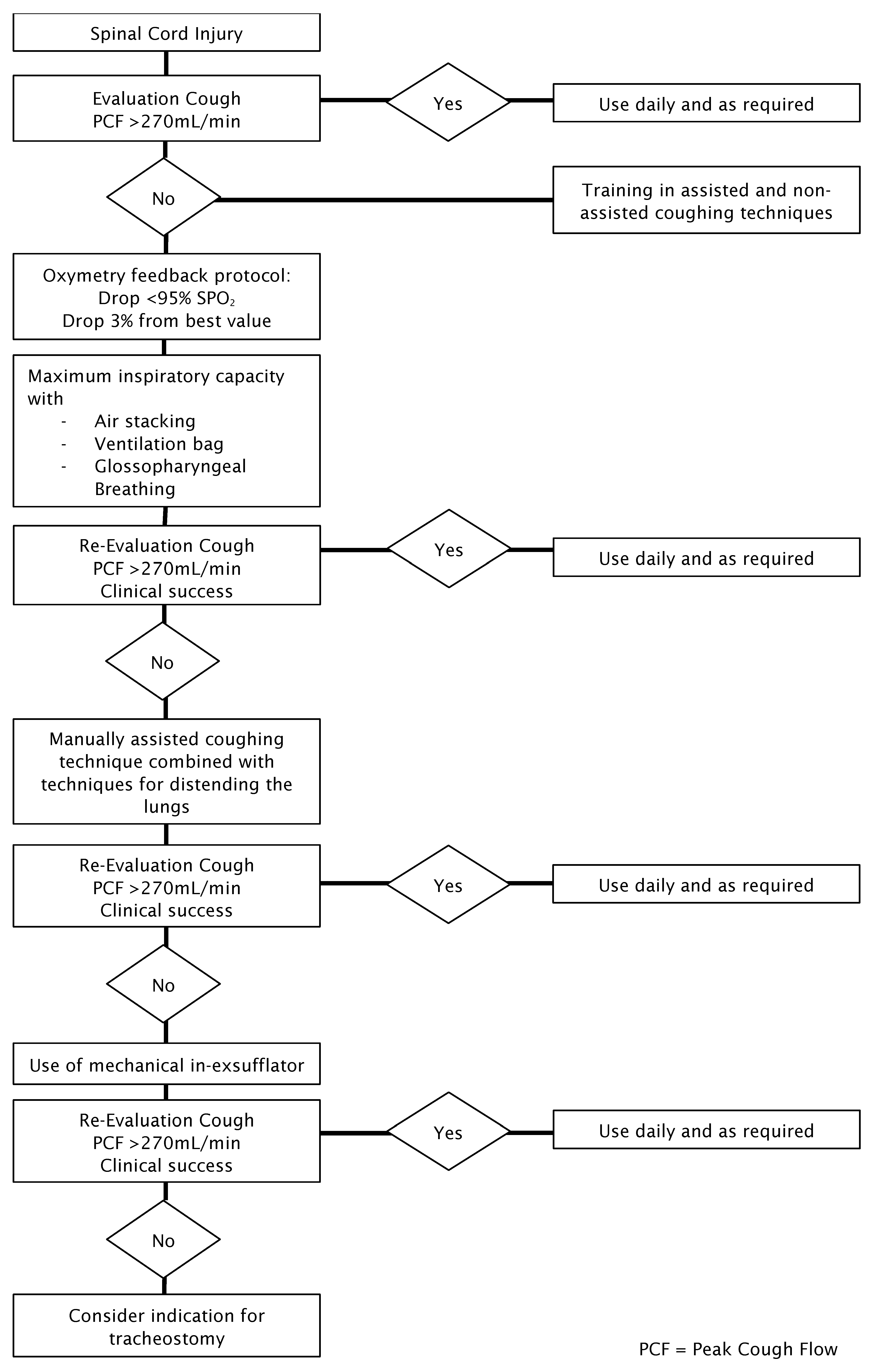
Figure 1. Flow diagram for cough and secretion management (modified according to the DGP guideline, “Non-Invasive and Invasive Ventilation as Therapy for Chronic Respiratory Insufficiency,” Revision 2017 [58]).
Jor 06 00013 g001
Figure 2. Practical recommendation for phonation.
Jor 06 00013 g002
Table 1. Participating organizations.
German Society for Paediatrics and Adolescent Medicine (DGKJ)
German Interdisciplinary Society for Out-of-Hospital Ventilation (DIGAB)
German Society for Neurology (DGN)
German Society for Pneumology and Respiratory Medicine (DGP)
Swiss Society of Pneumology (SGP)
German Society for Sleep Research and Sleep Medicine (DGSM)
Support Association for Paraplegics in Germany (Fördergemeinschaft für Querschnittgelähmte in Deutschland e.V.)
ParaHelp AG of the Paraplegic Group Nottwil, Switzerland
Table 2. Three-level scheme for graduating recommendations.
DescriptionExpression
Strong recommendationNeed/need not
RecommendationShould/should not
Open recommendationCan be considered/waived
Table 3. Determination of consensus strength.
Classification of Consensus Strength
Strong consensus>95% of those eligible to vote
Consensus>75–95% of those eligible to vote
Majority consensus>50–75% of those eligible to vote
No majority consensus<50% of those eligible to vote
Disclaimer/Publisher’s Note: The statements, opinions and data contained in all publications are solely those of the individual author(s) and contributor(s) and not of MDPI and/or the editor(s). MDPI and/or the editor(s) disclaim responsibility for any injury to people or property resulting from any ideas, methods, instructions or products referred to in the content.