Review

Gastroesophageal Reflux and Recurrent Wheezing in Preschool Children: An Update on Pathophysiology, Diagnosis, and Management


1
National Institute of Pediatric Tuberculosis and Respiratory Diseases, Dolný Smokovec, 05981 High Tatras, Slovakia
2
Department of Pathological Physiology, Jessenius Faculty of Medicine in Martin, Comenius University in Bratislava, 03601 Martin, Slovakia
3
Clinic of Pediatric Tuberculosis and Respiratory Diseases, Jessenius Faculty of Medicine in Martin, Comenius University in Bratislava, 05981 High Tatras, Slovakia
*
Author to whom correspondence should be addressed.
J. Respir. 2026, 6(2), 10; https://doi.org/10.3390/jor6020010
Submission received: 17 March 2026 / Revised: 27 April 2026 / Accepted: 18 May 2026 / Published: 19 May 2026

Abstract

Recurrent wheezing represents a significant cause of respiratory morbidity in preschool children. While viral infections in the context of immune dysregulation are primary drivers, gastroesophageal reflux disease (GERD) and laryngopharyngeal reflux (LPR) are increasingly recognized as critical triggers. The link between GERD and respiratory symptoms has historically been controversial; however, recent advances necessitate a re-evaluation of this relationship. This review critically analyzes the pathophysiological nexus between reflux and airway hyperresponsiveness, emphasizing the dual mechanism of microaspiration and vagal reflex, while integrating the concept of reverse causality (where respiratory effort actively exacerbates reflux). We highlight that the “respiratory reflux” phenotype in preschoolers is often characterized by non-acid and proximal episodes, which standard pH-metry fails to detect. Consequently, we discuss the diagnostic shift towards multichannel intraluminal impedance-pH (MII-pH) monitoring and the incorporation of novel metrics defined by the Lyon Consensus 2.0 (MNBI, PSPW index), alongside specific biomarkers such as pepsin. Finally, we propose a phenotype-driven management algorithm, differentiating between acid-suppressive therapy and alginate-based interventions, to mitigate disease burden and improve clinical outcomes in refractory cases.

1. Introduction: The Heterogeneity of Phenotypes and Clinical Context

Recurrent episodes of bronchial obstruction, characterized by wheezing, dyspnea, and cough, constitute one of the most frequent reasons for pediatric consultations and hospitalizations. Epidemiological data indicate that approximately one in three children will experience at least one acute wheezing episode before the age of three [1,2]. While the majority of these episodes are attributed to viral triggers (typically Rhinovirus or RSV) acting on genetically susceptible airways, a significant subset of patients presents with a “refractory phenotype.” These are preschool children with chronic cough or recurrent bronchospasms who exhibit a suboptimal response to standard anti–asthmatic therapy, including inhaled corticosteroids (ICSs) and bronchodilators [3,4].
In this clinical conundrum, Gastroesophageal Reflux Disease (GERD) emerges as a prevalent comorbidity. Studies consistently demonstrate a high coincidence of GERD in children with asthma, with prevalence rates ranging from 40% to 80% [5,6,7]. Recent cross-sectional analyses have further quantified this burden, revealing that verified pathological reflux is present in 30.2% to 45% of children with recurrent bronchial obstruction [8,9].
However, the nature of this association—whether causal or bi-directional—has been a subject of intense debate. Earlier reviews, such as Borrelli et al., concluded that evidence linking GERD to preschool wheezing was often inconsistent or inconclusive, largely due to the reliance on older diagnostic modalities [10]. This perspective is now shifting. Emerging data from 2024 and 2025, utilizing advanced diagnostics like Multichannel Intraluminal Impedance-pH (MII-pH) and molecular biomarkers, suggest that the “inconclusive” results of the past may have stemmed from the inability to detect non-acid and proximal reflux events [11].
This review provides an update on the GERD-wheezing relationship. We move beyond the traditional acid-centric view to explore the role of non-acid reflux, pepsin-mediated inflammation, and neurogenic pathways. Furthermore, we integrate the latest criteria from the Lyon Consensus 2.0 to propose a modern diagnostic and therapeutic roadmap for the pediatric practitioner. Despite these advances, the clinical paradigm must remain balanced; GERD is often a disease-modifying co-factor rather than the sole etiology, and isolated anti-reflux therapies do not universally resolve respiratory symptoms in all cohorts.

2. Materials and Methods

To provide an evidence-based update on the association between GERD and recurrent wheezing, we employed a narrative review approach underpinned by a systematic search strategy. The methodology was designed to prioritize studies utilizing objective diagnostic instrumentation over subjective symptom reporting.

2.1. Search Strategy and Data Sources

We conducted a targeted literature search across three major electronic databases: PubMed/MEDLINE, Scopus, and Web of Science. The search window covered the period from January 2011 to January 2026, with specific emphasis on data published in the last five years to reflect the diagnostic shift towards multichannel intraluminal impedance-pH (MII-pH) monitoring.

2.2. Keywords and Selection Criteria

The search algorithm combined Medical Subject Headings (MeSH) and free-text terms clustered into three domains:
  • Pathology: “Gastroesophageal Reflux”, “Laryngopharyngeal Reflux”, “Extra-esophageal reflux”, “Non-acid reflux”.
  • Clinical Presentation: “Recurrent wheezing”, “Preschool children”, “Bronchospasm”, “Airway hyperresponsiveness”.
  • Diagnostics & Biomarkers: “MII-pH”, “Impedance”, “Pepsin”, “Bronchoalveolar lavage”, “Lipid-laden macrophages”.

2.3. Inclusion and Exclusion Process

We screened titles and abstracts for relevance. To ensure the robustness of the reviewed evidence, we applied strict exclusion criteria:
  • Studies relying solely on parental questionnaires or clinical symptoms without instrumental verification were excluded.
  • Studies focusing exclusively on infants (<12 months) or adolescents were excluded unless they provided specific pathophysiological insights applicable to the preschool phenotype.
  • Duplicate records and non-English publications were removed.
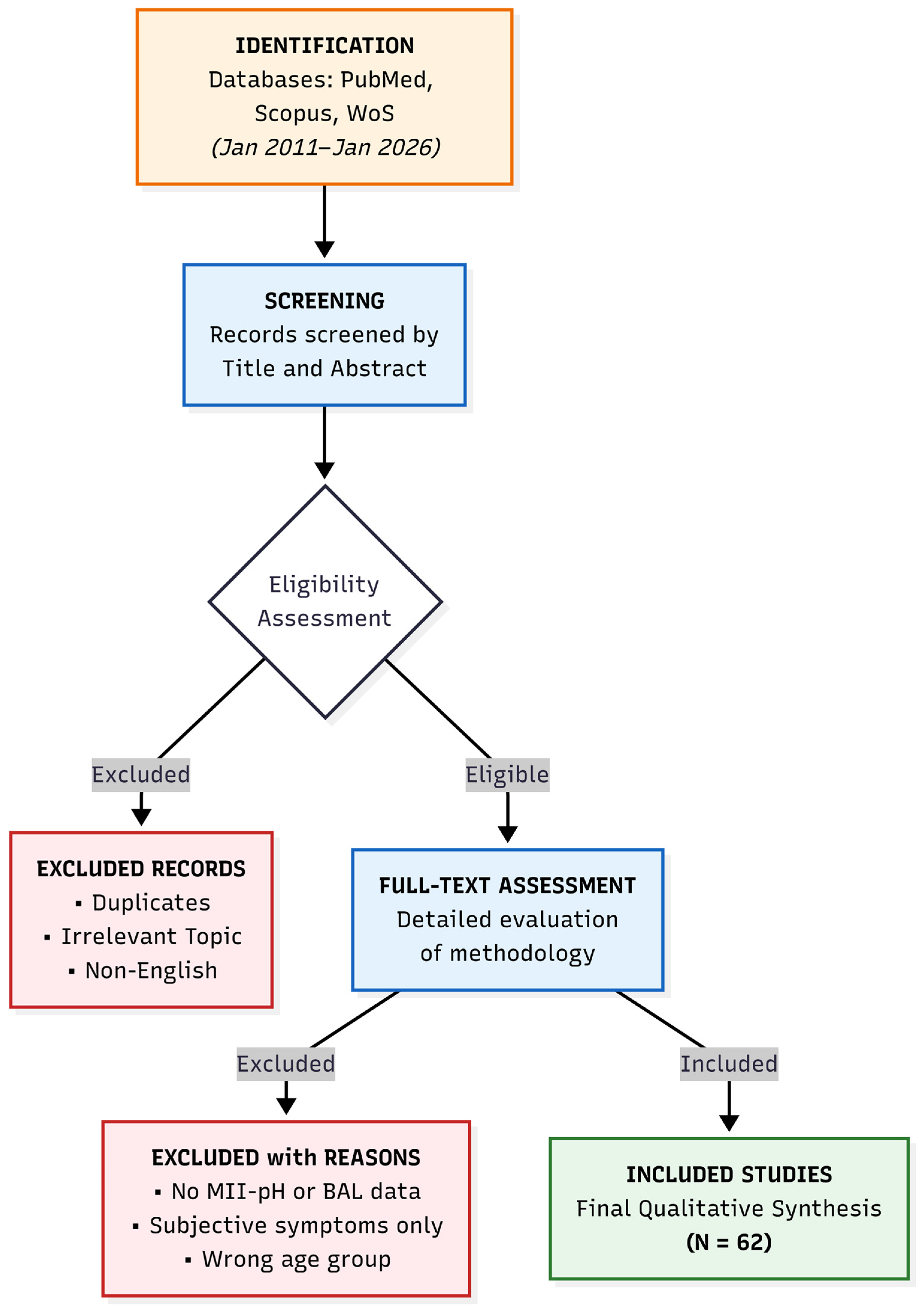
The final qualitative synthesis included 62 key studies that met the eligibility criteria. The selection workflow and attrition of records are detailed in Figure 1.

3. Clinical Manifestations and Long-Term Impact: The Spectrum of “Respiratory Reflux”

The clinical presentation of GERD in the preschool population is remarkably heterogeneous, often earning it the moniker of a “diagnostic chameleon.” Unlike adolescents or adults, where typical esophageal symptoms such as heartburn (pyrosis) and regurgitation dominate, preschool children frequently present with a predominantly extra-esophageal phenotype. This discrepancy often leads to significant diagnostic delays, with symptoms being misattributed to primary respiratory etiologies like viral-induced wheeze or poorly controlled asthma [12].

3.1. The “Silent” Presentation: Chronic Cough and Refractory Wheezing

Chronic cough and recurrent bronchospasm represent the most prevalent extra-esophageal manifestations of GERD in children aged 1–5 years [13]. To properly contextualize recurrent wheezing, it must be viewed within the broader spectrum of pediatric respiratory phenotypes, as highlighted by recent comprehensive systematic reviews and practical algorithms for acute and chronic childhood cough [14,15]. The cough associated with reflux is typically dry, non-productive, and nocturnal, often exacerbated by the supine position which facilitates gravitational reflux. However, a “barking” or bitonal cough may indicate laryngeal involvement (LPR). Studies utilizing objective monitoring have confirmed a robust correlation between these symptoms and reflux episodes. In children with recurrent wheezing, analyses have demonstrated a significantly higher burden of total, acidic, and weakly acidic reflux episodes compared to controls [3,10]. Crucially, this relationship is often observed in the absence of overt emesis, a condition referred to as “silent reflux.” In these cases, the refluxate reaches the proximal esophagus or hypopharynx, triggering airway hyperresponsiveness without causing the classic behavioral signs of gastrointestinal distress.

3.2. The Asthma-GERD Overlap: Causality or Comorbidity?

A critical aspect of the long-term impact is the bidirectional relationship between GERD and asthma. A large-scale longitudinal cohort study tracking over 85,000 children provided pivotal evidence, demonstrating that a diagnosis of GERD in the first year of life is an independent risk factor for the subsequent development of asthma in later childhood (6.5% vs. 3.7% in controls) [4]. Clinically, this translates to an odds ratio that warrants attention: for every 1000 infants with diagnosed GERD, approximately 28 additional cases of asthma occur compared to the general population. This data suggests that early exposure of the developing lung to gastric aspirate may induce chronic inflammation and airway remodeling, effectively lowering the threshold for future obstructive airway disease [12].

3.3. Recurrent Respiratory Infections

Beyond bronchospasm, GERD acts as a facilitator for recurrent respiratory tract infections (RRTIs). Lupu et al. conducted a comprehensive analysis in a pediatric cohort ranging from infancy to 18 years, revealing a striking prevalence of GERD in 83.5% of children presenting with recurrent wheezing and infections [12]. The pathophysiological basis likely involves the aspiration of non-sterile gastric contents, which disrupts the local immune homeostasis of the respiratory mucosa and alters the lung microbiome. A pivotal finding from this cohort was the reversibility of morbidity: the initiation of targeted anti-reflux therapy resulted in a significant reduction in the frequency of respiratory infections and wheezing episodes. This underscores that untreated GERD does not merely coexist with respiratory pathology but actively drives it.

3.4. Laryngopharyngeal Reflux (LPR) Specifics

In the preschool demographic, it is vital to distinguish classic GERD from LPR. While GERD is often associated with LES dysfunction and nocturnal symptoms, LPR often manifests during the day in the upright position and affects the upper airway. The distinguishing clinical features are summarized in Table 1.

4. The Pathophysiological Nexus: A Dual Mechanism of Injury

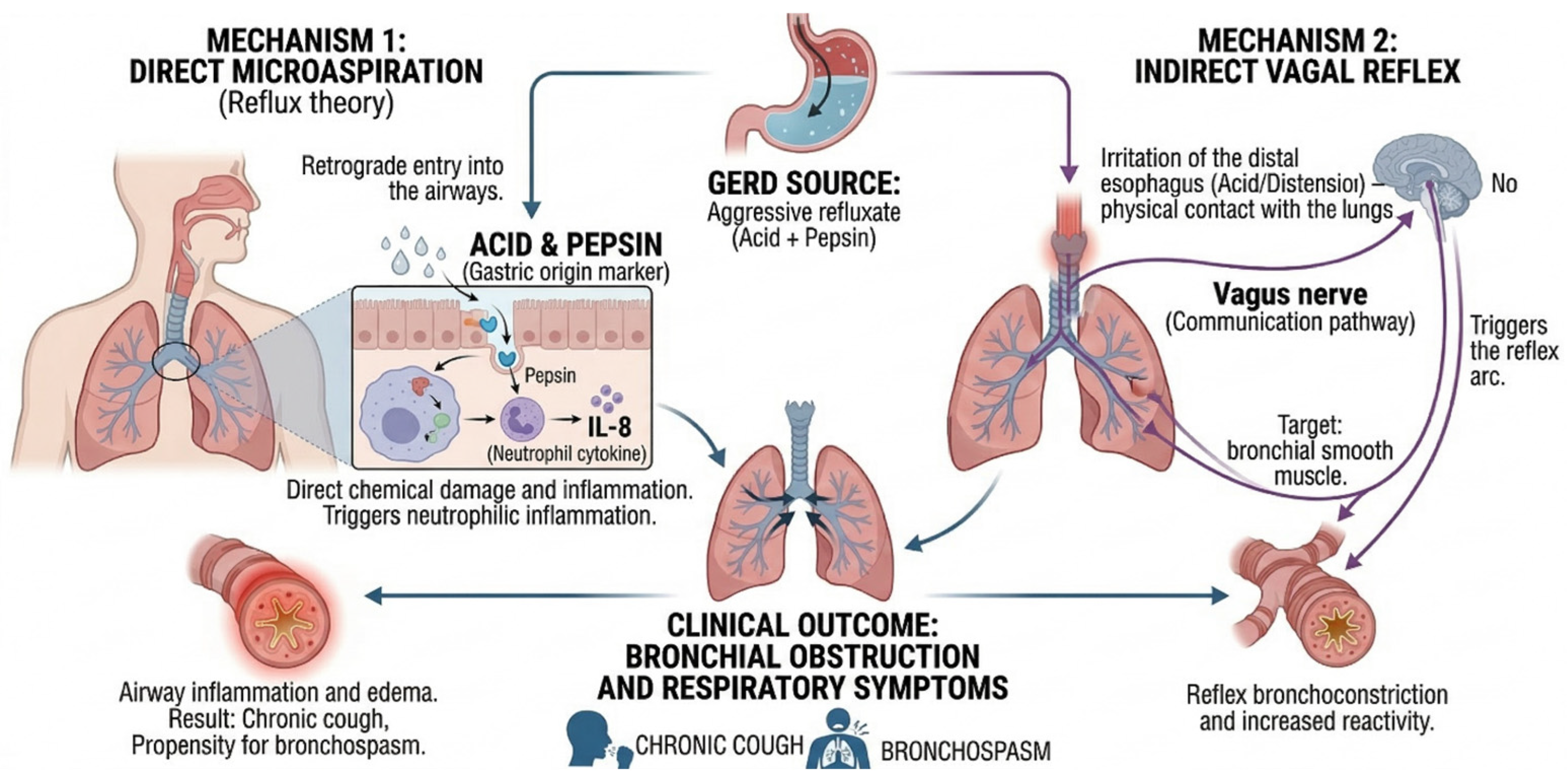
The interaction between the esophagus and the respiratory tract is intricate, mediated by their shared embryonic origin from the foregut and common autonomic innervation via the vagus nerve. Current scientific consensus suggests that GERD induces respiratory pathology through two primary afferent pathways, namely direct microaspiration and the vagal reflex arc, often operating simultaneously [16,17,18,19]. Furthermore, this relationship is increasingly understood as bidirectional, involving a “reverse” efferent loop where respiratory effort exacerbates reflux (Figure 2). It is important to note that while the mechanical aspects of microaspiration are well-established clinically, many molecular mechanisms—such as specific receptor pathways (TRPV1, ASICs)—are largely extrapolated from adult and animal models, representing emerging hypotheses rather than established evidence in the preschool demographic.

4.1. Theory of Direct Microaspiration (The “Reflux Theory”)

This mechanism postulates the retrograde movement of gastric content through the esophagus, breaching the Upper Esophageal Sphincter (UES), and penetrating the tracheobronchial tree. The refluxate is a complex and heterogeneous “chemical cocktail” containing not only hydrochloric acid and pepsin, but also bile salts, pancreatic enzymes, microorganisms, and particulate matter [19]. While the healthy lung possesses defense mechanisms, chronic microaspiration overwhelms these barriers. Emerging evidence indicates that besides acid and pepsin, factors such as bile-induced damage and secondary lung dysbiosis contribute significantly to respiratory epithelial injury. Pepsin, a proteolytic enzyme produced exclusively by gastric chief cells, is identified as a key aggressor. Although maximally active at pH 2.0, in vitro studies demonstrate that pepsin retains some activity up to pH 6.5 and maintains stability and the ability to reactivate up to pH 8.0 [20,21]. This characteristic is critical because it implies that pepsin can cause tissue damage even in weakly acidic or non-acid environments. Upon endocytosis via receptor-mediated uptake, pepsin is stored in acidic intracellular vesicles; this environment is thought to facilitate its intracellular reactivation and trigger downstream inflammatory injury [22]. Crucially, recent data demonstrates that pepsin is retained within epithelial cells for at least 36 h (the maximum duration is currently unknown) [23]. This prolonged intracellular retention elegantly explains the frequent temporal discordance observed in MII-pH monitoring, where clinical symptoms, such as chronic cough, may manifest hours after the objective reflux event.
Recent molecular studies indicate that pepsin is endocytosed by laryngeal and bronchial epithelial cells via receptor-mediated uptake. Once intracellular, it triggers a deleterious signaling cascade. This cascade is characterized by mitochondrial metabolic disruption, E-cadherin depletion, and the upregulation of pro-inflammatory cytokines (specifically IL-8) [24,25]. Consequently, GERD-induced wheezing is typically defined by neutrophilic inflammation [26,27]. Unlike the eosinophilic inflammation seen in atopic asthma, this neutrophilic phenotype is inherently less responsive to inhaled corticosteroids (ICSs), explaining the “steroid-resistant” clinical picture frequently observed in these children [28]. Furthermore, chronic exposure to lipid-rich gastric contents leads to the transformation of alveolar macrophages into lipid-laden macrophages (LLM), serving as a historically proposed cytologic marker of this process [29,30,31]. In summary, direct microaspiration drives a predominantly neutrophilic, steroid-resistant inflammatory response in the airways, actively mediated by prolonged intracellular pepsin retention.

4.2. Theory of the Vagal Reflex (The “Reflex Theory”)

This mechanism explains the occurrence of bronchospasm in the absence of aspiration. The distal esophagus and the bronchial tree share a dense network of vagal afferents. In adult models, this pathway is heavily mediated by acid-sensitive ion channels (ASICs) and Transient Receptor Potential Vanilloid 1 (TRPV1) receptors, leading to neurogenic inflammation via Substance P [32]. However, recent translational evidence (2021) cautions against direct extrapolation to preschoolers. Pediatric esophageal mucosa in non-erosive reflux disease exhibits deep-lying nerve fibers that do not co-express TRPV1 in the same pattern as adults, suggesting a distinct, evolving innervation framework [33]. Therefore, while neurogenic inflammation remains a plausible hypothesis, direct mechanistic links to neuropeptide release in preschool GERD remain unproven.
Instead, current evidence and guidelines from the American Gastroenterological Association (AGA) highlight mechanical distension as the primary driver of the vagal reflex in this age group. High-resolution MII-pH studies demonstrate that vagally mediated bronchospasm and chronic cough are frequently triggered by the volume burden and proximal extent of the refluxate, rather than its acidity [34,35]. Importantly, this distension-induced reflex is readily provoked by weakly acidic or non-acid reflux. This provides a robust physiological rationale for why children continue to wheeze despite aggressive acid suppression: Proton Pump Inhibitors (PPIs) alter the pH of the gastric juice but do not prevent the occurrence of volume reflux events that continue to stimulate esophageal mechanoreceptors [36,37]. In summary, the vagal reflex in preschoolers is primarily a volume-driven, distension-mediated bronchospastic response, underscoring why pure acid suppression frequently fails to provide relief.

4.3. The Concept of Reverse Causality (The Cough-Reflux Loop)

It is imperative to acknowledge the bidirectional physical dynamics between the thoracic and abdominal cavities. During episodes of acute bronchospasm or paroxysmal coughing, the generation of highly negative intrathoracic pressure dramatically increases the transdiaphragmatic pressure gradient. High-resolution manometry (HRM) confirms that this “suction effect” actively impairs the barrier function of the Lower Esophageal Sphincter (LES) and the crural diaphragm, effectively drawing gastric content into the esophagus [38].
While this establishes the physiological plausibility of a “Cough → Reflux” vicious cycle, recent objective MII-pH-manometry studies in pediatric cohorts dictate caution regarding causality. Objective intraesophageal pressure recordings reveal that while reflux events frequently precede and trigger cough, the reverse-cough directly inducing a new reflux episode is significantly less common [39,40]. Therefore, in the preschool phenotype, reverse causality should be viewed as a disease-modifying exacerbator (promoting transient LES relaxations during severe dyspnea) rather than the primary initiator of the pathology. This underscores the necessity of managing both ends of the axis concurrently without falsely assuming that aggressive asthma therapy alone will resolve the underlying reflux.

5. Comprehensive Diagnostic Approach: From pH-Metry to Multimodality

Diagnosing GERD-associated respiratory symptoms is notoriously challenging due to the absence of a singular “gold standard” capable of definitively confirming causality. Every diagnostic modality possesses inherent limitations; therefore, accurately phenotyping the patient and tailoring personalized therapy frequently requires a multimodal approach.

5.1. The Limitations of Conventional pH-Metry

Historically, 24-h esophageal pH-metry served as the cornerstone of GERD diagnostics. However, in the context of extra-esophageal manifestations, relying solely on pH-metry is analogous to utilizing a monochrome camera—it captures only one dimension of the pathology: acidic reflux (pH < 4.0). Recent clinical analyses have exposed the critical limitations of this unimodal view.
A pivotal study by Zenzeri et al. compared reflux characteristics in pediatric cohorts, revealing a dramatic phenotypic divergence based on symptomatology. While children with typical gastrointestinal symptoms (pyrosis, regurgitation) exhibited predominantly acidic reflux, children presenting with respiratory symptoms demonstrated a significantly higher frequency of weakly alkaline and non-acidic reflux episodes (mean 22.1 vs. 10.1; p < 0.001) [5]. This physiological distinctiveness provides the missing rationale for the frequent failure of Proton Pump Inhibitors (PPIs) in children with recurrent bronchial obstruction. PPIs merely neutralize the pH of the gastric aspirate; they do not halt the retrograde flow of the refluxate [41]. Consequently, aggressive factors like pepsin, which maintains functional stability at pH levels above 4.0, continue to be aspirated. Furthermore, the mechanical distension of the distal esophagus by the unsuppressed volume of the refluxate continues to trigger vagally mediated bronchoconstriction, regardless of the neutralized pH [35].

5.2. MII-pH, Lyon Consensus 2.0, and the Paradigm Shift

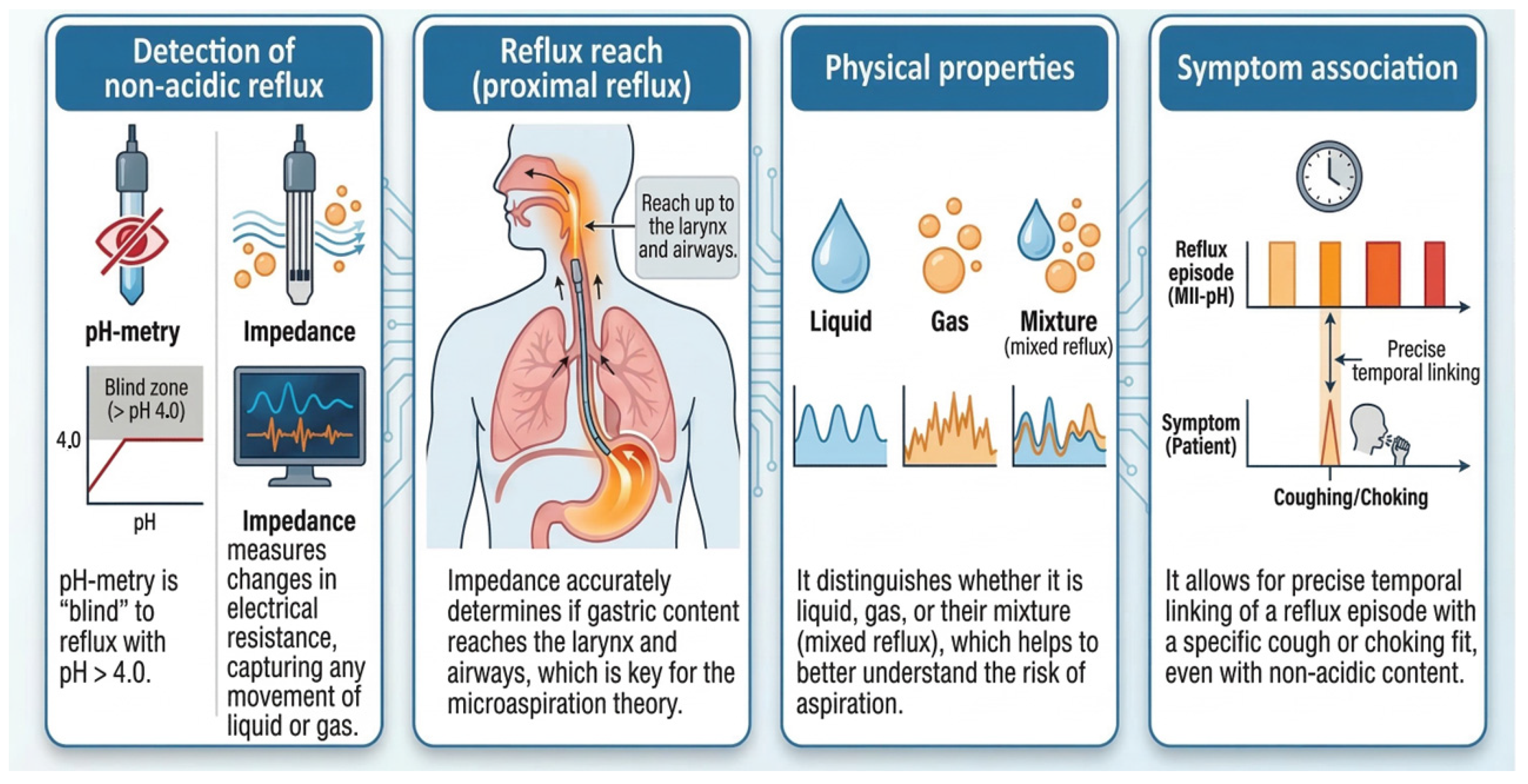
The current diagnostic gold standard for evaluating “respiratory reflux” is Multichannel Intraluminal Impedance-pH (MII-pH) monitoring. If conventional pH-metry is a monochrome snapshot, MII-pH represents high-definition multimodal imaging (Figure 3). It detects all modalities of reflux (acidic, weakly acidic, and non-acidic) and precisely maps its proximal extent, which is critical when evaluating the risk of microaspiration. While MII-pH is the physiological gold standard, its routine clinical feasibility is limited. It is an invasive, relatively expensive procedure that requires specialized equipment and expert interpretation, restricting its availability primarily to tertiary pediatric gastroenterology centers.
The diagnostic superiority of MII-pH is robustly documented. Pavić et al. demonstrated that MII-pH successfully identified GERD in 70% of preschool patients with recurrent wheezing, whereas isolated pH-metry detected pathological acid reflux in a mere 16% of the same cohort [3].
Crucially, the interpretation of MII-pH data is evolving with the updated Lyon Consensus 2.0 criteria [8]. Beyond simply counting reflux episodes, modern diagnostics in adults emphasize novel impedance metrics:
  • Mean Nocturnal Baseline Impedance (MNBI): A marker of longitudinal mucosal integrity and altered tight junctions.
  • Post-reflux Swallow-induced Peristaltic Wave (PSPW) index: A metric evaluating chemical clearance.
A Critical Pediatric Caveat: It must be explicitly noted that the Lyon 2.0 criteria were developed for adults. While the Lyon Consensus 2.0 highlights these metrics as powerful adjunctive tools, recent evidence (2024) dictates extreme caution when extrapolating to preschoolers. Systematic reviews and joint guidelines from NASPGHAN and ESPGHAN emphasize that standardized diagnostic criteria and validated normative values for MNBI and PSPW index are currently lacking for young children. Therefore, until robust pediatric reference ranges are established, these advanced metrics should be regarded as highly promising translational research tools rather than definitive clinical decision-making endpoints in pediatric extra-esophageal GERD [42,43].

5.3. Biomarkers of Microaspiration: The Search for Specificity

While MII-pH quantifies the presence of reflux in the esophagus, specific biomarkers are theoretically required to confirm microaspiration. However, the translation of these biomarkers into routine clinical practice remains highly controversial.
  • Pepsin (BAL and Salivary): Pepsin is conceptually appealing as a “gastric fingerprint.” Our previous research [11] and others [9,44] have demonstrated high detection rates of salivary and BAL pepsin in symptomatic children, even when standard pH-metry is negative (e.g., 90.9% salivary pepsin positivity in pH-negative patients). While this highlights the massive presence of non-acidic reflux, contemporary clinical practice updates, including those from the American Gastroenterological Association (AGA) [45], strongly caution against the routine clinical use of pepsin assays. The medical literature demonstrates substantial methodological heterogeneity, poor concordance with MII-pH, and significant overlap between symptomatic patients and healthy controls. With reported sensitivities around 43–78% and specificities as low as 50%, and critically, without universally accepted pediatric threshold (cut-off) values, pepsin currently lacks the reliability to serve as a standalone diagnostic tool [45,46].
  • Lipid-Laden Macrophages (LLM): Historically, the transformation of alveolar macrophages into LLMs following lipid phagocytosis was considered a key cytological marker of microaspiration. While some recent studies (e.g., Pavić et al., 2025) report correlations between LLM percentages and weakly acidic proximal events [3], current Evidence-Based Medicine paradigms increasingly view LLMI as obsolete. NASPGHAN and ESPGHAN explicitly recommend against using LLMs to diagnose reflux-related respiratory disease [43]. The index suffers from extremely low specificity, wide inter-observer variability, and an inability to reliably differentiate the aspiration of exogenous gastric contents from endogenous lipid accumulation occurring in a variety of other pulmonary pathologies [29].
  • While pepsin and LLM have historically dominated clinical research, keyword selection limited to these markers may fail to capture the breadth of new candidate biomarkers. Although bile is not found in all refluxate (restricted to concomitant gastroduodenal reflux), clinical and laboratory evidence supports the contribution of bile acids to pulmonary injury in both adults and children [47]. Additionally, glycerophospholipids demonstrate a correlation with MII-pH data [48], and pancreatic elastase along with cholesterol have recently been identified as biomarkers for gastroduodenal reflux in adult LPR patients [49]. A major limitation to the clinical implementation of these promising biomarkers is the requirement for highly sensitive, expensive assays, such as Liquid Chromatography-Mass Spectrometry (LC-MS), as opposed to standard, clinically feasible enzyme-based tests.
Consequently, the diagnosis of GERD-induced wheezing cannot rely on a single laboratory test. It requires a holistic synthesis of the clinical phenotype, therapeutic trials (where appropriate), and objective physiological evaluation via MII-pH.

6. Therapeutic Approaches: Moving Towards Phenotype-Driven Management

The primary therapeutic endpoint in GERD-associated respiratory disease extends beyond the healing of the esophageal mucosa; it fundamentally aims to control respiratory morbidity, reduce the frequency of wheezing exacerbations, and prevent long-term airway remodeling. Contemporary management encompasses lifestyle modifications, pharmacotherapy, and, in highly refractory cases, surgical intervention. However, the paradigm has shifted strictly away from empiric trials toward phenotype-driven therapy guided by objective baseline diagnostics.

6.1. Acid Suppression (PPIs): Lack of Extraesophageal Benefit and Quantifiable Risks

Proton Pump Inhibitors (PPIs) remain the cornerstone of therapy exclusively for the objectively verified acid-predominant phenotype. When pathological acid GERD is confirmed (e.g., via MII-pH), targeted acid suppression yields clinical benefits. For instance, in pediatric cohorts with recurrent respiratory tract infections and confirmed acidic GERD, short-term PPI therapy has been associated with significant symptomatic improvement [12].
However, current pediatric literature (2020–2025) strongly refutes the utility of empiric PPI therapy for isolated chronic cough or wheezing without proven GERD. Previously, it was hypothesized that PPIs might offer relief in non-acidic phenotypes via a “volume-reduction” mechanism (decreasing total gastric juice volume). Recent mechanistic and clinical reviews entirely dismiss this notion in children; PPIs effectively inhibit the parietal H+/K+-ATPase to raise gastric pH but do not significantly reduce gastric volume or mitigate volume-induced vagal reflexes [50,51]. Crucially, contemporary pediatric guidelines emphasize the quantifiable long-term risks associated with acid suppression. The reliance on prolonged PPI therapy in preschoolers is now highly discouraged due to → Infectious Complications: Large cohort studies demonstrate a significant increase in serious overall infections (adjusted Hazard Ratio [aHR] 1.34), lower respiratory tract infections (aHR 1.22), and ENT infections (aHR 1.47) in young children on PPIs [52,53]. Microbiome Dysbiosis: Prospective pediatric cohorts reveal that PPIs profoundly alter oral, gut, and lung microbiota. This includes the enrichment of Streptococcus species and the loss of beneficial taxa, which directly correlates with the increased susceptibility to respiratory infections and asthma exacerbations [53,54]. Therefore, guidelines dictate that PPIs should be used only for clear indications and limited to short courses (4–8 weeks). This must be followed by mandatory clinical reassessment and deprescribing to avoid iatrogenic morbidity [51,53,55].

6.2. Alginates and Mechanical Barriers: Efficacy vs. Pediatric Evidence Gaps

Given the limitations and risks of pure acid suppression, magnesium and sodium alginates are frequently utilized for Extra-Esophageal Reflux (EER) and Laryngopharyngeal Reflux (LPR) phenotypes. Mechanistically, alginates precipitate in the presence of gastric acid to form a viscous mechanical barrier (a “raft”) that physically impedes the proximal migration of acid, pepsin, and bile acids. While adult randomized controlled trials demonstrate that alginates are non-inferior to PPIs for LPR [56], direct clinical evidence for their chronic use in preschool children remains extremely limited. Current pediatric gastroenterology guidelines (including ESPGHAN/NASPGHAN aligned consensus) support the use of aluminum-free alginates primarily as a short-term, post-prandial rescue therapy for mild symptoms, rather than a proven long-term solution for EER [57,58]. While generally considered devoid of serious adverse effects in short-term infant use, there is no robust evidence supporting the prolonged efficacy of alginates specifically for extraesophageal respiratory outcomes in preschoolers. Clinicians must exercise caution with chronic, high-dose use, monitoring for potential GI upset (constipation or diarrhea) and considering the sodium or magnesium load, particularly in children with underlying renal or cardiac comorbidities [43,59].

6.3. Anti-Reflux Surgery: Strict Objective Indications

Anti-reflux surgery (e.g., fundoplication) is not a treatment for refractory wheezing per se. According to the latest multidisciplinary consensus, surgical intervention is strictly reserved for children who demonstrate objective, severe GERD (via endoscopy and MII-pH) that fails optimized maximal medical therapy, accompanied by life-threatening or major complications [45,60]. Isolated extraesophageal symptoms such as chronic cough or wheeze without robust objective GERD evidence show an unpredictable and generally poor response to fundoplication. Recognized surgical indications in the respiratory phenotype are limited to recurrent aspiration pneumonia, chronic lung aspiration, and severe apneic/bradycardic episodes (ALTEs) temporally related to feeds [61,62].

7. Clinical Recommendations: A Proposed Management Algorithm

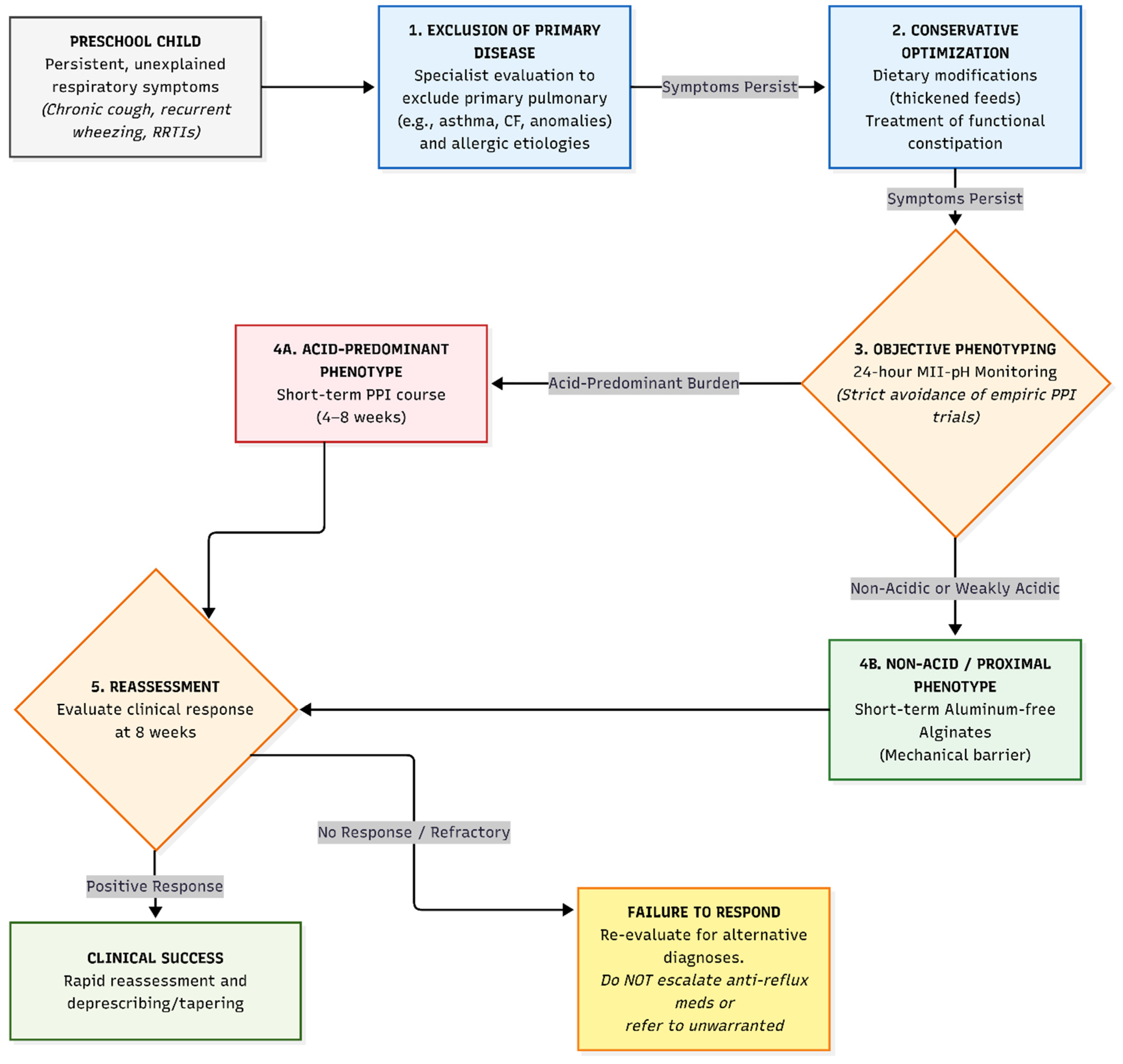
Synthesizing current evidence, we propose an expert-opinion-based diagnostic and therapeutic algorithm for the management of preschool children presenting with persistent, unexplained respiratory symptoms (Figure 4).
Limitations and Algorithm Positioning: This proposed algorithm is intended to serve as a specialized extension for a niche population (refractory cases after primary exclusion) and should be interpreted alongside primary guidelines, such as those from NASPGHAN/ESPGHAN and NICE. Clinicians must acknowledge the limitations of this approach, specifically the lack of robust pediatric normative data for advanced diagnostic indicators. Furthermore, an over-reliance on MII-pH data presents a potential risk of over-diagnosis, particularly given the limited support for MII-pH in perfectly predicting treatment responses. Therefore, if strict MII-pH criteria and clinical phenotyping do not align, practitioners must recognize when not to treat respiratory symptoms as reflux-related, avoiding unwarranted medical escalation.
  • Exclusion of Primary Disease: The absolute prerequisite is the systematic exclusion of primary pulmonary (asthma, anatomical anomalies) and allergic etiologies by a specialist.
  • Conservative Optimization: Initial management should focus on dietary modifications (thickened feeds) and treating functional constipation, which elevates intra-abdominal pressure. Notably, while positional therapies (e.g., head-of-bed elevation) are often suggested, recent EBM reviews show no proven respiratory efficacy for this measure in the 1–5-year age group, and it is restricted by safety guidelines [54].
  • Objective Phenotyping (MII-pH): We strongly advocate against prolonged empiric PPI trials. 24-h MII-pH monitoring should act as the primary objective gateway.
  • Phenotype-Driven Therapy: * Acid-Predominant: Short-term PPI course (4–8 weeks) followed by rapid reassessment and deprescribing.
    Non-Acid/Proximal Predominant: Short-term use of aluminum-free alginates as a mechanical barrier, with close clinical follow-up.
  • Reassessment: Failure to respond to targeted therapy within 8 weeks should trigger a re-evaluation for alternative diagnoses, rather than an endless escalation of anti-reflux medications or unwarranted surgical referral.

8. Conclusions and Future Directions

The intersection of GERD and pediatric respiratory disease, particularly recurrent preschool wheezing and chronic cough, is a complex, clinically significant entity driven by a dual pathophysiological mechanism: direct microaspiration and the distension-mediated vagal reflex arc. The central paradigm shift highlighted in this review is the recognition that this respiratory phenotype is predominantly fueled by weakly acidic or non-acidic reflux, rendering conventional pH-metry largely inadequate for accurate diagnosis.
Consequently, the clinical management of “respiratory reflux” must urgently transition from empiric therapeutic trials to phenotype-driven precision medicine. The routine, prolonged prescription of PPIs for isolated respiratory symptoms without objective verification is no longer justifiable. Evidence-based medicine demonstrates their lack of efficacy against non-acidic volume distension and highlights quantifiable, long-term risks, including microbiome dysbiosis and increased susceptibility to lower respiratory tract infections. While mechanical barriers, such as aluminum-free alginates, offer a rational physiological countermeasure for non-acidic and proximal reflux, their chronic application in the preschool demographic demands cautious clinical monitoring and ongoing reassessment to facilitate timely deprescribing.
Ultimately, while MII-pH monitoring stands as the current gold standard, future research must prioritize the pediatric validation of advanced diagnostic metrics, such as MNBI and the PSPW index (Lyon Consensus 2.0). Furthermore, rigorous, long-term randomized controlled trials are imperative to establish definitive safety profiles and efficacy cut-offs for both novel biomarkers and mechanical therapeutics in the vulnerable preschool population.

Author Contributions

Conceptualization: R.P. and P.K.; methodology: P.K. and R.P.; Formal analysis and investigation: P.K., T.S. and J.F.; writing—original draft preparation: P.K.; writing—review and editing: R.P., T.S. and P.K.; supervision: R.P. and J.F. All authors have read and agreed to the published version of the manuscript.

Funding

This research received no external funding.

Institutional Review Board Statement

Not applicable. This manuscript is a comprehensive review and does not involve primary research on human or animal subjects.

Informed Consent Statement

Not applicable.

Data Availability Statement

No new data were created or analyzed in this study. Data sharing is not applicable to this article.

Conflicts of Interest

The authors declare no conflicts of interest.

References

  1. Fainardi, V.; Santoro, A.; Caffarelli, C. Preschool Wheezing: Trajectories and Long-Term Treatment. Front. Pediatr. 2020, 8, 240. [Google Scholar] [CrossRef]
  2. Zhai, J.; Zou, Y.; Liu, J.; Jin, X.; Ma, C.; Li, J.; Guo, R.; Huang, B. Analysis of the Predicting Factors of Recurrent Wheezing in Infants. Ital. J. Pediatr. 2019, 45, 19. [Google Scholar] [CrossRef] [PubMed]
  3. Pavić, I.; Topalušić, I.; Močić Pavić, A.; Šarkanji Golub, R.; Hofman Jaeger, O.; Hojsak, I. Linking Gastroesophageal Reflux Characteristics to Airway Inflammation: Insights from Bronchoalveolar Lavage Cytology in Severe Preschool Wheeze. Life 2025, 15, 1561. [Google Scholar] [CrossRef]
  4. Cantarutti, A.; Barbiellini Amidei, C.; Valsecchi, C.; Scamarcia, A.; Corrao, G.; Gregori, D.; Giaquinto, C.; Ludvigsson, J.F.; Canova, C. Association of Treated and Untreated Gastroesophageal Reflux Disease in the First Year of Life with the Subsequent Development of Asthma. Int. J. Environ. Res. Public Health 2021, 18, 9633. [Google Scholar] [CrossRef]
  5. Zenzeri, L.; Quitadamo, P.; Tambucci, R.; Ummarino, D.; Poziello, A.; Miele, E.; Staiano, A. Role of Non-Acid Gastro-Esophageal Reflux in Children with Respiratory Symptoms. Pediatr. Pulmonol. 2017, 52, 669–674. [Google Scholar] [CrossRef]
  6. Pavić, I.; Šarkanji-Golub, R.; Hojsak, I. Diagnostic Utility of pH-MII Monitoring in Preschool Children with Recurrent Wheeze and Suspected Gastroesophageal Reflux Disease: A Prospective Study. Diagnostics 2023, 13, 3567. [Google Scholar] [CrossRef]
  7. Rosen, R.; Amirault, J.; Johnston, N.; Haver, K.; Khatwa, U.; Rubinstein, E.; Nurko, S. The Utility of Endoscopy and Multichannel Intraluminal Impedance Testing in Children with Cough and Wheezing. Pediatr. Pulmonol. 2014, 49, 1090–1096. [Google Scholar] [CrossRef]
  8. Gyawali, C.P.; Yadlapati, R.; Fass, R.; Katzka, D.; Pandolfino, J.; Savarino, E.; Sifrim, D.; Spechler, S.; Zerbib, F.; Fox, M.R.; et al. Updates to the Modern Diagnosis of GERD: Lyon Consensus 2.0. Gut 2024, 73, 361–371. [Google Scholar] [CrossRef]
  9. Abdallah, A.F.; El-Desoky, T.; Fathi, K.; Elkashef, W.F.; Zaki, A. Clinical Utility of Bronchoalveolar Lavage Pepsin in Diagnosis of Gastroesophageal Reflux among Wheezy Infants. Can. Respir. J. 2016, 2016, 9480843. [Google Scholar] [CrossRef] [PubMed]
  10. Borrelli, M.; Ponte, G.; Miele, E.; Maglione, M.; Caffarelli, C.; Santamaria, F. Preschool Wheezing and Gastro-Esophageal Reflux: --Causal or Casual Coincidence? Update from Literature. Children 2021, 8, 180. [Google Scholar] [CrossRef] [PubMed]
  11. Strachan, T.; Melter, J.; Barabasová, A.; Miškovská, M.; Ferenc, P.; Fábry, J.; Bánovčin, P. Pepsín v sekrétoch horných dýchacích ciest u detí ako marker extraezofágového refluxu. Ceskoslov. Pediatr. 2017, 72, 176–181. [Google Scholar]
  12. Lupu, V.V.; Stefanescu, G.; Buga, A.M.L.; Forna, L.; Tarca, E.; Starcea, I.M.; Mihai, C.M.; Florescu, L.; Cernomaz, A.T.; Mocanu, A.; et al. Is There a Potential Link between Gastroesophageal Reflux Disease and Recurrent Respiratory Tract Infections in Children? Diagnostics 2023, 13, 2310. [Google Scholar] [CrossRef]
  13. Chang, A.B.; Oppenheimer, J.J.; Weinberger, M.M.; Rubin, B.K.; Grant, C.C.; Weir, K.; Irwin, R.S. Management of Children with Chronic Wet Cough and Protracted Bacterial Bronchitis: CHEST Guideline and Expert Panel Report. Chest 2017, 151, 884–890. [Google Scholar] [CrossRef]
  14. Marseglia, G.L.; Manti, S.; Chiappini, E.; Brambilla, I.; Caffarelli, C.; Calvani, M.; Cardinale, F.; Cravidi, C.; Duse, M.; Martelli, A.; et al. Acute Cough in Children and Adolescents: A Systematic Review and a Practical Algorithm by the Italian Society of Pediatric Allergy and Immunology. Allergol. Immunopathol. 2021, 49, 155–169. [Google Scholar] [CrossRef]
  15. Marseglia, G.L.; Manti, S.; Chiappini, E.; Brambilla, I.; Caffarelli, C.; Calvani, M.; Cardinale, F.; Cravidi, C.; Duse, M.; Martelli, A.; et al. Chronic Cough in Childhood: A Systematic Review for Practical Guidance by the Italian Society of Pediatric Allergy and Immunology. Allergol. Immunopathol. 2021, 49, 133–154. [Google Scholar] [CrossRef] [PubMed]
  16. Hamed, N.F.; AlAlwan, A.A.; Alqahtani, F.J.; Alanazi, F.; Albalawi, M.M. The Bidirectional Relationship Between Gastroesophageal Reflux Disease (GERD) and Asthma in Pediatric and Adult Populations: A Systematic Review. Cureus 2025, 17, e99995. [Google Scholar] [CrossRef]
  17. Özdemir, P.; Erdinç, M.; Vardar, R.; Veral, A.; Akyıldız, S.; Özdemir, Ö.; Bor, S. The Role of Microaspiration in the Pathogenesis of Gastroesophageal Reflux-Related Chronic Cough. J. Neurogastroenterol. Motil. 2017, 23, 41–48. [Google Scholar] [CrossRef]
  18. Zhang, L.; Aierken, A.; Zhang, M.; Qiu, Z. Pathogenesis and Management of Gastroesophageal Reflux Disease-Associated Cough: A Narrative Review. J. Thorac. Dis. 2023, 15, 2314–2323. [Google Scholar] [CrossRef]
  19. Lee, A.S.; Lee, J.S.; He, Z.; Ryu, J.H. Reflux-Aspiration in Chronic Lung Disease. Ann. Am. Thorac. Soc. 2020, 17, 155–164. [Google Scholar] [CrossRef] [PubMed]
  20. Johnston, N.; Dettmar, P.W.; Bishwokarma, B.; Lively, M.O.; Koufman, J.A. Activity/Stability of Human Pepsin: Implications for Reflux Attributed Laryngeal Disease. Laryngoscope 2007, 117, 1036–1039. [Google Scholar] [CrossRef] [PubMed]
  21. Lechien, J.R.; Hans, S.; Simon, F.; Horoi, M.; Calvo-Henriquez, C.; Chiesa-Estomba, C.M.; Mayo-Yáñez, M.; Bartel, R.; Piersiala, K.; Nguyen, Y.; et al. Association Between Laryngopharyngeal Reflux and Media Otitis: A Systematic Review. Otol. Neurotol. 2021, 42, e801–e814. [Google Scholar] [CrossRef]
  22. Johnston, N.; Wells, C.W.; Blumin, J.H.; Toohill, R.J.; Merati, A.L. Receptor-Mediated Uptake of Pepsin by Laryngeal Epithelial Cells. Ann. Otol. Rhinol. Laryngol. 2007, 116, 934–938. [Google Scholar] [CrossRef] [PubMed]
  23. Niu, K.; Guo, C.; Teng, S.; Zhou, D.; Yu, S.; Yin, W.; Wang, P.; Zhu, W.; Duan, M. Pepsin Promotes Laryngopharyngeal Neoplasia by Modulating Signaling Pathways to Induce Cell Proliferation. PLoS ONE 2020, 15, e0227408. [Google Scholar] [CrossRef]
  24. West, J.B. Henry Cavendish (1731–1810): Hydrogen, Carbon Dioxide, Water, and Weighing the World. Am. J. Physiol.-Lung Cell. Mol. Physiol. 2014, 307, L1–L6. [Google Scholar] [CrossRef]
  25. Doukas, P.G.; Vageli, D.P.; Sasaki, C.T.; Judson, B.L. Pepsin Promotes Activation of Epidermal Growth Factor Receptor and Downstream Oncogenic Pathways, at Slightly Acidic and Neutral pH, in Exposed Hypopharyngeal Cells. Int. J. Mol. Sci. 2021, 22, 4275. [Google Scholar] [CrossRef]
  26. Moore, J.M.; Vaezi, M.F. Extraesophageal Manifestations of Gastroesophageal Reflux Disease: Real or Imagined? Curr. Opin. Gastroenterol. 2010, 26, 389–394. [Google Scholar] [CrossRef] [PubMed]
  27. Chen, S.; Wang, M.; Zhang, S.; Huang, X.; Sui, X.; Li, D.; Zhong, C.; Wu, W. The Complexity of Mucosal Damage in Gastroesophageal Airway Reflux Disease: A Molecular Perspective. Gastroenterol. Endosc. 2025, 3, 39–46. [Google Scholar] [CrossRef]
  28. Solidoro, P.; Patrucco, F.; Fagoonee, S.; Pellicano, R. Asthma and Gastroesophageal Reflux Disease: A Multidisciplinary Point of View. Minerva Med. 2017, 108, 350–356. [Google Scholar] [CrossRef]
  29. Orishchak, O.; Moise, A.; Al-Osamey, F.; Kaspy, K.; Daniel, S.J. Lipid-Laden Macrophage Index as a Marker of Aspiration in Children, Is It Reliable? A Scoping Review. Int. J. Pediatr. Otorhinolaryngol. 2024, 186, 112151. [Google Scholar] [CrossRef]
  30. Lawlor, C.M.; Choi, S.S. Lipid-Laden Macrophage Index as a Diagnostic Tool for Pediatric Aspiration: A Systematic Review. OTO Open 2023, 7, e33. [Google Scholar] [CrossRef]
  31. Zhu, Y.; Choi, D.; Somanath, P.R.; Zhang, D. Lipid-Laden Macrophages in Pulmonary Diseases. Cells 2024, 13, 889. [Google Scholar] [CrossRef]
  32. Canning, B.J.; Mazzone, S.B. Reflex Mechanisms in Gastroesophageal Reflux Disease and Asthma. Am. J. Med. 2003, 115, 45S–48S. [Google Scholar] [CrossRef]
  33. Nikaki, K.; Lee, C.; Ustaoglu, A.; Alessandrella, A.; Staiano, A.; Woodland, P.; Sifrim, D. Esophageal Mucosa Innervation in Children with Nonerosive Reflux Disease. Am. J. Gastroenterol. 2021, 116, 1727–1729. [Google Scholar] [CrossRef] [PubMed]
  34. Vaezi, M.F.; Katzka, D.; Zerbib, F. Extraesophageal Symptoms and Diseases Attributed to GERD: Where Is the Pendulum Swinging Now? Clin. Gastroenterol. Hepatol. 2018, 16, 1018–1029. [Google Scholar] [CrossRef] [PubMed]
  35. Herregods, T.V.K.; Pauwels, A.; Jafari, J.; Sifrim, D.; Bredenoord, A.J.; Tack, J.; Smout, A.J.P.M. Determinants of Reflux-Induced Chronic Cough. Gut 2017, 66, 2057–2062. [Google Scholar] [CrossRef] [PubMed]
  36. Bertin, L.; Savarino, V.; Marabotto, E.; Ghisa, M.; de Bortoli, N.; Savarino, E.V. Pathophysiology of Gastroesophageal Reflux Disease. Digestion 2026, 107, 185–201. [Google Scholar] [CrossRef]
  37. Zachariah, R.A.; Goo, T.; Lee, R.H. Mechanism and Pathophysiology of Gastroesophageal Reflux Disease. Gastrointest. Endosc. Clin. N. Am. 2020, 30, 209–226. [Google Scholar] [CrossRef]
  38. Zerbib, F.; Bredenoord, A.J.; Fass, R.; Kahrilas, P.J.; Roman, S.; Savarino, E.; Sifrim, D.; Vaezi, M.; Yadlapati, R.; Gyawali, C.P. ESNM/ANMS Consensus Paper: Diagnosis and Management of Refractory Gastro-Esophageal Reflux Disease. Neurogastroenterol. Motil. 2021, 33, e14075. [Google Scholar] [CrossRef]
  39. Blondeau, K.; Mertens, V.; Dupont, L.; Pauwels, A.; Farré, R.; Malfroot, A.; De Wachter, E.; De Schutter, I.; Hauser, B.; Vandenplas, Y.; et al. The Relationship between Gastroesophageal Reflux and Cough in Children with Chronic Unexplained Cough Using Combined Impedance-pH-Manometry Recordings. Pediatr. Pulmonol. 2011, 46, 286–294. [Google Scholar] [CrossRef]
  40. Rosen, R.; Amirault, J.; Giligan, E.; Khatwa, U.; Nurko, S. Intraesophageal Pressure Recording Improves the Detection of Cough during Multichannel Intraluminal Impedance Testing in Children. J. Pediatr. Gastroenterol. Nutr. 2014, 58, 22–26. [Google Scholar] [CrossRef]
  41. De Bruyne, P.; Ito, S. Toxicity of Long-Term Use of Proton Pump Inhibitors in Children. Arch. Dis. Child. 2018, 103, 78–82. [Google Scholar] [CrossRef]
  42. Pop, R.S.; Farcău, D.; Chiperi, L.E.; Dumitrașcu, D.L. The Utility of Novel pH-Impedance Monitoring Parameters (PSPW Index and MNBI) in Pediatric Gastroesophageal Reflux Disease Phenotypes-A Systematic Review. J. Clin. Med. 2024, 13, 3351. [Google Scholar] [CrossRef]
  43. Rosen, R.; Vandenplas, Y.; Singendonk, M.; Cabana, M.; DiLorenzo, C.; Gottrand, F.; Gupta, S.; Langendam, M.; Staiano, A.; Thapar, N.; et al. Pediatric Gastroesophageal Reflux Clinical Practice Guidelines: Joint Recommendations of the North American Society for Pediatric Gastroenterology, Hepatology, and Nutrition and the European Society for Pediatric Gastroenterology, Hepatology, and Nutrition. J. Pediatr. Gastroenterol. Nutr. 2018, 66, 516–554. [Google Scholar] [CrossRef] [PubMed]
  44. Sui, H.; Shen, H.; Zhang, C.; Wang, M.; Zhen, Z.; Zhang, J. Elevated Saliva Pepsin Concentration as a Risk Factor for Asthma in Children with Allergic Rhinitis: A Preliminary Study. J. Asthma Allergy 2024, 17, 391–397. [Google Scholar] [CrossRef] [PubMed]
  45. Chen, J.W.; Vela, M.F.; Peterson, K.A.; Carlson, D.A. AGA Clinical Practice Update on the Diagnosis and Management of Extraesophageal Gastroesophageal Reflux Disease: Expert Review. Clin. Gastroenterol. Hepatol. 2023, 21, 1414–1421.e3. [Google Scholar] [CrossRef]
  46. Han, F.; Li, X.; Song, Z.; Xie, J.; Wang, N.; Yao, J. The Association between Salivary Pepsin and Gastroesophageal Reflux Disease: A Meta-Analysis. Neurogastroenterol. Motil. 2024, 36, e14905. [Google Scholar] [CrossRef]
  47. Parikh, S.; Brownlee, I.A.; Robertson, A.G.; Manning, N.T.; Johnson, G.E.; Brodlie, M.; Corris, P.A.; Ward, C.; Pearson, J.P. Are the Enzymatic Methods Currently Being Used to Measure Bronchoalveolar Lavage Bile Salt Levels Fit for Purpose? J. Heart Lung Transplant. 2013, 32, 418–423. [Google Scholar] [CrossRef]
  48. Mahoney, L.B.; Esther, C.R.; May, K.; Rosen, R. Metabolomic Profiling of Extraesophageal Reflux Disease in Children. Clin. Transl. Sci. 2021, 14, 2025–2033. [Google Scholar] [CrossRef]
  49. De Vos, N.V.; Trelcat, A.; Antoine, M.; Dahma, H.; Muls, V.; Hans, S.; Saussez, S.; Lechien, J.R. Performance Study of Noninvasive Salivary Biomarkers in Laryngopharyngeal Reflux. J. Appl. Lab. Med. 2026, 11, 307–319. [Google Scholar] [CrossRef] [PubMed]
  50. Orel, R.; Benninga, M.A.; Broekaert, I.J.; Gottrand, F.; Papadopoulou, A.; Ribes-Koninckx, C.; Thomson, M.; Wilschanski, M.; Thapar, N. Drugs in Focus: Proton Pump Inhibitors. J. Pediatr. Gastroenterol. Nutr. 2021, 72, 645–653. [Google Scholar] [CrossRef]
  51. Dipasquale, V.; Cicala, G.; Spina, E.; Romano, C. A Narrative Review on Efficacy and Safety of Proton Pump Inhibitors in Children. Front. Pharmacol. 2022, 13, 839972. [Google Scholar] [CrossRef]
  52. Lassalle, M.; Zureik, M.; Dray-Spira, R. Proton Pump Inhibitor Use and Risk of Serious Infections in Young Children. JAMA Pediatr. 2023, 177, 1028–1038. [Google Scholar] [CrossRef]
  53. Alla, D.; Shah, D.J.; Seepana, M.; Salian, R.B.; Alla, S.S.M.; Krishna Mohanan, M.; Sabıroğlu, M.; Vegesna, M.S.S.; Singh, A.; Gupta, S.; et al. Safety of Proton Pump Inhibitors in Pediatric Population: A Systematic Review. Glob. Pediatr. Health 2024, 11, 2333794X241248967. [Google Scholar] [CrossRef]
  54. Zhang, Y.J.; Connearney, S.; Hester, L.; Du, M.; Catacora, A.; Akkara, A.; Wen, A.; Bry, L.; Alm, E.J.; Rosen, R. Longitudinal Microbiome Changes in Children Exposed to Proton Pump Inhibitors. Clin. Transl. Gastroenterol. 2024, 15, e1. [Google Scholar] [CrossRef]
  55. Yalçın, H.T.; Yalçın, N.; Allegaert, K. Real-World Safety Profile of Proton Pump Inhibitors in Infants as Reported in the FDA Adverse Event Reporting System (FAERS): Tiny Tummies, Key Decisions. Pharmaceuticals 2025, 18, 730. [Google Scholar] [CrossRef]
  56. Pizzorni, N.; Ambrogi, F.; Eplite, A.; Rama, S.; Robotti, C.; Lechien, J.; Schindler, A. Magnesium Alginate versus Proton Pump Inhibitors for the Treatment of Laryngopharyngeal Reflux: A Non-Inferiority Randomized Controlled Trial. Eur. Arch. Otorhinolaryngol. 2022, 279, 2533–2542. [Google Scholar] [CrossRef] [PubMed]
  57. Abdulkader, Z.M.; Woodley, F.; Lu, P.L. Extra-Esophageal Manifestations of Pediatric Gastroesophageal Reflux Disease: Updated Review. Curr. Treat. Options Pediatr. 2021, 7, 217–228. [Google Scholar] [CrossRef]
  58. Ablaza, T.J.J.; Crisostomo, E.A.; Uy, M.E.V. A Systematic Review on the Efficacy and Safety of Alginate–Based Liquid Formulations in Reducing Gastroesophageal Reflux in Neonates and Infants. Acta Med. Philipp. 2024, 58, 55–63. [Google Scholar] [CrossRef]
  59. Simon, M.; Levy, E.I.; Vandenplas, Y. Safety Considerations When Managing Gastro-Esophageal Reflux Disease in Infants. Expert Opin. Drug Saf. 2021, 20, 37–49. [Google Scholar] [CrossRef] [PubMed]
  60. Mostafa, I.A.; Hader, H.A.; Khan, S.A.; Hilal, A.M.; Gathradi, M.A.; Ibrahim, A.H.M. Anti-Reflux Surgery in Neonates and Infants: Analysis of Indications, Outcomes, and Link to Mortality among Primary and Secondary Gastroesophageal Reflux Patients. Ann. Pediatr. Surg. 2022, 18, 45. [Google Scholar] [CrossRef]
  61. Al-Refaie, M.A.; Alsurmi, M.M.; Obadiel, Y.A.; Jowah, H.M.; Alsharafy, K.M. Fundoplication for Pediatric Gastroesophageal Reflux Disease: Indications, Techniques, and Outcomes. Cureus 2024, 16, e72930. [Google Scholar] [CrossRef] [PubMed]
  62. Mitri, M.D.; Vastano, M.; Carmine, A.D.; Oriani, E.; Thomas, E.; Bisanti, C.; D’Antonio, S.; Catania, V.D.; Collautti, E.; Gargano, T.; et al. Surgical Management of Gastroesophageal Reflux in Neurologically Impaired Children: Fundoplication vs. Total Esophagogastric Dissociation. J. Clin. Med. 2025, 14, 1058. [Google Scholar] [CrossRef] [PubMed]
Figure 1. PRISMA-adapted flow diagram illustrating the study selection process. The search strategy focused on identifying studies with objective diagnostic criteria (MII-pH, BAL cytology, pepsin analysis) published between January 2011 and January 2026. Records relying solely on subjective symptom reporting or lacking instrumental verification were excluded to ensure the validity of the pathophysiological update.
Jor 06 00010 g001
Figure 2. Schematic representation of the dual pathophysiological nexus between GERD and bronchial obstruction. The left panel illustrates the Direct Microaspiration (Reflux) theory, where gastric contents breach the airways, causing pepsin-mediated neutrophilic inflammation. The right panel depicts the Indirect Vagal Reflex theory, demonstrating how esophageal distension triggers a neurogenic bronchospastic response without direct airway contamination. (Original illustration).
Jor 06 00010 g002
Figure 3. The diagnostic superiority of MII-pH monitoring. The schematic illustrates why MII-pH is analogous to “high-definition imaging” compared to standard pH-metry. It highlights MII-pH’s ability to capture non-acidic reflux, accurately determine its proximal extent into the airways, differentiate the physical state of the refluxate (liquid vs. mixed), and precisely correlate these episodes with respiratory symptoms.
Jor 06 00010 g003
Figure 4. Proposed evidence-based diagnostic and therapeutic algorithm for preschool children presenting with persistent, unexplained respiratory symptoms and suspected extra-esophageal reflux. The workflow emphasizes the fundamental exclusion of primary respiratory etiologies, the critical necessity of objective phenotyping via Multichannel Intraluminal Impedance-pH (MII-pH) monitoring over empiric proton pump inhibitor (PPI) trials, and the implementation of cautious, phenotype-driven therapy (acid suppression vs. mechanical barrier). Mandatory reassessment at 8 weeks aims to ensure timely deprescribing in responders and to prevent unwarranted medical escalation or surgical referral in refractory cases.
Jor 06 00010 g004
Table 1. Distinguishing Clinical Features of GERD vs. LPR in Preschool Children.
FeatureGERD (Gastroesophageal Reflux)LPR (Laryngopharyngeal Reflux)
Primary DefectLower Esophageal Sphincter (LES) DysfunctionUpper Esophageal Sphincter (UES) Dysfunction
Dominant SymptomsHeartburn, Regurgitation (if verbal), Epigastric painHoarseness, Chronic Cough, Throat Clearing, Globus sensation
Timing of EpisodesPredominantly Nocturnal/Supine positionPredominantly Daytime/Upright position
Respiratory LinkLower Airway (Wheezing, Bronchospasm)Upper Airway (Stridor, Laryngitis, Subglottic stenosis)
PathophysiologyProlonged acid exposure (Esophagitis)Intermittent micro-aspiration (Laryngeal irritation)
Diagnostic SensitivityHigh with standard pH-metryRequires MII-pH (proximal events) or Pepsin detection
Legend: GERD, Gastroesophageal Reflux Disease; LPR, Laryngopharyngeal Reflux; LES, Lower Esophageal Sphincter; UES, Upper Esophageal Sphincter; MII-pH, Multichannel Intraluminal Impedance-pH.
Disclaimer/Publisher’s Note: The statements, opinions and data contained in all publications are solely those of the individual author(s) and contributor(s) and not of MDPI and/or the editor(s). MDPI and/or the editor(s) disclaim responsibility for any injury to people or property resulting from any ideas, methods, instructions or products referred to in the content.