
Surface Modification of Titanium Implants with Chitosan–Hydroxyapatite Composite: A Review on Osseointegration and Bioactivity †

Abstract
1. Introduction
2. Osseointegration and Its Correlation with Surface Modification
3. Materials and Methods
4. Results
5. Discussion
6. Limitations and Future Scope of Ti Surface Modification
7. Conclusions
Author Contributions
Funding
Institutional Review Board Statement
Informed Consent Statement
Data Availability Statement
Conflicts of Interest
References
- Prasad, K.; Bazaka, O.; Chua, M.; Rochford, M.; Fedrick, L.; Spoor, J.; Symes, R.; Tieppo, M.; Collins, C.; Cao, A.; et al. Metallic Biomaterials: Current Challenges and Opportunities. Materials 2017, 10, 884. [Google Scholar] [CrossRef]
- Zindani, D.; Kumar, K.; Paulo Davim, J. Metallic biomaterials—A review. In Mechanical Behaviour of Biomaterials; Elsevier: Amsterdam, The Netherlands, 2019; pp. 83–99. [Google Scholar] [CrossRef]
- Niinomi, M. Design and development of metallic biomaterials with biological and mechanical biocompatibility. J. Biomed. Mater. Res. A 2019, 107, 944–954. [Google Scholar] [CrossRef]
- Smeets, R.; Stadlinger, B.; Schwarz, F.; Beck-Broichsitter, B.; Jung, O.; Precht, C.; Kloss, F.; Gröbe, A.; Heiland, M.; Ebker, T. Impact of Dental Implant Surface Modifications on Osseointegration. Biomed. Res. Int. 2016, 2016, 6285620. [Google Scholar] [CrossRef]
- Yang, T.; Qian, S.; Qiao, Y.; Liu, X. Cytocompatibility and antibacterial activity of titania nanotubes incorporated with gold nanoparticles. Colloids Surf. B Biointerfaces 2016, 145, 597–606. [Google Scholar] [CrossRef]
- Kuroda, K.; Okido, M. Hydroxyapatite coating of titanium implants using hydroprocessing and evaluation of their osteoconductivity. Bioinorg. Chem. Appl. 2012, 2012, 730693. [Google Scholar] [CrossRef]
- Mehdikhani-Nahrkhalaji, M.; Fathi, M.H.; Mortazavi, V.; Mousavi, S.B.; Hashemi-Beni, B.; Razavi, S.M. Novel nanocomposite coating for dental implant applications in vitro and in vivo evaluation. J. Mater. Sci. Mater. Med. 2012, 23, 485–495. [Google Scholar] [CrossRef] [PubMed]
- Park, Y.-J.; Choi, K.-H.; Hahn, B.-D.; Lee, Y.-C.; Song, J.-Y.; Park, Y.-T.; Kim, S.-G. New Bone Formation Between Bare Titanium Surface and Hydroxyapatite Coating by the Aerosol Deposition Technique in the Nasal Mucosal Penetration Model. J. Craniofacial Surg. 2013, 24, 632–635. [Google Scholar] [CrossRef] [PubMed]
- Ozhukil Kollath, V.; Chen, Q.; Closset, R.; Luyten, J.; Traina, K.; Mullens, S.; Boccaccini, A.; Cloots, R. AC vs. DC electrophoretic deposition of hydroxyapatite on titanium. J. Eur. Ceram. Soc. 2013, 33, 2715–2721. [Google Scholar] [CrossRef]
- Hung, K.-Y.; Lo, S.-C.; Shih, C.-S.; Yang, Y.-C.; Feng, H.-P.; Lin, Y.-C. Titanium surface modified by hydroxyapatite coating for dental implants. Surf. Coat. Technol. 2013, 231, 337–345. [Google Scholar] [CrossRef]
- Bryington, M.S.; Hayashi, M.; Kozai, Y.; Vandeweghe, S.; Andersson, M.; Wennerberg, A.; Jimbo, R. The influence of nano hydroxyapatite coating on osseointegration after extended healing periods. Dent. Mater. 2013, 29, 514–520. [Google Scholar] [CrossRef] [PubMed]
- Jing, W.; Zhang, M.; Jin, L.; Zhao, J.; Gao, Q.; Ren, M.; Fan, Q. Assessment of osteoinduction using a porous hydroxyapatite coating prepared by micro-arc oxidation on a new titanium alloy. Int. J. Surg. 2015, 24, 51–56. [Google Scholar] [CrossRef]
- Ding, M.; Shi, J.; Wang, W.; Li, D.; Tian, L. Early osseointegration of micro-arc oxidation coated titanium alloy implants containing Ag: A histomorphometric study. BMC Oral Health 2022, 22, 628. [Google Scholar] [CrossRef]
- Shi, Y.Y.; Li, M.; Liu, Q.; Jia, Z.J.; Xu, X.C.; Cheng, Y.; Zheng, Y.F. Electrophoretic deposition of graphene oxide reinforced chitosan–hydroxyapatite nanocomposite coatings on Ti substrate. J. Mater. Sci. Mater. Med. 2016, 27, 48. [Google Scholar] [CrossRef]
- Balagangadharan, K.; Dhivya, S.; Selvamurugan, N. Chitosan based nanofibers in bone tissue engineering. Int. J. Biol. Macromol. 2017, 104, 1372–1382. [Google Scholar] [CrossRef]
- Ahsan, S.M.; Thomas, M.; Reddy, K.K.; Sooraparaju, S.G.; Asthana, A.; Bhatnagar, I. Chitosan as biomaterial in drug delivery and tissue engineering. Int. J. Biol. Macromol. 2018, 110, 97–109. [Google Scholar] [CrossRef] [PubMed]
- Palla-Rubio, B.; Araújo-Gomes, N.; Fernández-Gutiérrez, M.; Rojo, L.; Suay, J.; Gurruchaga, M.; Goñi, I. Synthesis and characterization of silica-chitosan hybrid materials as antibacterial coatings for titanium implants. Carbohydr. Polym. 2019, 203, 331–341. [Google Scholar] [CrossRef] [PubMed]
- Abinaya, B.; Prasith, T.P.; Ashwin, B.; Viji Chandran, S.; Selvamurugan, N. Chitosan in Surface Modification for Bone Tissue Engineering Applications. Biotechnol. J. 2019, 14, e1900171. [Google Scholar] [CrossRef] [PubMed]
- Jiang, T.; Zhang, Z.; Zhou, Y.; Liu, Y.; Wang, Z.; Tong, H.; Shen, X.; Wang, Y. Surface Functionalization of Titanium with Chitosan/Gelatin via Electrophoretic Deposition: Characterization and Cell Behavior. Biomacromolecules 2010, 11, 1254–1260. [Google Scholar] [CrossRef]
- Kung, S.; Devlin, H.; Fu, E.; Ho, K.-Y.; Liang, S.-Y.; Hsieh, Y.-D. The osteoinductive effect of chitosan-collagen composites around pure titanium implant surfaces in rats. J. Periodontal Res. 2011, 46, 126–133. [Google Scholar] [CrossRef]
- Peng, Z.; Ao, H.; Wang, L.; Guo, S.; Tang, T. Quaternised chitosan coating on titanium provides a self-protective surface that prevents bacterial colonisation and implant-associated infections. RSC Adv. 2015, 5, 54304–54311. [Google Scholar] [CrossRef]
- Zhang, T.; Qin, X.; Gao, Y.; Kong, D.; Jiang, Y.; Cui, X.; Guo, M.; Chen, J.; Chang, F.; Zhang, M.; et al. Functional chitosan gel coating enhances antimicrobial properties and osteogenesis of titanium alloy under persistent chronic inflammation. Front. Bioeng. Biotechnol. 2023, 11, 1118487. [Google Scholar] [CrossRef]
- Zanca, C.; Carbone, S.; Patella, B.; Lopresti, F.; Aiello, G.; Brucato, V.; Pavia, F.C.; La Carrubba, V.; Inguanta, R. Composite Coatings of Chitosan and Silver Nanoparticles Obtained by Galvanic Deposition for Orthopedic Implants. Polymers 2022, 14, 3915. [Google Scholar] [CrossRef] [PubMed]
- Gaafar, M.S.; Yakout, S.M.; Barakat, Y.F.; Sharmoukh, W. Electrophoretic deposition of hydroxyapatite/chitosan nanocomposites: The effect of dispersing agents on the coating properties. RSC Adv. 2022, 12, 27564–27581. [Google Scholar] [CrossRef] [PubMed]
- Esteves, G.M.; Esteves, J.; Resende, M.; Mendes, L.; Azevedo, A.S. Antimicrobial and Antibiofilm Coating of Dental Implants—Past and New Perspectives. Antibiotics 2022, 11, 235. [Google Scholar] [CrossRef]
- Suo, L.; Jiang, N.; Wang, Y.; Wang, P.; Chen, J.; Pei, X.; Wang, J.; Wan, Q. The enhancement of osseointegration using a graphene oxide/chitosan/hydroxyapatite composite coating on titanium fabricated by electrophoretic deposition. J. Biomed. Mater. Res. B Appl. Biomater. 2019, 107, 635–645. [Google Scholar] [CrossRef]
- Zhang, T.; Zhang, X.; Mao, M.; Li, J.; Wei, T.; Sun, H. Chitosan/hydroxyapatite composite coatings on porous Ti6Al4V titanium implants: In vitro and in vivo studies. J. Periodontal Implant. Sci. 2020, 50, 392. [Google Scholar] [CrossRef] [PubMed]
- Pawlik, A.; Rehman, M.A.U.; Nawaz, Q.; Bastan, F.E.; Sulka, G.D.; Boccaccini, A.R. Fabrication and characterization of electrophoretically deposited chitosan-hydroxyapatite composite coatings on anodic titanium dioxide layers. Electrochim. Acta 2019, 307, 465–473. [Google Scholar] [CrossRef]
- Zarif, M.E.; Bita, B.; Yehia-Alexe, S.A.; Negut, I.; Gradisteanu Pircalabioru, G.; Andronescu, E.; Groza, A. Biological and Physicochemical Analysis of Sr-Doped Hydroxyapatite/Chitosan Composite Layers. Polymers 2024, 16, 1922. [Google Scholar] [CrossRef] [PubMed]
- Anil, S.; Anand, P.S.; Alghamdi, H.; Janse, J.A. Dental Implant Surface Enhancement and Osseointegration. In Implant Dentistry—A Rapidly Evolving Practice; Ilser, T., Ed.; InTech: Vienna, Austria, 2011. [Google Scholar] [CrossRef]
- Shi, X.; Xu, L.; Violin, K.B.; Lu, S. Improved osseointegration of long-term stored SLA implant by hydrothermal sterilization. J. Mech. Behav. Biomed. Mater. 2016, 53, 312–319. [Google Scholar] [CrossRef]
- Terheyden, H.; Lang, N.P.; Bierbaum, S.; Stadlinger, B. Osseointegration—Communication of cells. Clin. Oral Implant. Res. 2011, 23, 1127–1135. [Google Scholar] [CrossRef] [PubMed]
- Dehghanpour, A.A.; Rouhi, G. A Critical Review on the Effects of Dental Implants Micro-roughness on Osseointegration. Biomed. Mater. Devices 2025. [Google Scholar] [CrossRef]
- Guglielmotti, M.B.; Olmedo, D.G.; Cabrini, R.L. Research on implants and osseointegration. Periodontology 2019, 79, 178–189. [Google Scholar] [CrossRef]
- Osman, M.A.; Alamoush, R.A.; Kushnerev, E.; Seymour, K.G.; Watts, D.C.; Yates, J.M. Biological response of epithelial and connective tissue cells to titanium surfaces with different ranges of roughness: An in-vitro study. Dent. Mater. 2022, 38, 1777–1788. [Google Scholar] [CrossRef] [PubMed]
- Alshadidi, A.A.F.; Dommeti, V.K.; Aldosari, L.I.N.; Bin Hassan, S.A.; Okshah, A.; Merdji, A.; Roy, S. Influence of surface texturing and coatings on mechanical properties and integration with bone tissue: An in silico study. Front. Bioeng. Biotechnol. 2024, 12, 1439262. [Google Scholar] [CrossRef] [PubMed]
- Herrero-Climent, M.; Lázaro, P.; Vicente Rios, J.; Lluch, S.; Marqués, M.; Guillem-Martí, J.; Gil, F.J. Influence of acid-etching after grit-blasted on osseointegration of titanium dental implants: In vitro and in vivo studies. J. Mater. Sci. Mater. Med. 2013, 24, 2047–2055. [Google Scholar] [CrossRef] [PubMed]
- Teulé-Trull, M.; Altuna, P.; Arregui, M.; Rodriguez-Ciurana, X.; Aparicio, C. Antibacterial coatings for dental implants: A systematic review. Dent. Mater. 2025, 41, 229–247. [Google Scholar] [CrossRef]
- Oleshko, O.; Liubchak, I.; Husak, Y.; Korniienko, V.; Yusupova, A.; Oleshko, T.; Banasiuk, R.; Szkodo, M.; Matros-Taranets, I.; Kazek-Kęsik, A.; et al. In vitro biological characterization of silver-doped anodic oxide coating on titanium. Materials 2020, 13, 4359. [Google Scholar] [CrossRef]
- Page, M.J.; McKenzie, J.E.; Bossuyt, P.M.; Boutron, I.; Hoffmann, T.C.; Mulrow, C.D.; Shamseer, L.; Tetzlaff, J.M.; Akl, E.A.; Brennan, S.E.; et al. The PRISMA 2020 statement: An updated guideline for reporting systematic reviews. BMJ 2021, 372, n71. [Google Scholar] [CrossRef]
- Stevanović, M.; Djošić, M.; Janković, A.; Nešović, K.; Kojić, V.; Stojanović, J.; Grujić, S.; Bujagić, I.M.; Rhee, K.Y.; Mišković-Stanković, V. Assessing the Bioactivity of Gentamicin-Preloaded Hydroxyapatite/Chitosan Composite Coating on Titanium Substrate. ACS Omega 2020, 5, 15433–15445. [Google Scholar] [CrossRef]
- Li, B.; Zhao, P.; Liu, S.; Wang, D.; Li, H.; Liang, C.; Wang, H. Preparation and Properties of Hydroxyapatite/Chitosan Coatings on Ti Surface by Different Electrochemical Methods. J. Mater. Eng. Perform. 2025, 34, 7946–7954. [Google Scholar] [CrossRef]
- Mishra, R.R.; Kumar, R.; Sahoo, A.K.; Panda, A. Machinability behaviour of biocompatible Ti-6Al-4V ELI titanium alloy under flood cooling environment. Mater. Today Proc. 2020, 23, 536–540. [Google Scholar] [CrossRef]
- Km, R.; Sahoo, A.K.; Routara, B.C.; Panda, A.; Kumar, R. Study on machinability characteristics of novel additive manufactured titanium alloy (Ti-6Al-4V) fabricated by direct metal laser sintering. Proc. Inst. Mech. Eng. C J. Mech. Eng. Sci. 2023, 237, 865–885. [Google Scholar] [CrossRef]
- Han, X.; Ma, J.; Tian, A.; Wang, Y.; Li, Y.; Dong, B.; Tong, X.; Ma, X. Surface modification techniques of titanium and titanium alloys for biomedical orthopaedics applications: A review. Colloids Surf. B Biointerfaces 2023, 227, 113339. [Google Scholar] [CrossRef]
- Frosch, S.; Buchhorn, G.H. Considerations on the animal model and the biomechanical test arrangements for assessing the osseous integration of orthopedic and dental implants. MethodsX 2021, 8, 101352. [Google Scholar] [CrossRef] [PubMed]
- Bai, X.; Sandukas, S.; Appleford, M.; Ong, J.L.; Rabiei, A. Antibacterial effect and cytotoxicity of Ag-doped functionally graded hydroxyapatite coatings. J. Biomed. Mater. Res. B Appl. Biomater. 2012, 100, 553–561. [Google Scholar] [CrossRef]
- Fletcher, J.W.A.; Williams, S.; Whitehouse, M.R.; Gill, H.S.; Preatoni, E. Juvenile bovine bone is an appropriate surrogate for normal and reduced density human bone in biomechanical testing: A validation study. Sci. Rep. 2018, 8, 10181. [Google Scholar] [CrossRef]
- Blanc-Sylvestre, N.; Bouchard, P.; Chaussain, C.; Bardet, C. Pre-Clinical Models in Implant Dentistry: Past, Present, Future. Biomedicines 2021, 9, 1538. [Google Scholar] [CrossRef]
- ISO 23317:2015; Implants for Surgery—Materials—Simulated Body Fluid (SBF) Preparation Procedure and Test Method to Detect Apatite Formation in SBF for Initial Screening of Bone-Contacting Implant Materials. International Organization for Standardization (ISO): Geneva, Switzerland, 2025.
- Zhang, H.; Wu, Z.; Wang, Z.; Yan, X.; Duan, X.; Sun, H. Advanced surface modification techniques for titanium implants: A review of osteogenic and antibacterial strategies. Front. Bioeng. Biotechnol. 2025, 13, 1549439. [Google Scholar] [CrossRef] [PubMed]



| Author, Year | Study Type | Surface Modification | Key Findings |
|---|---|---|---|
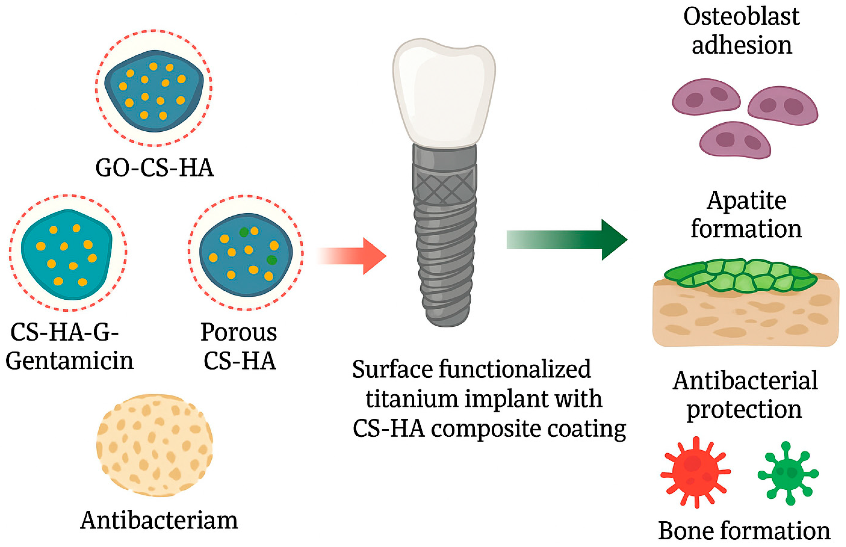
| Shi et al., 2016. [14] | In vitro | Experimental Group: CS-HA 1.0 Graphene oxide-loaded CS–HA (GO-CS-HA) GO-CS-HA nanocomposite via electrophoretic deposition (EPD) on CP-Ti Control group: HA-coated CP-Ti | After 5 days of incubation, the GO-CS-HA composite coating induced significant cytotoxicity towards MG63 cells. GO-CS-HA showed significant apatite formation when put in simulated body fluid (SBF). GO-CS-HA enhanced corrosion resistance, antimicrobial activity against Staphylococcus aureus, and osteoblast-like MG63 cell adhesion or proliferation versus other coatings. |
| Suo et al., 2019. [26] | In vitro & In vivo | Experimental Group: GO-HA, CS-HA and GO-CS-HA, by EPD on CP-Ti Control group: HA-coated CP-Ti | In vitro: GO-CS-HA stimulated osteoblast proliferation or differentiation. The relative proliferation rates of the cells on the CS-HA and GO-CS-HA were significantly higher than those on the HA and GO-HA-Ti samples from 5 to 9 days (p < 0.05) In vivo: Rat tibiae model GO-CS-HA improved bone–implant contact. Histological analysis showed quantitative bone area (BA) and bone-to-implant (BI) ratios. Both CS-HA and GO-CS-HA showed significantly higher BA and BI ratios than HA and GO-HA (p < 0.05). GO-CS-HA push-out force and the ultimate shear strength were significantly higher than other groups after 12 weeks of implantation |
| Stevanović et al. 2020. [41] | In vitro | Experimental Group: CS-HA, CS-HA with gentamicin via EPD on CP-Ti Control group: Uncoated CP-Ti | The ALP assay results indicated that the CS-HA-Gentamicin coating exhibited the highest ALP levels in the cell extract, which is the most widely recognised biochemical marker for osteoblast activity. CS-HA and CS-HA–Gentamicin increased ALP activity and were noncytotoxic to fibroblasts. |
| Zhang et al., 2020. [27] | In vitro & In vivo | Experimental Group: Porous Ti6Al4V implants coated with CS-HA composite (via SLS and EPD) Control group: Dense Ti6Al4V implants without any coating. | In vitro: CS-HA porous implants showed higher ALP activity (p < 0.05) and >85% cell viability, promoting early MC3T3-E1 differentiation and pore proliferation. In vivo: Rabbit femoral model implantation At week 4, porous CS-HA exhibited bone ingrowth, while dense Ti6Al4V showed only surface bone formation. At week 12, porous CS-HA showed thicker trabecular bone and increased bone ingrowth, whereas dense Ti6Al4V had limited bonding. |
| Zarif et al., 2024. [29] | In vitro | Experimental Group: HA CS-HA Strontium-doped HA (Sr-HA) Strontium-doped CS-HA (Sr-CS-HA) (generated by radio-frequency magnetron sputtering and assisted pulsed laser evaporation) Control group: Uncoated CP-Ti | Formation of Ca–P apatite layer was fastest and densest on Sr–CS-HA than others. Incorporated Sr into CS–HA layers; enhanced Fibroblasts (L929 cells) proliferation. Sr–CS-HA showed the strongest antibacterial effect against S. aureus, attributed to Sr ions and chitosan’s cationic charge disrupting bacterial membranes. |
| Li et al., 2025. [42] | In vitro | Experimental groups: CS-HA via in situ MAO CS-HA via two-step MAO + anodic oxidation (ANO) Control group: HA via MAO | HA showed the best cell (MC3T3-E1) proliferation. CS-HA via in situ MAO showed intermediate cell proliferation. CS-HA via two-step MAO + ANO showed the lowest cell proliferation. All CS-containing coatings improved antibacterial performance against E. coli vs. HA only. MAO + ANO (two-step) CS-HA exhibited better antibacterial activity than in situ MAO CS-HA. |
Disclaimer/Publisher’s Note: The statements, opinions and data contained in all publications are solely those of the individual author(s) and contributor(s) and not of MDPI and/or the editor(s). MDPI and/or the editor(s) disclaim responsibility for any injury to people or property resulting from any ideas, methods, instructions or products referred to in the content. |
© 2026 by the authors. Licensee MDPI, Basel, Switzerland. This article is an open access article distributed under the terms and conditions of the Creative Commons Attribution (CC BY) license (https://creativecommons.org/licenses/by/4.0/).
Share and Cite
Balang, A.; Bonithon, R. Surface Modification of Titanium Implants with Chitosan–Hydroxyapatite Composite: A Review on Osseointegration and Bioactivity. Mater. Proc. 2026, 31, 12. https://doi.org/10.3390/materproc2026031012
Balang A, Bonithon R. Surface Modification of Titanium Implants with Chitosan–Hydroxyapatite Composite: A Review on Osseointegration and Bioactivity. Materials Proceedings. 2026; 31(1):12. https://doi.org/10.3390/materproc2026031012
Chicago/Turabian StyleBalang, Amantle, and Roxane Bonithon. 2026. "Surface Modification of Titanium Implants with Chitosan–Hydroxyapatite Composite: A Review on Osseointegration and Bioactivity" Materials Proceedings 31, no. 1: 12. https://doi.org/10.3390/materproc2026031012
APA StyleBalang, A., & Bonithon, R. (2026). Surface Modification of Titanium Implants with Chitosan–Hydroxyapatite Composite: A Review on Osseointegration and Bioactivity. Materials Proceedings, 31(1), 12. https://doi.org/10.3390/materproc2026031012