
1. Introduction
Optical sensors have established themselves as highly sensitive and specific tools for detecting environmental pollutants [
1], clinical biomarkers [
2], and gases in various contexts [
2,
3]. Their ability to provide rapid, non-invasive measurements has driven their implementation in both environmental monitoring [
4,
5] and biomedical applications [
6]. However, the effectiveness of these sensors depends mainly on the interaction between light and the surface of the device, which has led to the incorporation of functional coatings at the nanometric scale that modify the optical and chemical properties of the fiber, increasing its sensitivity and range of application [
7,
8].
Several studies have explored the use of nanomaterials such as germanium and silver [
9], gold [
10], graphene [
11], combinations of silver and platinum [
12], and silver with zirconia [
13] to improve the response of optical devices. These studies have shown that the choice of material and deposition technique plays a crucial role in directly influencing the uniformity of the coating and the reproducibility of the sensor. Understanding and optimizing these factors is of utmost importance in our research. In this context, zinc oxide (ZnO) has attracted considerable interest due to its semiconductor properties, chemical stability, low cost, and compatibility with simple deposition methods, such as dip-coating, which allow the fiber surface to be modified in a controlled and uniform manner [
6,
13,
14].
Parameters such as stability and sensitivity are of great importance for optical sensors, as they largely depend on the coating applied to the optical fiber [
15,
16]. In [
17], a more detailed study of the effects of coatings can be found. Pacheco-Chacón (2021) demonstrated a temperature sensor before and after aluminum coating, which produced an increase in temperature sensitivity from approximately 35 to 120 pm/°C [
18].
In this study, the term nanoparticles (NPs) is used to refer to ZnO particles with an average size of less than 100 nm, characterized by atomic force microscopy (AFM), which justifies their designation as nanometric. The incorporation of these nanoparticles not only improves the interaction of light with the external environment but also increases detection sensitivity, a critical aspect for the development of advanced optical sensors [
19,
20,
21].
This study focuses on the development of a protocol for coating single-mode silicon fiber optics (SMF-28) by immersion, optimizing the chemical pretreatment of the surface and the deposition of ZnO nanoparticles to achieve a homogeneous and stable coating. The standardization of this procedure seeks to lay the foundations for the manufacturing of low-cost, high-sensitivity optical sensors with potential applications in both environmental monitoring and the biomedical field. In addition, this study contributes to establishing methodological criteria for the selection of immersion times, acid treatment, and nanoparticle deposition, which are fundamental elements for obtaining reproducible coatings with improved optical properties.
2. Materials and Methods
The experimental procedure was carried out using a single-mode silicon optical fiber (SMF-28, Thorlabs, Newton, NJ, USA) to modify its surface by coating it with zinc oxide (ZnO nanoparticles, previously synthesized in the laboratory) using the dip-coating technique. The protocol was divided into three stages, namely fiber preparation, ZnO solution preparation, and coating application, followed by characterization using atomic force microscopy (AFM) (Nanoscope, Bruker Corporation, Billerica, MA, USA).
To prepare the optical fiber, it was cut into segments of approximately 10 cm and subjected to a three-step chemical pretreatment. First, an initial wash was performed by immersion in distilled water and 99.9% absolute ethanol (
v/
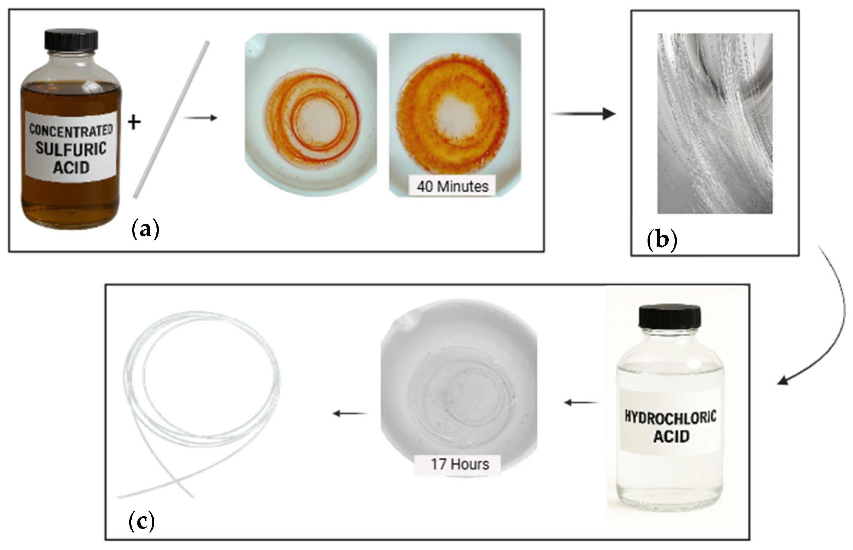
v) (generic laboratory supplier, Mexico) for 5 min to remove surface organic contaminants. Subsequently, as shown in
Figure 1, the fiber was immersed in concentrated sulfuric acid (H
2SO
4, 95–98%, Omnichen ACS, Puebla, México) for 40 min at room temperature, with occasional stirring, allowing the protective polymer coating to be removed and exposing the silica core of the optical fiber, as shown in
Figure 1a. After this step, a second wash was performed with 99.9% ethanol and distilled water, as shown in
Figure 1b. The fiber was then immersed in concentrated hydrochloric acid (HCl, 37%, Omnichen ACS, Puebla, México) for 17 h, generating a controlled attack that increased the surface roughness and promoted the adhesion of the nanoparticles. As shown in
Figure 1c, it was finally rinsed with abundant distilled water and 99.9% ethanol to remove acid residues and left to dry at room temperature. Immersion times were optimized experimentally to achieve the desired roughness without compromising fiber integrity. The acids used can be decanted and reused, following established safety and chemical waste management protocols.
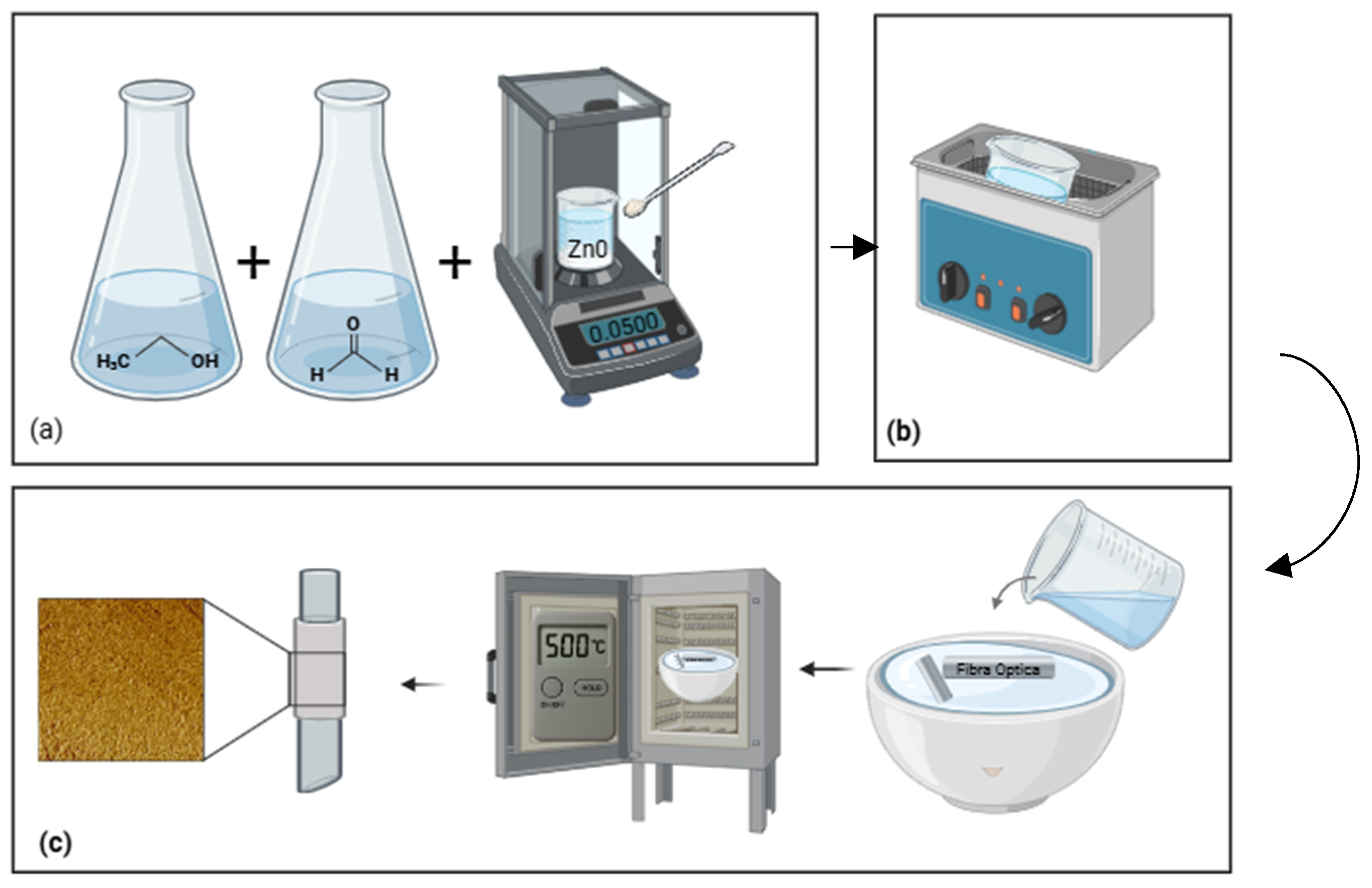
The ZnO solution was prepared in a 1:1 (
v/
v) mixture of 99.9% absolute ethanol and distilled water, using 5 mL of each solvent. In this solution, 0.05 g 0 were dispersed, which were previously synthesized in a laboratory and have an average size of less than 100 nm, as verified by AFM, shown in
Figure 2a. The suspension was sonicated in an ultrasonic cleaner (Branson 1510, Branson Ultrasonics Corporation, Danbury, CT, USA) for 5 min; the device only allows for control of the sonication time, not the power, to ensure homogeneous dispersion and minimize the formation of aggregates, as shown in
Figure 2b. Subsequently, the treated fibers were placed in a single immersion in a porcelain capsule and subjected to heat treatment in a muffle (Thermolyne Small Benchtop Muffle Furnace, Thermo Fisher Scientific, Dubuque, IA, USA) to 500 °C for 10 min as a drying procedure (
Figure 2c), which allowed for partial sintering of the nanoparticles on the fiber surface, ensuring adhesion without affecting its structure.
The coating was characterized by using atomic force microscopy (AFM) with Innova AFM equipment (Bruker) (Nanoscope, Bruker Corporation, Billerica, MA, USA).
The coated fibers were fixed to the sample holder with double-sided conductive tape and analyzed in tapping mode. The tip used was an RTESP-300 AFM probe (Bruker Nano Surfaces, USA) made of antimony-doped silicon (0.01–0.025 Ω·cm), with a cantilever 125 μm long, 40 μm wide, and 3.4 μm thick, a resonance frequency of 300 kHz, and a spring constant of 40 N/m. The back of the tip had a reflective aluminum coating, while the front had no coating.
Height and phase images were obtained over areas of 250 × 250 nm, using a scanning speed ranging from 0.5 to 1.5 kHz to obtain optimal images. Each sample was analyzed in three replicates to ensure reproducibility, and the images were used to calculate the average roughness (Ra), RMS roughness (Rq), minimum, maximum, and average height, and particle distribution on the fiber surface.
3. Results
This study was conducted in two stages, in which changes in the roughness of the optical fiber were evaluated, allowing progress to be made towards the controlled deposition of ZnO nanoparticles using the immersion technique. The samples were characterized using atomic force microscopy (AFM, Innova Bruker), which allowed the uniformity and roughness of the coating to be quantified and the three samples studied to be systematically compared (
Table 1).
These include particle count, density, RMS roughness (Rq), average arithmetic roughness (Ra), minimum, maximum, and average height, standard deviation of height, and immersion times in HCl and H2SO4, as well as the duration of heat treatment in a muffle furnace.
Sample 1, considered the most representative, had a total count of 416 particles with a density of 9.203 particles/µm2, an average height of 1.117 nm, and a standard deviation of 1.630 nm, indicating a highly homogeneous surface. The RMS roughness (Rq) and arithmetic mean (Ra) were 2.98 nm and 1.82 nm, respectively, showing that acid pretreatment with HCl for 17 h and H2SO4 for 40 min, together with exposure in a muffle furnace for 10 min, allowed for controlled nucleation of the nanoparticles and uniform adhesion. These conditions minimized the formation of aggregates and pronounced peaks, increasing the potential of the optical fiber for applications in high-sensitivity sensors.
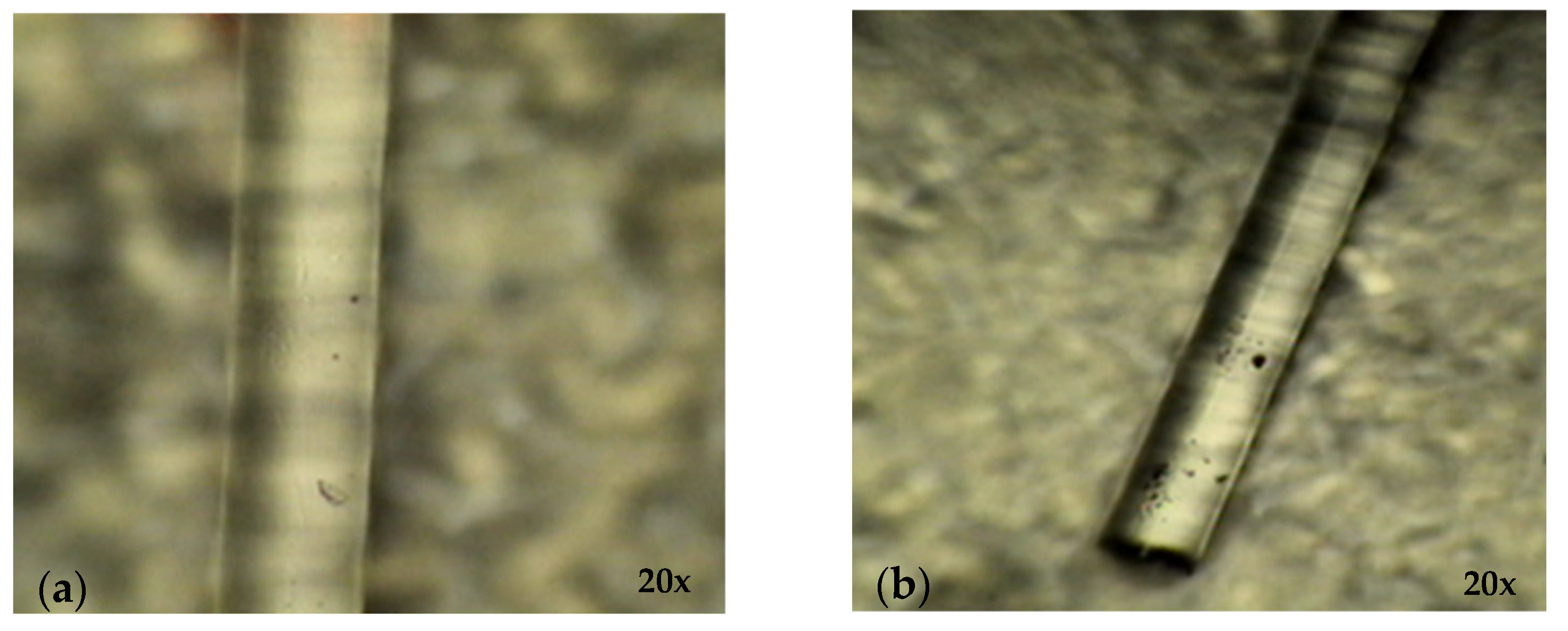
Figure 3 illustrates the evolution of the optical fiber, captured using a professional trinocular metallurgical optical microscope (AmScope T800M, designed/shipped from Irvine, CA, USA; manufactured in China) with an optical zoom of 20×.
Figure 3a shows the uncoated surface, while
Figure 3b, shows the surface after acid treatment, which exhibits an increase in surface roughness that favors the adhesion of nanoparticles.
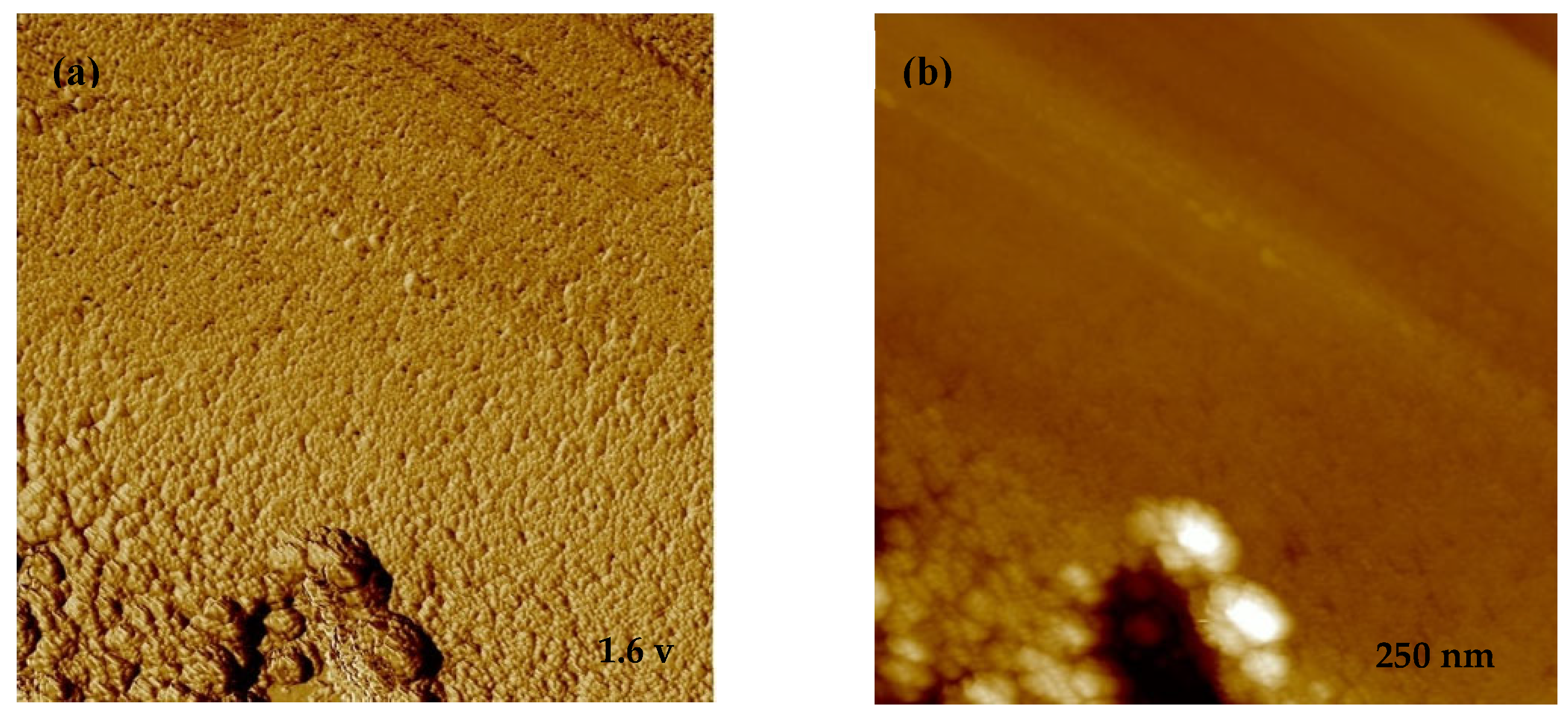
Figure 4 presents AFM images of Sample 1, where the height topography and voltage measurement show a homogeneous distribution of particles with an average size of 30 nm, confirming the effectiveness of the deposition protocol.
Sample 2 had a lower density (6.500 particles/µm2) and an increase in average height (3.450 nm) and standard deviation (4.220 nm), indicating less uniform surfaces with greater roughness. The Rq (6.20 nm) and Ra (2.30 nm) values show localized irregularities, resulting from shorter immersion times in HCl and H2SO4 (12 h and 40 min, respectively) and shorter exposure in the muffle furnace (7 min). These conditions resulted in a less homogeneous coating and highlight the sensitivity of the process to variations in treatment times.
Finally, Sample 3 showed the least favorable results, with a density of 4.120 particles/µm2, an average height of 5.980 nm, and a standard deviation of 6.780 nm, reflecting a highly irregular surface with significant aggregates. The roughness Rq (42.2 nm) and Ra (19.4 nm) confirm the formation of pronounced peaks, resulting from reduced immersion times in HCl (8 h) and muffle exposure (5 min), which did not allow for the adequate nucleation or uniform distribution of ZnO nanoparticles.
In summary, the results indicate that Sample 1 achieved optimal coating conditions, demonstrating that the combination of immersion times in acids and muffle exposure is decisive in obtaining uniform surfaces with low roughness.
Figure 4 of AFM and the comparison with
Figure 3 clearly show how the roughness generated during pretreatment favors the adhesion and homogeneous distribution of nanoparticles, which increases the potential of the optical fiber in the development of high-sensitivity and high-precision optical sensors, underlining the urgent need for further research in this area.
4. Discussion
The results obtained show that the combination of acid pretreatment and controlled exposure in a muffle furnace significantly influences the roughness and uniformity of the ZnO nanoparticle coating on single-mode optical fibers. Sample 1, with longer immersion times in H
2SO
4 and adequate muffle exposure, presented the highest particle density and lowest roughness in the coating, indicating that the prepared surface facilitated homogeneous nucleation and efficient adhesion of the nanoparticles. This behavior is consistent with previous reports where a controlled increase in surface roughness favors the interaction between light and the external medium, increasing the sensitivity of optical sensors [
7,
8,
9,
10,
11,
12,
13,
14,
15,
16,
17,
18,
19,
20,
21,
22].
On the other hand, Sample 2 and Sample 3, with shorter immersion and exposure times, exhibited lower particle densities and higher roughness in the coating, indicating less controlled nucleation and the formation of aggregates. This underscores the sensitivity of the coating process to variations in chemical and thermal treatment times but also underscores the potential for experimental optimization of these parameters, empowering researchers to enhance sensor performance.
The development of homogeneous coatings by immersion is a versatile method that can be adapted to different types of nanomaterials. It allows for the uniform deposition of nanoparticles on the fiber, is reproducible, and facilitates the control of parameters such as particle density and size. Surface roughness parameters (Ra and Rq) vary significantly depending on the deposition method and the intended application of the material.
Table 2 presents values that favor optical transmission. Kaim et al. [
23] reported Ra = 3.57 nm and Rq = 4.49 nm for films obtained by the sputtering technique, while Lee et al. [
24] achieved Ra = 3.12 nm and Rq = 3.97 nm using nanoporous structures. Sarıca et al. [
25] observed an increase in roughness in Mg-coated samples (Ra = 4.04 nm; Rq = 5.16 nm), and Duinong et al. [
26] obtained significantly higher values (Ra = 18.65 nm; Rq = 22.94 nm) after gamma irradiation. Each ZnO based study reports materials prepared according to the methodology and specific requirements of their intended application.
Smooth surfaces enhance light absorption and reduce scattering, whereas excessive roughness can lead to strong light dispersion. In this study, a ZnO coating was deposited on an optical fiber surface via chemical immersion, yielding Ra = 1.82 nm and Rq = 2.98 nm, which are considered indicative of a smooth surface. The resulting coating may be suitable for optical sensors, such as surface plasmon resonance (SPR) devices [
27], as it can enhance the surface plasmonic effect and improve sensor efficiency.
Furthermore, the results suggest that the optical properties of the fiber, such as the refractive index, reflectivity, and sensitivity to changes in the environment, could be improved by ZnO coatings. This discovery not only validates our research but also opens up the thrilling possibility of applying this type of coating in optical sensors and biosensors, where controlled interaction between light and functionalized surfaces is crucial. The potential for future applications is fascinating.
5. Conclusions
Optimizing acid pretreatment times and muffle exposure times was crucial to obtaining homogeneous, low-roughness ZnO coatings on single-mode optical fibers. Sample 1 had the highest particle density and most uniform surface, indicating controlled nucleation and efficient adhesion of the nanoparticles. These findings demonstrate that the combination of chemical and thermal treatments used improves the optical properties of the fiber, increasing its potential for applications in high-sensitivity, high-precision optical sensors and biosensors.
Author Contributions
Methodology, S.X.M.-L., J.L.C.-P., J.G.-G. and A.C.-U.; Validation, J.G.-G., M.V.-T., R.M.V.-C. and I.A.D.-S.; Formal analysis, M.V.-T., R.M.V.-C. and I.A.D.-S.; Investigation, S.X.M.-L., J.L.C.-P., J.G.-G. and A.C.-U.; Data curation, M.V.-T. and J.L.C.-P.; Writing—original draft, S.X.M.-L. and J.L.C.-P.; Writing—review & editing, S.X.M.-L., J.G.-G., A.C.-U., M.V.-T., R.M.V.-C. and I.A.D.-S.; Visualization, S.X.M.-L., J.G.-G., M.V.-T. and I.A.D.-S.; Supervision, J.G.-G., J.L.C.-P. and A.C.-U.; Project administration, S.X.M.-L. and J.L.C.-P. All authors have read and agreed to the published version of the manuscript.
Funding
This research received no external funding.
Institutional Review Board Statement
Not applicable.
Informed Consent Statement
Not applicable.
Data Availability Statement
Data are contained within the article.
Conflicts of Interest
The authors declare no conflicts of interest.
References
- Development of Cellulose Nanofiber Films Functionalized with Curcuminoids for Applications in Optical Sensors of Metallic Contaminants in Water. Master’s Thesis, University of Chile, Santiago, Chile, 2024.
- Castrejón, J.P. Development of a Porous Silicon-Based Optical Immunosensor Functionalized for the Detection of COVID-19 Biomarkers. Master’s Thesis, Autonomous University of the State of Morelos, Cuernavaca, Mexico, 2024. [Google Scholar]
- Garzón, G.; Santacoloma, C.; Galle, B.; Solarte, E. Strategies for measuring volcanic gases in Colombia using remote optical sensors. Bistua J. Fac. Basic Sci. 2008, 6, 32–40. [Google Scholar]
- Navarro-Villoslada, F.; Orellana, G.; Moreno-Bondi, M.C.; Vick, T.; Driver, M.; Hildebrand, G.; Liefeith, K. Fiber-optic luminescent sensors with composite oxygen-sensitive layers and anti-biofouling coatings. Anal. Chem. 2001, 73, 5150–5156. [Google Scholar] [CrossRef] [PubMed]
- Vázquez Sánchez, R.A. Chapter VI. Optical Fiber Sensors: Applications in Environmental Engineering. Universidad de Ciencias y Artes de Chiapas, Faculty of Humanities: Chiapas, Mexico, 2012. Available online: https://repositorio.unicach.mx/handle/20.500.12753/1575?locale-attribute=en (accessed on 9 September 2025).
- Liu, D.; Han, W.; Mallik, A.K.; Yuan, J.; Yu, C.; Farrell, G.; Semenova, Y.; Wu, Q. High sensitivity sol-gel silica coated optical fiber sensor for detection of ammonia in water. Opt. Express 2016, 24, 24179–24187. [Google Scholar] [CrossRef] [PubMed]
- Utzinger, U.; Richards-Kortum, R.R. Fiber optic probes for biomedical optical spectroscopy. J. Biomed. Opt. 2003, 8, 121. [Google Scholar] [CrossRef]
- Zeng, S.; Baillargeat, D.; Ho, H.-P.; Yong, K.-T. Nanomaterials enhanced surface plasmon resonance for biological and chemical sensing applications. Chem. Soc. Rev. 2014, 43, 3426. [Google Scholar] [CrossRef]
- Maciak, E. Low-Coherence Interferometric Fiber Optic Sensor for Humidity Monitoring Based on Nafion® Thin Film. Sensors 2019, 19, 629. [Google Scholar] [CrossRef]
- Allsop, T.D.P.; Neal, R.; Mou, C.; Kalli, K.; Saied, S.; Rehman, S.; Webb, D.J.; Culverhouse, P.F.; Sullivan, J.L.; Bennion, I. Fabrication and characterization of an ultrasensitive surface plasmon resonance sensor based on a multilayer corrugated optical fiber with D-shaped nanoscale coating. IEEE J. Quantum Electron. 2012, 48, 394–405. [Google Scholar] [CrossRef]
- Heidemann, B.R.; Chiamenti, I.; Oliveira, M.M.; Muller, M.; Fabris, J.L. Functionalized Long Period Grating—Plasmonic Fiber Sensor Applied to the Detection of Glyphosate in Water. J. Light. Technol. 2018, 36, 863–870. [Google Scholar] [CrossRef]
- Esfandiyari, M.; Norouzi, M.; Haghdoust, P.; Jarchi, S. Study of a Surface Plasmon Resonance Optical Fiber Sensor Based on Periodically Grating and Graphene. Silicon 2018, 10, 2711–2716. [Google Scholar] [CrossRef]
- Allsop, T.; Neal, R.; Davies, E.M.; Mou, C.; Bond, P.; Rehman, S.; Kalli, K.; Webb, D.J.; Calverhouse, P.; Bennion, I. Low refractive index gas sensing using a surface plasmon resonance fibre device. Meas. Sci. Technol. 2010, 21, 094029. [Google Scholar] [CrossRef]
- Lin, W.B.; Lacroix, M.; Chovelon, J.M.; Jaffrezic-Renault, N.; Gagnaire, H. Development of a fiber-optic sensor based on surface plasmon resonance on silver film for monitoring aqueous media. Sens. Actuators B Chem. 2001, 75, 203–209. [Google Scholar] [CrossRef]
- Liu, Y.; Xia, Q.; Zhou, A.; Wang, X.; Yuan, L. Multi-parameter sensing based on surface plasma resonance with tungsten disulfide sheets coated. Opt. Express 2020, 28, 6084. [Google Scholar] [CrossRef]
- Dong, X.; Du, H.; Sun, X.; Duan, J. Simultaneous Strain and Temperature Sensor Based on a Fiber Mach-Zehnder Interferometer Coated with Pt by Iron Sputtering Technology. Materials 2018, 11, 1535. [Google Scholar] [CrossRef]
- Cusano, A.; Iadicicco, A.; Pilla, P.; Contessa, L.; Campopiano, S.; Cutolo, A.; Giordano, M. Mode transition in high refractive index coated long period gratings. Opt. Express 2006, 14, 19. [Google Scholar] [CrossRef] [PubMed]
- Pacheco-Chacon, E.; Sierra-Hernandez, J.; Gallegos-Arellano, E.; Avila-Garcia, M.; Bianchetti, M.; Hernandez-Romano, I.; Lopez-Dieguez, Y.; Herrera-Piad, L.; Rojas-Laguna, R. An aluminum-coated asymmetric core-offset Mach-Zehnder interferometer temperature sensor. Opt. Fiber Technol. 2021, 65, 102591. [Google Scholar] [CrossRef]
- de Carvalho, D.M.S. Fabrication of Ultraviolet Sensors Based on ZTO and ZnO Nanowires. Master’s Thesis, NOVA University Lisbon, Lisbon, Portugal, 2025. [Google Scholar]
- Wang, X.; Liu, Q.; Bu, Y.; Liu, C.-S.; Liu, T.; Yan, X. Optical temperature sensing of rare-earth ion doped phosphors. RSC Adv. 2015, 5, 86219–86236. [Google Scholar] [CrossRef]
- Rivero, P.; Goicoechea, J.; Arregui, F. Optical Fiber Sensors Based on Polymeric Sensitive Coatings. Polymers 2018, 10, 280. [Google Scholar] [CrossRef] [PubMed]
- Li, C.; Yang, W.; Wang, M.; Yu, X.; Fan, J.; Xiong, Y.; Yang, Y.; Li, L. A Review of Coating Materials Used to Improve the Performance of Optical Fiber Sensors. Sensors 2020, 20, 4215. [Google Scholar] [CrossRef]
- Kaim, P.; Lukaszkowicz, K.; Szindler, M.; Szindler, M.M.; Basiaga, M.; Hajduk, B. The Influence of Magnetron Sputtering Process Temperature on ZnO Thin-Film Properties. Coatings 2021, 11, 1507. [Google Scholar] [CrossRef]
- Lee, S.; Joung, Y.-H.; Yoon, Y.-K.; Choi, W. Preparation of a ZnO Nanostructure as the Anode Material Using RF Magnetron Sputtering System. Nanomaterials 2022, 12, 215. [Google Scholar] [CrossRef]
- Sarica, E. The Effect of Mg Content on the Physical Properties of ZnO Films Deposited by Ultrasonic Spray Pyrolysis. Sak. Univ. J. Sci. 2023, 27, 603–613. [Google Scholar] [CrossRef]
- Duinong, M.; Rasmidi, R.; Chee, F.P.; Moh, P.Y.; Salleh, S.; Salleh, K.A.M.; Ibrahim, S. Effect of Gamma Radiation on Structural and Optical Properties of ZnO and Mg-Doped ZnO Films Paired with Monte Carlo Simulation. Coatings 2022, 12, 1590. [Google Scholar] [CrossRef]
- Liang, H.; Shen, T.; Feng, Y.; Liu, H.; Han, W. A D-Shaped Photonic Crystal Fiber Refractive Index Sensor Coated with Graphene and Zinc Oxide. Sensors 2020, 21, 71. [Google Scholar] [CrossRef] [PubMed]
| Disclaimer/Publisher’s Note: The statements, opinions and data contained in all publications are solely those of the individual author(s) and contributor(s) and not of MDPI and/or the editor(s). MDPI and/or the editor(s) disclaim responsibility for any injury to people or property resulting from any ideas, methods, instructions or products referred to in the content. |
© 2025 by the authors. Licensee MDPI, Basel, Switzerland. This article is an open access article distributed under the terms and conditions of the Creative Commons Attribution (CC BY) license (https://creativecommons.org/licenses/by/4.0/).