Proceeding Paper

Detection of Proteus spp. in Artificial Surface Samples and Estimation of the LOD of the Qualitative Microbiological Method †

by
Dragica Đurđević-Milošević
1,*,
Andrijana Petrović
1,
Jasmina Elez
1,
Vesna Kalaba
2 and
Goran Gagula
3
1
Institute of Chemistry, Technology and Microbiology, Prokupačka 41, 11000 Belgrade, Serbia
2
College of Health Sciences Prijedor, Nikole Pašića 4a, 79101 Prijedor, Bosnia and Herzegovina
3
Department of Food Technology & Biotechnology, Karlovac University of Applied Sciences, J.J. Strosmayer Square No.9, 47000 Karlovac, Croatia
*
Author to whom correspondence should be addressed.
Presented at the 5th International Electronic Conference on Applied Sciences (ASEC 2024), 4–6 December 2024; Available online: https://sciforum.net/event/ASEC2024.
Eng. Proc. 2025, 87(1), 83; https://doi.org/10.3390/engproc2025087083
Published: 25 June 2025
(This article belongs to the Proceedings of The 5th International Electronic Conference on Applied Sciences)

Abstract

Food contact surfaces can be a source of food contamination. Bacteria of the genus Proteus are known as opportunistic pathogens, often associated with faecal contamination and decomposition of organic matter. This study aimed to isolate Proteus spp. from surface samples (of dimensions 5 cm2 × 5 cm2). Three levels of artificially soiled aluminium foil were prepared using bacterial suspensions of Proteus hauseri ATCC 13315. Afterwards, the surface swabbing method for the detection of Proteus spp. was applied. The swab was homogenised with Eugon LT 100 broth, and 1 mL was transferred to the enrichment broth. After the incubation of the enrichment broth, streaking on the Brilliant Green Agar and Salmonella Shigella Agar was performed. The characteristic colonies were confirmed by biochemical reactions. The number of positive findings of Proteus hauseri on the applied level of contamination was used for calculation by the PODLOD_ver12.xls ECEL program by Wilrich and Wilrich. This program estimates the probability of detection (POD) function and the limit of detection (LOD) of qualitative microbiological methods. The results of the detection of Proteus hauseri in surface samples showed LOD50 = 24.60 [48.96; 97.45] CFU in 1 mL of swab rinse, and LOD95 = 106.30 [211.59; 421.15] CFU in 1 mL of swab rinse. The applied method for isolation of Proteus spp. from the surface samples can be used for well-contaminated surfaces.

1. Introduction

Bacteria of the genus Proteus indicate faecal contamination of water or soil, and may play an important role in the decomposition of organic material [1]. Proteus bacteria are known as opportunistic pathogens in human and animals that can cause a variety of infections and are often isolated from the intestines of clinically healthy humans and animals without symptoms of disease [2]. Kozlovska [3] reported that Proteus bacteria-associated pathology in humans and animals has increased significantly in recent years with intestinal, respiratory, and urinary tract diseases and involvement in the development of kidney and bladder stones. For example, bacteria from Proteus species have been isolated in 70% of cases of bacterial urinary stones [4].
Bacteria of the genus Proteus are responsible for food spoilage. Of the 60 tested samples of minced meat from the Alkarkh area of Bagdad Province, Iraq, 5 samples (8.33%) were positive for Proteus spp. [5]. Yu et al. [6] isolated P. mirabilis from 29 out of 80 broiler carcasses with a mean contamination level of 2.25 ± 0.50 lg CFU/g. Wang et al. [7] reported that P. mirabilis was responsible for a case of food poisoning that occurred in August 2008 in a group of 13 individuals who consumed stewed pork balls in brown sauce in Beijing. Also, there are growing concerns about antimicrobial resistance and the transfer of antimicrobial resistance genes in the environment. Ma et al. [8] isolated 89 P. mirabilis strains from 347 samples of retail meat products in food markets across China and reported that high contamination of P. mirabilis can serve as a source for various clinically important antimicrobial resistance genes. Sanches et al. [9] tested the antimicrobial resistance of 32 P. mirabilis strains isolated from chicken carcasses for antimicrobial resistance and discovered that 25 (78.13%) of the strains were multidrug-resistant. Handling of contaminated meat poses a risk of surface contamination and health hazards.
The presence of Proteus spp. on the surface of food processing equipment indicates inadequate sanitation and hygiene practices [10]. Microbiological contamination of surfaces in food production facilities and food-handling areas challenges the prevention of cross-contamination [11]. Therefore, assessing the microbiological purity of surfaces that come into contact with food necessitates a rigorous methodological approach. The objective of the study was to design a method to determine the limit of detection of Proteus spp. from artificially contaminated surface samples.

2. Materials and Methods

2.1. Artificial Contamination of Surfaces

The prepared bacterial suspension with a density of 0.5 McF was decimally diluted, and 1 mL of dilutions 10−6 and 10−7 was transferred to a Petri plate in duplicate and poured with Plate Count Agar (PCA) (HiMedia, Maharashtra, India) following ISO 4833-1 [12]. The Petri plates were incubated at 37 °C for 72 h. After incubation, the number of cells of P. hauseri in suspension was counted to estimate the cell count on the contaminated surfaces.
The three levels of artificially contaminated aluminium foil were prepared using dilutions of a suspension of P. hauseri ATCC 13315 and spread with Z-shaped rods within a stainless-steel frame (5 cm × 5 cm) for each surface. For the 1st level of contamination, 20 contaminated surfaces (5 cm × 5 cm) spreading 0.1 mL of dilution 10−5 were prepared. For the 2nd level of contamination, 10 surfaces spreading 0.25 mL of dilution 10−5 were prepared. For the 3rd level of contamination, 5 surfaces spreading 0.2 mL of dilution 10−4 of a suspension of P. hauseri were prepared. One aluminium foil surface with no artificial contamination (blank control) was also prepared for testing.

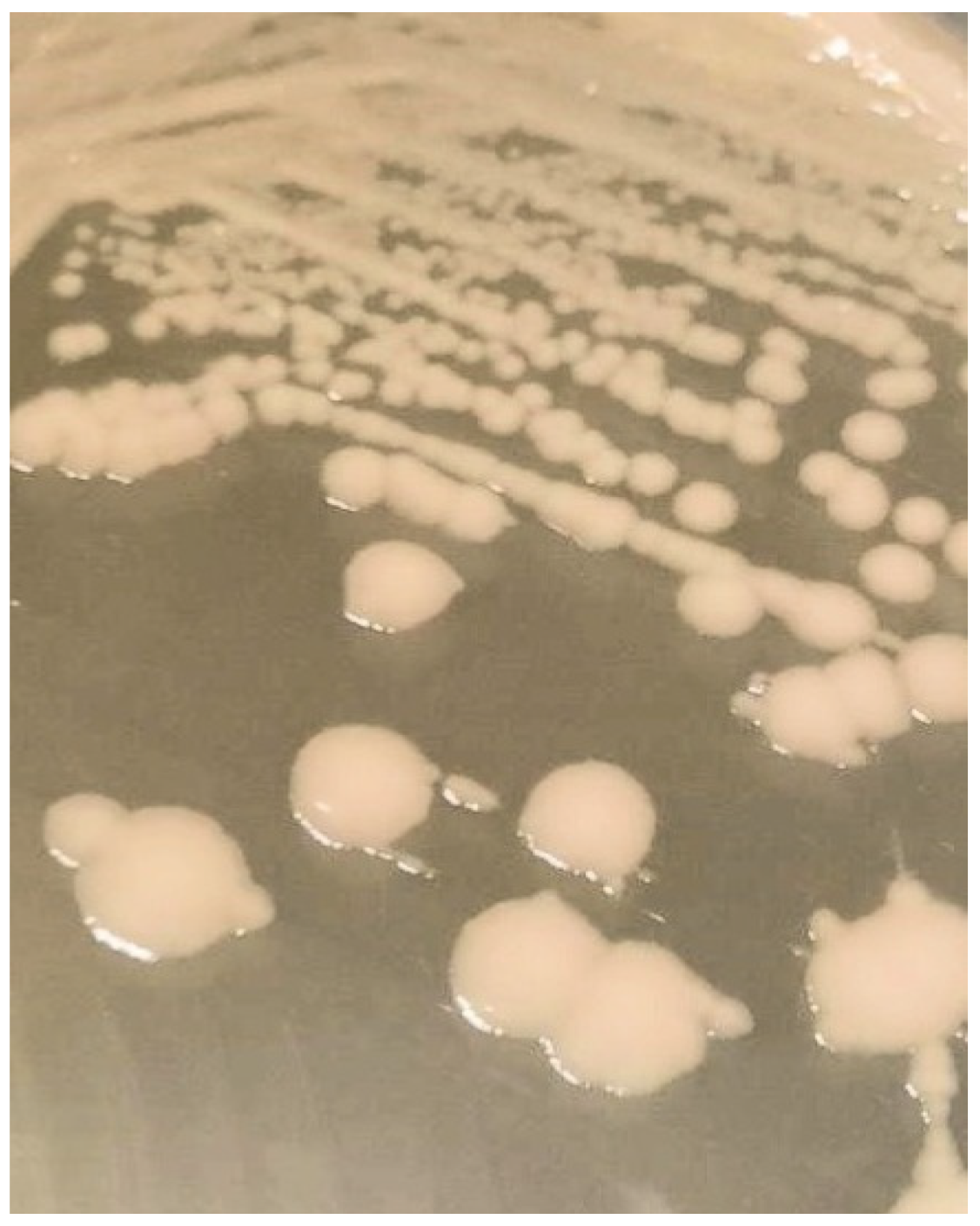
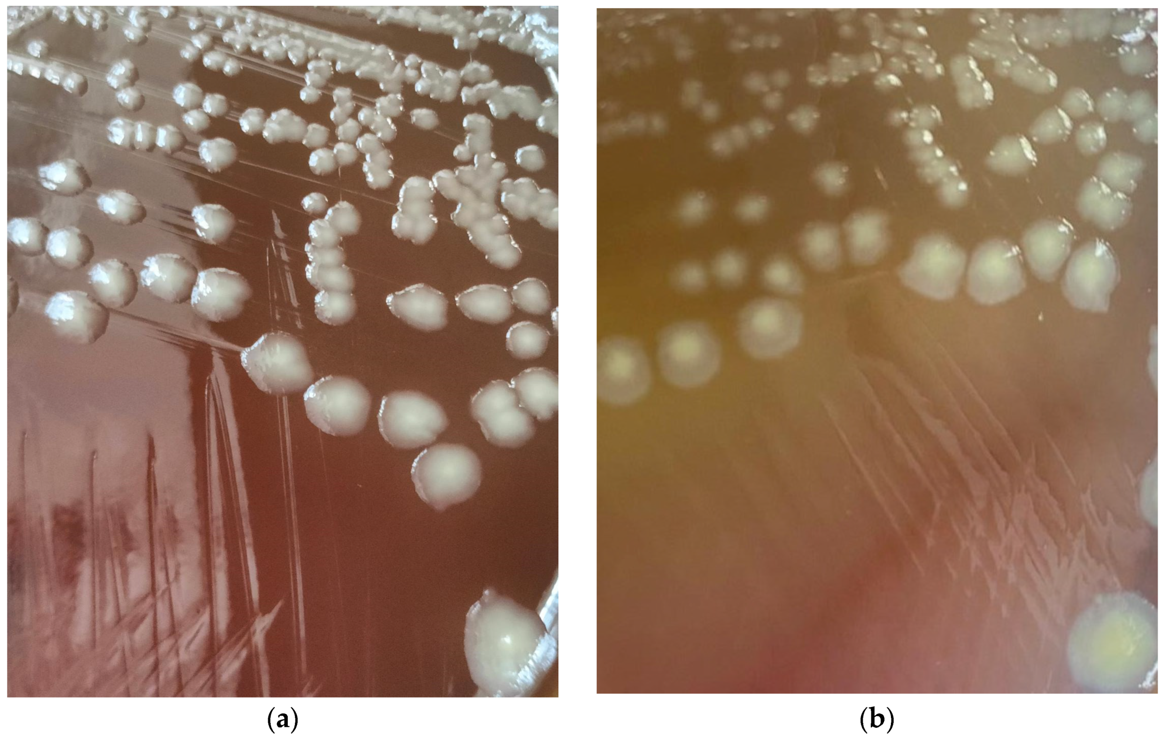
2.2. Surface Sampling, Isolation and Identification of Proteus spp.

After swabbing the surface with a cotton swab stick following the standardised method ISO 18593 [13], the determination of Proteus spp. was performed. The swab was homogenised with 25 mL Eugenic LT 100 broth (HiMedia, India), and 1 mL of broth was transferred to Nutrient broth (Torlak, Belgrade, Serbia). After incubation of the Nutrient broth for 18 ± 2 h at 37 ± 1 °C, streaking on the Brilliant Green Agar (BGA) (HiMedia, India) and Salmonella Shigella (SS) Agar (Oxoid, UK) was performed. BGA and SS plates were incubated at 37 ± 1 °C for 24 ± 2 h. Picked colonies were streaked to Tryptone Soy Agar (TSA) (Oxoid, Basingstoke, UK) and incubated at 37 ± 1 °C for 24 ± 2 h. The colonies from TSA agar were subjected to biochemical reactions and identification by an API test (bioMérieux, Crappone, France) as well as a haemolysis test on a Blood agar plate (Torlak, Belgrade, Serbia).

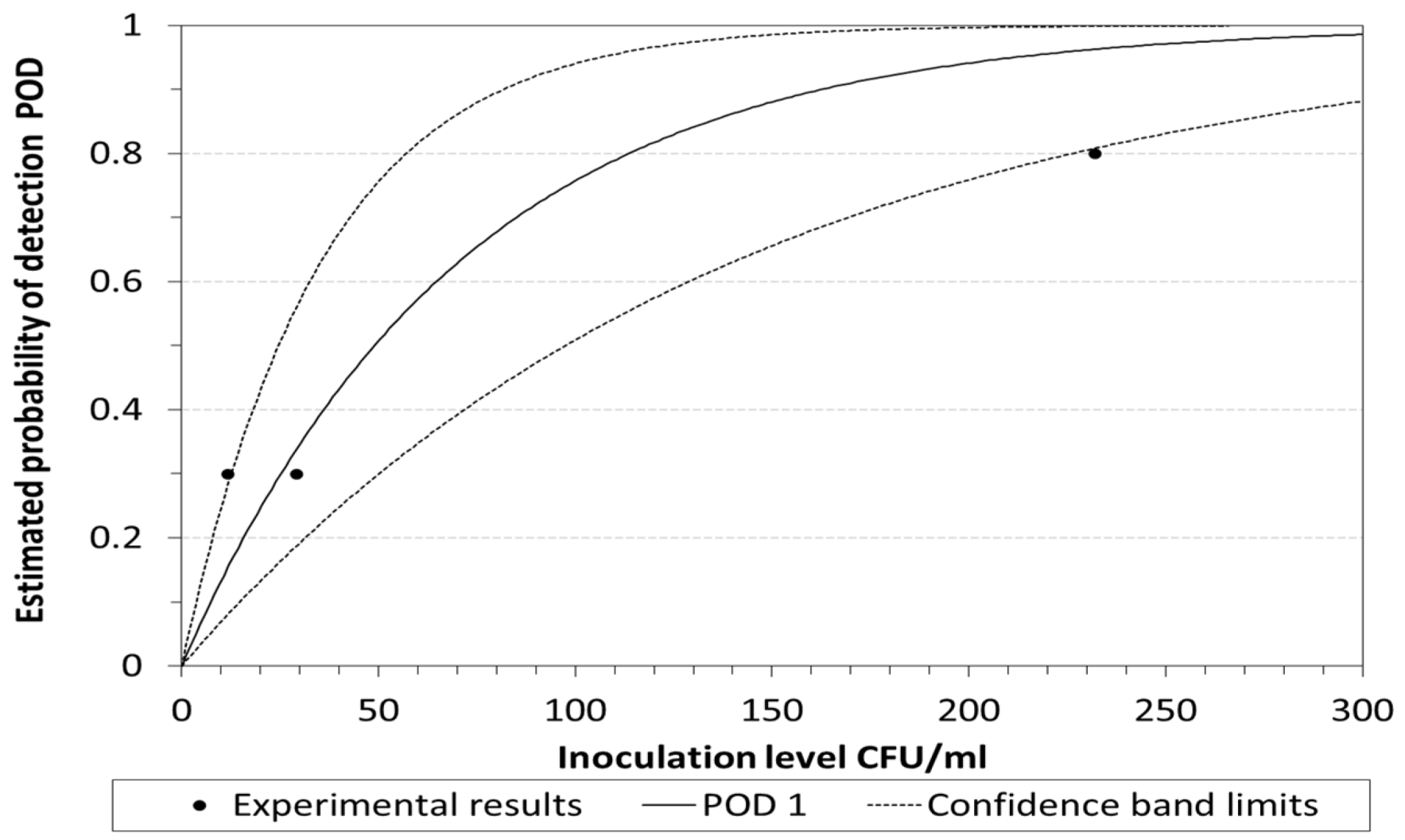
2.3. POD and LOD Calculation

The number of positive findings of P. hauseri on the applied level of contamination was used for calculation by the adopted PODLOD_ver12.xls ECEL program by Wirlich and Wilrich [14]. This program estimates the probability of detection (POD) function and the limit of detection (LOD) of qualitative microbiological methods. The probability of detection (POD) function, p(d), is the functional relationship between the probability p of obtaining a measurement result 1 and the contamination of the test material, quantitatively expressed as the number of CFUs of the microorganism of the defined type per unit of weight or volume, d (CFU/g or CFU/mL) [15]. The calculation of POD and LOD of detected Proteus hauseri from artificially contaminated surfaces was changed in part of the unit, i.e. CFU/cm2 instead of CFU/g or CFU/mL.

3. Results and Discussion

The artificially contaminated aluminium foils simplified isolation and detection because the surfaces were contaminated with known monoculture P. hauseri ATCC 13315. The isolated colonies show non-swarming on the TSA (Figure 1) and Blood agar plate (Figure 2a) after 24 ± 2 h of incubation. Growth of P. hauseri on the Blood agar plates after 26 h of incubation (Figure 2b) showed no haemolysis. After prolonged incubation of plates for five days, alpha haemolysis appeared, indicating mild swarming. Both used media had 5.0 g/L of sodium chloride (NaCl). Nissanka et al. [16] investigated the growth of P. mirabilis on Nutrient agar plates prepared with a range of sodium NaCl concentrations from 0.1 to 5.0 g/L. They concluded that P. mirabilis did not swarm on modified Nutrient agar with a NaCl concentration of ≤0.66 g/L.
Due to its non-swarming nature, many isolates of P. hauseri from naturally contaminated samples and mixed cultures can be missed [17]. Swarming is the main micromorphological characteristic of Proteus bacteria, which enables the bacteria to migrate on a solid surface and form a biofilm, particularly on the surface of a urinary catheter [18]. Kishore [19] noted that P. penneri may be non-swarming on first isolation. Some psychotropic drugs can inhibit swarming in Proteus, and swarming appears to be a sign of the pathogenicity of the species [20].
In addition to morphological characteristics, the detection identification of Proteus spp. also requires knowledge of the diversity of biochemical characteristics of the members of this genus [21]. The genus Proteus also includes P. alimentorum sp. nov. [22], P. appendicitidis [23], P. cibi and P. faecis [24], P. columbae [25], P. genom sp. 4, P. genom sp. 6, and many unclassified Proteus species. P. hauseri can be distinguished from P. mirabilis, P. penneri, and P. myxofaciens as it is positive for indole production, and the other three are negative [17].
The test results of the artificially contaminated surfaces are presented in Table 1. The number of tested surfaces and the number of surfaces with detected Proteus hauseri are used for the calculation of POD (Figure 3) and LOD, following Wirlich and Wilrich [15].
The LOD calculation of results showed LOD50 = 48.96 [24.60; 97.45] CFU of P. hauseri in 1 mL of swab rinse and LOD95 = 211.59 [106.30; 421.15] CFU in 1 mL of swab rinse. The tested 1 mL of swab rinse is equivalent to 1 cm2 because the swab sample from 25 cm2 was homogenised in 25 mL of Eugenic LT 100 broth, and 1 mL of homogenate was used for enrichment. So, based on applied analysis and calculation, the applied method is less suitable for low-level contaminations. The reasons for such results and the low levels of positive findings can vary and could include many factors. One of the reasons could be the efficiency of the swab stick, depending on the shape of the swab head and its material. Jansson et al. [26] evaluated the sampling efficiencies for 15 different swabs made of cotton (5), flocked nylon (3), and foam (7) and concluded that the recovery varied substantially between swabs made of the same material. The choice of the suitable type of swab will increase the swab efficiency, according to Keeratipibul et al. [27]. Another reason could be the loss of the bacterial suspension on a Z-shaped rod during the spreading on the aluminium foil and, therefore, the absence of quantitative application of the P. hauseri suspension. Đurđević-Milošević et al. [28] reported that after sampling from artificially contaminated aluminium foil and comparing the result with the estimated number of Escherichia coli in the overnight suspension, the recovery of Escherichia coli after swab sampling was less than 60% on PCA. The estimated LOD based on the analysis of an artificially contaminated surface may be lower for naturally contaminated surfaces, due to the loss during the preparation of the contaminated surfaces.
Antibiotic resistance of Proteus spp. is a growing trend that is a cause of worry regarding public health [29], especially due to biofilm formation [30]. However, the isolation and identification of Proteus spp. are important for monitoring the effects of surface disinfection, preventive hygienic action, and biocontrol.

4. Conclusions

The methods of surface sampling and isolation of microorganisms play a key role in the inspection and control of surface safety in the food industry. Surface swabs were taken from artificially contaminated surfaces with P. hauseri ATCC 13315 suspension. The estimated limit of detection of an applied method for isolation of Proteus spp. from the surface samples indicates this method can be used on well-contaminated surfaces. Swarming on solid culture media usually indicates the presence of Proteus spp. However, the absence of swarming is not a reliable indicator of the absence of Proteus spp., and special care should be taken when isolating Proteus spp. from mixed cultures, and this should be followed by biochemical tests for identification. Future work should include more species of the genus Proteus, isolation from mixed cultures, and sampling with different types of swab sticks.

Author Contributions

Conceptualization, data curation, writing—original draft preparation, supervision, D.Đ.-M.; data curation, methodology, validation, writing—review and editing, A.P.; formal analysis, investigation, writing—review and editing, J.E.; investigation, writing—review and editing, V.K.; writing—review and editing, G.G. All authors have read and agreed to the published version of the manuscript.

Funding

This research received no external funding.

Institutional Review Board Statement

Not applicable.

Informed Consent Statement

Not applicable.

Data Availability Statement

All the data used in the experiment are available in the present article.

Conflicts of Interest

The authors declare no conflicts of interest.

References

  1. Drzewiecka, D. Significance and Roles of Proteus spp. Bacteria in Natural Environments. Microbial Ecol. 2016, 72, 741–758. [Google Scholar] [CrossRef] [PubMed]
  2. Peng, Y.; Wang, Y.; Kang, X.; Cai, X. Genomic insights of the emerging human pathogen Proteus appendicitidis sp. nov. Heliyon 2024, 10, e37114. [Google Scholar] [CrossRef] [PubMed]
  3. Kozlovska, G. Bioecology and pathogenicity of Proteus bacteria: A literature review. Ukr. J. Vet. Sci. 2023, 14, 91–107. [Google Scholar] [CrossRef]
  4. Prywer, J.; Olszynski, M. Bacterially induced formation of infectious urinary stones: Recent developments and future challenges. Curr. Med. Chem. 2017, 24, 292–311. [Google Scholar] [CrossRef]
  5. Al-Kubaisi, M.S.; Al-Deri, A.H. Isolation of Proteus spp. bacterial pathogens from raw minced meat in Alkarkh area, Baghdad provelance. Int. J. Health Sci. 2022, 6, 4196–4204. [Google Scholar] [CrossRef]
  6. Yu, Z.; Joossens, M.; Van den Abeele, A.-M.; Kerkhof, P.-J.; Houf, K. Isolation, characterization and antibiotic resistance of Proteus mirabilis from Belgian broiler carcasses at retail and human stool. Food Microbiol. 2021, 96, 103724. [Google Scholar] [CrossRef]
  7. Wang, Y.; Zhang, S.; Yu, J.; Zhang, H.; Yuan, Z.; Sun, Y.; Zhang, L.; Zhu, Y.; Song, H. An outbreak of Proteus mirabilis food poisoning associated with eating stewed pork balls in brown sauce, Beijing. Food Control 2010, 21, 302–305. [Google Scholar] [CrossRef]
  8. Ma, W.Q.; Han, Y.Y.; Zhou, L.; Peng, W.Q.; Mao, L.Y.; Yang, X.; Wang, Q.; Zhang, T.J.; Wang, H.N.; Lei, C.W. Contamination of Proteus mirabilis harbouring various clinically important antimicrobial resistance genes in retail meat and aquatic products from food markets in China. Front. Microbiol. 2022, 16, 1086800. [Google Scholar] [CrossRef]
  9. Sanches, M.S.; Baptista, A.A.S.; de Souza, M.; Menck-Costa, M.F.; Koga, V.L.; Kobayashi, R.K.T.; Rocha, S.P.D. Genotypic and phenotypic profiles of virulence factors and antimicrobial resistance of Proteus mirabilis isolated from chicken carcasses: Potential zoonotic risk. Braz. J. Microbiol. 2019, 50, 685–694. [Google Scholar] [CrossRef]
  10. Wang, Y.; Pan, X. Bacteria: Proteus. In Encyclopedia of Food Safety; Motarjemi, Y., Ed.; Academic Press: Cambridge, MA, USA, 2014; pp. 486–489. [Google Scholar]
  11. Butucel, E.; Balta, I.; Ahmadi, M.; Dumitrescu, G.; Morariu, F.; Pet, I.; Stef, L.; Corcionivoschi, N. Biocides as Biomedicines against Foodborne Pathogenic Bacteria. Biomedicines 2022, 10, 379. [Google Scholar] [CrossRef]
  12. ISO 4833-1; Microbiology of the Food Chain. Horizontal Methods for Enumeration of Microorganisms. Part 1: Colony Count at 30 °C by the Pour Plate Technique. ISO—International Organisation for Standardization: Geneva, Switzerland, 2013.
  13. ISO 18593; Microbiology of the Food Chain. Horizontal Methods for Surface Sampling. ISO—International Organisation for Standardization: Geneva, Switzerland, 2018.
  14. POD LOD, Version 12, 2024-03-05, according to Wilrich, C.; Wilrich, P.-T. Estimation of the POD function and LOD of a qualitative microbiological measurement method. J. AOAC Int. 2009, 92, 1763–1772. Available online: https://www.statistics-wilrich.info (accessed on 14 February 2025).
  15. Wilrich, C.; Wilrich, P.-T. Estimation of the POD function and LOD of a qualitative microbiological measurement method. J. AOAC Int. 2009, 92, 1763–1772. [Google Scholar] [CrossRef] [PubMed]
  16. Nissanka, M.C.; Weerasekera, M.; Dilhari, A. Optimization of NaCl Concentration in a General Purpose Medium to Inhibit the Swarming Motility of Proteus mirabilis. In Proceedings of the International Research Symposium of the Faculty of Allied Health Sciences, Galle, Sri Lanka, 10 November 2023; p. 72. [Google Scholar]
  17. Ostwal, K.; Shah, P.; Rebecca, L.; Shaikh, N.; Inole, K. A tale of two novel Proteus species—Proteus hauseri and Proteus penneri. Int. J. Curr. Microbiol. Appl. Sci. 2016, 5, 84–89. [Google Scholar] [CrossRef]
  18. Różalski, A.; Torzewska, A.; Moryl, M.; Kwil, I.; Maszewska, A.; Ostrowska, K.; Drzewiecka, D.; Zablotni, A.; Palusiak, A.; Siwinska, M.; et al. Proteus sp.—An opportunistic bacterial pathogen—Classification, swarming growth, clinical significance and virulence factors. Acta Univ. Lodz. Folia Biol. Oecolog. 2012, 8, 1–17. [Google Scholar] [CrossRef]
  19. Kishore, J. Isolation, identification & characterization of Proteus penneri—A missed rare pathogen. Indian J. Med. Res. 2012, 135, 341–345. [Google Scholar]
  20. Muñoz-Criado, S.; Muñoz-Bellido, J.L.; Alonso-Manzanares, M.A.; Gutiérrez-Zufiaurre, M.N.; García-Rodríguez, J.A. Psychotropic drugs inhibit swarming in Proteus spp. and related genera. Clin. Microbiol. Infect. 1998, 4, 447–449. [Google Scholar] [CrossRef]
  21. O’Hara, C.M.; Brenner, F.W.; Steigerwalt, A.G.; Hill, B.C.; Holmes, B.; Grimont, P.A.; Hawkey, P.M.; Penner, J.L.; Miller, J.M.; Brenner, D.J. Classification of Proteus vulgaris biogroup 3 with recognition of Proteus hauseri sp. nov., nom. rev. and unnamed Proteus genomospecies 4, 5 and 6. Int. J. Syst. Evol. Microbiol. 2000, 50, 1869–1875. [Google Scholar] [CrossRef]
  22. Dai, H.; Wang, Y.; Fang, Y.; Huang, Z.; Kan, B.; Wang, D. Proteus alimentorum sp. nov., isolated from pork and lobster in Ma’anshan city, China. Int. J. Syst. Evol. Microbiol. 2018, 68, 1390–1395. [Google Scholar] [CrossRef]
  23. He, J.; Zhang, Z.; Qu, H.; Chen, G.; Zhou, J.; Zhou, W.; Peng, Y.; Cai, X. Proteus appendicitidis sp. nov., isolated from the appendiceal pus of an appendicitis patient in Yongzhou, China. Arch. Microbiol. 2024, 206, 171. [Google Scholar] [CrossRef]
  24. Dai, H.; Chen, A.; Wang, Y.; Lu, B.; Wang, Y.; Chen, J.; Huang, Y.; Li, Z.; Fang, Y.; Xiao, T.; et al. Proteus faecis sp. nov., and Proteus cibi sp. nov., two new species isolated from food and clinical samples in China. Int. J. Syst. Evol. Microbiol. 2019, 69, 852–858. [Google Scholar] [CrossRef]
  25. Dai, H.; Wang, Y.; Fang, Y.; Xiao, T.; Huang, Z.; Kan, B.; Wang, D. Proteus columbae sp. nov., isolated from a pigeon in Ma’anshan, China. Int. J. Syst. Evol. Microbiol. 2018, 68, 552–557. [Google Scholar] [CrossRef] [PubMed]
  26. Jansson, L.; Akel, Y.; Eriksson, R.; Lavander, M.; Hedman, J. Impact of swab material on microbial surface sampling. J. Microbiol. Methods 2020, 176, 106006. [Google Scholar] [CrossRef] [PubMed]
  27. Keeratipibul, S.; Laovittayanurak, T.; Pornruangsarp, O.; Chaturongkasumrit, Y.; Takahashi, H.; Techaruvichit, P. Effect of swabbing techniques on the efficiency of bacterial recovery from food contact surfaces. Food Control 2017, 77, 139–144. [Google Scholar] [CrossRef]
  28. Đurđević-Milošević, D.; Petrović, A.; Ncube, T.; Gagula, G. Surface swab sampling and recovery of Escherichia coli. In Proceedings of the Scientific Conference SANUS 2023, Prijedor, Bosnia and Herzegovina, 23–24 June 2023; pp. 271–279. [Google Scholar]
  29. Facciolà, A.; Gioffrè, M.E.; Chiera, D.; Ferlazzo, M.; Virgà, A.; Laganà, P. Evaluation of antibiotic resistance in Proteus spp: A growing trend that worries Public Health. Results of 10 Years of Analysis. New Microbiol. 2022, 45, 269–277. [Google Scholar]
  30. Ghamdi, A.; Zabermawi, N.; Mohamed Aly, M. Biofilm formation of foodborne pathogens and strategies of its prevention and biocontrol: A review. Appl. Food Biotechnol. 2025, 12, 1–8.e4. [Google Scholar]
Figure 1. Non-swarming colonies of P. hauseri on TSA after 26 h of incubation.
Engproc 87 00083 g001
Figure 2. Colonies of P. hauseri on the Blood agar plate: (a) after 26 h of incubation; (b) after 5 days of incubation.
Engproc 87 00083 g002
Figure 3. Estimated POD curve of the combined ideal method and its 95% confidence band.
Engproc 87 00083 g003
Table 1. Results of detecting Proteus hauseri from artificially contaminated surfaces.
Level of Surface
Artificial
Contamination
Estimated Artificial Surface
Contamination
(CFU/25 cm2)
Estimated Artificial Surface
Contamination
(CFU/cm2)
Number
of
Tested
Surfaces
Number of
Surfaces with
Detected
Proteus hauseri
Blank control0010
I29011.6206
II72529103
III580023254
Disclaimer/Publisher’s Note: The statements, opinions and data contained in all publications are solely those of the individual author(s) and contributor(s) and not of MDPI and/or the editor(s). MDPI and/or the editor(s) disclaim responsibility for any injury to people or property resulting from any ideas, methods, instructions or products referred to in the content.

