Proceeding Paper

Electrospinning Poly(acrylonitrile) (PAN) Nanofiber Mats with Mushroom Mycelium Powder †

1
Faculty of Engineering and Mathematics, Bielefeld University of Applied Sciences and Arts, 33619 Bielefeld, Germany
2
School of Engineering, Moi University, Eldoret 30100, Kenya
3
Department of Physical and Biophysical Chemistry (PC III), Faculty of Chemistry, Bielefeld University, 33615 Bielefeld, Germany
4
Department of Apparel Engineering and Textile Processing, Berlin University of Applied Sciences—HTW Berlin, 12459 Berlin, Germany
5
Department of Textile and Paper Engineering, Universitat Politècnica de València, Pza Ferrandiz y Carbonell s/n, 03801 Alcoy, Spain
*
Author to whom correspondence should be addressed.
Presented at the 5th International Electronic Conference on Applied Sciences, 4–6 December 2024; https://sciforum.net/event/ASEC2024.
Eng. Proc. 2025, 87(1), 45; https://doi.org/10.3390/engproc2025087045
Published: 11 April 2025
(This article belongs to the Proceedings of The 5th International Electronic Conference on Applied Sciences)

Abstract

:
Electrospinning is a technique to produce nanofiber mats for diverse applications. In biomedicine in particular, the addition of an antibacterial agent can be advantageous. Here, we report on the needleless electrospinning of nanofiber mats using poly(acrylonitrile) (PAN) blended with different mushroom mycelium powders, which have antibacterial and other functional properties. While PAN blended with Pleurotus ostreatus (oyster mushroom) powder could be electrospun well, PAN blended with Ganoderma lucidum (reishi mushroom) powder was nearly impossible to spin. The PAN/P. ostreatus nanofiber mats showed a morphology after electrospinning similiar to pure PAN; however, the carbon yield was lower. This indicates the possibility of embedding P. ostreatus powder in PAN nanofiber mats for biotechnological or biomedical applications.

1. Introduction

Recently, the utilization of different electrospinning techniques has become increasingly popular. They enable the production of nanofiber mats, usually with arbitrarily oriented nanofibers, which can be used for a broad range of applications, such as filtration, biomedicine, or biotechnology [1,2,3]. In these cases, the large surface-to-volume ratio of nanofibrous membranes is often advantageous as compared to macroscopic textile fabrics or other structures [4,5]. Additionally, the spinning process enables the integration of metallic or ceramic nanoparticles, the blending of different polymers, and even the preparation of non-polymeric nanofibers through the calcination of the polymer used as a spinning agent, making these nanofiber mats potentially useful as sensors [6,7,8].
In the fields of biomedicine and biotechnology, antibacterial properties are highly valuable [9]. Amongst natural materials with antibacterial properties, different edible mushrooms can be found in the literature [10,11,12]. In addition, the chitin in mushroom mycelium cell walls may also improve the nanofiber mats’ mechanical properties [13]. This is why a recent study investigated the electrospinnability of polymer solutions of poly(acrylonitrile) (PAN) with embedded Pleurotus ostreatus (oyster mushroom) and Ganoderma lucidum (reishi mushroom) powder. The PAN/P. ostreatus nanofiber mats were further stabilized and carbonized. All samples were examined by confocal laser scanning microscopy (CLSM), scanning electron microscopy (SEM), and Raman microscopy, revealing morphologies similarto pure PAN directly after electrospinning but modified morphologies after stabilization and carbonization, as well as lower carbon yields. This study shows for the first time that electrospinning PAN/P. ostreatus nanofiber mats with a wire-based process is possible.

2. Materials and Methods

The mushroom mycelium powders used in this study were Pleurotus ostreatus (oyster mushroom) powder (Somatem GmbH, Nidda, Germany) and Ganoderma lucidum (reishi mushroom) powder (GoApollo GmbH, Vienna, Austria).
Electrospinning solutions were prepared using 8 g of PAN (X-PAN copolymer, Dralon, Dormagen, Germany), 42 g of dimethyl sulfoxide (DMSO) (min 99.9%, S3 chemicals, Bad Oeynhausen, Germany), and 1.35 g of mushroom powder. The small additional fraction of mycelium powder did not significantly alter the spinning solution viscosity.
Electrospinning was performed using a wire-based machine, the “Nanospider Lab” (Elmarco Ltd., Liberec, Czech Republic), applying a voltage of 60–70 kV, which led to currents of 0.04–0.06 mA. The carriage speed was 100 mm/s, and the distance between the electrode and substrate was set to 240 mm/200 mm for the samples S1/S2 (containing P. ostreatus) or R1/R2 (containing G. lucidum), respectively. The nozzle diameter was 0.9 mm, the temperature was 23–24 °C, and the relative humidity was 32%, and spinning was performed for 1 h. Samples R1 and R2 were repeatedly electrospun due to insufficient results, but only the first R1 sample had some nanofibrous parts, while the other samples were only electrosprayed.
The samples were characterized by confocal laser scanning microscopy (CLSM) (VK-8710, Keyence, Neu-Isenburg, Germany), scanning electron microscopy (SEM) (Phenom ProX G3 Desktop SEM, Thermo Fisher Scientific, Waltham, MA, USA), and Raman microscopy (WITec alpha300 apyron, Ulm, Germany). The spectra were acquired using a 532 nm laser with a power output of 10 mW, an integration time of 3 s, and a 50× objective. Spectral data processing was performed using an in-house written MATLAB script (MATLAB 2023b).
The samples containing P. ostreatus were further stabilized in a muffle oven B150 (Nabertherm, Lilienthal, Germany) at 280 °C for 1 d, approached with a heating rate of 1 K/min. Carbonization was performed in a tube furnace CTF 12/TZF 12 (Carbolite Gero Ltd., Sheffield, UK) at 500 °C for 1 h under a constant nitrogen flow of 160 mL/min, approached with a heating rate of 1 K/min. Carbonization is used to improve the strength and conductivity of PAN nanofiber mats and is a necessary prerequisite for their use in many advanced applications, such as biomedical sensors or composite scaffolds made from carbon nanofibers and a hydrogel [14]. Carbonizing nanofiber mats that contain different nanoparticles has been shown to improve the bendability of carbon nanofiber mats compared to carbonizing pure PAN nanofiber mats [15,16]. In addition, carbonizing, as well as embedding, different biopolymers has been shown to improve the growth of mammalian cells on the respective nanofiber mats [17,18].

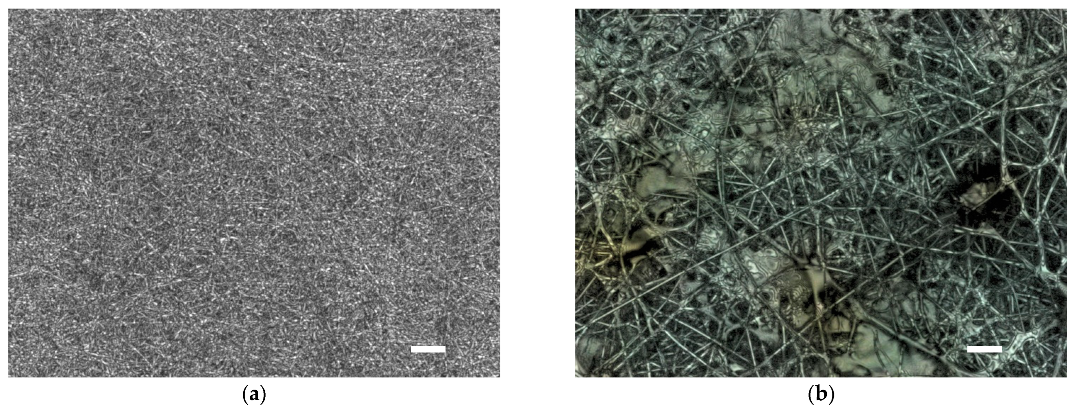
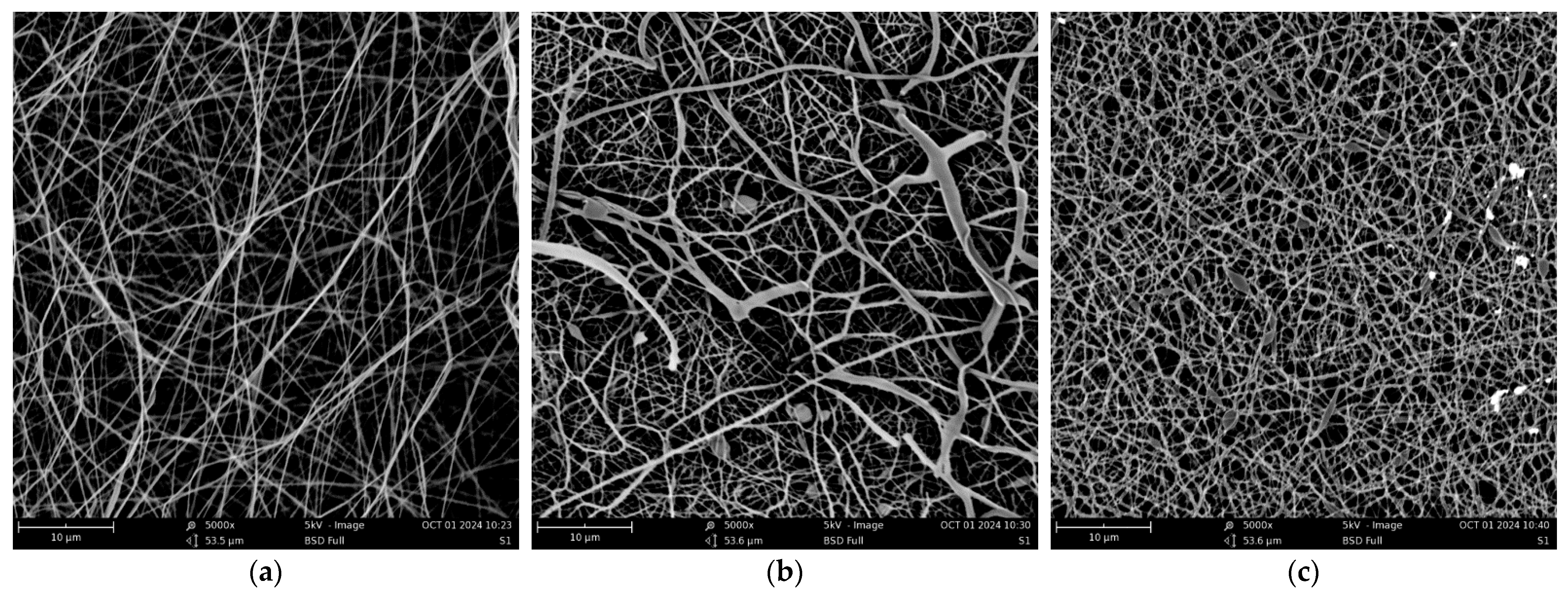
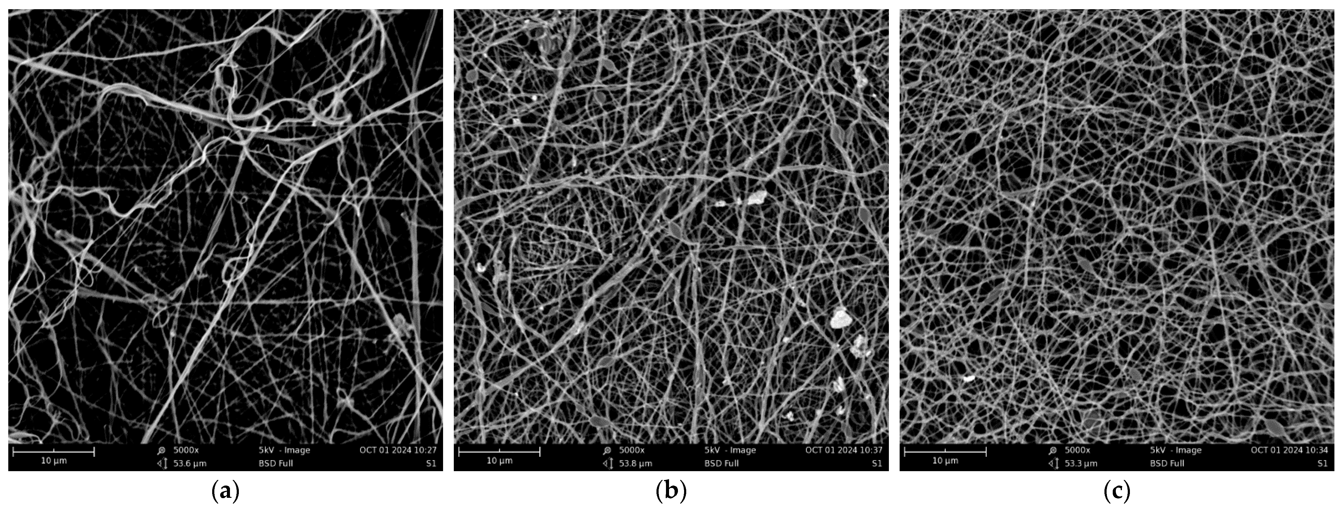
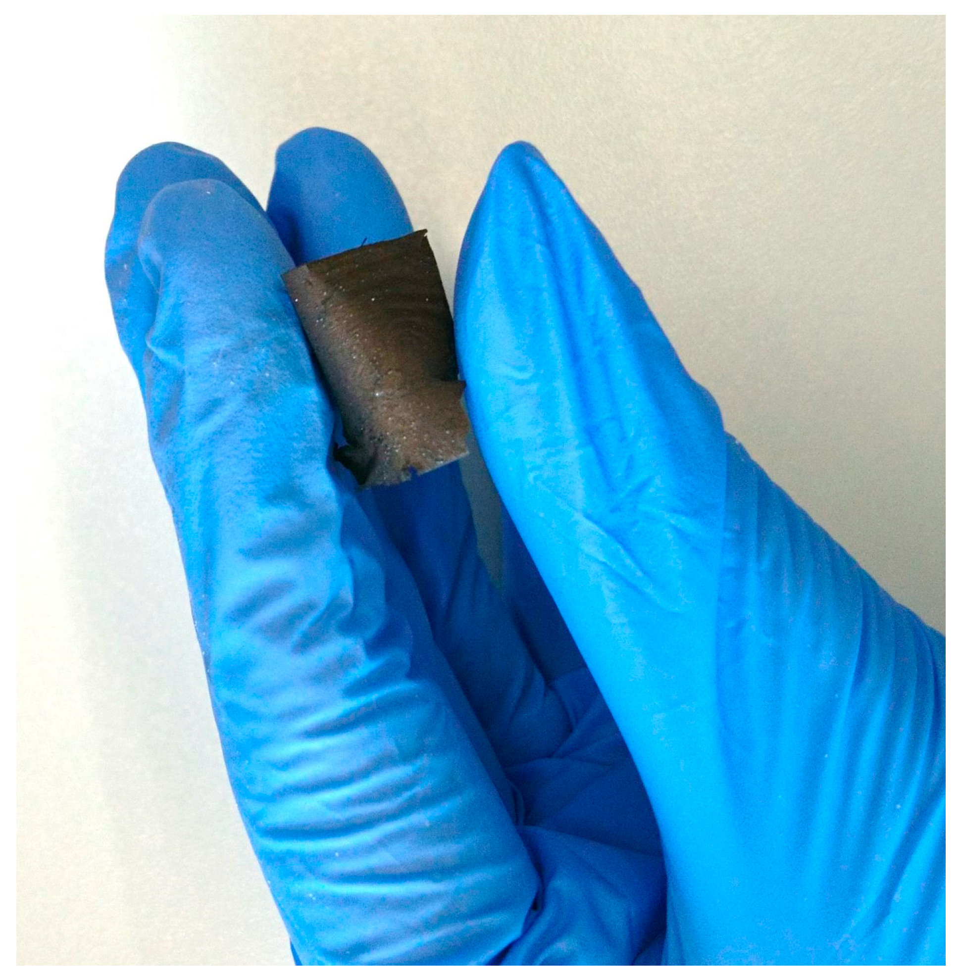
3. Results and Discussion

The samples S1 and S2 showed a mass per unit area of 7.2 g/m2 and 6.2 g/m2, respectively, meaning they are relatively thick nanofiber mats that can be extensively handled without large risk of breaking.
CLSM images of the samples S1 and R1 (the latter taken on one of the few mostly fibrous areas of the sample) are depicted in Figure 1. While sample S1 shows a typical nanofibrous structure, similar to pure PAN, sample R1 shows large non-fibrous areas between the relatively thick fibers with diameters of some micrometers. This shows that even the areas of sample R1 which looked fibrous contain no nanofiber mats. Apparently, the G. lucidum powder impedes electrospinning with the wire-based technique and in the composition of polymer solution chosen here. That is why further experiments—stabilization and carbonization—were only performed with samples containing P. ostreatus.
Next, Figure 2 shows SEM images of sample S1 directly after electrospinning, after stabilization, and after carbonization. Obviously, the originally straight nanofibers (Figure 2a) become more crinkled after carbonization (Figure 2c), while stabilization apparently leads to melting and re-solidification of some parts (Figure 2b), which seem to vanish after carbonization. This behavior is different from the stabilization of pure PAN and can be explained by the thermal decomposition of chitin, which is known to occur in a broad temperature range starting around 200 °C, depending on the molecular weight and degree of deacetylation [19].
SEM images of sample S2 show a very similar nanofibrous structure, with a higher irregularity of the fibers in the as-spun sample (Figure 3a), as expected for a shorter spinning distance and thus a shorter time for the solvent to evaporate. The carbonized S2 sample (Figure 3c), however, is highly similar to the carbonized S1 sample (Figure 2c).
Regarding the mechanical properties, the carbonized samples are found to be bendable (Figure 4), as opposed to carbonized pure PAN nanofiber mats [15,16].
For a chemical analysis of the samples, Raman microscopy was used. The results for the as-spun sample S1 are given in Figure 5. Figure 5a shows the wide-field view of the PAN material with the designated area in red that has been scanned in order to obtain the false-color image in Figure 5b, which indicates the PAN concentration, measured at the peak position of the Raman spectrum of the nanofiber mat (Figure 5c). The low concentration of the fungi impeded their detection by Raman microscopy. Here, we see a certain inhomogeneity, as expected due to blending PAN with P. ostreatus powder. The topography (inset in Figure 5c) was recorded simultaneously during the Raman scan using the True Surface Module.
Next, Raman microscopy was performed on the carbonized S1 sample (Figure 6). Besides the expected color change in the sample in the optical image (Figure 6a), the false-color map of one of the typical carbon peaks again shows some irregularities (Figure 6b). The full spectrum (Figure 6c) reveals a relatively high ratio of the peaks at 1320 cm−1 (D, defective domains) and 1560 cm−1 (G, graphitic ordered domains) of ID/IG = 1.47, i.e., a high number of defects during carbonization. This is expected since 500 °C is not sufficient for full carbonization of PAN, and nanofibers with higher orders need stretching or at least fixation during stabilization and carbonization [20].
Due to the relatively large amount of mycelium powder, the carbon yield of the samples is relatively low, in the range of 39% (S1) to 37% (S2), compared to typical values for pure PAN, which are around 50–70% [21]. As chitin decomposes at elevated temperatures, it contributes less to the overall carbon yield compared to pure PAN fibers, which primarily consist of a carbon-rich backbone [22,23]. Nevertheless, the blending of PAN spinning solutions with a suitable mycelium powder (e.g., from P. ostreatus) may be used to modify the morphological, physical, and chemical properties of nanofiber mats for different applications.

4. Conclusions

Nanofiber mats were electrospun by a wire-based technique, using PAN/mushroom mycelium powder/DMSO spinning solutions. The most significant result is that P. ostreatus powder could be integrated in the spinning solution without problems, while G. lucidum powder impeded electrospinning with the setup and spinning solution used in this study. The carbon yield was reduced compared to pure PAN nanofiber mats due to the decomposition of chitin, which is the main part of the mushroom mycelium. Nevertheless, P. ostreatus powder can be used to modify the physical and chemical properties of nanofiber mats, as compared to pure PAN, for potential use in biotechnological or biomedical applications.

Author Contributions

Conceptualization, L.S.; methodology, all authors; validation, all authors; formal analysis, E.S.; investigation, N.S.M., E.S., U.G. and L.S.; writing—original draft preparation, A.E.; writing—review and editing, all authors; visualization, U.G. and E.S. All authors have read and agreed to the published version of the manuscript.

Funding

The article was written during a research stay of Nonsikelelo Sheron Mpofu at Bielefeld University of Applied Sciences and Arts (HSBI). The research stay was funded through the New Horizons Fellowship from HSBI’s Central Gender and Diversity Officer. The Raman microscope was funded by the Deutsche Forschungsgemeinschaft (DFG) in the scope of “Großgeräteaktion für Hochschulen für Angewandte Wissenschaften (HAW) 2022”, grant number INST 235/37-1.

Institutional Review Board Statement

Not applicable.

Informed Consent Statement

Not applicable.

Data Availability Statement

All data are included in this paper.

Conflicts of Interest

The authors declare no conflicts of interest. The funders had no role in the design of the study; in the collection, analyses, or interpretation of data; in the writing of the manuscript; or in the decision to publish the results.

Abbreviations

The following abbreviations are used in this manuscript:
PANPoly(acrylonitrile)

References

  1. Adeli-Sardou, M.; Yaghoobi, M.M.; Torkzadeh-Mahani, M.; Dodel, M. Controlled release of lawsone from polycaprolactone/gelatin electrospun nanofibers for skin tissue regeneration. Int. J. Biol. Macromol. 2019, 124, 478–491. [Google Scholar] [CrossRef] [PubMed]
  2. Yeo, M.; Ki, G.H. Anisotropically Aligned Cell-Laden Nanofibrous Bundle Fabricated via Cell Electrospinning to Regenerate Skeletal Muscle Tissue. Small 2018, 14, e1803491. [Google Scholar] [CrossRef] [PubMed]
  3. Roche, R.; Yalcinkaya, F. Electrospun Polyacrylonitrile Nanofibrous Membranes for Point-of-Use Water and Air Cleaning. ChemistryOpen 2019, 8, 97–103. [Google Scholar] [CrossRef] [PubMed]
  4. Xiong, J.; Zhou, M.J.; Zhang, H.N.; Quan, Z.Z.; Wang, R.W.; Yin, X.H. Sandwich-structures fibrous membranes with low filtration resistance for effective PM2.5 capture via one-step needleless electrospinning. Mater. Res. Express 2019, 6, 035027. [Google Scholar] [CrossRef]
  5. Yalcinkaya, F. A review on advanced nanofiber technology for membrane distillation. J. Eng. Fiber. Fabr. 2019, 14, 1558925018824901. [Google Scholar] [CrossRef]
  6. Rasouli, R.; Barhoum, A.; Bechelany, M.; Dufresne, A. Nanofibers for Biomedical and Healthcare Applications. Macromol. Biosci. 2019, 19, e1800256. [Google Scholar] [CrossRef]
  7. Horne, J.; McLoughlin, L.; Bridgers, B.; Wujcik, E.K. Recent developments in nanofiber-based sensors for disease detection, immunosensing, and monitoring. Sens. Actuators Rep. 2020, 2, 100005. [Google Scholar] [CrossRef]
  8. Chen, X.W.; Li, H.; Xu, Z.T.; Lu, L.J.; Pan, Z.F.; Mao, Y.C. Electrospun Nanofiber-Based Bioinspired Artificial Skins for Healthcare Monitoring and Human-Machine Interaction. Biomimetics 2023, 8, 223. [Google Scholar] [CrossRef]
  9. Mei, L.; Ren, Y.M.; Gu, Y.C.; Li, X.L.; Wang, C.; Du, Y.; Fan, R.R.; Gao, X.; Chen, H.F.; Tong, A.P.; et al. Strengthened and Thermally Resistant Poly(lactic acid)-Based Composite Nanofibers Prepared via Easy Stereocomplexation with Antibacterial Effects. ACS Appl. Mater. Inter. 2018, 10, 42992–43002. [Google Scholar] [CrossRef]
  10. Reis, F.S.; Pereira, E.; Barros, L.; Sousa, M.J.; Martins, A.; Ferreira, I.C.F.R. Biomolecule profiles in inedible wild mushrooms with antioxidant value. Molecules 2011, 16, 4328–4338. [Google Scholar] [CrossRef]
  11. Mishra, V.; Tomar, S.; Yadav, P.; Vishwakarma, S.; Singh, M.P. Elemental Analysis, Phytochemical Screening and Evaluation of Antioxidant, Antibacterial and Anticancer Activity of Pleurotus ostreatus through In Vitro and In Silico Approaches. Metabolites 2022, 19, 821. [Google Scholar] [CrossRef] [PubMed]
  12. Andrjc, D.C.; Knez, Z.; Marevci, M.K. Antioxidant, antibacterial, antitumor, antifungal, antiviral, anti-inflammatory, and nevro-protective activity of Ganoderma lucidum: An overview. Front. Pharmacol. 2022, 13, 934982. [Google Scholar]
  13. Qiu, Z.H.; Wu, X.L.; Gao, W.; Zhang, J.X.; Huang, C.Y. High temperature induced disruption of the cell wall integrity and structure in Pleurotus ostreatus mycelia. Appl. Microbiol. Biotechnol. 2018, 102, 6627–6636. [Google Scholar] [CrossRef] [PubMed]
  14. Serag, E.; Eltawila, A.M.; Salem, E.M.; El-Maghraby, A.; Abd El-Aziz, A.M. Development of an innovative cylindrical carbon nanofiber/gelatin-polycaprolactone hydrogel scaffold for enhanced bone regeneration. Int. J. Biol. Macromol. 2025, 306, 141250. [Google Scholar] [CrossRef]
  15. Trabelsi, M.; Mamun, A.; Klöcker, M.; Sabantina, L.; Großerhode, C.; Blachowicz, T.; Ehrmann, A. Increased Mechanical Properties of Carbon Nanofiber Mats for Possible Medical Applications. Fibers 2019, 7, 98. [Google Scholar] [CrossRef]
  16. Brockhagen, B.; Hellert, C.; Grothe, T.; Güth, U.; Storck, J.L.; Ehrmann, A.; Wortmann, M. Freestanding Flexible Carbon Nanofiber Mats for Energy Storage Applications. Mater. Proc. 2025, 21, 1. [Google Scholar] [CrossRef]
  17. Wehlage, D.; Blattner, H.; Mamun, A.; Kutzli, I.; Diestelhorst, E.; Rattenholl, A.; Gudermann, F.; Lütkemeyer, D.; Ehrmann, A. Cell growth on electrospun nanofiber mats from polyacrylonitrile (PAN) blends. AIMS Bioeng. 2020, 7, 43–54. [Google Scholar] [CrossRef]
  18. Wehlage, D.; Blattner, H.; Sabantina, L.; Böttjer, R.; Grothe, T.; Rattenholl, A.; Gudermann, F.; Lütkemeyer, D.; Ehrmann, A. Sterilization of PAN/Gelatine Nanofibrous Mats for Cell Growth. Tekstilec 2019, 62, 78–88. [Google Scholar] [CrossRef]
  19. Barbosa, H.F.G.; Francisco, D.S.; Ferreira, A.P.G.; Cavalheiro, É.T.G. A new look towards the thermal decomposition of chitins and chitosans with different degrees of deacetylation by coupled TG-FTIR. Carbohydr. Polym. 2019, 225, 115232. [Google Scholar] [CrossRef]
  20. Storck, J.L.; Hellert, C.; Brockhagen, B.; Wortmann, M.; Diestelhorst, E.; Frese, N.; Grothe, T.; Ehrmann, A. Metallic Supports Accelerate Carbonization and Improve Morphological Stability of Polyacrylonitrile Nanofibers during Heat Treatment. Materials 2021, 14, 4686. [Google Scholar] [CrossRef]
  21. Sabantina, L.; Böttjer, R.; Wehlage, D.; Grothe, T.; Klöcker, M.; García-Mateos, F.; Rodríguez-Mirasol, J.; Cordero, T.; Ehrmann, A. Morphological study of stabilization and carbonization of polyacrylonitrile/TiO2 nanofiber mats. J. Eng. Fibers Fabr. 2019, 14, 1558925019862242. [Google Scholar] [CrossRef]
  22. Yang, H.P.; Yan, R.; Chen, H.P.; Lee, D.H.; Zheng, C.G. Characteristics of hemicellulose, cellulose and lignin pyrolysis. Fuel 2007, 86, 1781–1788. [Google Scholar] [CrossRef]
  23. Escalante, J.; Chen, W.-H.; Tabatabaei, M.S.; Hoang, A.T.; Kwon, E.E.; Lin, Y.-Y.A.; Saravanakumar, A. Pyrolysis of lignocellulosic, algal, plastic, and other biomass wastes for biofuel production and circular bioeconomy: A review of thermogravimetric analysis (TGA) approach. Renew. Sustain. Energy Rev. 2022, 169, 112914. [Google Scholar] [CrossRef]
Figure 1. Confocal laser scanning microscopy (CLSM) images of samples spun with an electrode–substrate distance of 240 mm: (a) sample S1 (containing P. ostreatus); (b) sample R1 (containing G. lucidum). Scale bars correspond to 10 µm.
Engproc 87 00045 g001
Figure 2. Scanning electron microscopy (SEM) images of sample S1 with a nominal magnification of 5000×: (a) as-spun; (b) stabilized; (c) carbonized.
Engproc 87 00045 g002
Figure 3. Scanning electron microscopy (SEM) images of sample S2 with a nominal magnification of 5000×: (a) as-spun; (b) stabilized; (c) carbonized.
Engproc 87 00045 g003
Figure 4. Bending test with the carbonized sample S1.
Engproc 87 00045 g004
Figure 5. Raman microscopy characterization of sample S1: (a) wide-field optical microscopy with the area for Raman microscopy marked; (b) false-color map from a scan at 2253 cm−1; (c) mean Raman spectrum (topography as inset).
Engproc 87 00045 g005
Figure 6. Raman microscopy characterization of the carbonized sample S1: (a) wide-field optical microscopy with the area for Raman microscopy marked; (b) false-color map from a scan at 1360 cm−1; (c) mean Raman spectrum with D and G bands at 1320 cm−1 and 1560 cm−1, respectively.
Engproc 87 00045 g006
Disclaimer/Publisher’s Note: The statements, opinions and data contained in all publications are solely those of the individual author(s) and contributor(s) and not of MDPI and/or the editor(s). MDPI and/or the editor(s) disclaim responsibility for any injury to people or property resulting from any ideas, methods, instructions or products referred to in the content.

