Proceeding Paper

Microfabricated Gold Aptasensors for the Label-Free Electrochemical Assay of Oxytetracycline Residues in Milk †

1
Department of Chemistry, National and Kapodistrian University of Athens, 157 71 Athens, Greece
2
Functional Interfaces Research Group, Institute of Materials and Environmental Chemistry, Research Centre for Natural Sciences, Magyar tudósok krt. 2., 1117 Budapest, Hungary
3
Biomedical Research Foundation, Academy of Athens, 4 Soranou Ephessiou Street, 115 27 Athens, Greece
4
Department of Nanoengineering, University California San Diego (UCSD), La Jolla, CA 92093-0448, USA
5
Institute of Nanoscience and Nanotechnology, NCSR “Demokritos”, Aghia Paraskevi, 15 310 Athens, Greece
*
Author to whom correspondence should be addressed.
Presented at the 10th International Electronic Conference on Sensors and Applications (ECSA-10), 15–30 November 2023; Available online: https://ecsa-10.sciforum.net/.
Eng. Proc. 2023, 58(1), 1; https://doi.org/10.3390/ecsa-10-16018
Published: 15 November 2023

Abstract

:
In this work, we describe a new type of electrochemical aptasensor for the label-free detection of oxytetracycline (OTC). Thin-film gold electrodes were fabricated through sputtering gold on a Kapton film, followed by the immobilization of a thiol-modified aptamer on the electrode surface. The selective capture of OTC at the aptamer-functionalized electrodes was monitored electrochemically with the use of the [Fe(CN)6]4−/[Fe(CN)6]3− redox probe. Different experimental variables were studied, through which the metrological features for OTC determination were derived. Finally, the developed sensor was implemented to achieve the detection of OTC in a spiked milk sample.

1. Introduction

Oxytetracycline (OTC) is a commonly used antibiotic in veterinary medicine; residues of OTC can be present in animal-derived food and are, therefore, consumed by humans [1]. The consumption of, and long-term exposure to, antibiotics may induce antibiotic resistance and can be harmful to human health [2]; therefore, maximum residue limits (MRLs) in foodstuff of animal origin have been set by the European Union for antibiotics [3]; in particular, an MRL of 100 µg/L for OTC residues in milk has been established.
Τherefore, it is essential to develop methods for the sensitive and specific detection of OTC in the environment and food products [4]. To achieve this goal, chromatographic methods have been predominantly used, but these require expensive, laboratory-based instrumentation and trained personnel that preclude their implementation in the field for on-site analysis [5]. On the other hand, biosensors offer an alternative and highly attractive approach for antibiotic residue monitoring, due to their portability and low cost. Amongst the different types of bioreceptors used in various biosensing devices, aptamers (single-stranded oligonucleotides capable of binding to an analyte with high affinity due to the 3D structural arrangement they conform to) present numerous advantages. As a result, many aptasensors have been already developed for the detection of several antibiotics, including OTC [6,7]. Furthermore, the implementation of electrochemical sensing in aptamer-based assays offers some important advantages, such as a high sensitivity, low cost and portable equipment, applicability to on-site analysis, and scope for miniaturization [8].
Herein, the fabrication and application of electrochemical gold-based aptasensors for the label-free assay of OTC is described. Thin-film gold electrodes were fabricated through sputtering gold on a Kapton film. Subsequently, thiol-modified aptamers were immobilized onto the electrodes via the exploitation of the interaction of sulfur with gold. The selective capture of OTC to the aptamer-functionalized electrodes was monitored electrochemically using cyclic voltammetry (CV), differential-pulse voltammetry (DPV), and electrochemical impedance spectroscopy (EIS), with [Fe(CN)6]4−/[Fe(CN)6]3− as a redox probe (Figure 1). The decrease in the charge transfer of the redox probe could be related to the OTC concentration.

2. Experiment

2.1. Reagents and Materials

Reagents were of analytical grade and obtained from Sigma-Aldrich (Burlington, MA, USA). The aptamer sequence was: 5′-GGA ATT CGC TAG CAC GTT GAC GCT GGT GCC CGG TTG TGG TGC GAG TGT TGT GTG GAT CCG AGC TCC ACG TG/3ThioMC3-D/-3′ and was purchased from Integrated DNA Technologies Inc. (Coralville, IA, USA). The aptamer was diluted in phosphate buffer (PB) (10 mM, pH 7.4) containing 1 mM MgCl2. OTC was purchased from Thermo Fisher Scientific (Waltham, MA, USA), and a stock solution of 100 mg/L was prepared in DMSO:PB (50:50 v/v).
OTC standard calibration solutions containing 0, 25, 50, 100, 200, 400, and 600 ng/mL of OTC were prepared in PB (pH 7.4). A milk matrix was prepared by dissolving 1.00 g of low-fat dried milk powder in 50 mL of PB (pH 7.4), centrifuging, and reconstituting the supernatant solution to a final volume of 100.0 mL with PB. OTC matrix-matched standard calibration solutions containing 0, 25, 50, 100, 200, 400, and 600 ng/mL of OTC were prepared in milk matrix.
A spiked milk sample was prepared as follows: 1.00 g of low-fat dried milk powder was spiked with 10 μg of OTC, dissolved in 50 mL of PB (pH 7.4), and centrifuged; the supernatant solution was reconstituted to a final volume of 10.0 mL with PB.
All electrochemical measurements were performed using a solution containing 5 mM [Fe(CN)6]4−/[Fe(CN)6]3− in 0.1 M KCl.

2.2. Instrumentation

A Metrohm Autolab PGSTAT12 Potentiostat/Galvanostat equipped with the GPES Software v. 4.9 (Metrohm, Switzerland) was used for all the electrochemical measurements. Measurements were carried out in a standard electrochemical cell consisting of a Ag/AgCl reference electrode, a Pt counter electrode, and the microfabricated thin-film gold electrode as the working electrode. The measurement conditions were: DPV: scan from −0.1 to +0.4 V, scan rate 10 mV/s, step 5 mV, pulse amplitude 25 mV, modulation time 50 ms, interval time 0.5 s; CV: scan from −0.5 to +0.5 V, scan rate 50 mV/s; EIS: DC potential +0.25 V, AC potential 10 mV, frequency range 100,000 Hz to 0.1 Hz.
Circular dichroism (CD) spectra were recorded (200–320 nm range, 2 nm bandwidth, 50 nm/min scan rate) using a Jasco J-1500 CD spectrometer (Tokyo, Japan) thermostated at 25 °C.
Sputtering was performed with a CV401 system (Cooke Vacuum Products, W. Redding, CT, USA), and atomic force microscopy (AFM) images were taken with an SPM SMENA instrument.

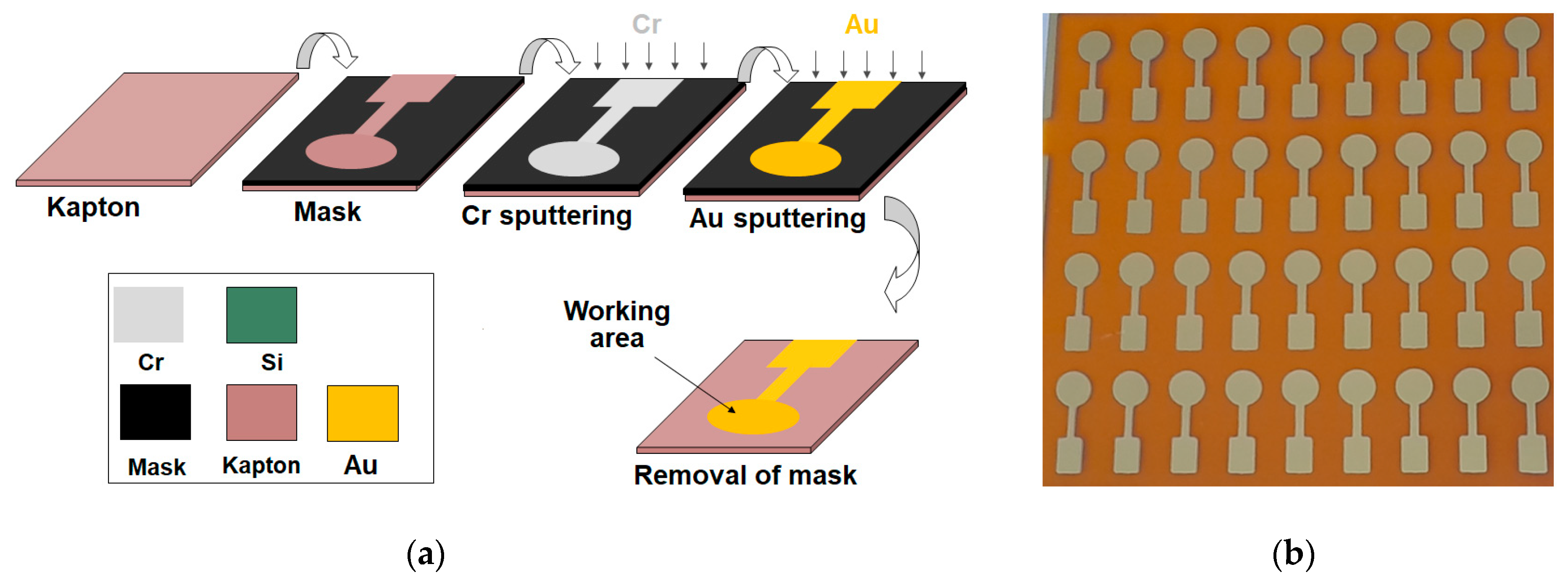
2.3. Fabrication of the Thin-Film Gold Sensors

The thin-film gold electrodes were fabricated via metal sputtering. A film of flexible Kapton® HN polyimide film (50 μm thicknesses, from RS) was covered with a metal mask with the electrode pattern, and Cr and Au were sputtered on the wafer at a nominal thickness of 5 and 100 nm, respectively. A schematic diagram of the fabrication process of the gold sensors and an array of gold electrodes are illustrated in Figure 2a,b, respectively.

2.4. Experimental Protocol

The aptamer stock solution was heated for 5 min at 95 °C and allowed to cool down to room temperature. Aptamer solutions of 5, 10, 20, and 40 μM were prepared in PB containing 1 mM MgCl2. An amount of 10 μL of the aptamer solution was drop-casted onto the thin-film gold working electrode surface and was left for 12 h at 4 °C in a humidity chamber. The aptamer-modified electrodes were thoroughly washed with PB to remove unbound aptamers. Subsequently, 5 μL of mercaptoethanol solution (1 mM in 1:2 (v/v) water/ethanol) was added at the aptamer-modified electrode and was left for 30 min at room temperature.
OTC detection was accomplished through incubating 60 μL of the standard solution or milk sample onto the sensor surface for 30 min at room temperature. Then, the electrode was washed thoroughly with PB to remove unbound OTC and subjected to electrochemical analysis.

3. Results and Discussion

AFM imaging of the thin-film gold electrodes indicates that the gold film is grainy in structure, with a roughness of ca. 9 nm and a grain diameter of ca. 15 nm (Figure 3a). These data demonstrate an exceptionally smooth surface morphology, which is suitable for the reproducible and uniform attachment of the aptamer, as opposed to the commonly used thick-film screen-printed gold deposits that exhibit a much rougher surface and, consequently, lower device-to-device uniformity.
The CD spectrum of the aptamer before and after the addition of different concentrations of OTC is illustrated in Figure 3b. The spectrum reveals that the aptamer adopts a parallel G-quadruplex conformation (maximum at 260 nm, minimum at 240 nm), while incubation with increasing OTC concentrations results in a shift of the peak minimum from ca. 240 nm to ca. 250 nm, coupled with a decrease in the peak maximum at ca. 270 nm and the appearance of a peak at ca. 290 nm, indicative of an analyte-induced folding of the aptamer into a hybrid G quadruplex or a mixture of hybrid and antiparallel quadruplexes [9].
The different steps of the aptasensor preparation and the assay were studied using EIS and CV (Figure 4). EIS demonstrated that the charge transfer resistance of the bare electrode increased upon successive immobilization of the aptamer, mercaptoethanol, and OTC (Figure 4a). This was corroborated by the respective CVs, which showed that the redox current decreased as the bare electrode was successively treated with aptamer, mercaptoethanol, and OTC (Figure 4b). These results suggest that the aptamer is successfully immobilized on the electrode surface and that the OTC is effectively bound to the immobilized aptamer. The concentration of the aptamer was studied, and a 20 μM solution was found to produce the highest sensitivity and repeatability.
Analytical measurements were performed via recording the DPV oxidation current of the [Fe(CN)6]4−/[Fe(CN)6]3− redox probe at different OTC concentrations in the range 0–600 μg/L. Increasing concentrations of OTC induced a reduction in the DPV current as a result of the aptamer blocking of the electrode surface. The % signal reduction, I%, was calculated as: I% = (io − i)/io (where io is the DPV current at the aptamer-modified electrode in the absence of OTC, and i is the DPV current at the aptamer-modified electrode in the presence of OTC).
In order to assess possible milk matrix interference, the matrix effect was calculated as ME = I%,st/I%,mm (where = I%,mm is the signal reduction in a matrix-matched OTC standard, and I%,st is the signal reduction in the same OTC standard in PB) (Table 1). These data indicate that a more pronounced matrix effect existed at lower OTC concentrations, so a matrix-matched calibration plot is recommended for quantitative assays.
Typical DPV traces are illustrated in Figure 5, and the linear–linear (I%,mm vs. [OTC]) and linear–log (I%,mm vs. log[OTC]) calibration plots are shown as inserts. The limit of detection (LOD) of OTC was ~5 μg/L (calculated as the OTC concentration that produced a statistically different signal from the blank). The repeatability between sensors (calculated as the % relative standard deviation from five different aptasensors) was 16% at the 100 μg/L OTC level.
Preliminary experiments were carried out for the determination of OTC in a milk sample spiked with 100 μg/L OTC. The recovery was calculated as R% = I%,sp/I%mm,100 (where I%,sp is the % signal reduction in the spiked milk sample, and I%mm,100 is the % signal reduction in a 100 μg/L matrix-matched standard of OTC). The mean recovery (n = 3) was calculated as 95%.

Author Contributions

Conceptualization, A.E., G.T., J.W., T.S. and Z.K.; methodology, A.E., G.T., J.W., T.S. and Z.K.; validation, V.M., A.A., K.J., N.M. and D.S.; investigation, V.M., A.A., K.J., N.M. and D.S.; data curation, K.J., N.M. and D.S.; writing—original draft preparation, A.E. and D.S.; writing—review and editing, A.E., G.T., J.W. and Z.K.; supervision, A.E., G.T., J.W. and Z.K.; project administration, A.E.; funding acquisition, A.E., G.T., J.W. and Z.K. All authors have read and agreed to the published version of the manuscript.

Funding

This work was supported by the European Union’s Horizon 2020 Research and Innovation Programme under the Marie Sklodowska-Curie Grant Agreement No. 101007299.

Institutional Review Board Statement

Not applicable.

Informed Consent Statement

Not applicable.

Data Availability Statement

Data sharing is not applicable to this article.

Conflicts of Interest

The authors declare no conflict of interest.

References

  1. Landers, T.F.; Cohen, B.; Wittum, T.E.; Larson, E.L. A Review of Antibiotic Use in Food Animals: Perspective, Policy, and Potential. Public Health Rep. 2012, 127, 4–22. [Google Scholar] [CrossRef] [PubMed]
  2. Virto, M.; Santamarina-García, G.; Amores, G.; Hernández, I. Antibiotics in Dairy Production: Where Is the Problem? Dairy 2022, 3, 541–564. [Google Scholar] [CrossRef]
  3. Commission Regulation (EU), No. 37/2010. Available online: https://eur-lex.europa.eu/legal-content/EN/TXT/?uri=CELEX%3A32010R0037 (accessed on 13 November 2023).
  4. Sversut, R.A.; da Silva, A.A.; Mazon Cardoso, T.F.; Kassab, N.M.; do Amaral, M.S.; Nunes Salgado, H.R. A Critical Review of Properties and Analytical Methods for the Determination of Oxytetracyline in Biological and Pharmaceutical Matrices. Crit. Rev. Anal. Chem. 2017, 47, 154–171. [Google Scholar] [CrossRef] [PubMed]
  5. Peris-Vicente, J.; Peris-García, E.; Albiol-Chiva, J.; Durgbanshi, A.; Ochoa-Aranda, E.; Carda-Broch, S.; Bose, D.; Esteve-Romero, J. Liquid chromatography, a valuable tool in the determination of antibiotics in biological, food and environmental samples. Microchem. J. 2022, 177, 107309. [Google Scholar] [CrossRef]
  6. Mehlhorn, A.; Rahimi, P.; Joseph, Y. Aptamer-Based Biosensors for Antibiotic Detection: A Review. Biosensors 2018, 8, 54. [Google Scholar] [CrossRef] [PubMed]
  7. Liang, G.; Song, L.; Gao, Y.; Wu, K.; Guo, R.; Chen, R.; Zhen, J.; Pan, L. Aptamer Sensors for the Detection of Antibiotic Residues—A Mini-Review. Toxics 2023, 11, 513. [Google Scholar] [CrossRef] [PubMed]
  8. Evtugyn, G.; Porfireva, A.; Tsekenis, G.; Oravczova, V.; Hianik, T. Electrochemical Aptasensors for Antibiotics Detection: Recent Achievements and Applications for Monitoring Food Safety. Sensors 2022, 22, 3684. [Google Scholar] [CrossRef] [PubMed]
  9. Kejnovská, Ι.; Renčiuk, D.; Palacký, J.; Vorlíčková, M. CD Study of the G-Quadruplex Conformation. Methods Mol. Biol. 2019, 2035, 25–44. [Google Scholar] [CrossRef] [PubMed]
Figure 1. The principle of the aptamer-based assay for OTC at the microfabricated gold electrodes.
Engproc 58 00001 g001
Figure 2. (a) Schematic diagram of the fabrication process of the thin-film gold electrodes, (b) photograph of an array of 36 electrodes.
Engproc 58 00001 g002
Figure 3. (a) AFM image of the thin-film gold electrode surface, (b) CD spectrum of the aptamer before and after the addition of OTC.
Engproc 58 00001 g003
Figure 4. (a) EIS spectra, (b) CVs at a bare gold electrode and gold electrodes after the successive immobilization of aptamer, conditioning with mercaptoethanol, and incubation with 200 μg/L OTC. Aptamer concentration: 20 μM.
Engproc 58 00001 g004
Figure 5. DPV traces obtained with the aptasensor at different matrix-matched OTC standards in the concentration range 0–600 μg/L. The linear–linear and linear–log calibration plots are shown as inserts.
Engproc 58 00001 g005
Table 1. Study of the milk matrix effect.
[OTC] (μg/L)ME
251.25
501.23
1001.15
2001.15
4001.11
6001.05
Disclaimer/Publisher’s Note: The statements, opinions and data contained in all publications are solely those of the individual author(s) and contributor(s) and not of MDPI and/or the editor(s). MDPI and/or the editor(s) disclaim responsibility for any injury to people or property resulting from any ideas, methods, instructions or products referred to in the content.

