Proceeding Paper

An Optical Fiber Sensor System for Uranium Detection in Water †

1
Department of Engineering, University of Campania Luigi Vanvitelli, Via Roma 29, 81031 Aversa, Italy
2
Department of Chemistry, University of Pavia, Via Taramelli 12, 27100 Pavia, Italy
*
Author to whom correspondence should be addressed.
Presented at the 2nd International Electronic Conference on Biosensors, 14–18 February 2022; Available online: https://sciforum.net/event/IECB2022.
Eng. Proc. 2022, 16(1), 10; https://doi.org/10.3390/IECB2022-12296
Published: 17 February 2022
(This article belongs to the Proceedings of The 2nd International Electronic Conference on Biosensors)

Abstract

:
A simple to realize optical fiber sensor specific for uranium(VI) detection in water is reported to demonstrate the sensing approach’s capability to determine uranyl (UO22+) in water solution in the ppb range. The proposed sensor was obtained by combining a specific receptor layer for uranium to a gold thin film at which the surface plasmon resonance (SPR) phenomenon takes place via optical fiber. In particular, an SPR D-shaped plastic optical fiber (POF) probe was used for signal transduction. The proposed optical-chemical sensing method is attractive because, in principle, it can be applied directly in the field, giving an analytical response in a fast and not overly expensive manner.

1. Introduction

The concentration of uranium in non-contaminated environmental waters is very low, often lower than 1 ppb in freshwater and around 3 ppb in sea water [1], so it is not of environmental or health concern. However, it can be much higher at contaminated sites. In particular, the by-product of the uranium enrichment process, depleted uranium, has been applied as armor-piercing ammunition in several international military conflicts because of its high density, hardness, and pyrophoric properties [2]. The testing and use of such ammunitions have led to the release of depleted uranium into the environment at several locations worldwide [3]. In this situation, the use of marker-free sensing devices for monitoring the uranium content of natural and contaminated waters can be of particular interest. Even if several analytical methods are currently available for the determination of uranium at the very low concentrations present in environmental waters, the proposed sensing method is attractive because, in principle, it can be applied on-site directly, giving an analytical response in a faster and not overly expensive manner.
In the present investigation, an optical fiber sensor for uranyl detection in water, based on surface plasmon resonance (SPR) transduction, is proposed. More specifically, an SPR D-shaped plastic optical fiber (POF) [4] has been covered by a specific receptor monolayer. This optical platform was developed by our research group more than ten years ago, and presents advantages in terms of low cost and small dimensions with respect to the classical Kretschman configuration [4]. It has already been applied to the detection of many different molecular species [5]. In some cases, it has been applied to detect different metal ions, such as copper(II) [6] and iron(III) [7]. In [6,7], strong and selective ligands for these metal ions were fixed at the SPR interface as a monolayer.
In this work, for the uranium(VI) detection, 11-mercaptoundecylphosphonic acid (MUPA) was used as a receptor monolayer, which is commercially available and possesses a sulfide terminal group that can be easily fixed to the gold surface of the SPR POF probe. In a previous research, the same receptor has been demonstrated to be suitable for a uranyl sensor with electrochemical transduction [8]. As a preliminary investigation, experimental results have been reported for a uranyl sensor based on the SPR POF platform combined with MUPA.

2. Materials and Methods

2.1. Reagents

For this study, 11-mercaptoundecylphosphonic acid (MUPA) was purchased from Sigma-Aldrich, as was uranyl (UO22+) standard (uranyl nitrate, standard for ICPOES, 1000 mg L−1). The lower concentration standards were obtained daily by dilution with Milli-Q water. Other reagents were always of the purest grade available and were used as received.

2.2. Instruments and Experimental Setup

The same experimental setup already used in [4,6,7] was employed here. The white light source (Halogen lamp HL-2000-LL manufactured by Ocean Optics, Dunedin, FL, USA) presents an emission range from 360 nm to 1700 nm, whereas the spectrometer FLAME-S-VIS-NIR-ES (manufactured by Ocean Optics, Dunedin, FL, USA) has a detection range from 350 nm to 1023 nm. The transmission spectra were displayed by Spectra Suite software (Ocean Optics, Dunedin, FL, USA). The spectra were normalized by the MATLAB software (MathWorks, Natick, MA, USA) using, as reference for normalization, the spectrum acquired with air as a surrounding medium over the gold surface (D-POF-bare) or MUPA-derivatized gold surface (D-POF-MUPA). All measurements were performed at 25 °C.

2.3. SPR-POF Platforms Preparation

The SPR platform was realized by exploiting a plastic optical fiber (POF) with a core of poly-methylmethacrylate (PMMA) of 980 µm and a cladding of fluorinated polymer of 10 µm (a total diameter of 1 mm), embedded in a resin support and erased as previously described [7] to produce a D-shaped region via polishing. The multilayer interface was realized as described for similar sensors. First, a Microposit S1813 photoresist, with a refractive index higher than that of the POF core, was deposited on the exposed POF by spinning at 6000 rpm for 60 s and polymerizing. Then, a gold film 60 nm thick was sputtered on the photoresist layer by a Bal-Tec SCD 500 machine.
The platforms produced in this way are indicated as SPR-D-POF-bare. The procedure is reported schematically in Figure 1.

2.4. MUPA Deposition

The selected ligand for uranium(VI) (i.e., MUPA) was immobilized as a monolayer on the gold surface, taking advantage of the presence of the sulfide group terminal group in MUPA according to the procedure previously described for an electrochemical sensor of uranyl [8]. The gold film was contacted overnight with a solution containing 2.5 mM MUPA in water. The modified POF platform was then abundantly rinsed with ethanol and Milli-Q water before use. The sensors produced in this way are indicated as SPR-D-POF-MUPA.

2.5. Measurements

About 50 µL of the aqueous sample solution were dropped over the flat sensing region and incubated at room temperature for ten minutes. The spectrum was registered and normalized, and the minimum transmission wavelength (the resonance wavelength) was evaluated.
The quantity of analytical interest is the resonance wavelength variations with respect to the resonance wavelength of the blank solution (∆λris). The function ∆λris versus uranyl concentration (c) were fitted by the Hill equation [9], using the software OriginPro (Origin Lab. Corp., Northampton, MA, USA). When the measured signal (∆λris (c)) is directly proportional to the concentration of the analyte adsorbed on the layer in contact with the gold surface, the response of the sensor is as follows:
∆λris (c) = λc − λ0 = ∆λmax · (c/(K + c))
The symbols λc and λ0 indicate the resonance wavelength at 0 and c uranyl concentration, respectively. ∆λmax is the value of the maximum resonance wavelength variation at increasing concentration of uranyl (c) (i.e., the value at saturation when c is much lower than K). Notice that according to the Langmuir adsorption model, K corresponds to the reciprocal of the affinity constant of the ion UO22+ for MUPA forming the thin receptor layer at the surface of gold. It can be evaluated, together with ∆λmax, from Equation (1). Conversely, Equation (1) can be used as a standardization curve once the parameters are known.

3. Results

The optical sensitivity (bulk sensitivity) of the platforms was determined by measuring λris at the bare platform in dielectrics with different refractive indexes, as previously reported [4]. Water-glycerol solutions were used for this experiment, obtaining a sensitivity of 2500 nm RIU−1 in the wavelength range considered.
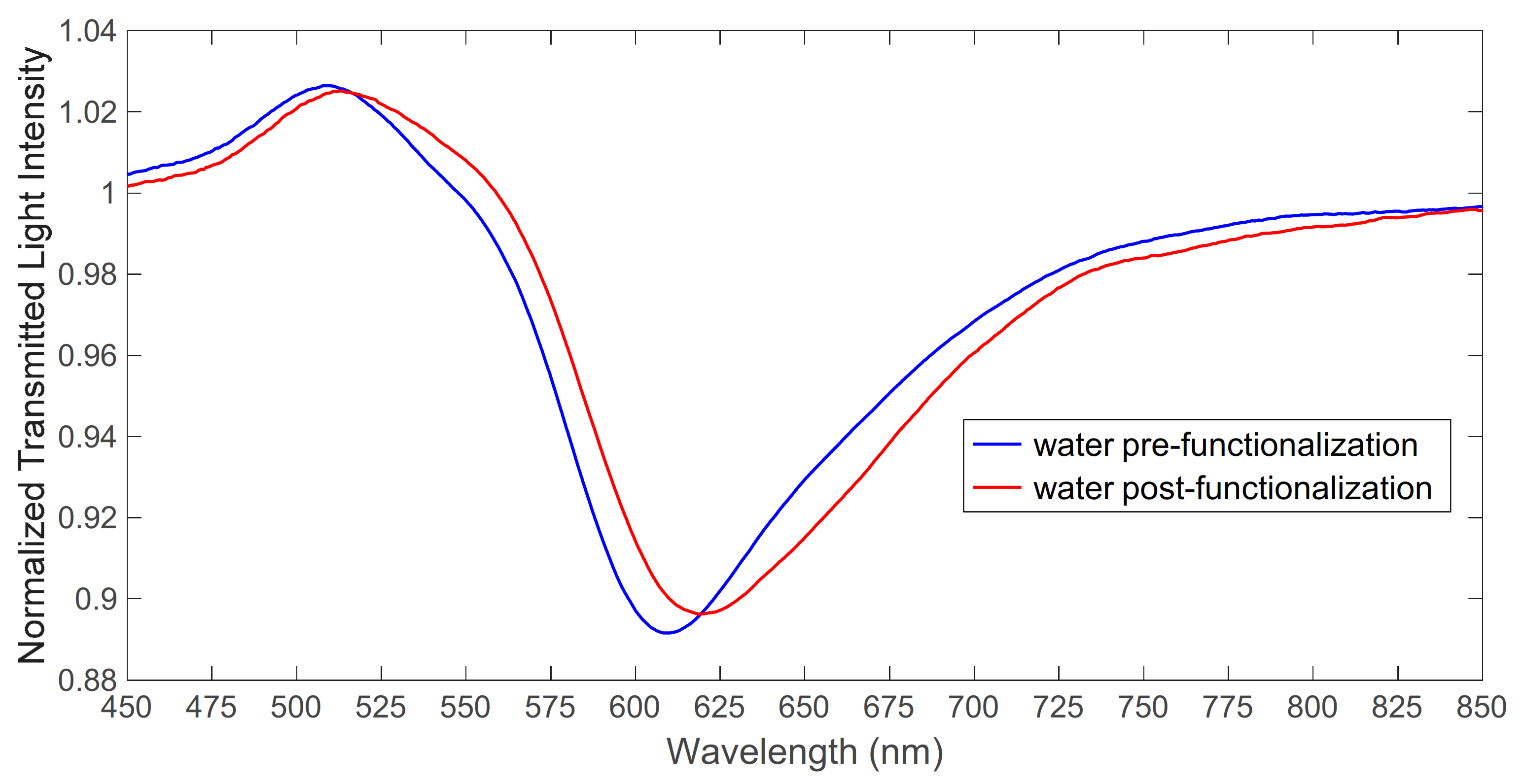
The presence of the receptor layer on the gold surface was demonstrated by the SPR method by comparing the transmission spectrum of the bare platform (SPR-D-POF-bare) in water with that of the MUPA-derivatized platform (SPR-D-POF-MUPA) in water. An example is reported in Figure 2, in which it is seen that the spectrum of SPR-D-POF-MUPA normalized on the spectrum in air is similar to that of the SPR-D-POF-bare normalized on the spectrum in air, but with the resonance wavelength shifted to higher values. This clearly indicates the presence of some receptor molecules at the gold surface producing a refractive index higher than that of pure water.
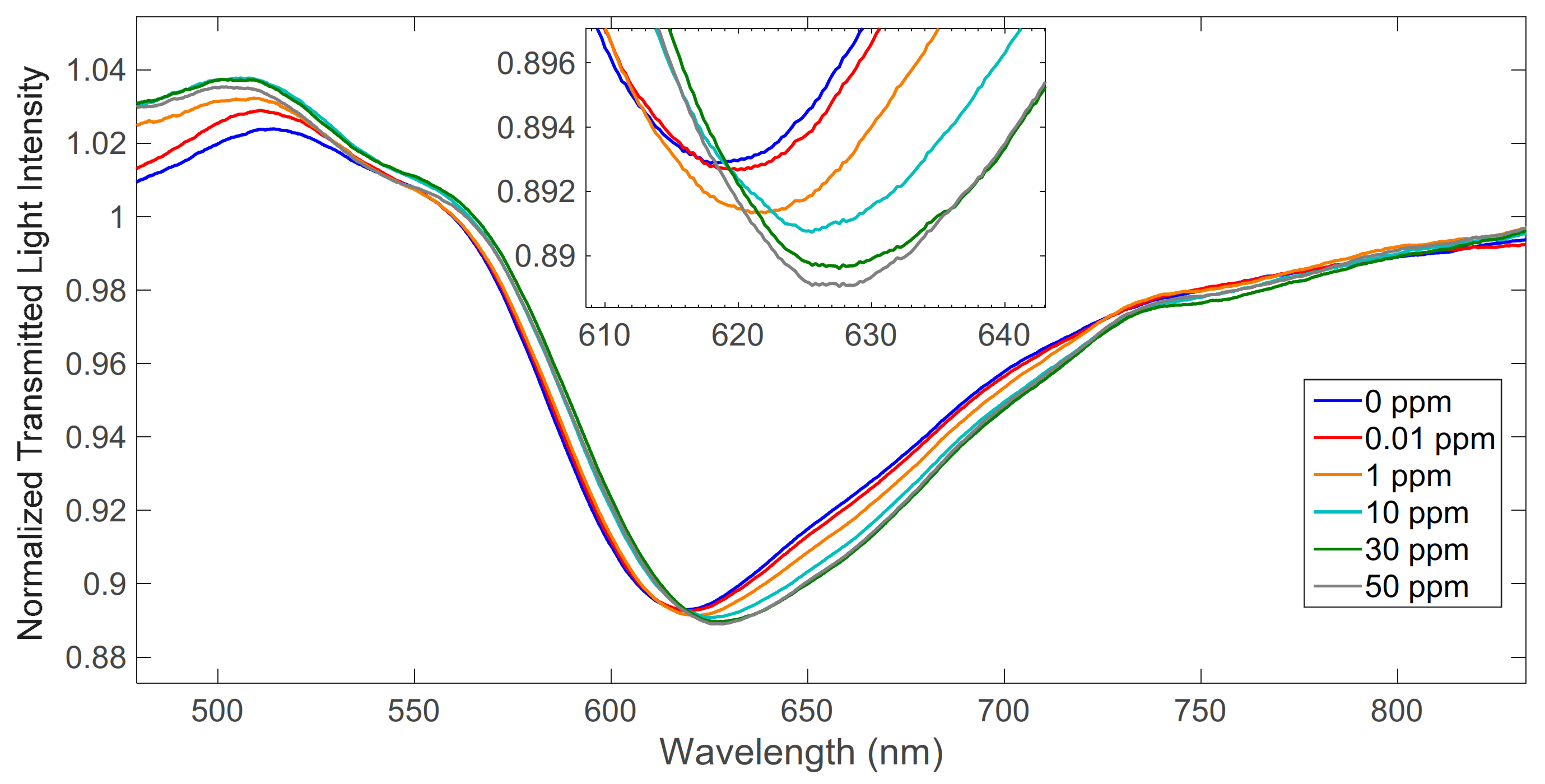
The spectra of SPR-D-POF-MUPA in 0.1 M NaNO3 solution containing different concentrations of uranium(VI), normalized on the corresponding spectra in air, are reported in Figure 3. As shown in Figure 3, when the analyte concentration increases, the resonance wavelength increases and shifts to the right; in fact, the refractive index in contact with the gold nanofilm increases with the uranium concentration.

4. Conclusions

The developed SPR sensor for uranium(VI) is based on an optical platform that is fast and easy to prepare, including the deposition of the receptor, a monolayer of a well-known commercially available complexing agent for uranyl. When deposited on the gold surface, its affinity for uranyl is sufficiently high to reach a LOD of a few μg L−1, suitable for detection in slightly contaminated waters. However, this limit could be improved by optimizing the procedure of the monolayer deposition in order to have a higher density of complexing sites at the sensor surface or by selecting a stronger ligand.

Author Contributions

Conceptualization: M.P. and N.C.; methodology: M.P. and N.C.; validation: M.P., D.M., G.A., A.P., L.Z. and N.C.; formal analysis: M.P., D.M., G.A., A.P., L.Z. and N.C.; investigation: M.P., D.M., G.A., A.P., L.Z. and N.C.; writing—original draft preparation: M.P., D.M., G.A., A.P., L.Z. and N.C.; writing—review and editing: M.P., D.M., G.A., A.P., L.Z. and N.C.; supervision: M.P. All authors have read and agreed to the published version of the manuscript.

Funding

This research received no external funding.

Institutional Review Board Statement

Not applicable.

Informed Consent Statement

Not applicable.

Data Availability Statement

The data are available on reasonable request from the corresponding author.

Conflicts of Interest

The authors declare no conflict of interest.

References

  1. Alberti, G.; Biesuz, R.; Pesavento, M. Determination of the total concentration and speciation of Uranium in natural waters by the Resin Titration method. Microchem. J. 2007, 86, 166–173. [Google Scholar] [CrossRef]
  2. Danesi, P.R.; Bleise, A.; Burkart, W.; Cabianca, T. Isotopic composition and origin of uranium and plutonium in selected soil samples collected in Kosovo. J. Environ. Radioact. 2003, 64, 121–131. [Google Scholar] [CrossRef]
  3. Saleh, I.H.; Abdel-Halim, A.A. Determination of depleted uranium using a high-resolution gamma-ray spectrometer and its applications in soil and sediments. J. Taibah Univ. Sci. 2016, 10, 205–211. [Google Scholar] [CrossRef]
  4. Cennamo, N.; Massarotti, D.; Conte, L.; Zeni, L. Low Cost Sensors Based on SPR in a Plastic Optical Fiber for Biosensor Implementation. Sensors 2011, 11, 11752–11760. [Google Scholar] [CrossRef] [PubMed]
  5. Cennamo, N.; Pesavento, M.; Zeni, L. A review on simple and highly sensitive plastic optical fiber probes for bio-chemical sensing. Sens. Actuators B Chem. 2021, 331, 129393. [Google Scholar] [CrossRef]
  6. Pesavento, M.; Profumo, A.; Merli, D.; Cucca, L.; Zeni, L.; Cennamo, N. An Optical Fiber Chemical Sensor for the Detection of Copper(II) in Drinking Water. Sensors 2019, 19, 5246. [Google Scholar] [CrossRef] [PubMed] [Green Version]
  7. Cennamo, N.; Alberti, G.; Pesavento, M.; D’Agostino, G.; Quattrini, F.; Biesuz, R.; Zeni, L. A Simple Small Size and Low Cost Sensor Based on Surface Plasmon Resonance for Selective Detection of Fe(III). Sensors 2014, 14, 4657–4661. [Google Scholar] [CrossRef] [PubMed]
  8. Merli, D.; Protti, S.; Labò, M.; Pesavento, M.; Profumo, A. A ω-mercaptoundecylphosphonic acid chemically modied gold electrode for uranium determination in waters in presence of organic matter. Talanta 2016, 151, 119–125. [Google Scholar] [CrossRef] [PubMed]
  9. Garcia-Calzon, J.A.; Diaz-Garcia, M.E. Characterization of binding sites in molecularly imprinted polymers. Sens. Actuators B Chem. 2007, 123, 1180–1194. [Google Scholar] [CrossRef]
Figure 1. Preparation of the SPR platform based on D-shaped POF.
Engproc 16 00010 g001
Figure 2. Transmission spectra of SPR-D-POF-bare and SPR-D-POF-MUPA in water normalized to the spectrum of the corresponding platform in air.
Engproc 16 00010 g002
Figure 3. SPR spectra obtained at different uranium concentrations (in mg L−1 (ppm)).
Engproc 16 00010 g003
Publisher’s Note: MDPI stays neutral with regard to jurisdictional claims in published maps and institutional affiliations.