Proceeding Paper

Hyaluronic Acid for Wound Healing: Experience in Deep-Burn Rat Model †

Institute for Problems of Cryobiology and Cryomedicine of NAS of Ukraine, 61016 Kharkiv, Ukraine
*
Author to whom correspondence should be addressed.
Presented at the 6th International Electronic Conference on Applied Sciences, 9–11 December 2025; Available online: https://sciforum.net/event/ASEC2025.
Eng. Proc. 2026, 124(1), 111; https://doi.org/10.3390/engproc2026124111
Published: 23 April 2026
(This article belongs to the Proceedings of The 6th International Electronic Conference on Applied Sciences)

Abstract

Hyaluronic acid (HA), a major extracellular matrix component, is used therapeutically to aid healing and deliver drugs to injury sites. Burns create serious clinical and aesthetic problems needing fast skin repair to prevent complications. This study compared 1.8% pharmaceutical-grade HA with panthenol-containing gel (PCG) in deep-burn healing in rats against spontaneous healing. HA slightly accelerated wound closure from day 3 compared to PCG; both induced granulation by day 7 and epithelialization by day 28. HA caused early collagen drop (day 3), later matched PCG levels with abnormal distribution, and both exceeded control by day 28. HA normalized systemic leukocyte counts by day 14 while strongly increasing local leukocyte infiltration in the wound area. HA dual immune effect depends on source and properties; further research is required for clinical use in wound healing.

1. Introduction

Thermal injuries represent a serious medical, social, and economic problem. Complexity arises not from frequency but from severity of tissue damage, prolonged treatment duration, and high mortality in extensive burns. Restoration of skin integrity in a short time after injury—when regenerative capacity remains unexhausted—is essential for correcting systemic disturbances. Both synthetic skin substitutes and biological wound dressings (e.g., based on collagen or chitosan) are used for skin recovery [1,2].
Hyaluronic acid (HA) is currently one of the most promising and widely applied agents for wound healing and aesthetic medicine [3]. As an extracellular matrix component, HA directly participates in healing processes [4]. It also serves as a natural, biocompatible, degradable carrier for hydrophilic therapeutic agents to injury site. However, questions remain about HA application in wound healing: a number of studies demonstrate the significant role of exogenous HA in this treatment, for example, a systematic review of several clinical trials where HA was used to treat various skin injuries including chronic with fairly high efficacy [5]; at the same time, a recent Cochrane review on chronic trophic ulcers in diabetic patients found insufficient evidence of the efficacy of HA application [6].
This ambiguity persists in burn healing, with efficacy varying by formulation and molecular weight, and no consensus on pure HA use [7]. A similar situation is observed in the field of HA application for burn healing—varied efficacy, different additional compounds and no unequivocal consensus about its use for this goal in pure form [7].
An additional question for non-compromised research is how to select the optimal form by physicochemical properties to avoid complications. Strong immunogenicity of low-molecular-weight (LMW) and branching HA, and the opposite for high-molecular-weight (HMW) and more linear forms, is a well-known fact today [8,9]. Although HMW-HA is generally considered less immunogenic than LMW-HA, we selected HA with a molecular weight of 2.3 MDa—higher than that commonly used in many wound healing studies—to reduce potential immunogenicity in a burn model (which typically elicits strong local and systemic inflammation).
D-Panthenol-containing remedies are widely used for burn treatment, especially at home, and the history of their applications for skin defect treatment dates back almost 80 years. After conversion to pantothenic acid (known as vit B5), it supports skin metabolism, cell proliferation, and epidermal differentiation; enhances hydration and barrier restoration; contributes to more effective epithelial repair; accelerates re-epithelialization; and improves barrier function [10]. Usually, D-panthenol is applied in hydrophobic forms, like creams and ointments. Use of such treatment for early-stage burns is not recommended; therefore, we chose hydrophilic panthenol-containing gel (PCG) for our experiments.
Critically, direct comparisons of pure topical HMW-HA versus marketed PCG (e.g., Pantestin or Bepanthen-like OTC products [10,11]) in deep dermal burn models are notably absent, with prior work focusing on combined HA formulations (e.g., with iodine [12]) or mild burn scenarios. While HA has been investigated in various burn models, often in advanced formulations (e.g., combined with stem cells, anti-inflammatory conjugates, etc.), we did not identify studies directly comparing plain topical HA to PCG in deep dermal burns. Our study addresses this gap by evaluating pure 2.3 MDa HMW-HA against PCG, revealing unexpected dual immunomodulation and early healing advantages over standard comparators.
A separate task in the treatment of extensive skin damage, particularly in burns, is the prevention of pathological scarring [13], which significantly affects patient quality of life after wound closure. The efficacy of D-panthenol in the field of curation of scar formation after burn and other skin injuries is still debatable [11]. In turn, HA, as a key extracellular compound, plays an essential role in normal scar formation [14]. Thus, the influence of PCG and HA on extracellular matrix restoration was also studied by quantifying type I and III collagens: type I predominates in physiological dermis, and type III plays the main role in reparative and regenerative processes during skin recovery [15].

2. Materials and Methods

2.1. Materials

Hyaluronic acid was used as a commercially available pharmaceutical-grade injectable form (Diart®—1.8% sodium hyaluronate, non-stabilized, non-animal origin, obtained via bacterial fermentation; molecular weight 2.3 MDa; YURiA-Pharm, Ukraine). As a comparator, PCG, a pharmaceutical-grade commercially available OTC gel (“Pantestin-Darnytsia”, Darnytsia, Ukraine), was used, which contained 50 mg of D-panthenol per g.

2.2. Experimental Animals

White outbred male rats weighing 200–300 g (n = 70) were used. Animals were kept under standard vivarium conditions of the Institute for Problems of Cryobiology and Cryomedicine of NASU. Experiments were approved by the Bioethics Committee of the Institute, conducted in accordance with Ukrainian Law “On Protection of Animals from Cruel Treatment,” European Convention ETS 123 (Strasbourg, 1986), and General Principles of Experimentation adopted by the 5th National Congress on Bioethics (Kyiv, 2013). Animals underwent a 7–14-day quarantine pre-experiment and were housed individually post-burn (temperature of 22–24 °C, humidity of 50–60%, 12 h light/dark cycle) to prevent cross-contamination or self-injury.

2.3. Experimental Design

Deep dermal burns were modeled by applying a 200 °C copper plate (2.5 × 2.5 cm) to thigh skin for 10 s. This method produces standardized full-thickness skin burns [16]. Burn depth was confirmed via pilot histological biopsies immediately post-injury, showing consistent deep dermal involvement.
Animals were randomly assigned to groups, and observers performing planimetric measurements, histological analysis, and collagen quantification were blinded to group assignments. Animal groups (n = 12–15/group): 1—intact; 2—control (spontaneous healing); 3—PCG group (comparator); 4—HA group. Treatments (0.5 g PCG or HA) were applied uniformly using a sterile spatula to cover the entire wound surface, 24 h post-burn. No occlusive dressing was used to allow natural drying, as in open burn models. Daily monitoring included signs of infection or distress.
The observation period was 28 days. Planimetric measurements, tail vein blood sampling for leukocyte count, and sacrifice of some animals for histology, depending on the task, were performed on days 3, 7, 14, 21, 28.
Macroscopic monitoring included wound area measurement via ImageJ v. 1.5b (National Institutes of Health, USA)software after photography until complete restoration, accounting for surface relief. The percentage of animals with fully closed wounds was recorded at each stage.

2.4. Histological Studies

Skin samples were fixed in 10% neutral formalin and embedded in paraffin. Deparaffinized 6–8 μm sections stained with hematoxylin–eosin for general morphology. Analysis performed under a light microscope, Inverted Tissue Culture Microscope 40X-800X + 5MP Digital Camera (Amscope, Irvine, CA, USA).
Type I and III collagen content was assessed by PicroSirius Red staining (0.1% Direct Red 80 in picric acid, (St. Louis, MO, USA, Sigma-Aldrich) under polarized light using the LSM 500 META microscope (Jena, Germany, Carl Zeiss). Quantitative analysis was performed using ImageJ threshold analysis [17]. Three fields per section were averaged.

2.5. Statistical Analysis

Data were processed using the Mann–Whitney nonparametric test in Origin 9.5. Results were expressed as M ± SEM; differences considered significant at p < 0.05.

3. Results

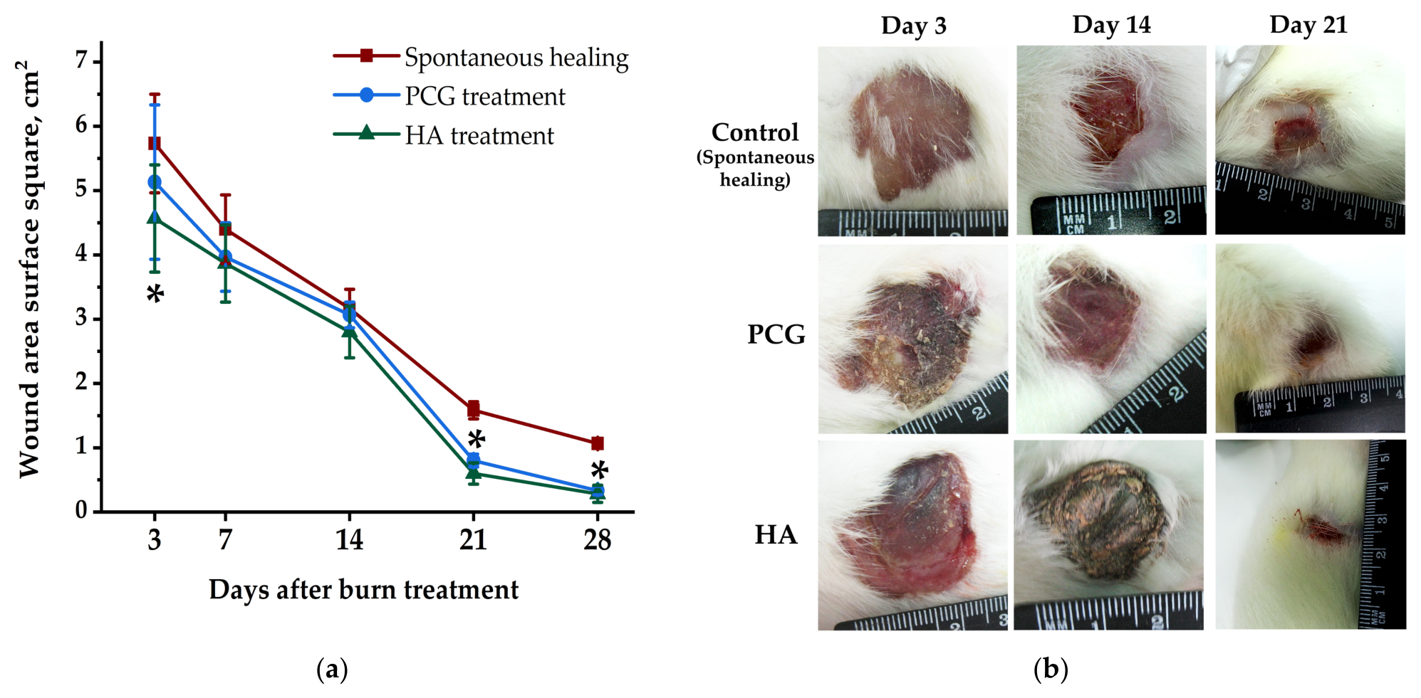
In the control group, wound area decreased slowly throughout the experiment; complete healing was absent even on day 28 and a significant reduction was observed only by day 21. PCG application produced no marked difference from control until days 21 and 28 (significant only then). HA-group showed similar dynamics but a significant difference from control appeared already on day 3, then on days 21 and 28; on day 21, HA also slightly but significantly outperformed PCG (Figure 1a, Table 1).
The visual picture was nearly identical in PCG and HA groups: granulation tissue appeared from day 7, there was no plasmorrhea, and wounds were dry. By day 28, burns were epithelialized, but a full epidermal layer was not formed; healing was completed by dense scar replacement (Figure 1b).
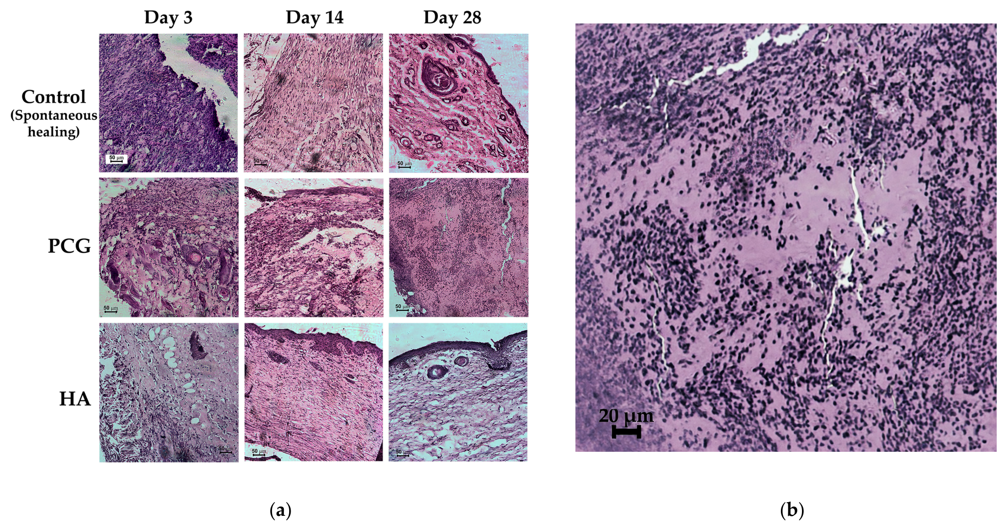
In the control group on day 3, the inflammatory infiltrate contained neutrophils, macrophages (lymphocytes predominant), moderate fibrin, multiple hemorrhages, and no newly formed vessels were present (Figure 2a, Table 1).
In the PCG group on day 14, leukocyte infiltration was less pronounced; angiogenesis foci with thin-walled vertically oriented vessels, increased fibroblasts, parallel collagen fibers, and marginal epithelium growth were observed (Figure 2a, Table 1).
In the HA group on day 14, a strong inflammatory reaction persisted (mostly neutrophils/macrophages); edematous subcutaneous fat, solitary fibroblasts, and marginal epithelium growth were shown (Figure 2a,b; Table 1).
On day 28 after HA treatment, complete epithelialization, granulation tissue with predominant inflammatory cells, histiocytes, fibroblasts, reduced vessels, chaotic dermal collagen arrangement, and significant epithelial growth with partial microrelief disturbance were observed (Figure 2a, Table 1).
Under physiological conditions, skin contains ~70% type I collagen; injury alters its state and content. Rapid inflammation and slow reparative transition after thermal damage promote scar formation. Controlled production of types I and III collagen is crucial for physiological healing without pathological scars [18]. PicroSirius Red enables qualitative, quantitative, and 3D structural analysis.
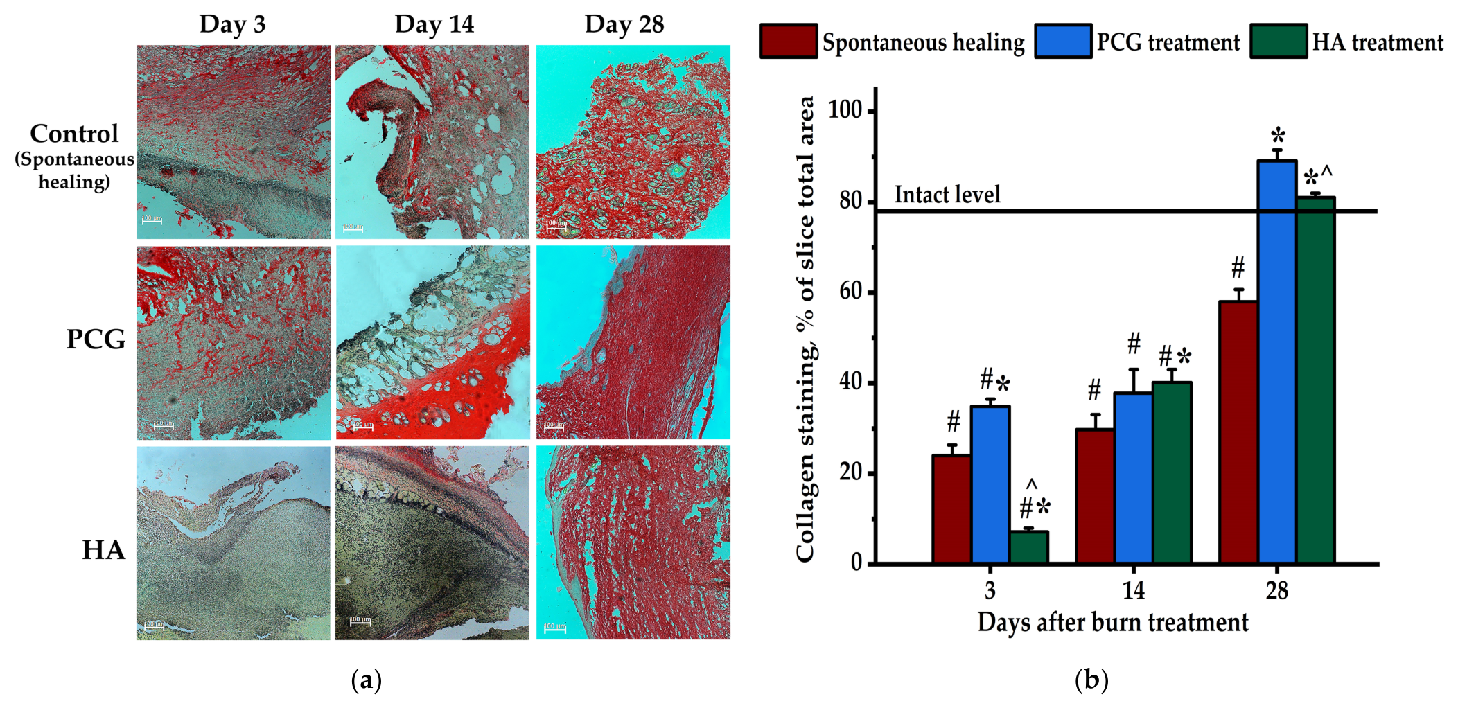
Figure 3a shows collagen distribution in rat derma after burn treatment with PCG or HA. Quantitative ImageJ analysis of types I/III collagen amount is presented in Figure 3b. On day 3, PCG partially prevented collagen degradation, and its content was 2.2 times below intact and 50% above control. After HA treatment, paradoxical collagen drop was observed on day 3: 11 times lower than in the intact group, 3.3—below control, 4.9—below PCG (significant vs. all groups). On day 14, the HA effect was neutralized: total collagen matched the level in the PCG group, but accumulated mainly in superficial dermis.
By the end of the experiment, control collagen remained 35% below normal despite a 2 times increase from day 14. Both PCG and HA normalized collagen content, exceeding control by 1.5 and 1.4 times respectively (Figure 3).
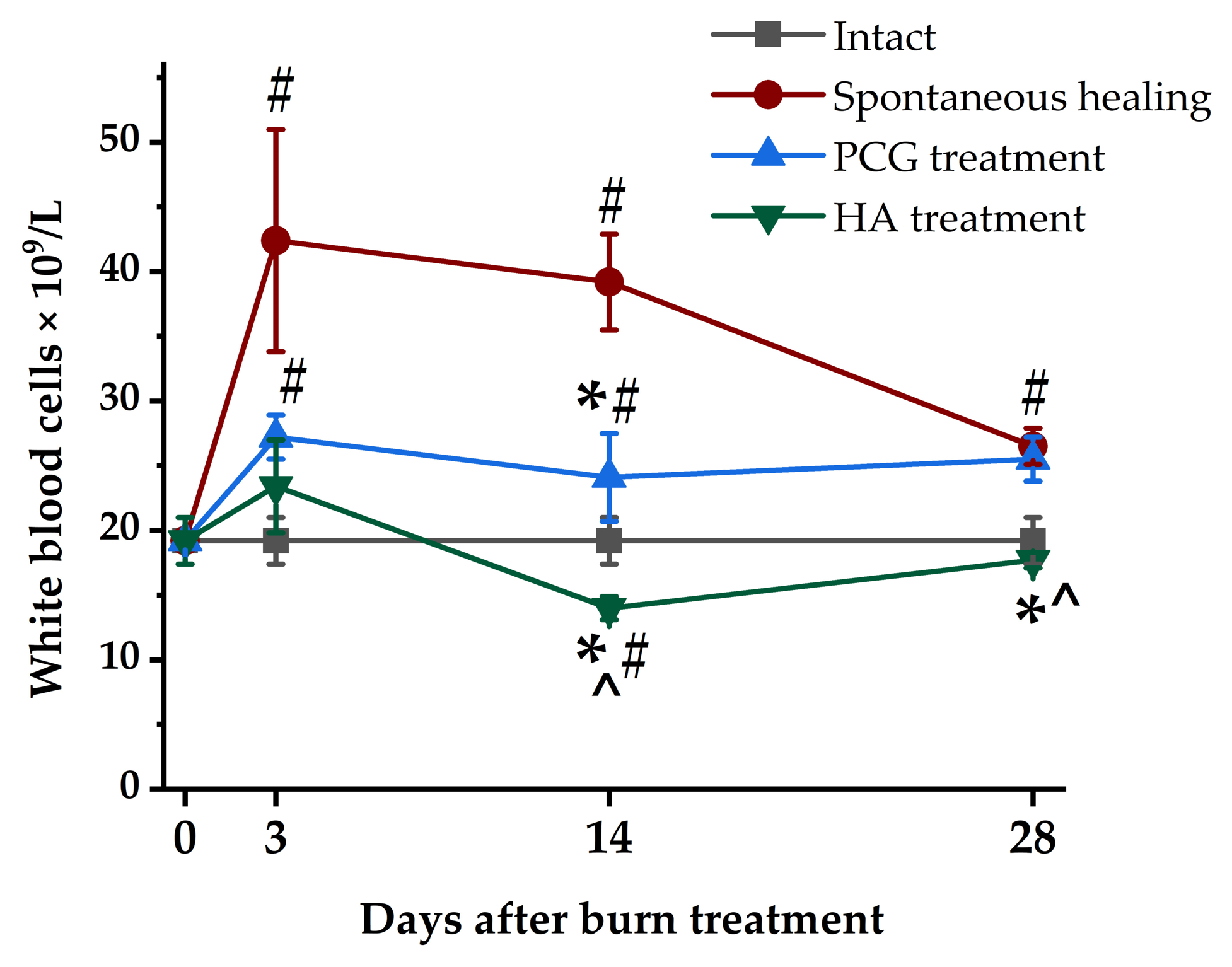
Thermal injury caused strong local immune cell infiltration and peripheral blood leukocytosis: on day 3, in the control group, the blood leukocyte number was 2.2 times higher than in the intact group, in the PCG group—1.4 times; HA application almost completely prevented this spike (Figure 4).
On day 14, in the control group, leukocytosis persisted and PCG partially reduced it. At the same time, HA caused its substantial drop to normal values (significant vs. control and others). On day 28, in the control and PCG groups, leukocyte number remained above normal, and in the HA group, it was at a normal level (Figure 4).
Notably, on day 14, the lowest peripheral leukocyte count was observed in the HA group, and comparing the local and general immune response at this stage of the experiment, interesting features emerged: this group had the most significant level of leukocyte infiltration in area of injury and the lowest level of leukocytes in blood.

4. Discussion

The results demonstrate that pure HMW-HA provides modest but significant advantages over marketed PCG in early deep-burn healing in rats: accelerated wound closure (significant from day 3 vs. control, slight superiority over PCG on day 21, as shown in Figure 1 and Table 1 and Table 2), absent plasmorrhea, and dry wounds from day 7—outcomes better than typical OTC panthenol moisturizing role in mild burns [10,11]. Unlike prior HA studies on combined or LMW forms [5,7,8], this work reveals a novel paradoxical profile for 2.3 MDa HMW-HA: early collagen degradation (day 3 drop 3.3× below control, as evidenced by Figure 3 and Table 2) followed by normalization with chaotic distribution, and dual immune effect (systemic leukocyte normalization by day 14 vs. strong local neutrophil/macrophage infiltration persisting to day 28, as per Figure 4 and Table 2).
Contrary to expectations based on HMW-HA’s typical systemic anti-inflammatory effects, we observed strong stimulation of the local inflammatory reaction after HMW-HA application for burn treatment. As is well-known today, HA is able to both stimulate and suppress immune response in skin, depending on the source and structural features (molecular weight, polymer chain length, degree of branching) [19,20].
According to some researchers, HMW-HA generally has anti-inflammatory effects (often systemic), while its interactions with receptors like CD44 and the receptor for hyaluronan-mediated motility) (RHAMM/HMMR) allow for nuanced local modulation (e.g., anti-inflammatory signaling, receptor clustering, or context-dependent responses in tissues). This contrasts with LMW-HA fragments, which are typically pro-inflammatory [8,21,22]. In fact, the bulk of the literature on HMW-HA (typically >1–2 MDa, like our 2.3 MDa) describes it as anti-inflammatory at both systemic and local levels in most wound models—suppressing excessive macrophage activation, promoting M2 polarization (pro-resolving), reducing cytokine release, and limiting neutrophil/macrophage influx or persistence [23].
Thus, the results obtained may apply only to the specific drug used in our work and we may propose some possible mechanisms for the realization of such effects. HMW-HA primarily engages CD44 for anti-inflammatory signaling, suppressing NF-κB, promoting IL-10/Arg1 pathway in macrophages. However, in acute injury sites with high local HA turnover (common in burns due to hyaluronidase upregulation), partial degradation can occur, leading to mixed-size fragments. These interact variably with RHAMM, which can promote local cell recruitment, migration, and persistent inflammation in some contexts—especially if the environment has ongoing tissue damage or hypoxia. RHAMM-HA interactions often drive fibroblast/myofibroblast activity and inflammatory cell retention in wounds, contributing to prolonged infiltrate without systemic spillover [21,22,24].
An additional problem in our research may be caused by high HA concentration (1.8%) in the applied topical gel, which might create a viscous barrier that traps inflammatory cells locally or delays their efflux/lymphatic drainage, leading to apparent persistence. High local HA density can sometimes sustain macrophage activation in vitro if not fully anti-inflammatory polarized [19].
Nevertheless, this problem requires further, more detailed studies on the safety of HA use in the treatment of acute skin injuries accompanied by a significant reaction in the patient’s immune system. At the same time, taking into account existing risks, it can be concluded that HA is a suitable carrier for delivering therapeutic agents to the injury area.
To clarify the novelty and advantages of pure HMW-HA over marketed PCG and previously reported HA studies, the key findings are summarized in Table 2.
These findings highlight the unique advantages of pure 2.3 MDa HMW-HA in deep-burn healing and underscore the need for further clinical translation.

5. Conclusions

Pure HMW-HA gel shows advantages over marketed panthenol products in early-phase deep burn healing, with unique immunomodulatory properties warranting further mechanistic studies (e.g., CD44/RHAMM pathways) and clinical translation for optimized formulations.

Author Contributions

Conceptualization, D.C. and O.P.; methodology, D.C. and O.P.; software, D.C. and O.R.; validation, D.C., O.R. and O.P.; formal analysis, D.C. and O.P.; investigation, D.C., O.R. and S.B.; resources, O.R. and S.B.; data curation, D.C. and O.R.; writing—original draft preparation, D.C.; writing—review and editing, D.C. and O.P.; visualization, D.C., O.R. and S.B.; supervision, O.P.; project administration, D.C. All authors have read and agreed to the published version of the manuscript.

Funding

This research received no external funding.

Institutional Review Board Statement

Experiments were conducted in accordance with Ukrainian Law “On Protection of Animals from Cruel Treatment,” European Convention ETS 123 (Strasbourg, 1986), and General Principles of Experimentation adopted by the 5th National Congress on Bioethics (Kyiv, 2013). The animal study protocol was approved by the Institutional Ethics Committee of the Institute for Problems of Cryobiology and Cryomedicine of NAS of Ukraine (protocol no 3/15 March 2023).

Informed Consent Statement

Not applicable.

Data Availability Statement

Data are available upon request.

Conflicts of Interest

The authors declare no conflicts of interest.

Abbreviations

The following abbreviations are used in this manuscript:
HAHyaluronic acid
HMWHigh molecular weight
LMWLow molecular weight
PCGPanthenol-containing gel
RHAMMReceptor for hyaluronan-mediated motility

References

  1. Rowan, M.P.; Cancio, L.C.; Elster, E.A.; Burmeister, D.M.; Rose, L.F.; Natesan, S.; Chan, R.K.; Christy, R.J.; Chung, K.K. Burn wound healing and treatment: Review and advancements. Crit. Care 2015, 19, 243. [Google Scholar] [CrossRef]
  2. Liang, Y.; He, J.; Guo, B. Functional hydrogels as wound dressing to enhance wound healing. ACS Nano 2021, 15, 12687–12722. [Google Scholar] [CrossRef] [PubMed]
  3. Aya, K.L.; Stern, R. Hyaluronan in wound healing: Rediscovering a major player. Wound Repair Regen. 2014, 22, 579–593. [Google Scholar] [CrossRef]
  4. Antoszewska, M.; Sokolewicz, E.M.; Barańska-Rybak, W. Wide use of hyaluronic acid in the process of wound healing—A rapid review. Sci. Pharm. 2024, 92, 23. [Google Scholar] [CrossRef]
  5. Voigt, J.; Driver, V.R. Hyaluronic acid derivatives and their healing effect on burns, epithelial surgical wounds, and chronic wounds: A systematic review and meta-analysis of randomized controlled trials. Wound Repair Regen. 2012, 20, 317–331. [Google Scholar] [CrossRef]
  6. Roehrs, H.; Stocco, J.G.D.; Pott, F.; Blanc, G.; Meier, M.J.; AL Dias, F. Dressings and topical agents containing hyaluronic acid for chronic wound healing. Cochrane Database Syst. Rev. 2023, 2023, CD012215. [Google Scholar] [CrossRef]
  7. Longinotti, C. The use of hyaluronic acid based dressings to treat burns: A review. Burns Trauma 2014, 2, 162–168. [Google Scholar] [CrossRef]
  8. Cyphert, J.M.; Trempus, C.S.; Garantziotis, S. Size matters: Molecular weight specificity of hyaluronan effects in cell biology. Int. J. Cell Biol. 2015, 2015, 563818. [Google Scholar] [CrossRef]
  9. Lee, B.M.; Park, S.J.; Noh, I.; Kim, C.H. The effects of the molecular weights of hyaluronic acid on the immune responses. Biomater. Res. 2021, 25, 27. [Google Scholar] [CrossRef] [PubMed]
  10. Proksch, E.; de Bony, R.; Trapp, S.; Boudon, S. Topical use of dexpanthenol: A 70th anniversary article. J. Dermatol. Treat. 2017, 28, 766–773. [Google Scholar] [CrossRef] [PubMed]
  11. Bagheri, M.; Werres, M.; Fuchs, P.C.; Seyhan, H.; Lefering, R.; Grieb, G.; Schiefer, J.L. Which moisturizer to use in scar therapy after burn injuries? A subjective and objective skin and scar evaluation after topical treatment with dexpanthenol, aloe vera, and plant oil. Medicina 2023, 59, 1874. [Google Scholar] [CrossRef] [PubMed]
  12. Arokh Forghani, S.; Bagheri, T.; Naderi Gharahgheshlagh, S.; Hoveidamanesh, S.; Ghadimi, T.; Ghorbanian Kelachayeh, S.; Irilouzadian, R. The effect of hyaluronic acid and iodine complex gel compared to Vaseline on deep second-degree burn wound in rats. Int. Wound J. 2024, 21, e14738. [Google Scholar] [CrossRef]
  13. Finnerty, C.C.; Jeschke, M.G.; Branski, L.K.; Barret, J.P.; Dziewulski, P.; Herndon, D.N. Hypertrophic scarring: The greatest unmet challenge after burn injury. Lancet 2016, 388, 1427–1436. [Google Scholar] [CrossRef]
  14. Saadati, F.; Bahrulolum, H.; Talebi, M.; Karimi, M.; Bozorgchami, N.; Ghale, R.A.; Zafar, S.; Aghighi, Y.; Asiaei, E.; Tabandeh, F. Advances and principles of hyaluronic acid production, extraction, purification, and its applications: A review. Int. J. Biol. Macromol. 2025, 312, 143839. [Google Scholar] [CrossRef]
  15. Gordon, M.K.; Hahn, R.A. Collagens. Cell Tissue Res. 2010, 339, 247–257. [Google Scholar] [CrossRef] [PubMed]
  16. Abdullahi, A.; Amini-Nik, S.; Jeschke, M.G. Animal models in burn research. Cell. Mol. Life Sci. 2014, 71, 3241–3255. [Google Scholar] [CrossRef] [PubMed]
  17. Lattouf, R.; Younes, R.; Lutomski, D.; Naaman, N.; Godeau, G.; Senni, K.; Changotade, S. Picrosirius red staining: A useful tool to appraise collagen networks in normal and pathological tissues. J. Histochem. Cytochem. 2014, 62, 751–758. [Google Scholar] [CrossRef]
  18. Takeo, M.; Lee, W.; Ito, M. Wound healing and skin regeneration. Cold Spring Harb. Perspect. Med. 2015, 5, a023267. [Google Scholar] [CrossRef]
  19. Rayahin, J.E.; Buhrman, J.S.; Zhang, Y.; Koh, T.J.; Gemeinhart, R.A. High and low molecular weight hyaluronic acid differentially influence macrophage activation. ACS Biomater. Sci. Eng. 2015, 1, 481–493. [Google Scholar] [CrossRef]
  20. D’Agostino, A.; Stellavato, A.; Busico, T.; Papa, A.; Tirino, V.; Papaccio, G.; La Gatta, A.; De Rosa, M.; Schiraldi, C. In vitro analysis of the effects on wound healing of high- and low-molecular weight chains of hyaluronan and their hybrid H-HA/L-HA complexes. BMC Cell Biol. 2015, 16, 19. [Google Scholar] [CrossRef]
  21. Tolg, C.; McCarthy, J.B.; Yazdani, A.; Turley, E.A. Hyaluronan and RHAMM in wound repair and the “cancerization” of stromal tissues. BioMed Res. Int. 2014, 2014, 103923. [Google Scholar] [CrossRef] [PubMed]
  22. Monslow, J.; Govindaraju, P.; Puré, E. Hyaluronan—A functional and structural sweet spot in the tissue microenvironment. Front. Immunol. 2015, 6, 231. [Google Scholar] [CrossRef] [PubMed]
  23. Indrawati, D.W.; Setiawatie, E.M.; Rahayu, R.P.; Manurung, R.B.S.; Manurung, M.R.A.; Aljunaid, M.A. A PRISMA-based systematic review on the effect of high- and low-molecular-weight hyaluronic acid on IL-1, IL-6, IL-10, CD44, p53, and RHAMM in cellular response related to bone regeneration. Int. J. Biol. Macromol. 2026, 337, 149440. [Google Scholar] [CrossRef] [PubMed]
  24. Misra, S.; Hascall, V.C.; Markwald, R.R.; Ghatak, S. Interactions between hyaluronan and its receptors (CD44, RHAMM) regulate the activities of inflammation and cancer. Front. Immunol. 2015, 6, 201. [Google Scholar] [CrossRef]
Figure 1. Dynamics of wound area reduction in deep dermal burns treated with PCG or HA compared to untreated control: (a) wound surface square; (b) representative pictures of wound healing process depending on observation term. Data are mean ± SEM (n = 12–15/group). *—p < 0.05 as compared to control (spontaneous healing) group.
Engproc 124 00111 g001
Figure 2. (a) Representative histological sections of burn wounds stained with hematoxylin–eosin at days 3, 14, and 28 post-treatments with PCG and HA (scale bar = 50 μm; magnification ×100); (b) Representative picture of massive leukocyte infiltration in wound area at day 14 after HA application (scale bar = 20 μm; magnification ×100).
Engproc 124 00111 g002
Figure 3. Picrosirius red staining of collagen I & III types in derma on day 3, 14, and 28 after burn treatment with PCG or HA: (a) Distribution of collagen in derma layers (representative images; scale bar = 100 μm, magnification ×40); (b) quantitative analysis with ImageJ of collagen amount in derma (Data are mean ± SEM (n = 12–15/group)). #—p < 0.05 compared to intact level; *—p < 0.05 compared to control group; ^—p < 0.05 compared to PCG group.
Engproc 124 00111 g003
Figure 4. Peripheral blood leukocyte counts on day 0, 3, 14, and 28 after burn treatment with PCG or HA (Data are mean ± SEM (n = 12–15/group). #—p < 0.05 compared to intact level; *—p < 0.05 as compared to control group; ^—p < 0.05 compared to PCG group.
Engproc 124 00111 g004
Table 1. Summary of key planimetric, macroscopic and histological observations after burn treatment with PCG or HA.
DayControl (Spontaneous)PCGHA
3Plasmorrhea + strong inflammation
No area reduction
Necrosis of all layers, pyknosis, plethoric vessels, leukocytic infiltrate, edema
Reduced plasmorrhea, drier
No area reduction vs. control
Less infiltration than control
No plasmorrhea, dry
Significant area reduction vs. control
Infiltrate (neutrophils/macrophages > lymphocytes), fibrin, hemorrhages, no new vessels
14Purulent-necrotic wound, hemorrhage, granulation forming, plethoric vessels, persistent inflammation + leukocytes, solitary fibroblasts, marginal epithelium under scabDry, granulation from day 7, less leukocyte infiltrate, angiogenesis (thin vertical vessels), more fibroblasts, parallel collagen, marginal epitheliumDry, granulation from day 7, significant area reduction, strong persistent inflammation (neutrophils/macrophages), edematous fat, solitary fibroblasts, marginal epithelium
21Significant area reduction vs. baselineSignificant area reduction vs. controlSignificant area reduction vs. control + slight vs. PCG
28Epithelialized
Incomplete epithelium, immature granulation, proliferating fibroblasts, increased vessels, normal remodeling
Epithelialized, dry
Complete epithelium, thickened, all layers + derivatives, no microrelief (prolonged remodeling), dense scar
Epithelialized, dry
Complete epithelium, inflammatory granulation, histiocytes + fibroblasts, reduced vessels, chaotic collagen, partial microrelief disturbance
Table 2. Advantages and novel aspects of pure high-molecular-weight hyaluronic acid compared to marketed PCG and prior HA studies in burn wound healing (based on present results).
ParameterMarketed PCGReported HA Studies (Combined or LMW Forms)Our Pure HMW-HA (2.3 MDa)Novelty/Advantage in This Study
Formulation OTC hydrophilic gel, dexpanthenol-focusedOften combined (iodine, silver, esters, etc.)Pure pharmaceutical-grade gelFirst direct comparison of pure HMW-HA vs panthenol in deep-burn model
Early wound closureSimilarly to control until late daysVariable, often acceleratedSignificant reduction from day 3 vs. control; slight superiority over PCG on day 21Earlier onset than standard PCG
Collagen (early phase)Partial preservationUsually, anti-degradativeParadoxical strong drop on day 3, then normalization with chaotic distributionUnexpected effect for HMW-HA
Immune responseMild anti-inflammatoryMostly systemic/local anti-inflammatoryDual: systemic normalization + strong local stimulationNovel paradoxical local pro-inflammation despite HMW
Applicability in deep burnsLimited data, mainly mild/superficial burnsFocus on combined therapiesTested in standardized deep dermal rat modelAddresses gap in pure HA for severe injury
Disclaimer/Publisher’s Note: The statements, opinions and data contained in all publications are solely those of the individual author(s) and contributor(s) and not of MDPI and/or the editor(s). MDPI and/or the editor(s) disclaim responsibility for any injury to people or property resulting from any ideas, methods, instructions or products referred to in the content.