1. Introduction
Understanding how cell–cell interactions contribute to tissue organization is essential for explaining the formation, maintenance, and dynamics of multicellular systems [
1,
2,
3]. Cells exert forces, change shape, and respond to their neighbors, and these interactions—both at direct contact and over longer distances—play a central role in shaping global tissue architecture. Such forces underlie a wide range of biological processes, including morphogenesis, wound healing, and collective migration.
In the previous study [
4], we proposed a minimal model in which each cell is represented as a single point and interacts with others through a remote force and a contact force. The remote force acts between all cell pairs and depends only on the distance between them. The contact force acts only between connected cell pairs. Despite the lack of explicit representation of cell shape or polarity, this model successfully reproduced experimentally observed multicellular pattern formation, including scattered, network-like, and clustered configurations. These results demonstrate that a simple combination of a remote force and a contact force can account for diverse spatial organizations in epithelial cell populations. In the remainder of this paper, we refer to this model as the single-particle model.
In this study, we extend the previous model by representing each cell with two particles connected by an internal link. Each particle is subject to interactions with other cells, and the internal link maintains the coherence of the cell. The model retains the two types of interaction: a remote force acting between all cell pairs and a contact force acting only between physically connected pairs. This two-particle configuration introduces internal structure while preserving the minimal nature of the original model.
This extension enables the model to represent key cellular features such as shape deformation and orientation. Unlike the single-particle model, the two-particle model allows each cell to elongate and reorient in response to external forces, capturing directional dependencies in cell–cell interactions. While the model described in this study treats the two particles symmetrically, the model structure allows future extensions in which front–rear asymmetry is explicitly introduced. These capabilities provide a more expressive yet still tractable framework for studying how local physical interactions give rise to multicellular organization.
Two-particle models are also proposed in the context of collective cell migration, particularly to capture behaviors such as contact inhibition of locomotion and substrate-mediated traction forces [
5,
6]. In those models, each cell is composed of two connected particles, with motility driven by internal crawling forces and polarity updated through local mechanical feedback. The surrounding substrate is modeled as an elastic medium that transmits intercellular forces indirectly. In contrast, our model does not rely on active motility mechanisms or substrate coupling. Instead, it focuses on the role of direct pairwise interactions—specifically, remote and contact forces—in shaping multicellular structures. This formulation allows us to isolate and analyze the effects of intercellular physical interactions on pattern formation without the confounding influence of external media or complex polarity dynamics.
The remainder of this article is organized as follows.
Section 2 describes the two-particle model in detail, including the definitions of interaction forces and the update procedure.
Section 3 presents simulation experiments that illustrate how the model reproduces diverse multicellular patterns. Finally,
Section 4 summarizes the main findings and discusses possible directions for future work.
2. Two-Particle Cell Model
2.1. Cell Representation and Force Overview
In the proposed model, we consider a set of cells. Each cell is represented in two-dimensional space by a pair of particles connected by a linear spring. These two particles, denoted by and , together define the physical extent and intrinsic polarity of the cell. The two particles are treated symmetrically and governed by the same physical laws.
Each cell undergoes deformation and movement as a result of forces acting on its two constituent particles. Internally, the spring force connecting the particles enables dynamic changes in cell shape. Externally, each particle interacts with particles from other cells (and the surrounding environment). These interactions drive the coordinated motion of the two particles, resulting in both translational displacement and morphological variation at the cell level. The model includes four types of forces in total: one internal force (spring) and three external forces (repulsive, contact, and remote).
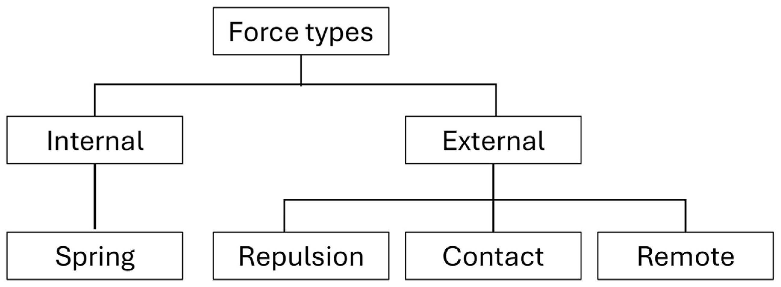
Figure 1 summarizes the classification of force types used in our model. At the top level, forces are divided into internal and external categories. The internal force is a spring that connects the two particles within a single cell. External forces govern interactions between particles of different cells and are further classified based on both their functional roles (repulsive or attractive) and their interaction ranges. Specifically, the repulsive force acts over short distances and prevents physical overlap between neighboring particles. The contact force is attractive and also short-range, acting only between physically adjacent particles to promote interactions through cell–cell adhesion. The remote force is attractive and long-range, allowing non-contacting particles from different cells to influence each other over extended distances. While the present model includes only a limited set of force types, the framework is designed to accommodate additional interaction mechanisms.
2.2. Equations of Motion
We assume overdamped dynamics and describe the motion of each particle
in cell
by the following equation.
where
denotes the particle index, and
is the mobility coefficient. In this expression,
denotes the internal spring force acting within cell
, maintaining its physical structure. The term
represents the short-range repulsive force that prevents overlap between particles of different cells. The term
corresponds to an attractive contact force that acts between physically contacting particles from different cells. Finally,
describes a long-range attractive force acting between non-contacting particles of different cells. These four components together determine the net displacement of each particle under overdamped conditions. Note in the rest of this paper that the mobility coefficient
is set to unity without loss of generality, effectively rescaling the time unit.
2.3. Force Models
Each particle is subject to four types of forces: the internal spring force and three types of external forces—repulsive, contact, and remote. These forces are mathematically defined as follows.
To simplify the notation in the following expressions, we first define the distance and direction between two particles
and
from cells
and
.
2.3.1. Spring Force
The spring force
acts between the two particles of cell
to maintain their natural length.
2.3.2. Repulsive Force
The repulsive force
prevents overlap between particles of different cells.
where
is the repulsion strength and
is the repulsion cutoff distance.
2.3.3. Contact Force
Two particles from different cells are considered to enter physical contact when their distance becomes less than or equal to . Once contact is established, it is maintained as long as their separation remains strictly less than an upper threshold . A contact history is stored and updated accordingly. A pair is considered to be in contact if it has an established contact and has not yet exceeded the separation, if the pair (i,k) and (j,l) is in contact.
The contact force is defined as follows.
where
is the contact attraction strength.
2.3.4. Remote Force
The remote force
acts between particles of different cells.
where
is the remote attraction strength and
is the decay length.
3. Simulation
3.1. Simulation Setup
The goal of the simulation experiments is to verify that the two-particle cell model can reproduce the same density-dependent multicellular patterns previously observed in the single-particle model [
4]. In particular, we aim to reproduce the transition from scattered clusters at low cell density to network-like configurations at intermediate density, and finally to large, connected aggregates at high density.
Simulations were conducted in a square domain without periodic boundary conditions. The number of cells
was varied to control the overall cell density. Initial cell positions were generated by a spatial Poisson process, resulting in randomly distributed cells that may include occasional particle overlaps. For each cell, the orientation of its internal link was initialized randomly. The model was numerically integrated using the forward Euler method under overdamped dynamics, with a fixed time step
for at least 300 simulation steps. The parameter values used for the spring, repulsion, contact, and remote forces are listed in
Table 1.
3.2. Density-Dependent Pattern Formation
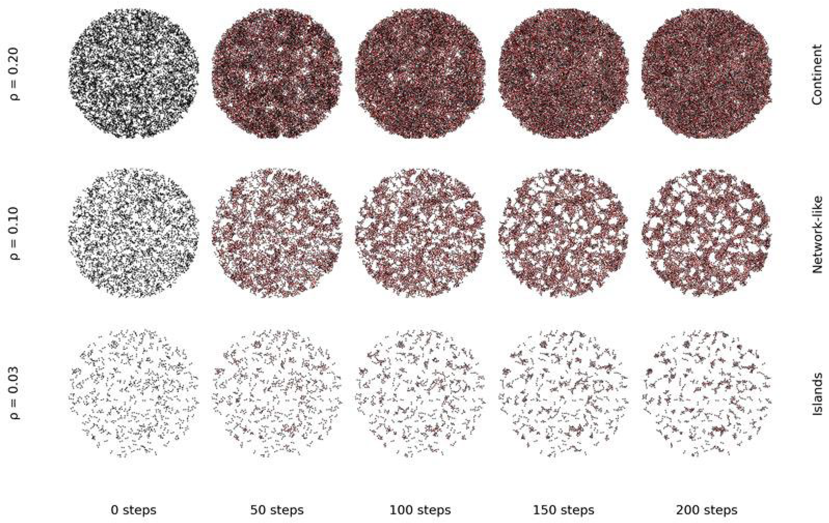
Figure 2 shows representative simulation results obtained under three nondimensionalized cell density conditions, denoted by
. In each snapshot, the two-particle model represents each cell as a black short line with dots at both ends, indicating its constituent particles, while red lines denote links formed by contact between cells. Snapshots are sampled at time steps 0, 50, 100, 150, and 200. At low density
, cells spontaneously form small, isolated clusters composed of a few connected cells (islands). At intermediate density
, they organize into extended, network-like structures characterized by branching connections and local alignment. At high density
, the system evolves into a large aggregate (continent’) in which the domain is densely packed with cells, leaving only a few small regions unoccupied.
These behaviors closely match those obtained using the previous single-particle model [
4], confirming that the two-particle model preserves the essential mechanisms of density-dependent pattern formation. In addition, the two-particle structure reveals geometric information not accessible in the earlier model. For example, in network-like states, cells tend to align locally along their elongation axes, resulting in chain-like or anisotropic contact arrangements. Although the current model treats the two particles symmetrically, these spatial arrangements suggest that orientation may play a role in modulating connectivity. A systematic analysis of such orientation effects will be explored in future work.
4. Conclusions
In this study, we extended a single-particle model by introducing a two-particle representation for each cell. The new model retains the original formulation of remote and contact forces, while incorporating an internal structure that enables cell shape deformation and orientation. Simulation results confirmed that this extended model successfully reproduces the same density-dependent multicellular patterns observed in the previous single-particle model, including scattered, network-like, and aggregated configurations.
In addition to this validation, the two-particle structure reveals geometric and directional features that are not accessible in the original model. For instance, in network-like states, cells often exhibit local alignment or chain-like arrangements, suggesting that orientation may influence contact topology. These preliminary observations point to the broader expressive potential of the two-particle model. In future work, we plan to explore extensions that incorporate front–rear asymmetry, directed motility, or polarity-dependent interactions to further investigate the role of orientation in multicellular organization.
Author Contributions
Conceptualization, X.W. and T.N.; modeling, X.W.; simulation, X.W.; writing, X.W. and T.N. All authors have read and agreed to the published version of the manuscript.
Funding
This research was funded by JSPS KAKENHI Grant Number JP21H04876.
Institutional Review Board Statement
Not applicable.
Informed Consent Statement
Not applicable.
Data Availability Statement
Data is contained within the article.
Conflicts of Interest
The authors declare no conflict of interest.
References
- Friedl, P.; Gilmour, D. Collective cell migration in morphogenesis, regeneration and cancer. Nat. Rev. Mol. Cell Biol. 2009, 10, 445–457. [Google Scholar] [CrossRef] [PubMed]
- Mehes, E.; Vicsek, T. Collective motion of cells: From experiments to models. Integr. Biol. 2014, 6, 831–854. [Google Scholar] [CrossRef]
- Szabo, B.; Szollosi, G.J.; Gonci, B.; Juranyi, Z.; Selmeczi, D.; Vicsek, T. Phase transition in the collective migration of tissue cells: Experiment and model. Phys. Rev. E 2006, 74, 061908. [Google Scholar] [CrossRef] [PubMed]
- Nakano, T.; Okaie, Y.; Kinugasa, Y.; Koujin, T.; Suda, T.; Hiraoka, Y.; Haraguchi, T. Roles of remote and contact forces in epithelial cell structure formation. Biophys. J. 2020, 118, 1466–1478. [Google Scholar] [CrossRef] [PubMed]
- Zimmermann, J.; Camley, B.A.; Rappel, W.-J.; Levine, H. Contact inhibition of locomotion determines cell-cell and cell-substrate forces in tissues. Proc. Natl. Acad. Sci. USA 2016, 113, 2660–2665. [Google Scholar] [CrossRef] [PubMed]
- Schnyder, S.K.; Molina, J.J.; Tanaka, Y.; Yamamoto, R. Collective motion of cells crawling on a substrate: Roles of cell shape and contact inhibition. Sci. Rep. 2017, 7, 5163. [Google Scholar] [CrossRef] [PubMed]
| Disclaimer/Publisher’s Note: The statements, opinions and data contained in all publications are solely those of the individual author(s) and contributor(s) and not of MDPI and/or the editor(s). MDPI and/or the editor(s) disclaim responsibility for any injury to people or property resulting from any ideas, methods, instructions or products referred to in the content. |