Proceeding Paper

A High-Sensitivity Electrochemical Sensor Based on Polyaniline/Sodium Alginate Composite for Pb and Cd Detection †

LaMaCoP, Faculty of Sciences of Sfax, Sfax 3018, Tunisia
*
Author to whom correspondence should be addressed.
Presented at the 5th International Electronic Conference on Biosensors, 26–28 May 2025; Available online: https://sciforum.net/event/IECB2025.
Eng. Proc. 2025, 106(1), 2; https://doi.org/10.3390/engproc2025106002
Published: 12 August 2025

Abstract

Water pollution remains one of the most pressing global environmental challenges, posing significant threats to ecosystems and human health. Among the various pollutants, heavy metal contamination is particularly concerning, even at trace concentrations, due to its bioaccumulative and toxic effects. The Efficient detection of heavy metals is therefore essential for effective environmental monitoring and public health protection. In this study, we present the development of an advanced electrochemical sensor based on polyaniline (PANI) incorporated into a sodium alginate (SA) matrix. The PANI/SA composite was synthesized via in-situ polymerization, improving both the material’s electrical conductivity and mechanical stability. The Scanning Electron microscopy (SEM) analysis confirmed a porous, interconnected structure favorable for electrochemical activity. Excellent sensitivity, stability, selectivity and rapid response times for Pb2+ and Cd2+ detection were demonstrated by the sensor that was created by fusing the high conductivity of PANI with the biocompatibility and gel-like qualities of SA. Notably, the sensor modified with 10 µL of PANI/SA suspension achieved a sensitivity of 3.183 µA µM−1 cm−2 for Cd2+ detection, representing an eightfold increase compared to the sensor using 5 µL (0.394 µA µM−1 cm−2). These results highlight the potential of the PANI/SA-based sensor for real-time and low-level heavy metal ion monitoring in environmental applications.

1. Introduction

Water contamination by heavy metals has become one of the most alarming environmental challenges worldwide, posing serious risks to ecosystems, human health, and sustainable development [1,2,3]. Heavy metals such as lead (Pb) and cadmium (Cd) are naturally present in the Earth’s crust, but industrial activities, agricultural runoff, and urban wastewater have dramatically increased their concentrations in water bodies. Unlike organic pollutants, heavy metals are non-biodegradable, tend to bioaccumulate through the food chain, and can cause severe toxic effects even at trace levels, including neurological disorders, organ damage, and carcinogenic impacts. Therefore, effective monitoring and accurate detection of heavy metals in water sources are crucial for environmental protection and public health safety [4,5].
Several analytical methods, including atomic absorption spectroscopy (AAS), high-performance liquid chromatography (HPLC) [6], and X-ray fluorescence (XRF), have been widely used for heavy metal detection. While these techniques offer high precision and sensitivity, they often require expensive instrumentation, complex sample preparation, skilled operators, and are not suitable for on-site or real-time monitoring. In contrast, electrochemical sensors have emerged as attractive alternatives due to their low cost, simple operation, portability, and ability to provide rapid and sensitive detection under field conditions [7].
Conducting polymers have attracted significant interest in the design of advanced electrochemical sensors owing to their excellent electrical properties and tunable structures. In this context, polyaniline (PANI) is one of the most promising conducting polymers because of its high electrical conductivity, environmental stability, and ease of synthesis [8]. Combining PANI with natural biopolymers such as sodium alginate (SA) can further enhance the sensor’s performance by providing a biocompatible, stable matrix with good ion exchange capacity. SA is an abundant, non-toxic polysaccharide widely used for its gel-forming ability and chemical functionality [9,10,11].
The main objective of this work is to develop and characterize an electrochemical sensor based on a PANI/SA composite for the sensitive detection of Pb(II) and Cd(II) ions in aqueous solutions. The composite was synthesized via in-situ polymerization and used to modify a glassy carbon electrode, which was then tested using electrochemical techniques. Electrochemical characterization demonstrated that the PANI/SA-based sensor exhibits high sensitivity, stability, and low detection limits, confirming its potential as a practical tool for real-time monitoring of heavy metal pollution in water.

2. Materials and Methods

2.1. Materials

Aniline monomer (C6H5NH2) was supplied by Sigma-Aldrich, and sodium alginate (SA) powder was obtained from a commercial supplier. Ammonium persulfate (APS) was used as the oxidizing agent, while hydrochloric acid (HCl, 1 M) was prepared for maintaining the acidic medium during polymerization process. Standard stock solutions of lead nitrate [Pb(NO3)2] and cadmium nitrate [Cd(NO3)2] were used to prepare heavy metal ion solutions for electrochemical measurements. All aqueous solutions were prepared with deionized water.

2.2. Preparation of PANI/SA

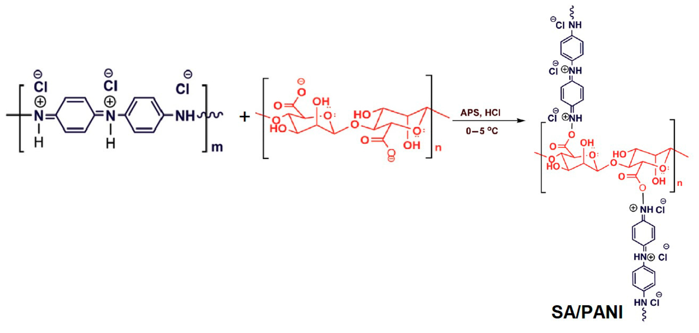
The PANI/SA composite was synthesized via an in-situ oxidative polymerization process, as presented in Scheme 1. First, 0.05 g of sodium alginate was dissolved in 50 mL of distilled water under constant stirring at 500 rpm for 10 min at room temperature (25 °C) to ensure complete dispersion. Subsequently, 1 mL of aniline was added dropwise into 20 mL of the prepared SA solution and stirred continuously for 30 min to allow homogeneous mixing. The resulting mixture was then cooled to a temperature range of 0–5 °C using an ice bath to control the polymerization kinetics. A freshly prepared solution of ammonium persulfate (APS), with a mass ratio of APS to aniline of 1.3:1, dissolved in 5 mL of 1 M HCl, was slowly added dropwise under continuous stirring, maintaining the reaction temperature within the 0–5 °C range. The polymerization process was allowed to continue for a period of 24 h. The resulting green PANI/SA composite suspension was centrifuged at 5000 rpm and underwent multiple washes with deionized water to remove any residual reagents and unreacted monomers. Finally, the product was dried at room temperature.
For electrode modification, a screen printed carbon electrode (SPCE) was first rinsed with distilled water. The dried PANI/SA composite was then redispersed in a small amount of deionized water to form a homogeneous ink. A measured volume of this dispersion was drop-cast onto the clean SPCE surface and left to dry at ambient conditions, forming a uniform PANI/SA film coating on the electrode. This resulting modified electrode served as the working electrode for all electrochemical measurements.

2.3. Characterization

The synthesized composite was characterized using Fourier Transform Infrared Spectroscopy (FTIR) (Perkin Elmer spectrometer, PerkinElmer Inc., Waltham, MA, USA) to identify functional groups and confirm successful polymerization. UV-Visible spectroscopy (UV-Vis) was employed to analyze the optical properties of the composite and monitor the characteristic absorption peaks of PANI and SA. The Scanning Electron microscopy (SEM) measurements were carried out using TS quanta 250 scanning electron microscope to study the composite’s morphology. Electrochemical measurements were carried out using a DropSens µStat 400 Potentiostat (Metrohm DropSens, Oviedo, Asturias, Spain), operated by the Dropview 8400 data acquisition software (version 2.1). Cyclic Voltammetry (CV) and Differential Pulse Voltammetry (DPV) were recorded for performance evaluation.
All electrochemical measurements were carried out using the modified SPCE. Experiments were performed in an acetate buffer solution (pH 4.5) to maintain stable experimental conditions. The CV scans were typically conducted within a potential window of −0.8 V to +0.8 V at various scan rates, while DPV was applied to detect trace concentrations of Pb(II) and Cd(II) ions. Unless otherwise specified, all experiments were conducted at room temperature.
This combination of synthesis, surface electrode modification, and advanced characterization technique provides a robust framework for evaluating the PANI/SA sensor’s structural, optical, and electrochemical properties and demonstrates its practical feasibility for heavy metal detection in aqueous environments.

3. Results and Discussion

3.1. Structural and Optical Characterization

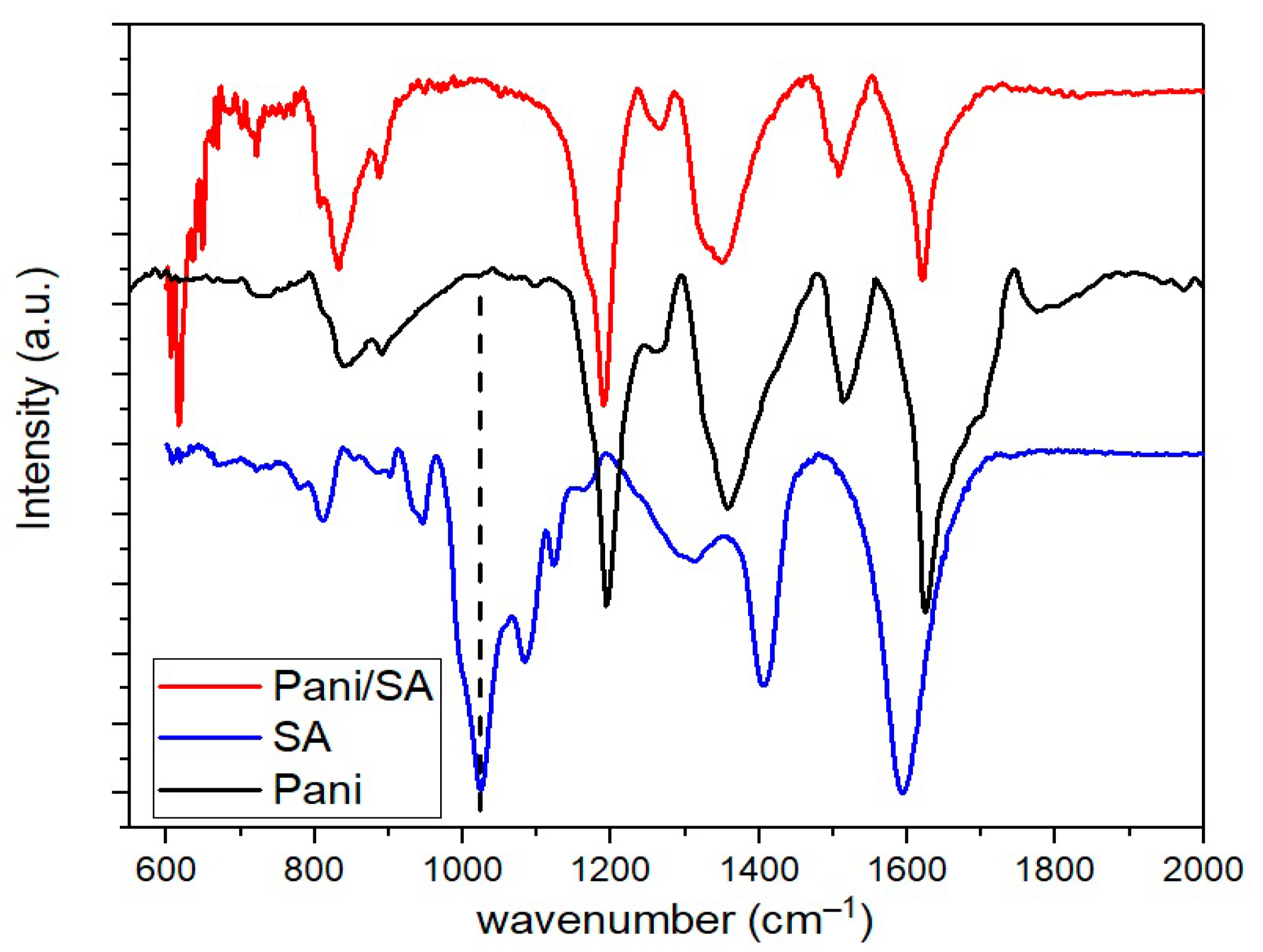
The successful formation of the PANI/SA composite was initially confirmed by Fourier Transform Infrared (FTIR) spectroscopy. As given in Figure 1, the FTIR spectrum of pure sodium alginate (SA) exhibits characteristic absorption bands at 1600 cm−1 and 1419 cm−1, attributed to the asymmetric and symmetric stretching vibrations of the carboxylate groups (–COO), respectively. A prominent peak at 1035 cm−1 corresponds to the C–O–C stretching vibration of the polysaccharide backbone [11,12,13]. Following in situ polymerization, the spectrum of the PANI/SA composite displays new absorption bands around 1560 cm−1 and 1480 cm−1, assigned to the quinoid and benzenoid ring stretching modes of polyaniline (PANI), respectively. These peaks are indicative of the formation of the conductive emeraldine salt form of PANI. Additional characteristic bands appear at 1240 cm−1, corresponding to C–N+ stretching vibrations in the polaron structure, and at 1180 cm−1, attributed to the –NH+= bending vibration, both of which reflect π-electron delocalization along the conjugated PANI backbone. Notably, the slight shifts and broadenings of certain peaks in the composite spectrum, compared to those of pure components, suggest strong intermolecular interactions between the PANI chains and the SA matrix. These observations support effective incorporation of PANI within the alginate network, likely through hydrogen bonding and physical entanglement, which contributes to the structural integrity and functional performance of the composite.
This interpretation is consistent with the proposed reaction mechanism (Scheme 2), which illustrates how hydrogen bonding can occur between the –OH groups of sodium alginate and the amine groups of polyaniline during the in situ polymerization process. These interactions enhance structural integration and facilitating efficient charge transfer pathways within the composite [8,14].
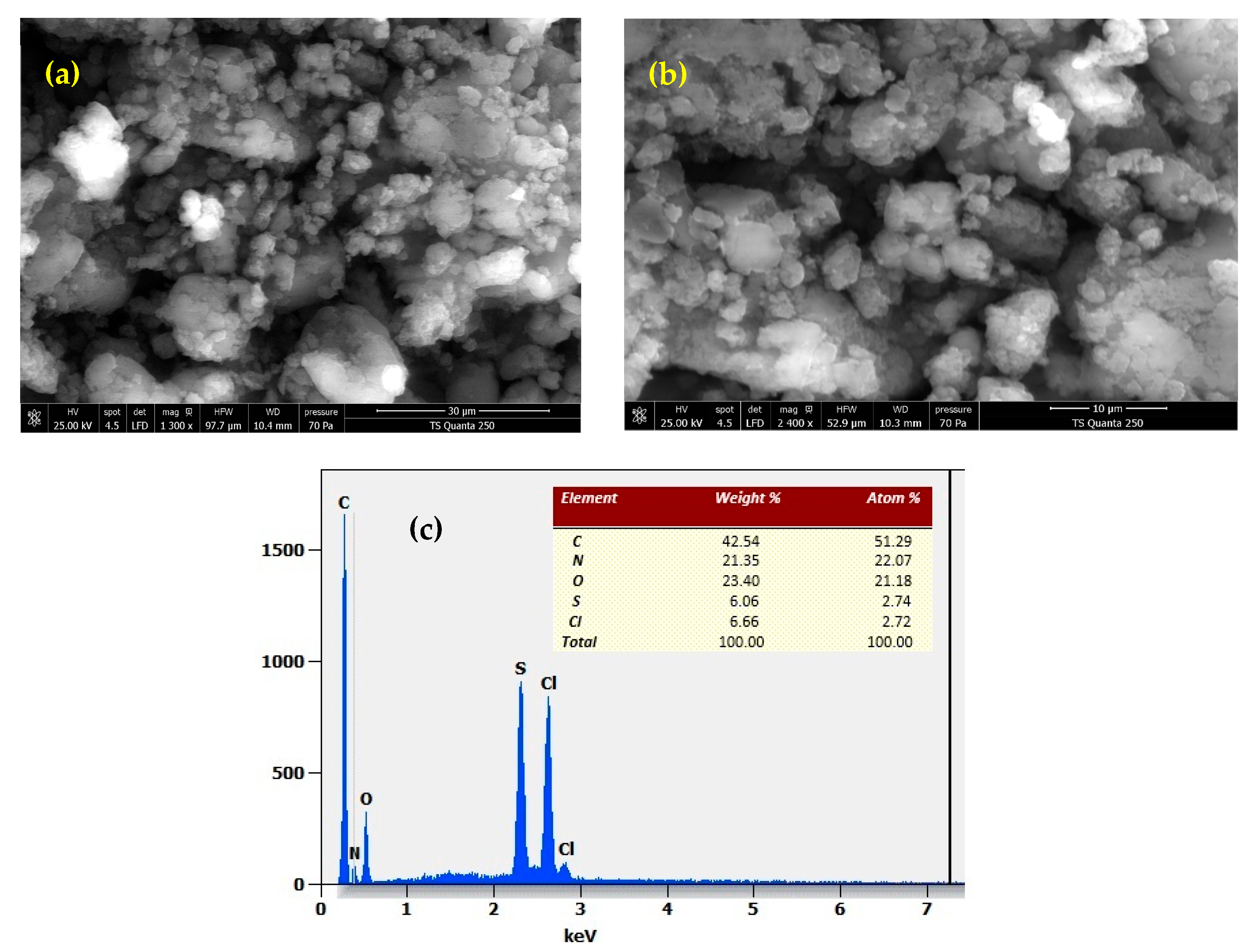
To gain deeper insights into the structural organization of the PANI/SA composites, scanning electron microscopy (SEM) combined with energy-dispersive X-ray spectroscopy (EDX) was employed. This analysis offers valuable information about the surface morphology and microstructural characteristics of the polymeric material. As shown in Figure 2a,b, the SEM images captured at magnifications ranging from ×1300 to ×5000 reveal a typical cauliflower-like morphology, consisting of interconnected globular aggregates dispersed within a porous biopolymeric matrix [11,15]. This distinctive structure, commonly observed in polyaniline-based composites, is highly beneficial for electrochemical applications due to the increased accessible surface area, which facilitates efficient ion diffusion and electron transport [8]. Moreover, the homogeneous distribution of PANI within the alginate framework, with no signs of phase separation or structural degradation, suggests good compatibility between the two components and successful composite formation.
Complementary EDX spectra confirmed the composite’s elemental composition, showing dominant signals for carbon (C), oxygen (O), and nitrogen (N), consistent with contributions from the polysaccharide backbone of sodium alginate and the amine/imine structures of polyaniline [16]. Furthermore, the presence of sulfur (S) and chlorine (Cl) signals supports successful doping, likely originating from sulfuric or hydrochloric acid used during oxidative polymerization, critical to enhancing the conductivity of the PANI phase. Together, the SEM and EDX analyses validate the formation of a structurally integrated and chemically homogeneous SA–PANI composite, suitably engineered for applications in sensing and energy storage systems.
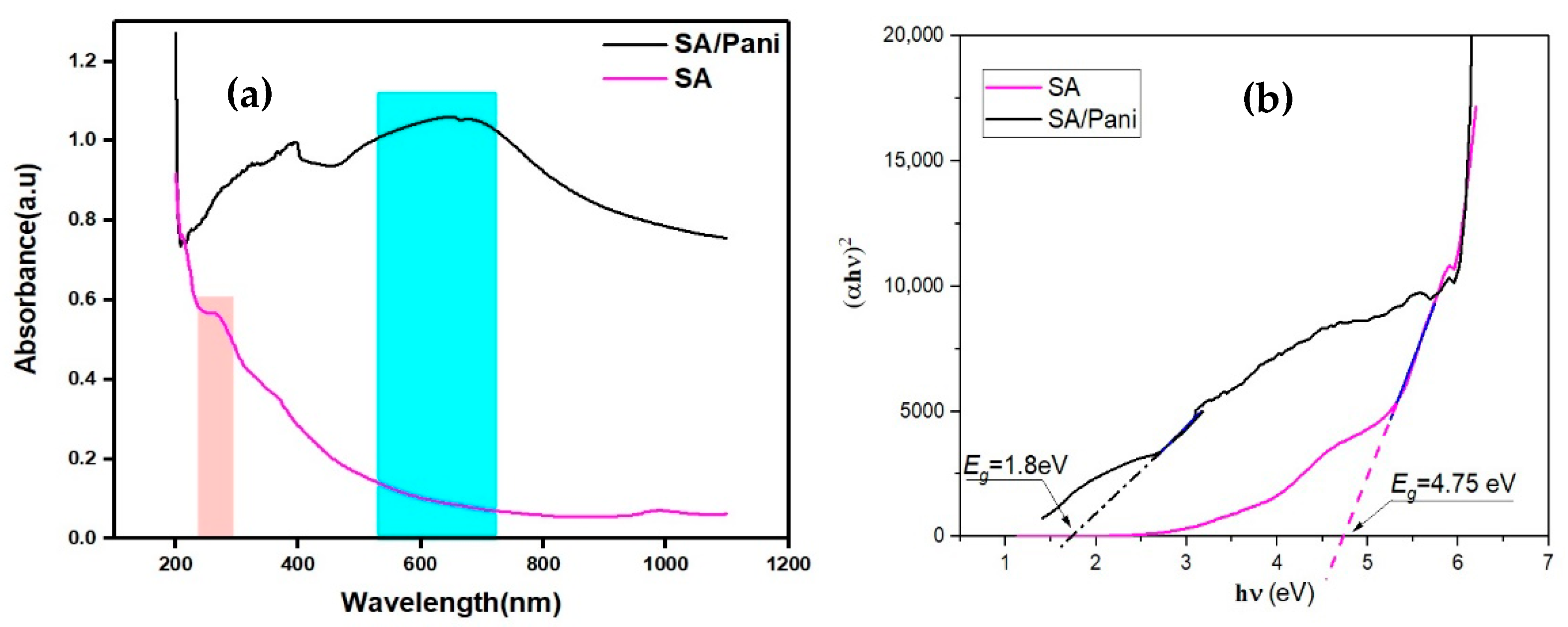
UV–Visible spectroscopy provided clear evidence of the successful formation of the PANI/SA composite (Figure 3a). The UV–Vis spectrum showed characteristic absorption bands of PANI, typically observed at 320–350 nm (π–π transition of the benzenoid ring) and 600–650 nm (polaron–π transition associated with the quinoid structure) [17]. The presence of these bands, along with slight shifts compared to pure PANI, indicates strong interactions between PANI and sodium alginate (SA), which can influence the composite’s electronic structure and improve charge transfer pathways. As expected for an insulating biopolymer, pure SA alone exhibits very low absorbance in the visible region.
The Tauc plot derived from the absorbance data further highlights these findings (Figure 3b). I reveals that pure SA possesses a wide optical band gap of approximately 4.75 eV, while the PANI/SA composite displays a significantly reduced band gap of about 1.8 eV. This reduction confirms the introduction of new electronic states associated with the conjugated structure of PANI. Together, these results demonstrate that the PANI/SA composite exhibits enhanced light-harvesting and electronic properties, making it a promising candidate for applications such as sensors, flexible electronics, and energy storage devices.

3.2. Electrochemical Characterization

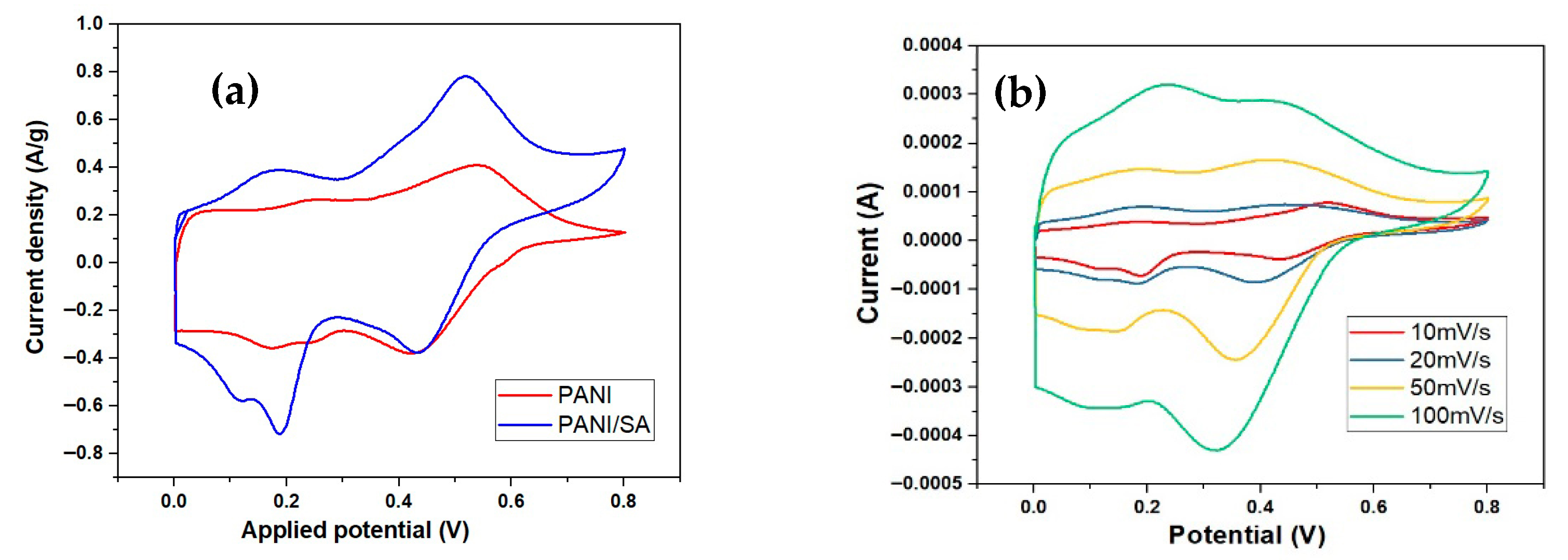
The electrochemical behavior of the PANI and PANI/SA-modified electrode (SPCE) was investigated by cyclic voltammetry (CV) in H2SO4 1 M. Figure 4a shows typical CV curves comparing the bare SPCE and the PANI/SA-modified SPCE. The modified electrode exhibited well-defined redox peaks corresponding to the reversible oxidation and reduction of the PANI backbone, demonstrating enhanced electrochemical activity. Referring to SA/PANI composite, the cyclic voltammetry curve exhibits a quasi-rectangular shape with clearly defined redox features, highlighting the strong pseudocapacitive behavior of the electrode. Specifically, two pairs of redox peaks, labeled A/A1 and B/B1, can be observed, corresponding to two distinct reversible electrochemical transitions. Peaks A (around 0.2 V) and A1 correspond to the reversible transition of polyaniline between its semi-conductive leucoemeraldine form and its conductive polaronic emeraldine salt form (ES). Peaks B (around 0.55 V) and B1 are attributed to the subsequent redox transformation from the emeraldine form to the fully oxidized pernigraniline state [18,19]. Notably, even at a relatively high scan rate of 100 mV s−1, the CV curve retains its well-defined redox peaks, demonstrating that the SA/PANI network structure facilitates rapid proton/electron transport and offers a short diffusion path for efficient redox activity.
Moreover, the current response increased linearly with the scan rate (as given in Figure 4b), indicating a surface-controlled electrochemical process and efficient electron transfer between the electrode surface and the electrolyte.

3.3. Detection of Heavy Metals (Pb(II) and Cd(II))

To evaluate the performance of our sensor in terms of lead and cadmium detection signals, initial tests were carried out using an unmodified (bare) screen-printed carbon electrode (SPCE). The measurements were performed by varying the potential in the range of −1.5 V to 0 V at a scan rate of 50 mV s−1, in an acetate buffer solution (5 mL, pH 4) containing different concentrations of Cd2+ (ranging from 20 µM to 200 µM). Figure 5 shows the electrochemical response of the unmodified SPCE electrode in the presence of Cd2+ ions. It can be observed that the unmodified electrode responds only at relatively high Cd2+ concentrations, with a detectable signal appearing in the potential window between −1.3 V and −1.1 V [20,21]. Moreover, the peak current remains relatively low, with a maximum intensity of approximately 25 µA at −1.2 V, highlighting the limited sensitivity of the bare electrode for cadmium detection.
From Figure 5, a detectable electrochemical signal was observed within the studied potential range when testing in acetate buffer, for Cd2+ concentrations ranging from 20 µM to 200 µM. It can be noted that the peak current intensity increases with increasing concentrations of Cd2+ ions.
In a second series of experiments, measurements were carried out using two modified electrodes, prepared by drop-casting 5 µL and 10 µL of the SA/PANI suspension onto separate SPCEs. After drying, both electrodes were immersed in an acetate buffer solution, and Differential Pulse Voltammetry (DPV) measurements were conducted for various concentrations of added cadmium ions (ranging from 20 µM to 200 µM).
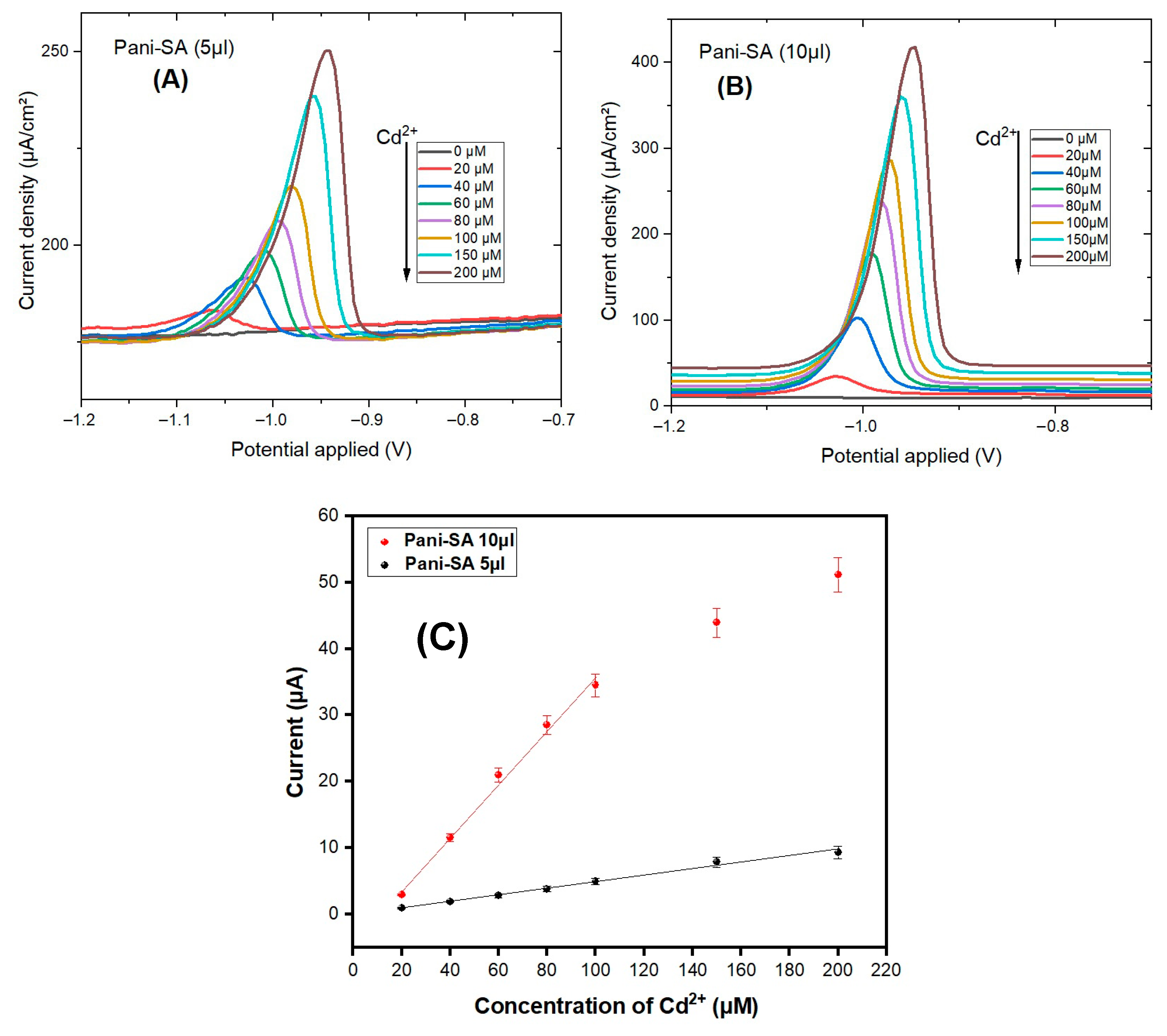
Figure 6 clearly shows the response of the biosensor, which was able to detect even a very low cadmium concentration (20 µM) within a potential window ranging from −1.2 V to −0.8 V (Figure 6A,B). When testing pure acetate buffer alone, no signal was detected within this potential range. As Cd2+ ions were progressively added to the acetate solution, an oxidation peak appeared at approximately −0.98 V, confirming the successful detection. This peak became more pronounced as the ion concentration increased. A maximum current of 50 µA was obtained at a potential of −0.9 V, which is significantly higher than the response obtained with the bare electrode. Besides, a shift towards positive potential was noted with increasing Cd2+ concentration indicating an enhanced of electrochemical kinetics and modified interfacial conditions at the electrode surface. As the concentration of Cd2+ increases, the greater availability of electroactive species at the electrode/electrolyte interface promotes more efficient electron transfer. Consequently, the system requires a lower overpotential for the reduction reaction to proceed, resulting in the observed positive shift. Similar behavior has been reported in the literature for metal ion detection using modified electrodes [22].
Moreover, the sensor exhibited a linear response to Cd2+ concentration in range of 20 µM to 200 µM in acetate buffer solution (pH 4.0). Figure 6C presents the linear correlation between the current density and cadmium concentration, demonstrating the good sensitivity and quantitative capability of the SA/PANI-based sensor.
As shown in Figure 6C, the electrode modified with 5 µL of the PANI/SA suspension exhibits a sensitivity of 0.394 µA µM−1 cm−2. Increasing the deposition volume to 10 µL leads to a remarkable eightfold enhancement, achieving a sensitivity of 3.183 µA µM−1 cm−2. This improvement reflects the beneficial effect of increased loading of the electroactive material on the electrode surface, enhancing ion interaction and charge transfer efficiency.
A comparative overview of various Cd2+ electrochemical sensors is presented in Table 1. The PANI/SA-modified screen-printed carbon electrode (SPCE) developed in this study demonstrates a significantly higher sensitivity than several previously reported systems, such as a recent study employing nanopipet-based ITIES sensing platforms with 1,10-phenanthroline as ionophore achieved a sensitivity of 0.127 pA µM−1, highlighting the capability of nanostructured platforms in complex matrices [23]. While the APTE-Mono@GC sensor exhibits an even higher sensitivity (~3 µA µM−1), its fabrication requires sophisticated procedures making it less practical for routine applications [24].
In contrast, the PANI/SA-modified SPCE benefits from a simple, low-cost, and scalable fabrication process, relying on drop-casting of biopolymer-based material. Its excellent sensitivity, combined with its eco-friendly composition and ease of preparation, positions it as a promising and practical candidate for the electrochemical detection of Cd2+ in environmental monitoring.

3.4. Selectivity, Stability, and Reproducibility

The selectivity of the sensor for lead ion detection was investigated in the presence of both lead and cadmium ions. Measurements were conducted in a 0.1 M acetate buffer solution containing a fixed concentration of Cd2+ (100 µM), while varying the concentration of Pb2+ ions from 0 to 100 µM. Figure 7 clearly shows two distinct peaks, indicating the presence of each metal ion in the solution. The first peak, appearing at −1.0 V, corresponds to cadmium ions, while the second peak, at −0.8 V, is attributed to lead ions. These results demonstrate that the SA/PANI-based sensor is capable of selectively detecting Pb2+ ions even in the presence of Cd2+, confirming its high selectivity for heavy metal ion discrimination in mixed solutions.
Stability tests showed that the PANI/SA-modified electrode maintained over 97% of its initial current response after two weeks of storage under ambient conditions, demonstrating good operational and storage stability. Reproducibility was confirmed by repeated measurements with multiple independently prepared electrodes indicating excellent fabrication consistency and measurement repeatability.

4. Conclusions

In this work, an advanced electrochemical sensor based on a polyaniline (PANI) and sodium alginate (SA) composite was successfully developed and characterized for the sensitive detection of heavy metal ions in aqueous environments. The composite material was synthesized via in-situ polymerization, effectively combining the excellent electrical conductivity of PANI with the biocompatibility and ion-exchange properties of SA. Structural and optical analyses confirmed the successful incorporation of PANI within the SA matrix, while electrochemical measurements demonstrated significant enhanced redox activity, improved charge transfer efficiency, and good stability.
The PANI/SA-modified glassy carbon electrode exhibited remarkable performance for the detection of lead (Pb(II)) and cadmium (Cd(II)) ions, achieving low detection limits, good linearity, high selectivity, and excellent reproducibility. These results highlight the potential of this biopolymer-conducting polymer composite as a promising and cost-effective sensing platform for real-time, monitoring of toxic heavy metals in water.
Future work will focus on optimizing the sensor’s performance for use in real water samples, exploring the detection capability of other heavy metal species, and investigating its long-term operational stability under different environmental conditions. Overall, the present study demonstrates that combining natural biopolymers with conducting polymers offers a sustainable and efficient route toward the development of next-generation electrochemical sensors for environmental monitoring and public health protection.

Author Contributions

Conceptualization, N.G. and R.W.; methodology and investigation, R.W.; validation, N.G.; writing—original draft preparation, N.G.; writing—review and editing, M.A.; supervision, R.M. and M.A. All authors have read and agreed to the published version of the manuscript.

Funding

This research received no external funding.

Data Availability Statement

The data presented in this study are available on request from the corresponding author due to (the large volume of data).

Conflicts of Interest

The authors declare no conflict of interest.

References

  1. Nriagu, J.O.; Pacyna, J.M. Quantitative assessment of worldwide contamination of air, water and soils by trace metals. Nature 1998, 333, 134–139. [Google Scholar] [CrossRef]
  2. Johri, N.; Jacquillet, G.; Unwin, R. Heavy metal poisoning: The effects of cadmium on the kidney. BioMetals 2010, 23, 783–792. [Google Scholar] [CrossRef]
  3. Triunfante, P.; Soares, M.E.; Santos, A.; Tavares, S.; Carmo, H.; Bastos, M.D.L. Mercury fatal intoxication: Two case reports. Forensic Sci. Int. 2009, 184, e1–e6. [Google Scholar] [CrossRef]
  4. Aragay, G.; Pons, J.; Merkoçi, A. Recent Trends in Macro-, Micro-, and Nanomaterial-Based Tools and Strategies for Heavy-Metal Detection. Chem. Rev. 2011, 111, 3433–3458. [Google Scholar] [CrossRef] [PubMed]
  5. Cui, L.; Wu, J.; Ju, H. Electrochemical sensing of heavy metal ions with inorganic, organic and bio-materials. Biosens. Bioelectron. 2015, 63, 276–286. [Google Scholar] [CrossRef] [PubMed]
  6. Okano, G.; Igarashi, S.; Yamamoto, Y.; Saito, S.; Takagai, Y.; Ohtomo, T.; Kimura, T.; Ohno, T.; Oka, Y. HPLC-spectrophotometric detection of trace heavy metals via ‘cascade’ separation and concentration. Int. J. Environ. Anal. Chem. 2015, 95, 135–144. [Google Scholar] [CrossRef]
  7. Moretto, L.M.; Kalcher, K. (Eds.) Environmental Analysis by Electrochemical Sensors and Biosensors: Fundamentals; Springer: New York, NY, USA, 2014. [Google Scholar] [CrossRef]
  8. Kushwaha, C.S.; Singh, V.K.; Shukla, S.K. Electrochemically triggered sensing and recovery of mercury over sodium alginate grafted polyaniline. New J. Chem. 2021, 45, 10626–10635. [Google Scholar] [CrossRef]
  9. Sun, J.; Tan, H. Alginate-Based Biomaterials for Regenerative Medicine Applications. Materials 2013, 6, 1285–1309. [Google Scholar] [CrossRef] [PubMed]
  10. Lee, K.Y.; Mooney, D.J. Alginate: Properties and biomedical applications. Prog. Polym. Sci. 2012, 37, 106–126. [Google Scholar] [CrossRef]
  11. Majhi, D.; Patra, B.N. Polyaniline and sodium alginate nanocomposite: A pH-responsive adsorbent for the removal of organic dyes from water. RSC Adv. 2020, 10, 43904–43914. [Google Scholar] [CrossRef]
  12. Yu, Y.; Zhihuai, S.; Chen, S.; Bian, C.; Chen, W.; Xue, G. Facile Synthesis of Polyaniline−Sodium Alginate Nanofibers. Langmuir 2006, 22, 3899–3905. [Google Scholar] [CrossRef] [PubMed]
  13. Ke, Y.-C.; Chen, T.-C.; Tang, R.-C.; Lin, J.-N.; Lin, F.-H. Development of resveratrol with thiolated alginate as a supplement to prevent nonalcoholic fatty liver disease (NAFLD). APL Bioeng. 2022, 6, 016102. [Google Scholar] [CrossRef] [PubMed]
  14. Zhang, X.; Sheng, N.; Wang, L.; Tan, Y.; Liu, C.; Xia, Y.; Nie, Z.; Sui, K. Supramolecular nanofibrillar hydrogels as highly stretchable, elastic and sensitive ionic sensors. Mater. Horiz. 2019, 6, 326–333. [Google Scholar] [CrossRef]
  15. Ke, X.; Yang, F.; Huang, H.; Zhao, R.; Huang, J.; Ji, J.; Li, L. Stable polyaniline/sodium alginate/graphene oxide composite hydrogels as binder-free supercapacitor electrodes. Ionics 2022, 28, 5633–5641. [Google Scholar] [CrossRef]
  16. Karthik, R.; Meenakshi, S. Removal of Cr(VI) ions by adsorption onto sodium alginate-polyaniline nanofibers. Int. J. Biol. Macromol. 2015, 72, 711–717. [Google Scholar] [CrossRef]
  17. Li, Y.; Zhao, X.; Xu, Q.; Zhang, Q.; Chen, D. Facile Preparation and Enhanced Capacitance of the Polyaniline/Sodium Alginate Nanofiber Network for Supercapacitors. Langmuir 2011, 27, 6458–6463. [Google Scholar] [CrossRef]
  18. Song, E.; Choi, J.-W. Conducting Polyaniline Nanowire and Its Applications in Chemiresistive Sensing. Nanomaterials 2013, 3, 498–523. [Google Scholar] [CrossRef] [PubMed]
  19. Zhang, S.; Sun, G.; He, Y.; Fu, R.; Gu, Y.; Chen, S. Preparation, Characterization, and Electrochromic Properties of Nanocellulose-Based Polyaniline Nanocomposite Films. ACS Appl. Mater. Interfaces 2017, 9, 16426–16434. [Google Scholar] [CrossRef]
  20. Deshmukh, M.A.; Celiesiute, R.; Ramanaviciene, A.; Shirsat, M.D.; Ramanavicius, A. EDTA_PANI/SWCNTs nanocomposite modified electrode for electrochemical determination of copper (II), lead (II) and mercury (II) ions. Electrochim. Acta 2018, 259, 930–938. [Google Scholar] [CrossRef]
  21. Wang, D.; Zhang, F.; Tang, J. Sodium Alginate Decorated Carbon Nanotubes-Graphene Composite Aerogel for Heavy Metal Ions Detection. Electrochemistry 2015, 83, 84–90. [Google Scholar] [CrossRef]
  22. Garg, N.; Deep, A.; Sharma, A.L. Simultaneous detection of Lead and Cadmium using a composite of Zeolite Imidazole Framework and Reduced Graphene Oxide (ZIF-67/rGO) via electrochemical approach. Environ. Eng. Res. 2022, 28, 220269. [Google Scholar] [CrossRef]
  23. Ahmed, M.M.N.; Bodowara, F.S.; Zhou, W.; Penteado, J.F.; Smeltz, J.L.; Pathirathna, P. Electrochemical detection of Cd(II) ions in complex matrices with nanopipets. RSC Adv. 2022, 12, 1077–1083. [Google Scholar] [CrossRef]
  24. Levanen, G.; Dali, A.; Leroux, Y.; Lupoi, T.; Betelu, S.; Michel, K.; Ababou-Girard, S.; Hapiot, P.; Dahech, I.; Cristea, C.; et al. Specific electrochemical sensor for cadmium detection: Comparison between monolayer and multilayer functionalization. Electrochim. Acta 2023, 464, 142962. [Google Scholar] [CrossRef]
  25. El-Fatah, G.A.; Magar, H.S.; Hassan, R.Y.A.; Mahmoud, R.; Farghali, A.A.; Hassouna, M.E.M. A novel gallium oxide nanoparticles-based sensor for the simultaneous electrochemical detection of Pb2+, Cd2+ and Hg2+ ions in real water samples. Sci. Rep. 2022, 12, 20181. [Google Scholar] [CrossRef] [PubMed]
  26. Mohammed, R.H.R.; Hassan, R.Y.A.; Mahmoud, R.; Farghali, A.A.; Hassouna, M.E.M. Electrochemical determination of cadmium ions in biological and environmental samples using a newly developed sensing platform made of nickel tungstate-doped multi-walled carbon nanotubes. J. Appl. Electrochem. 2024, 54, 657–668. [Google Scholar] [CrossRef]
Scheme 1. Schematic illustration of the preparation of PANI/SA by oxidative polymerization of ANI in the presence of Sodium Alginate.
Engproc 106 00002 sch001
Figure 1. FTIR spectra of PANI, SA and the composite PANI/SA.
Engproc 106 00002 g001
Scheme 2. Tentative mechanism for the growth of PANI particles on SA by oxidative polymerization in presence of APS.
Engproc 106 00002 sch002
Figure 2. (a,b) SEM image (c) EDX spectra of PANI-SA composite.
Engproc 106 00002 g002
Figure 3. (a) UV–Vis absorbance spectra of sodium alginate (SA) and the PANI/SA composite, (b) Tauc plots of SA and PANI/SA.
Engproc 106 00002 g003
Figure 4. (a) Cyclic voltammograms of the modified electrodes with PANI/SA and PANI at 10 mV s−1 in 1 M H2SO4, and (b) cyclic voltammograms of the PANI/SA-modified electrode at different scan rates in 1 M H2SO4.
Engproc 106 00002 g004
Figure 5. DPV curves of Cd2+ ions recorded with the unmodified SPCE electrode in acetate buffer solution at pH 4.3.
Engproc 106 00002 g005
Figure 6. DPV curves for different concentrations of Cd2+ ions in acetate buffer solution at pH 4.3 using (A) 5 µL of PANI/SA and (B) 10 µL of PANI/SA; (C) Calibration curves for Cd2+ ions obtained using the PANI/SA-modified electrode in acetate buffer.
Engproc 106 00002 g006
Figure 7. DPV curves obtained with the SA/PANI-modified electrode in the presence of 100 µM Cd2+ ions and varying concentrations of Pb2+ ions in acetate buffer solution at pH 4.3.
Engproc 106 00002 g007
Table 1. A summary and comparison of the sensitivity values for the detection of Cd(II) from the present work with previous reports.
ElectrodeCompositionSensitivity (µA µM−1)Ref.
PANI/SA PANI/SA on SPCE0.52This work
APTE-Mono@GCMonolayer of APTE on GCE via diazonium grafting3.00[24]
Ga2O3NPs/CPEGallium oxide nanoparticles mixed in carbon paste1.250[25]
NiWO4/MWCNT/CPENickel tungstate + multiwalled carbon nanotubes in carbon paste0.038[26]
Nanopipet/ITIESnanopipet-based ITIES sensing platforms with 1,10-phenanthroline0.127 × 10−3[23]
Disclaimer/Publisher’s Note: The statements, opinions and data contained in all publications are solely those of the individual author(s) and contributor(s) and not of MDPI and/or the editor(s). MDPI and/or the editor(s) disclaim responsibility for any injury to people or property resulting from any ideas, methods, instructions or products referred to in the content.