Proceeding Paper

Application of the Reduced Graphene Oxide–Multiwalled Carbon Nanotubes Composite for Development of the Electrochemical Aptasensor for Oxytetracycline Detection †

1
IMD Laboratories Co., R&D Section, Lefkippos Technology Park, NCSR Demokritos, 15341 Agia Paraskevi, Greece
2
Institute of Materials and Environmental Chemistry, HUN-REN Research Centre for Natural Sciences, Magyar Tudósok Krt. 2, 1117 Budapest, Hungary
3
Faculty of Mathematics, Physics and Informatics, Comenius University, Mlynská Dolina F1, 84248 Bratislava, Slovakia
4
Physics Department, College of Science and General Studies, Alfaisal University, Riyadh 11533, Saudi Arabia
5
Center for Nanophase Materials Sciences, Oak Ridge National Laboratory, Oak Ridge, TN 37831-6496, USA
*
Author to whom correspondence should be addressed.
Presented at the 5th International Electronic Conference on Biosensors, 26–28 May 2025; Available online: https://sciforum.net/event/IECB2025.
Eng. Proc. 2025, 106(1), 16; https://doi.org/10.3390/engproc2025106016
Published: 21 November 2025

Abstract

Excessive use of oxytetracycline (OTC) in veterinary medicine has increased the presence of antibiotics in food, which accelerates the development of antimicrobial resistance. We report the development of a highly sensitive electrochemical aptasensor for OTC detection, based on a glassy carbon electrode (GCE) modified with reduced graphene oxide (rGO) and multiwalled carbon nanotubes (MWCNTs) nanocomposite. DNA aptamers specific to OTC were covalently attached to the nanocomposite surface via carbodiimide chemistry. Differential pulse voltammetry (DPV) showed a decrease in peak current due to the binding of OTC to the aptamers. The sensor exhibited a limit of detection (LOD) of 1.72 ng/mL, which is below the maximum residue limit (MRL) for OTC (100 ng/mL) established by European Union. The sensor has been tested on a spiked milk sample.

1. Introduction

Excessive application of oxytetracycline (OTC), a broad-spectrum antibiotic of the tetracycline group in veterinary medicine, has increased the background contamination of food products and may be a leading cause behind decreased antibiotic efficiency. For instance, the contamination of milk and milk products with OTC leads to increased risks of the development of the antimicrobial resistance (AMR) of bacteria, which are harmful for consumers. To mitigate potential issues, the European Union established the maximum residue limit (MRL), restricting OTC in milk, as 100 µg/kg (~100 ng/mL) [1]. Antibiotics decompose slowly in the environment and tend to accumulate rapidly in organisms of the ecosystem, posing direct or indirect risks to human health, including a risk of the growth of AMR [2]. To control food quality, the development of cost-efficient, high-sensitivity methods for OTC detection in food products, including milk, which should be integrated into the food production/processing chain, is urgently needed.
High-performance liquid chromatography (HPLC) can be used to detect the presence of multiple antibiotics simultaneously. However, it is time-consuming, and its prohibitively high cost makes it difficult for its implementation as a standard analytical tool at every milk production facility [3]. Similar problems make gas chromatography–mass spectrometry (GC-MS) [4] and liquid chromatography–mass spectroscopy (LC-MS) unsuitable for routine or on-site antibiotic testing, despite their excellent sensitivity [5]. Enzyme-linked immunosorbent assay (ELISA) is commonly used for rapid screening of antibiotics in milk and meat, but their cross-reactivity and limited specificity often result in requiring additional chromatographic tests [6].
As an alternative to the conventional high-cost methods, a new line of low-cost biosensors is emerging. They build on the surface of a transducer that converts the chemical signal (affinity interactions) into electrical, optical, or acoustic (mass sensitive) signals, and an analytical instrument (potentiostats, spectrometers, or network analyzers). The sensing surface of such a sensor is modified with immobilized receptors (antibodies or nucleic acid aptamers), which ensure selective interaction with antibiotics [6]. The advantage of aptamers over antibodies is the possibility of their in vitro structural/compositional optimization using combinatorial chemistry approaches, including SELEX (Systematic evolution of ligands by exponential enrichment) [7]. In this method, the library (around 1013–1015 molecules) of the random sequences of the synthetic single-stranded DNA (ssDNA) (typically 30–60 nucleotides) is allowed to interact with the target of interest. ssDNA sequences bind to the target with various affinities. The complexes of the ssDNA-target are then dissociated. The ssDNA sequences are multiplied via polymerase chain reaction (PCR) and used in the next cycle of selection. After several cycles (around ten), the sequence with the highest affinity to the target is obtained. The SELEX was used for the development of DNA aptamers that specifically bind OTC. The SELEX protocol was further modified to include magnetic beads (Flu-Mag-SELEX), Toggle-SELEX, graphene oxide-based SELEX, and others [8]. For example, Niazi et al. [9] selected the aptamers for a tetracycline group of antibiotics using a random sequence of 1015 ssDNA, using Toggle-SELEX combined with Flu-Mag-SELEX. The magnetic beads coated with antibiotics allowed the development of DNA aptamers with high affinity to tetracycline or OTC after four rounds with the dissociation constant, KD = 9.6 nM (4.41 ng/mL). Aptamers are more stable than antibodies and can be synthesized with high purity and yield. Aptamers can also be chemically modified to increase their stability or functionality for surface immobilization. The increased interest in DNA-aptamer-based biosensors (aptasensors) for antibiotic detection was discussed in several comprehensive reviews [6,10,11]. The sensitivity of these sensors is usually better than the MRL. Current sensor development trends indicate growing interest in low-cost electrochemical methods which integrate nanocomposite materials. This is related to the improved electrical properties, increased surface area, presence of COOH functional groups, and possibility to increase surface concentration of aptamers [12].
Here, we report the development of an electrochemical aptasensor based on carbon nanocomposites for the sensitive detection of OTC in milk. By utilizing a glassy carbon electrode (GCE) modified with reduced graphene oxide–multiwalled carbon nanotubes (rGO-MWCNTs) composites, we achieved enhanced surface area and improved electron transfer properties. The aptasensor was evaluated using differential pulse voltammetry (DPV), providing a sensitive and straightforward approach for OTC detection.

2. Materials and Methods

2.1. Chemicals

The DNA aptamers, (APT) specific to oxytetracycline (OTC) modified at 5′ end by amino group (5′-amino-(CH2)6-TTT TTT TTT GGG GGC ACA CAT GTA GGT GCT GTC CAG GTG TGG TTG TGG T-3′), were obtained from GeneCust (Boynes, France). The aptamer reported by Niazi et al. [9] was further modified to include an additional dT9 sequence to provide better conformational flexibility of the aptamers. The aptamers have been dissolved in TE buffer (10 mM Tris-HCl, 1 mM EDTA, pH 8) in a concentration of 0.1 mM. Graphene oxide (GO), multiwalled carbon nanotubes (MWCNTs), OTC, tetracycline (TET), kanamycin (KAN), penicillin G (PEN), chloramphenicol (CAP), 1-ethyl-3-(3-dimethylaminopropyl) carbodiimide (EDC), N-hydroxysuccinimide (NHS), ethanolamine, Tris–HCl, ethylenediaminetetraacetic acid (EDTA), Tween 20, K3[Fe(CN)6], and K4[Fe(CN)6] have been purchased from Sigma-Aldrich (Darmstadt, Germany). NaCl, KCl, HCl, NaOH, MgCl2, and CaCl2 were from Slavus (Bratislava, Slovakia). The experiments were performed in Tris-HCl (20 mM, pH 7.6, contained 100 mM NaCl, 2 mM MgCl2, 5 mM KCl, and 1 mM CaCl2, 0.02% Tween 20). Deionized (DI) water with a resistance of 18 MΩ·cm, prepared by Purelab Classic UV (Elga, High Wycombe, UK), was used for the preparation of all aqueous solutions. The pH of the solution was adjusted by 0.1 M HCl or 0.1 M NaOH using the pH meter LE438 Mettler-Toledo (Greifensee, Switzerland).

2.2. Preparation of the Biosensor and Electrochemical Measurements

The 2 mm diameter glassy carbon electrodes (GCE) (CH Instruments, Austin, TX, USA) were used for biosensor preparation. The bare GCE was polished with 1.0 μm and 0.3 μm alumina slurry (CH Instruments) to obtain a mirror surface, then ultrasonicated for 3 min in DI water and dried under nitrogen gas. The GCE was electrochemically cleaned in 0.5 M H2SO4 aqueous solution by using cyclic voltammetry (CV) in the voltage range between 0.2 and 1.5 V. The Ag/AgCl reference electrode underwent a scan rate of 100 mV/s until the characteristic cyclic voltammogram was obtained. The electrodeposition was performed from 1 mg/mL GO-MWCNTs at 1.6 V for 30 min, forming a thin film on top of the GCE. Electrochemical reduction in GO was performed by CV, in a potential range between −0.2 to 1.0 V, with a scan rate of 100 mV/s, in 0.1 M KH2PO4 solution. After 60 cycles, the GO was reduced and rGO-MWCNTs on the working electrode were obtained. After washing the rGO-MWCNTs with DI, the COOH-functionalized MWCNTs were activated with the incubation of 100 µL of 20 mM EDC/50 mM NHS for 120 min. Then, 100 μL of 5 μM amino-modified OTC aptamers were added on the surface of the nanofabricated GCE. In addition, 100 μL of 0.1 M ethanolamine (ETA) was added at the sensing surface, blocking the free COO sites. Then, OTC was tested at different concentrations of 0.3, 0.5, 1, 5, 10, and 30 ng/mL. Figure 1 shows the scheme of sensor preparation. It has been adopted from paper by Jia et al. [13] with a similar design but in which detection of Salmonella was reported.
The biosensor was tested in milk spiked by OTC. For this purpose, the 1.5% fat milk from the local supermarket was used. The milk was thermostated at 40 °C for 30 min, diluted with ethanol in a 1:3 v/v ratio and centrifuged at 5000 rpm for 5 min. The supernatant, which is mainly ethanol and water mixture, were separated from the sediment, diluted 10-fold with the OTC binding buffer, and spiked by various concentrations of OTC [14].
Differential pulse voltammetry (DPV) was performed from −0.2 to 0.6 V in a 5 mM [Fe(CN)6]3−/4− solution at a scan rate of 100 mV/s. All electrochemical measurements were performed using CHI 410 potentiostat (CH Instruments) or µAutolab III/Fra2 (Metrohm, Herisau, Switzerland) in the three-electrode configuration consisting of the GCE working electrode, Ag/AgCl reference, and the platinum counter electrode (CH Instruments). All the experiments were performed at room temperature (~22 °C).

3. Results and Discussion

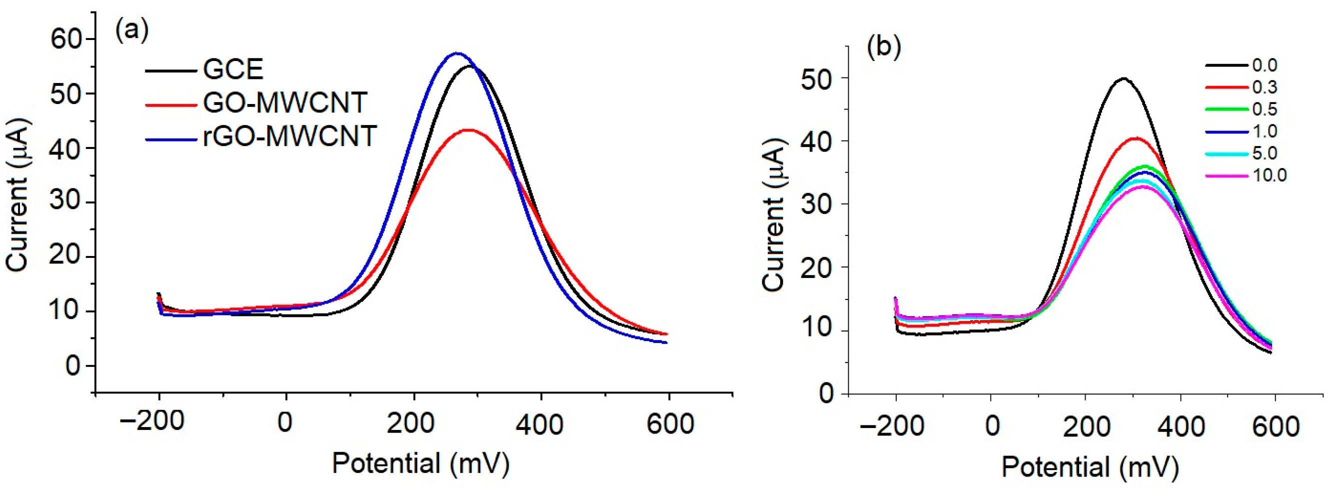
In the first series of experiments, we studied the DPV of the modified GCE in the presence of OTC. Figure 2a shows DPV of the bare, GO/MWCNTs and rGO/MWCNTs modified GCE in the presence of 5 mM [Fe(CN)6]3−/4− redox probe. A typical current peak at approx. 300 mV vs. Ag/AgCl reference electrode for bare GCE corresponds to reduction in [Fe(CN)6]4−. The value of current decreased after electrodeposition of the GO-MWCNTs on the surface of GCE, due to blocking of the electron transfer to the electrode surface from the redox probe caused by the negatively charged GO in GO/MWCNTs composite. The reduction in GO (rGO/MWCNTs) increased the current due to elimination of the negatively charged groups of GO. The immobilization of the aptamers, and covering the bare COO sites by ethanolamine (ETA), reduced the amplitude of the current by several µA (Figure 2b) due to increase in the negative change in the electrode surface caused by negatively charged aptamers. Then, the aptasensor was incubated for 30 min in a series of increasing concentrations, from 0.3 to 30 ng/mL OTC, followed by gentle rinsing with a binding buffer. Blocking the charge transfer through the sensing surface by OTC leads to a decrease in current amplitude and its slight shift toward higher potential. The isoelectric point of OTC is 5.2 [15]. Therefore, at pH 7.6, the OTC is negatively charged. This also contributes to the decrease in the current amplitude and modify the electrode potential. Thus, a more positive voltage is required for reduction in [Fe(CN)6]4−.
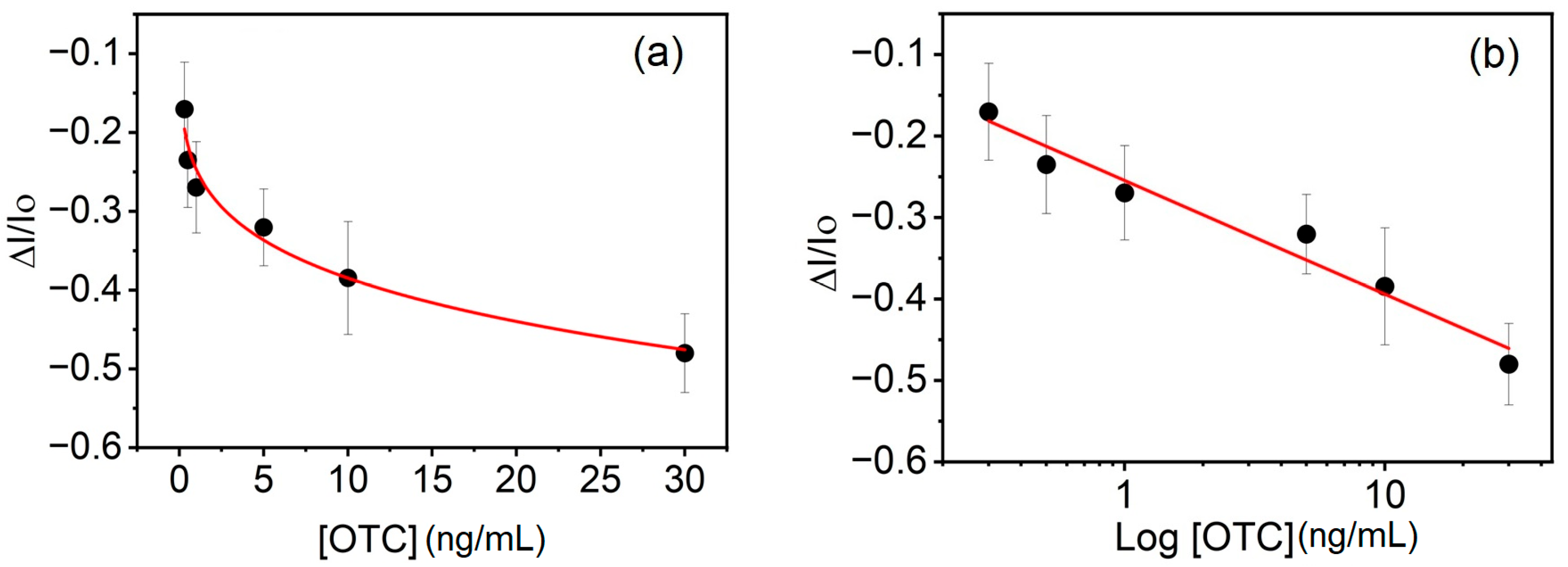
Based on the results presented in Figure 2b, we constructed the plot of the normalized changes in the current ΔI/I0 vs. concentration of OTC. Here, ΔI = I − I0, where I is the current amplitude at a certain concentration of OTC and I0 is the current for the bare aptasensor without OTC. As can be seen from Figure 3a, the value ΔI/I0 decreases with increasing OTC concentration. We find that the Langmuir model does not describe the observed changes, whereas the Freundlich adsorption isotherm, ΔI/I0 = KFC1/n, where KF and n are the constants and C is the concentration of OTC, describes the observed adsorption well. It suggests the heterogeneous nature of the surfaces, and it can be interpreted that the binding of OTC to the aptamers is combined with physical adsorption [16]. We estimated the value of the Freundlich adsorption constant KF to be −(0.25 ± 0.01) (mL/ng)1/n and n = 5.18 ± 0.47, R2 = 0.97.
A linear dependence of the relative changes in the current ΔI/I0 on the Log[OTC] (note logarithmic scale of concentration axes) can be seen on a calibration plot ΔI/I0 vs. Log[OTC] (Figure 3b). Using this plot, we estimated the limit of detection (LOD) using a formula LOD = 10a, where a = 3.3σ/b, σ is the standard deviation of the intercept, and b is the slope of the linear regression line [17,18]. The LOD = 1.72 ng/mL is about 58 times lower than the MRL established for OTC by the EU (100 ng/mL) [1]. This is also better than LOD reported for the electrochemical aptasensor based on a gold screen-printed electrode (LOD = 14 ng/mL) [19] and MWCNTs, gold nanoparticles (AuNPs), rGO, and a chitosan sensor (LOD = 4.2 ng/mL) [2]. Better sensitivity (LOD = 0.23 ng/mL) was achieved using GCE grafted by diazonium salts with covalently attached aptamers [20]. The high sensitivity of impedimetric aptasensor using covalent organic framework nanospheres, with a LOD of 7.4 fg/mL, has been reported by Yuan et al. [21]. However, the sensor response saturated at 10 pg/mL OTC, which limits its practical use. A comprehensive review of electrochemical aptasensors for OTC detection is available elsewhere [6,10]. The review of most recent publications (2023–2025) on electrochemical aptasensors for OTC detection is presented in Table 1.
As can be seen from Table 1, the highest sensitivity for OTC detection was obtained from the nanocomposites based on covalent organic frameworks (COF) [21]. However, even for sensing surfaces based on gold layers covered by Kapton film [24,27], the LOD was below the MRL. The LOD obtained in our work is better than those based on MOF(Fe); however, the dynamic range should be improved. Still, the sensitivity of all sensors presented is sufficient for practical applications, even for diluted food samples. Most of the sensors have been validated in milk with sufficient recovery. Typically, 10x diluted food samples were analyzed. Further effort should, therefore, be focused on the design of the sensing surface for the improvement of its antifouling properties [17,28]. This allows the application of the sensor directly in raw milk.
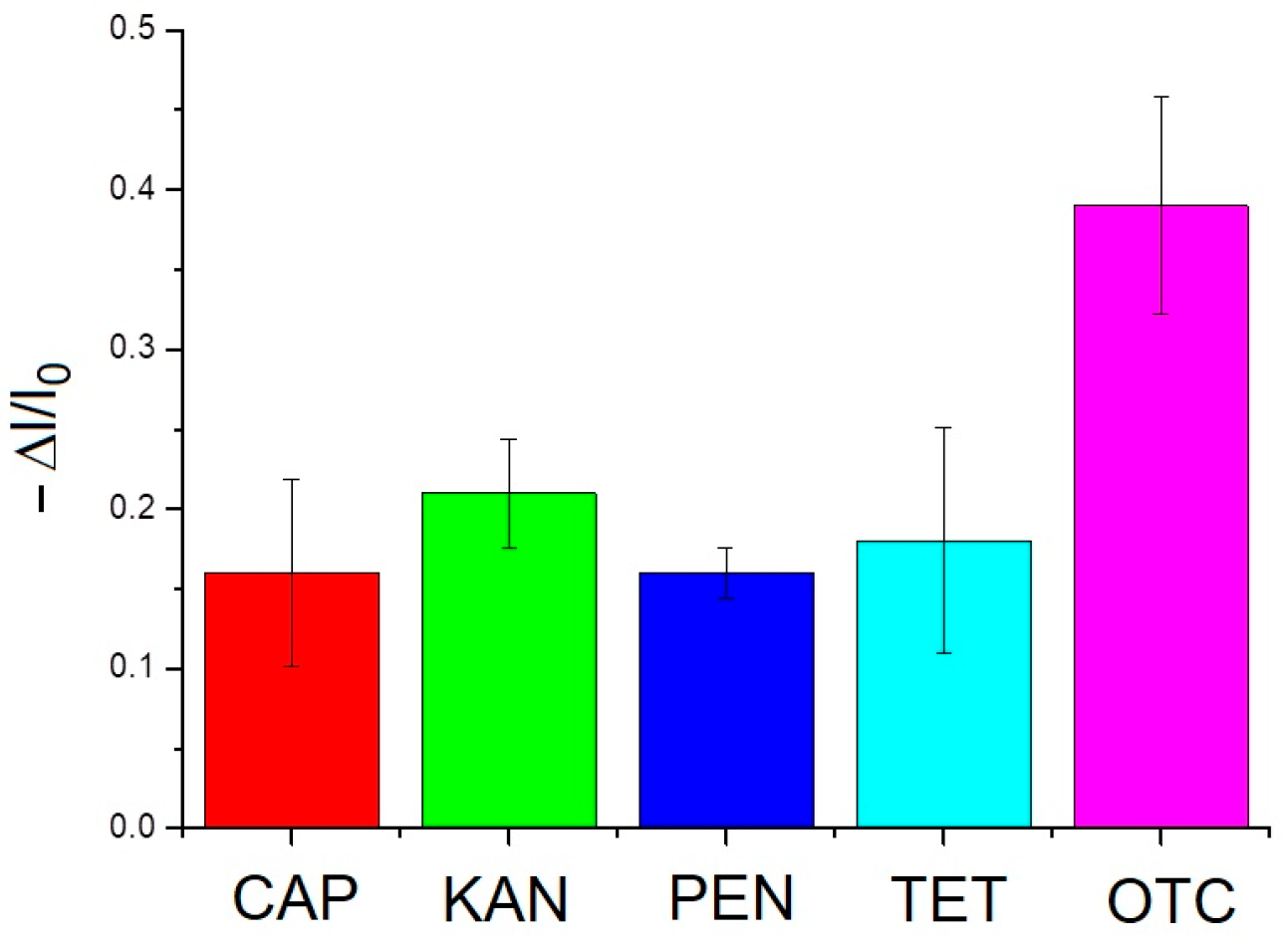
We also analyzed the selectivity of the sensor response in presence of 10 ng/mL of other antibiotics. Figure 4 shows the sensor response for OTC, chloramphenicol (CAP), kanamycin (KAN), penicillin G (PEN), and tetracycline (TET). While a higher response for OTC is evident, this is not OTC-specific, suggesting some nonspecific binding of CAP, KAN, PEN, and TET to the aptamers.
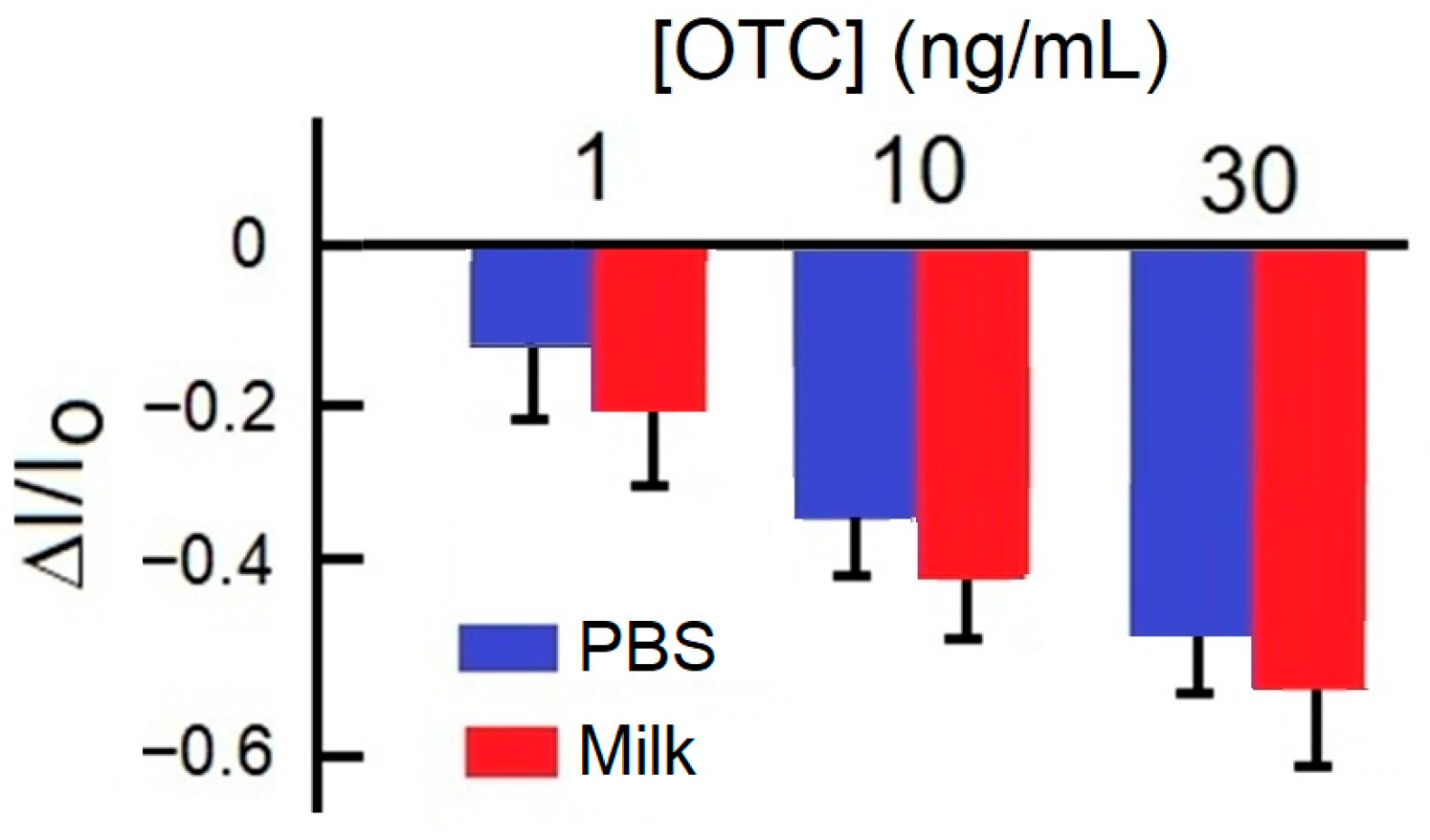
We performed experiments on the OTC charge transfer detection in spiked milk samples. Figure 5 shows the sensor response in PBS and in milk samples spiked by 1, 10, and 30 ng/mL OTC. The changes in the current amplitude measured are higher for OTC spiked milk compared with those in PBS. This can be explained as the influence of other milk components (caseins or lipids) on the sensor response [19]. The best recovery of 109% and the lowest relative standard deviation (RSD = 10.5%) was observed for 30 ng/mL, the highest OTC concentration tested. This means that the accuracy of measurements is improved at a higher OTC concentration, which is associated with the higher current amplitude changes. Considering rather high sensitivity and relatively large MRL for OTC detection, the sensor can be used in substantially diluted milk samples, which also helps to reduce the effect of the milk components.

4. Conclusions

We developed a highly sensitive electrochemical aptamer-based biosensor for detection oxytetracycline using GCE with electrodeposited and reduced graphene oxide–multiwalled carbon nanotubes (rGO/MWCNTs) composite. The sensor demonstrated high sensitivity in OTC detection with the LOD of 1.72 ng/mL. The binding of OTC to the sensor surface can be characterized by the Freundlich adsorption isotherm. This suggests the heterogeneous nature of the surfaces, and it can be interpreted that the binding of OTC to the aptamers is combined with physical adsorption. The sensor was tested in spiked milk sample with 109% recovery. We speculate that the observed effect can arise from the nonspecific adsorption of milk compounds, such as proteins and lipids, to the sensing surface. Further experiments are needed to improve the antifouling properties of the sensor as well as to increase the sensor’s dynamic range.

Author Contributions

Conceptualization, T.H.; methodology, T.H.; validation, M.K., C.Z., M.P. and K.S.; formal analysis, T.H., K.S., M.K. and M.P.; investigation, M.K., C.Z., M.P. and K.S.; resources, T.H.; writing—original draft preparation, T.H. and K.S.; writing—review and editing, T.H., K.S., Z.K., L.B. and I.N.I.; supervision, T.H. and I.N.I.; project administration, T.H.; funding acquisition, T.H., L.B., Z.K. and I.N.I. All authors have read and agreed to the published version of the manuscript.

Funding

This research was funded by the European Union’s Horizon 2020 research and innovation program through the Marie Skłodowska-Curie grant agreement No. 101007299, the Science Agency VEGA, project No. 1/0445/23. A portion of this research was conducted at the Center for Nanophase Materials Sciences, which is a DOE Office of Science User Facility, project No. CNMS2024-B-02709.

Institutional Review Board Statement

Not applicable.

Informed Consent Statement

Not applicable.

Data Availability Statement

The data will be available upon request.

Conflicts of Interest

Authors Minas Kakos, Charalampos Zacharopoulos and Leda Bousiakou were employed by the company IMD Laboratories Co., Athens, Greece. The remaining authors declare that the research was conducted in the absence of any commercial or financial relationships that could be construed as a potential conflict of interest.

References

  1. Commission Regulation (EU). No 37/2010 of 22 December 2009 on pharmacologically active substances and their classification regarding maximum residue limits in foodstuffs of animal origin. Off. J. Eur. Union. 2010, 15, 1–72. [Google Scholar]
  2. Akbarzadeh, S.; Khajehsharifi, H.; Hajihosseini, S. Detection of oxytetracycline using an electrochemical label-free aptamer-based biosensor. Biosensors 2022, 12, 468. [Google Scholar] [CrossRef] [PubMed]
  3. Ashraf, D.; Morsi, R.; Usman, M.; Meetani, M.A. Recent advances in the chromatographic analysis of emerging pollutants in dairy milk: A review (2018–2023). Molecules 2024, 29, 1296. [Google Scholar] [CrossRef] [PubMed]
  4. Chan, C.L.; Wai, H.K.F.; Wu, P.; Lai, S.W.; Chan, O.S.K.; Tun, H.M. A universal LC-MS/MS method for simultaneous detection of antibiotic residues in animal and environmental samples. Antibiotics 2022, 11, 845. [Google Scholar] [CrossRef]
  5. Wang, B.; Xie, K.; Lee, K. Veterinary drug residues in animal-derived foods: Sample preparation and analytical methods. Foods 2021, 10, 555. [Google Scholar] [CrossRef]
  6. Evtugyn, G.; Porfireva, A.; Tsekenis, G.; Oravczova, V.; Hianik, T. Electrochemical aptasensors for antibiotics detection: Recent achievements and applications for monitoring food safety. Sensors 2022, 22, 3684. [Google Scholar] [CrossRef]
  7. Stoltenburg, R.; Reinemann, C.; Strehlitz, B. SELEX–A (r)evolutionary method to generate high-affinity nucleic acid ligands. Biomol. Eng. 2007, 24, 381–403. [Google Scholar] [CrossRef]
  8. Tian, R.; Sun, J.; Ye, Y.; Lu, X.; Sun, X. Screening strategy of aptamers and its application in food contaminants determination. Trends Anal. Chem. 2024, 175, 117710. [Google Scholar] [CrossRef]
  9. Niazi, J.H.; Lee, S.J.; Gu, M.B. Single-stranded DNA aptamers specific for antibiotics tetracyclines. Bioorg. Med. Chem. 2008, 16, 7245–7253. [Google Scholar] [CrossRef]
  10. Singh, B.; Bhat, A.; Dutta, L.; Pati, K.R.; Korpan, Y.; Dahiya, I. Electrochemical biosensors for the detection of antibiotics in milk: Recent trends and future perspectives. Biosensors 2023, 13, 867. [Google Scholar] [CrossRef]
  11. Pisoschi, A.M.; Iordache, F.; Stanca, L.; Rosu, P.M.; Ciocirlie, N.; Geicu, I.O.; Bilteanu, L.; Serban, A.I. Electrochemical nanosensors applied to the assay of some food components—A review. Chemosensors 2025, 13, 272. [Google Scholar] [CrossRef]
  12. Shirsat, M.D.; Hianik, T. Electrochemical detection of heavy metal ions based on nanocomposite materials. J. Compos. Sci. 2023, 7, 473. [Google Scholar] [CrossRef]
  13. Jia, F.; Duan, N.; Wu, S.; Dai, R.; Wang, Z.; Li, X. Impedimetric Salmonella aptasensor using a glassy carbon electrode modified with an electrodeposited composite consisting of reduced graphene oxide and carbon nanotubes. Microchim. Acta 2016, 183, 337–344. [Google Scholar] [CrossRef]
  14. Sharma, A.; Istamboulie, G.; Hayat, A.; Catanante, G.; Bhand, S.; Marty, J.L. Disposable and portable aptamer functionalized impedimetric sensor for detection of kanamycin residue in milk sample. Sens. Actuators B 2017, 245, 507–515. [Google Scholar] [CrossRef]
  15. Tempfli, D.; Borbás, E.; Pataki, H.; Csicsák, D.; Völgyi, G.; Sinkó, B.; Takács-Novák, K. Revisit of solubility of oxytetracycline polymorphs. An old story in the light of new results. Eur. J. Pharm. Sci. 2020, 149, 105328. [Google Scholar] [CrossRef]
  16. Wang, J.; Guo, X. Adsorption isotherm models: Classification, physical meaning, application and solving method. Chemosphere 2020, 258, 127279. [Google Scholar] [CrossRef] [PubMed]
  17. Davoudian, K.; Spagnolo, S.; Chan, E.; Hianik, T.; Thompson, M. Acoustic wave sensor detection of an ovarian cancer biomarker with antifouling surface chemistry. Sensors 2024, 24, 7884. [Google Scholar] [CrossRef] [PubMed]
  18. Ambruser, D.A.; Pry, T. Limit of blank, limit of detection and limit of quantification. Clin. Biochen. Rev. 2008, 29 (Suppl. S1), S49–S52. [Google Scholar]
  19. Kourti, D.; Geka, G.; Nemtsov, L.; Ahmadi, S.; Economou, A.; Thompson, M. Electrochemical aptasensor with antifouling properties for label-free detection of oxytetracycline. Sensors 2024, 24, 5488. [Google Scholar] [CrossRef]
  20. Yang, Y.; Yan, W.; Guo, Y.; Wang, X.; Zhang, F.; Yu, L.; Guo, C.; Fang, G. Sensitive and selective electrochemical aptasensor via diazonium-coupling reaction for label-free determination of oxytetracycline in milk samples. Sens. Actuators Rep. 2020, 2, 100009. [Google Scholar] [CrossRef]
  21. Yuan, R.; Fu, Z.; He, Y.; Deng, Y.; Xi, J.; Xing, X.; He, H. Size-controlling preparation of covalent organic framework nanospheres for electrochemical impedimetric aptasensing of oxytetracycline. Talanta 2023, 265, 124834. [Google Scholar] [CrossRef]
  22. Wang, Q.; Li, P.; Wen, H.-M.; Hu, K.-J.; Huang, Z.-Y.; Chen, J. A HOF-based electrochemical aptasensor for highly sensitive and selective detection of trace oxytetracycline. Inorganic Chem. Commun. 2023, 156, 111213. [Google Scholar] [CrossRef]
  23. Song, P.; Qu, P.; Wang, M.; Wang, A.-J.; Xue, Y.; Mei, L.-P.; Feng, J.-J. Self-checking dual-modal aptasensor based on hybrid Z-scheme heterostructure of Zn-defective CdS/ZnS for oxytetracycline detection. Anal. Chim. Acta 2023, 1274, 341542. [Google Scholar] [CrossRef] [PubMed]
  24. Machairas, C.; Anagnostoupoulos, A.; Soulis, D.; Economou, A.; Jakab, K.; Melios, N.; Keresztes, Z.; Tsekenis, G.; Wang, J.; Speliotis, T. Microfabricated gold aptasensors for the label-free electrochemical assay of oxytetracycline residues in milk. Eng. Proc. 2023, 58, 1. [Google Scholar] [CrossRef]
  25. Song, J.; Fan, X.; Ee, L.Y.; Lin, X.; Zhang, D.; Li, S.F.Y.; Huang, M. Novel dopamine-derived carbon modified Fe-based metal- organic framework enhanced aptasensor rendering ultrasensitive electrochemical detection of oxytetracycline. Chem. Eng. J. 2024, 499, 156373. [Google Scholar] [CrossRef]
  26. Guan, F.; Dong, Y.; Wang, L.; Cai, D.; Guo, Y.; Zhao, S.; Sheng, Q. An electrochemical aptamer sensor based on AuNPs/ErGO/Cu-MOF nanocomposites for the detection of oxytetracycline in foodstuff. Microchem. J. 2025, 208, 112579. [Google Scholar] [CrossRef]
  27. Bizinti, C.; Soulis, D.; Kourti, D.; Geka, G.; Kokkinos, C.; Thompson, M.; Nemtsov, L.; Speliotis, T.; Economou, A. Microfabricated electrochemical aptasensing chip modified with dual-function antifouling linker for single-drop label-free assay of oxytetracycline in milk. Mirochim. Acta 2025, 192, 527. [Google Scholar] [CrossRef]
  28. Spagnolo, S.; Davoudian, K.; De La Franier, B.; Kocsis, R.; Hianik, T.; Thompson, M. Nanoparticle-enhanced acoustic wave biosensor detection of Pseudomonas aeruginosa in food. Biosensors 2025, 15, 146. [Google Scholar] [CrossRef]
Figure 1. The scheme of sequential steps of assembly of rGO/MWCNTs aptamer sensor.
Engproc 106 00016 g001
Figure 2. The DPV of (a) bare, GO-MWCNTs and rGO-MWCNTs modified GCE, and (b) DPV of GCE modified by rGO-MWCNTs-APT-ETA and by its incubation with various concentrations of OTC in ng/mL (see the inset).
Engproc 106 00016 g002
Figure 3. The plot of the relative changes in the current amplitude ΔI/I0 vs. the concentration of OTC (a) in a linear scale of OTC concentration. The red line is the fit according to the Freundlich adsorption isotherm. (b) The calibration plot in a semilogarithmic scale. The red line is a linear regression fit: ΔI/I0 = −(0.25 ± 0.01) − (0.14 ± 0.01) log[OTC], R2 = 0.96. Results are mean ± SD obtained from three independent experiments at each OTC concentration.
Engproc 106 00016 g003
Figure 4. The comparison of the sensor response following incubation with various antibiotics at the concentration of 10 ng/mL. The results are mean ± SD obtained from three experiments in each series. CAP—chloramphenicol; KAN—kanamycin; PEN—penicillin G; TET—tetracycline; and OTC—oxytetracycline.
Engproc 106 00016 g004
Figure 5. The elative changes in the current amplitude ΔI/I0 in PBS and in milk spiked by OTC at the concentrations of 1, 10, and 30 ng/mL.
Engproc 106 00016 g005
Table 1. The comparison of the properties of the electrochemical aptasensors for detection oxytetracycline published between 2023–2025.
Sensing SurfaceDetection MethodDynamic Range
ng/mL
LOD,
ng/mL
Sample Type/
Recovery %
Reference
SPE Au electrodeDPV1–50014 (PBS)/10 (Milk)Milk/124[19]
Au/COF nanospheresEIS10−5–10−27.4 × 10⁻⁶Milk/95.7–107[21]
Au/HOFCV, EIS(0.01–0.9) × 10−39.4 × 10⁻⁴-[22]
FTO/Zn-defective CdS/ZnS heterostructurePEC
EC
0.01–501.86 × 10⁻³
3.08 × 10⁻³
-[23]
Au/Kapton filmDPV1.5–35Milk/95[24]
GCE/MOF(Fe)@PDA/SPEDPV/
CA
4.6–46003.17/2.56Tap water/98.2/99.4[25]
SPCE/AuNPs/ErGO/Cu-MOFCV0.1–1050.03Milk/87–110[26]
Au/Kapton filmDPV1.4–2.77Milk/107–110[27]
GCE/rGO/MWCNTsDPV1–301.72Milk/109This work
AuNPs: gold nanoparticles; CA: chronoamperometry; COF: covalent organic framework; CV: cyclic voltammetry; EC: electrochemical; EIS: electrochemical impedance spectroscopy; ErGO: electrochemically reduced graphene oxide; FTO: fluorine doped tin oxide; GCE: glassy carbon electrode; HOF: hydrogen organic frameworks; MOF: metal–organic frameworks; PDA: polydopamine; PEC: photoelectrochemical; SPCE: screen-printed carbon electrode; and SPE: screen-printed electrode.
Disclaimer/Publisher’s Note: The statements, opinions and data contained in all publications are solely those of the individual author(s) and contributor(s) and not of MDPI and/or the editor(s). MDPI and/or the editor(s) disclaim responsibility for any injury to people or property resulting from any ideas, methods, instructions or products referred to in the content.