


Proceeding Paper

Contribution to the Chemistry of Randia echinocarpa  †
by
Dalia I. Díaz-Arellano
,
Armando Talavera-Alemán
,
Gabriela Rodríguez-García
,
Rosa E. del Río
* and
Mario A. Gómez-Hurtado
*
Instituto de Investigaciones Químico Biológicas, Universidad Michoacana de San Nicolás de Hidalgo, Morelia 58030, Michoacán, Mexico
*
Authors to whom correspondence should be addressed.
Presented at the 29th International Electronic Conference on Synthetic Organic Chemistry, 14–28 November 2025; Available online: https://sciforum.net/event/ecsoc-29.
Chem. Proc. 2025, 18(1), 94; https://doi.org/10.3390/ecsoc-29-26674
Published: 11 November 2025

Abstract

Plants from the Randia genus (Rubiaceae) are used in Mexican traditional medicine, where diseases such as diabetes, cancer, and chronic inflammation are treated with these plants. Particularly, Randia echinocarpa Sessé & Moc. ex DC (endemic in Mexico) is used in the northern region to treat kidney diseases and stomach disorders, while in the central area, this plant is used to treat circulatory and lung diseases, cancer, diabetes, and malaria. Previous research on this plant has highlighted the antibacterial potential of extracts from leaves and stems and nematicidal and antioxidant activities for fruit extracts. Phytochemical studies of this plant have been poorly explored, though the presence of mannitol, triterpene, and phytosterol compounds as main components in extracts from fruits was described. In this research, the chemical study of leaves and fruits of R. echinocarpa is described. The presence of gardenoside as the main component of the methanolic extract from leaves was determined after a phytochemical analysis. This compound could be directly related to the use of R. echinocarpa in traditional medicine since scientific reports suggested its potential as an anti-inflammatory and a pain suppressor, as well as its inhibitory effect on free fatty acid (FFA)-induced cellular steatosis. In addition, β-gardiol was isolated from fruit extract. A chemical correlation of β-gardiol with gardenoside was performed by enzymatic hydrolysis. Other components, including ursolic acid, stigmasterol, sitosterol, and D-mannitol, were also identified. All compounds were characterized by their physical and spectroscopic data.

1. Introduction

The Randia genus comprises around 160 species, which are concentrated in tropical and subtropical regions of the American Continent [1]. The ethnomedicinal background of species from the Randia genus is well known. Several diseases, including circulatory and lung diseases, cancer, diabetes, and malaria, are treated with these plants [2,3,4], and their biological potential as nematicidal, antioxidant, and antimicrobial agents has been scientifically described [5,6,7]. Several of these species grow in the Mexican territory, of which 44 species are endemic, including R. echinocarpa [8], whose traditional use in medicine is to treat kidney and stomach diseases by using the fruits [9], while the leaves are used to treat circulatory and lung diseases, as well as cancer, diabetes, and peptic ulcers [7,10]. Phytochemical studies on fruits from R. echinocarpa described the presence of D-mannitol, triterpenes, and phytosterol compounds as main components [9]. Due to the biological importance of this plant, research aimed at determining the phytochemical composition of R. echinocarpa becomes pertinent. Thus, in this paper, the isolation and structural characterization of the main components from the leaves and fruits of R. echinocarpa are described. Herein, gardenoside (1) resulted as a major component from leaves. The presence of D-mannitol, β-sitosterol, stigmasterol, and ursolic acid was also determined in leaf extracts. For its part, β-gardiol (2) was the main component of fruits. All compounds were characterized by their physical and spectroscopic data. The biological potential of compounds 1 and 2, previously described, could be related to the ethnomedicinal use of this plant.

2. Materials and Methods

2.1. General Procedures

Melting points (uncorrected) were measured on a Fisher-Johns apparatus (Fisher Scientific Co., Pittsburgh, PA, USA). Optical rotations were determined on a Perkin-Elmer 341 polarimeter (Perkin-Elmer, Waltham, MA, USA). UV-vis spectra were measured on a Genesys 10S spectrophotometer (Thermo Fisher Scientific Inc., Waltham, MA, USA). IR spectra were measured using a Thermo Scientific Nicolet IS10 spectrophotometer (Thermo Fisher Scientific Inc., Waltham, MA, USA) with the ATR technique. The NMR spectra were acquired on a Varian Mercury 400 spectrometer (Varian Inc., Palo Alto, CA, USA) from CDCl3 solutions using TMS as the internal reference. Column chromatography was performed using Merck silica gel 60 (230–400 mesh), and the solvents used as the mobile phase were distilled before their use.

2.2. Plant Material

Leaves and fruits of Randia echinocarpa Moc. & Sessé ex DC. were collected in the municipality of Huetamo de Núñez, Michoacán, Mexico (18°45′52.7″ N, 100°59′01.0″ W), at 540 m above sea level in November 2022. The plant material was dried in the shade and identified by Prof. Rosa Isabel Fuentes Chávez and Norma Patricia Reyes Martínez from the Herbario de la Facultad de Biología (EBUM) at UMSNH, where a specimen was deposited with voucher number EBUM-3648.

2.3. Extraction and Isolation

A batch of leaves (365 g) and fruits (1.5 kg) were separately macerated in MeOH (8 L each) for three days at room temperature. Each extract was individually filtered and evaporated to yield 46 g of leaf extract as a green, viscous material and 130 g of fruit extract as a bluish, viscous material. In addition, bipartition of both extracts was separately achieved; thus, a batch of leaves or fruit extract (20 g) was suspended in H2O (200 mL) and extracted with EtOAc (3 × 200 mL). After evaporation of the organic layer, 1 g of green oil was obtained in each case.

2.4. Gardenoside (1)

Isolated from MeOH extract from leaves (20 g) by column chromatography using CH2Cl2-MeOH-H2O mixtures (76:24:0.05; 74:26:0.05; 70:30:0.05; 200 mL each) as the eluent and collecting fractions of 15 mL each. This procedure yielded 5.6 g of 1 as a yellowish oil. [α]D = −106 (c 0.7, MeOH) Lit. [11] [α]D −84.6 (c 0.52, MeOH); IR νmax (cm−1): 3345, 2927, 1689, 1638, 1291, 1072, 863; UV (MeOH) λmax (log ε) nm: 236 (3.95); 1H and 13C NMR are concordant with the literature [12,13] (see Table S1 and Figures S1–S5).

2.5. β-Gardiol (2)

Isolated from the EtOAc fraction from fruits (3 g) after column chromatography using CH2Cl2-EtOAc mixtures in an ascending order of polarity (100 mL each) as the mobile phase, collecting fractions of 10 mL each. In fractions 1:1, 2:3, 3:7, 1:4, and 1:9, compound 2 was obtained as colorless needles (190 mg). m.p. 125–127 °C (lit. [14] 122 °C); [α]D = +212 (c 0.5, MeOH), Lit. [14] [α]D = +209 (c 0.07, MeOH), Lit. [15] +211 [α]D + (c 0.3, MeOH); 1H and 13C NMR are concordant with the literature (Table S1 and Figures S6 and S7) [11,14].

2.6. D-Mannitol

Isolated as a precipitate (25 g) from the MeOH extract from leaves before evaporation. White amorphous solid. Crystallization with EtOAc-MeOH yielded colorless needles; m.p. 163–164 °C (lit. [16] 167–170 °C). The 1H and 13C NMR data are consistent with the literature [17].

2.7. Mixture of β-Sitosterol and Stigmasterol

Isolated from the EtOAc fraction (3 g) from leaves after column chromatography using CH2Cl2-EtOAc mixtures in an ascending order of polarity (100 mL each) as the mobile phase, collecting fractions of 10 mL each. Fraction 9:1 yielded a mixture of β-sitosterol and stigmasterol (50 mg) as yellow crystals. m.p. 136–138 °C (lit. [16] 136–138 °C). 1H NMR data are concordant with the literature [18].

2.8. Ursolic Acid

Isolated from the EtOAc fraction (3 g) of the leaves after column chromatography using hexane–EtOAc mixtures in an ascending order of polarity (200 mL each) as the mobile phase, collecting fractions of 10 mL each. In fractions 4:1 and 7:3, ursolic acid was obtained as an amorphous solid. m.p. 233–235 °C (80 mg). 1H NMR data are concordant with the literature [19].

2.9. Enzymatic Hydrolysis of Gardenoside (1)

A batch of 1 (100 mg) was dissolved in H2O at 37 °C and stirred for 15 min. After, 6.6 mg of β-glucosidase enzyme from almonds (Aldrich) was added and stirred for two hours. After H2O was evaporated and the crude reaction was column chromatographed using silica gel as the stationary phase and a CHCl3-MeOH-H2O mixture (95:5:0.05, 500 mL) as the eluent, collecting fractions of 10 mL each. Fractions 21–34 yielded compound 2 (16 mg) as colorless needles.

3. Discussion

Gardenoside (1) (Figure 1) was isolated from leaves of R. echinocarpa as a yellowish oil with a levogyre optical activity of [α]D = −106 (c 0.7, MeOH). In the IR spectrum, two intense vibrational bands were highlighted at 1689 and 1291 cm−1, which were attributed to C=O and C-O vibrations, suggesting an oxidized natural product. The 1H NMR spectrum (see Figure S1) showed three signals from vinyl protons. The first one (H-3) was observed as a doublet (J = 1.4 Hz) at δ 7.38 assigned to a vinyl proton at β-position from a captodative system, as well as two doublet of doublets signals at δ 6.15 (J = 5.7, 2.8 Hz) and δ 5.73 (J = 5.7, 1.7 Hz) assigned to H-6 and H-7, respectively. A doublet signal was observed at δ 5.79 (J = 2.5 Hz), which was attributed to the acetal proton H-1. A resonance at δ 4.64 as a doublet (J = 7.9 Hz) was attributed to the anomeric proton (H-1′) from a glycosidic portion. The resonance pattern observed in the range of δ 3.88–3.18 suggested the presence of glucose as the sugar moiety [20,21]. A multiplet at δ 3.69 was observed and assigned to the allylic proton H-5. An AB spin system (J = 12.0 Hz) was observed at δ 3.57 and attributed to the CH2-10. In the aliphatic region, two signals were observed, including a singlet at δ 3.70 from the -OCH3 moiety and a doublet of doublets at δ 2.61 (J = 8.5, 2.5 Hz) from the H-9 proton. The 13C NMR showed 17 signals (see Figure S2), suggesting the presence of a glucosylated monoterpenoid compound. A carbonyl carbon from an ester group was observed at δ 168.8. Four resonances from vinyl carbons were observed at δ 151.9 (C-3), 135.8 (C-7), 135.6 (C-6), and 111.5 (C-4). The resonance of the acetal carbon (C-1) was observed at δ 94.2. In contrast, the anomeric carbon appeared at δ 99.8, and the remaining carbon signals from a glucose motif were observed at δ 78.3 (C-3′), 77.9 (C-5′), 74.6 (C-2′), 71.5 (C-4′), and 62.7 (C-6′). An OH-based carbon was observed at δ 86.2 (C-8). The signal attributed to CH2-10 was observed at δ 67.0. The resonances at δ 52.3 and 38.8 were related to the bridgehead carbons C-9 and C-5, respectively. The -OCH3 carbon signal was observed at δ 51.7. The assignment of the 1H and 13C signals suggested the presence of the glucoside iridoid 1 and was supported by 2D NMR experiments (see Supplementary Materials, Figures S3–S5). Additionally, the experimental data agreed with those previously described for compound 1 [11,12,13], which was previously isolated from Gardenia jasminoides [22], Rothmannia wittii [23], Genipa americana [24], Randia spinosa [12], and Xeromphis nilotica [11]. The described biological potential of compound 1 involves the inhibitory capabilities of COX-2, iNOS, and IL-6 [25], thereby indicating its potential as an anti-inflammatory and pain suppressor [26]. Additionally, its inhibitory effect on free fatty acids (FFA)-induced cellular steatosis has been described [27]. According to these backgrounds, compound 1 could be directly related to the use of R. echinocarpa in traditional medicine.
Column chromatography of the extract from fruits allowed the isolation of β-gardiol (2) (Figure 1) as colorless needles (m.p. 125–127 °C) with [α]D = +212 (c 0.5, MeOH). The 1H NMR spectrum (Figure S6) exhibited two resonances as a doublet of doublets at δ 5.91 (J = 5.6, 2.2 Hz) and 5.74 (J = 5.6, 2.0 Hz) from the vinylic protons H-6 and H-7, respectively. A signal from the acetal proton (H-1) was observed at δ 5.49 (d, J = 5.8 Hz). In addition, three resonances from OH-basis protons were observed and included the assignment to H-3 at δ 5.35 (d, J = 2.4 Hz), as well as those doublet signals (J = 9.4 Hz) from CH2-10 at δ 3.77 and 3.52. These signals, together with those observed at δ 3.50 (m) and 2.64 (dd, J = 9.2, 5.8 Hz) from bridgehead protons H-5 and H-9, respectively, suggested an iridoid skeleton, as that determined for compound 1. In addition, the resonance of H-4 was observed as a doublet of doublets (J = 9.2, 2.4 Hz), and a resonance as a singlet was observed at δ 3.72, which was assigned to a -OCH3 moiety. The 13C NMR spectrum (see Figure S7) exhibited eleven signals, suggesting the presence of a functionalized iridoid. Herein, a C=O signal from an ester moiety was observed at δ173.0 (C-11), while the resonances from the vinylic C-6 and C-7 were observed at δ 138.0 and 135.4, respectively. The acetal carbon resonance (C-1) appeared at δ 101.2, while the rest of the oxygen-bearing carbons were observed at δ 93.8 (C-8), 90.2 (C-3), and 75.0 (C-10), respectively. The -OCH3 carbon signal appeared at δ 52.3, the C-4 signal was observed at δ 49.0, while the resonances from bridgehead carbons C-9 and C-5 were observed at δ 48.8 and 40.4, respectively. These data agreed with those previously described for β-gardiol (2) [11,14,15]. Compound 2 has been identified in Rothmannia globosa [15], Burchellia bubaline [14], Rothmannia wittii [28], and Xeromphis nilotica [11]. The antimicrobial potential of 2 against Cladosporium cladosporioides and C. sphaerospermum [29] and as an anticancer agent was previously described [30].
A chemical correlation of compound 2 with iridoid 1 was achieved enzymatically. Herein, β-glucosidase, as the hydrolytic enzyme of compound 1, was used. After the reaction, extraction, and column chromatography purification processes, colorless needles were obtained whose physical and spectroscopic data agreed with those obtained herein for compound 2; thus, the close structural relationship between these compounds was evidenced (Table S1).
In addition, the isolation of D-mannitol was feasible by precipitation from the methanolic extract of leaves as a white precipitate, whose crystallization with EtOAc-MeOH gave colorless needles with m.p. 163–164 °C (lit. [16] 167–170 °C). The 1H and 13C NMR patterns agreed with the literature (see Figures S8 and S9) [17]. Also, the isolation of the mixture of β-sitosterol and stigmasterol as yellow crystals. m.p. 136–138 °C (lit. [16] 136–138 °C) was feasible after column chromatography from the EtOAc fraction of the methanolic extract of leaves as previously described (Figure S10) [18]. Ursolic acid was isolated from this fraction as an amorphous powder (see Figures S11 and S12). The 1H and 13C NMR patterns of these compounds agreed with the literature [19]. Together with compounds 1 and 2, these compounds may be related to the ethnomedicinal use of R. echinocarpa as the potential of D-mannitol as a hypotensive agent in the brain and eyes [31]. For its part, β-sitosterol and stigmasterol are described as anti-inflammatory compounds, while the antimicrobial, anticancer, anti-inflammatory, and antiviral activities of ursolic acid have been described [32,33,34].

4. Conclusions

The chemical study of R. echinocarpa enabled the isolation of two iridoids (1 and 2), which have known biological potential and are directly related to the ethnomedicinal uses of this plant. As seen, the chemical composition of fruits and leaves from R. echinocarpa involves secondary metabolites as the main components with directly related biosynthetic pathways. Considering the abundance of compounds 1 and 2 in R. echinocarpa, these iridoids represent an interesting option for future research aimed at the preparation of derivatives of chemical and biological interest, as well as a deeper biological exploration of 1 and 2 to increase the knowledge about their medicinal potential.

Supplementary Materials

The following supporting information can be downloaded at https://www.mdpi.com/article/10.3390/ecsoc-29-26674/s1, Figure S1. 1H NMR spectrum of gardenoside (1) in CD3OD (400 MHz); Figure S2. 13C NMR spectrum of gardenoside (1) in CD3OD (100 MHz); Figure S3. COSY NMR spectrum of gardenoside (1) in CD3OD (400 MHz); Figure S4. NOESY NMR spectrum of gardenoside (1) in CD3OD (400 MHz); Figure S5. HETCOR NMR of gardenoside (1) in CD3OD (400/100 MHz); Figure S6. 1H NMR spectrum of β-gardiol (2) in CD3OD (400 MHz); Figure S7. 13C NMR spectrum of β-gardiol (2) in CD3OD (100 MHz); Figure S8. 1H NMR spectrum of D-mannitol in D2O (400 MHz); Figure S9. 13C NMR spectrum of D-mannitol in D2O (100 MHz); Figure S10. 1H NMR spectrum of β-sitosterol and stigmasterol in CDCl3 (400 MHz); Figure S11. 1H NMR spectrum of ursolic acid in DMSOd6 (400 MHz); Figure S12. 13C NMR spectrum of ursolic acid in DMSOd6 (100 MHz); Table S1. NMR data for compounds 1 and 2.

Author Contributions

Conceptualization, R.E.d.R. and M.A.G.-H.; methodology, D.I.D.-A.; Formal analysis, A.T.-A. and G.R.-G.; investigation, D.I.D.-A.; resources, R.E.d.R. and M.A.G.-H.; writing—original draft preparation, M.A.G.-H.; writing—review and editing, R.E.d.R., A.T.-A. and G.R.-G.; supervision R.E.d.R. and M.A.G.-H.; funding acquisition, R.E.d.R. and M.A.G.-H. All authors have read and agreed to the published version of the manuscript.

Funding

This research was partially funded by CIC-UMSNH and FCCHT123_ME-4.1-0008.

Institutional Review Board Statement

Not applicable.

Informed Consent Statement

Not applicable.

Data Availability Statement

The original contributions presented in this study are included in this article. Further inquiries can be directed to the corresponding author.

Acknowledgments

D.I.D.-A. is grateful to SECIHTI for the doctoral scholarship 813417. We thank Rosa Isabel Fuentes Chávez and Norma Patricia Reyes Martínez, Facultad de Biología, UMSNH, for identifying the plant material.

Conflicts of Interest

The authors declare no conflicts of interest.

References

  1. Ojeda-Ayala, M.; Gaxiola-Camacho, S.M.; Delgado-Vargas, F. Phytochemical composition and biological activities of the plants of the genus Randia. Bot. Sci. 2022, 100, 779–796. [Google Scholar] [CrossRef]
  2. Méndez, V.L.M.; Hernández, M.M.R. Evaluación de la toxicidad del fruto de Randia monantha Benth. Rev. Méd. UV 2009, 9, 42–45. [Google Scholar]
  3. Erbano, M.; Duarte, M.R. Macro and microscopic analysis of the leaf and stem of Randia armata (Sw.) DC., Rubiaceae. Lat. Am. J. Pharm. 2011, 30, 1239–1243. [Google Scholar]
  4. Gallardo-Casas, C.A.; Guevara-Balcázar, G.; Morales-Ramos, E.; Tadeo-Jiménez, Y.; Gutiérrez-Flores, O.; Jiménez-Sánchez, N.; Valadez-Omaña, M.T.; Valenzuela-Vargas, M.T.; Castillo-Hernández, M.C. Ethnobotanic study of Randia aculeata (Rubiaceae) in Jamapa, Veracruz, Mexico, and its anti-snake venom effects on mouse tissue. J. Venom. Anim. Toxins incl. Trop. Dis. 2012, 18, 287–294. [Google Scholar] [CrossRef]
  5. López-Aroche, U.; Salinas-Sánchez, D.O.; Mendoza de Gives, P.; López-Arellano, M.E.; Liébano-Hernández, E.; Valladares-Cisneros, G.; Arias-Ataide, D.M.; Hernández-Velázquez, V. In vitro nematicidal effects of medicinal plants from the Sierra de Huautla, Biosphere Reserve, Morelos, Mexico against Haemonchus contortus infective larvae. J. Helminthol. 2008, 82, 25–31. [Google Scholar] [CrossRef]
  6. Cuevas-Juárez, E.; Yuriar-Aredondo, K.Y.; Pío-León, J.F.; Montes-Avila, J.; López-Angulo, G.; Díaz-Camacho, S.P.; Delgado-Vargas, F. Antioxidant and α-glucosidase inhibitory properties of soluble melanins from the fruits of Vitex mollis Kunth, Randia echinocarpa Sessé et Mociño, and Crescentia alata Kunth. J. Funct. Foods 2014, 9, 78–88. [Google Scholar] [CrossRef]
  7. Salinas-Sánchez, D.O.; Arteaga-Najera, G.L.; León-Rivera, I.; Dorado-Ramírez, O.; Valladares-Ceniceros, M.G.; Navarro-García, V.M. Antimicrobial activity of medicinal plants from the Huautla sierra biosphere reserve in Morelos (México). Polibotánica 2009, 28, 213–225. [Google Scholar]
  8. Villaseñor, J.L. Checklist of the native vascular plants of Mexico. Rev. Mex. Biodiv. 2016, 87, 559–902. [Google Scholar] [CrossRef]
  9. Bye, R.; Linares, E.; Mata, R.; Albor, C.; Castañeda, P.C.; Delgado, G. Ethnobotanical and phytochemical investigation of Randia echinocarpa (Rubiaceae). Anales Inst. Biol. Univ. Nac. Autón. México Ser. Bot. 1991, 62, 87–106. [Google Scholar]
  10. Martínez-Moreno, D.; Reyes-Matamoros, J.; Andrés-Hernández, A.R.; Pérez-Espinoza, L. Flora útil de la comunidad “Rancho el Salado” en Jolalpan, México. Rev. Iberoam. Cienc. 2016, 3, 1–15. [Google Scholar]
  11. Farid, H.A.R.; Kunert, O.; Haslinger, E.; Seger, C. Isolation and structure elucidation of iridoide and coumarin derivatives from Xeromphis nilotica (Rubiaceae). Monatsh. Chem. 2002, 133, 1453–1458. [Google Scholar] [CrossRef]
  12. Hamerski, L.; Furlan, M.; Siqueira, S.D.H.; Cavalheiro, A.J.; Eberlin, M.N.; Tomazela, D.M.; da Silva, B.V. Iridoid glucosides from Randia spinosa (Rubiaceae). Phytochemistry 2003, 63, 397–400. [Google Scholar] [CrossRef] [PubMed]
  13. Chaudhuri, R.K.; Afifi-Yazar, F.Ü.; Sticher, O.; Winkler, T. 13C NMR spectroscopy of naturally occurring iridoid glucosides and their acylated derivatives. Tetrahedron 1980, 36, 2317–2326. [Google Scholar] [CrossRef]
  14. Drewes, S.E.; Horn, M.M.; Munro, O.Q.; Ramesar, N.; Ochse, M.; Bringmann, G.; Peters, K.; Peters, E.-M. Stereostructure, conformation and reactivity of β- and α-gardiol from Burchellia bubaline. Phytochemistry 1999, 50, 387–394. [Google Scholar] [CrossRef]
  15. Jensen, S.R. Iridoids in Rothmannia globosa. Phytochemistry 1983, 22, 1761–1765. [Google Scholar] [CrossRef]
  16. Branco, A.; Santos, J.D.G.; Pimentel, M.M.A.M.; Osuna, J.T.A.; Lima, L.S.; David, J.M. D-Mannitol from Agave sisalana biomass waste. Ind. Crop. Prod. 2010, 32, 507–510. [Google Scholar] [CrossRef]
  17. Eswaraiah, M.C.; Elumalai, A. Isolation of phytoconstituents from the stems of Mussaenda erythrophylla. Pharm. Sin. 2011, 2, 132–142. [Google Scholar]
  18. Shwe, H.H.; Thein, W.W.; Win, S.S.; Pe, N.N.; Win, T. Structural characterization of stigmasterol and β-sitosterol from the roots of Premna herbacea Roxb. Int. Eur. Ext. Enablement Sci. Eng. Manag. 2019, 7, 195–201. [Google Scholar]
  19. Ludeña, H.M.A.; Tupa, Q.A.L.; Huamán, Q.R.I.; Serrano, F.C.A.; Robles, C.J. A simple method to obtain ursolic acid. Results Chem. 2021, 3, 100144. [Google Scholar] [CrossRef]
  20. Ávila-Román, J.; Quevedo-Tinoco, L.; Oliveros-Ortiz, A.J.; García-Gil, S.; Rodríguez-García, G.; Motilva, V.; Gómez-Hurtado, M.A.; Talero, E. Synthesis and bioevaluation of new stable derivatives of chrysin-8-C-glucoside that modulate the antioxidant Keap1/Nrf2/HO-1 pathway in human macrophages. Pharmaceuticals 2024, 17, 1388. [Google Scholar] [CrossRef]
  21. Quevedo-Tinoco, L.; Rodríguez-García, G.; del Río, R.E.; Talero, E.; Motilva, V.; Cerda-García-Rojas, C.M.; Joseph-Nathan, P.; Gómez-Hurtado, M.A. Strategy for the vibrational circular dichroism study of a glycosyl flavonoid evaluated as its peracetate. The case of bioactive 7-O-β-D-glucopyranosylchrysin. J. Mol. Struct. 2021, 1225, 129147. [Google Scholar] [CrossRef]
  22. Zhang, H.; Feng, N.; Xu, Y.-T.; Li, T.-X.; Gao, X.-M.; Zhu, Y.; Song, S.Y.; Wang, Y.-N.; Wu, H.-H. Chemical constituents from the flowers of wild Gardenia jasminoides Ellis. Chem. Biodivers. 2017, 14, e1600437. [Google Scholar] [CrossRef] [PubMed]
  23. Wisetsai, A.; Schevenels, F.T.; Faksri, K.; Tontapha, S.; Amornkitbamrung, V.; Lekphrom, R. New iridoid glucosides from the roots of Rothmannia witti (Craib) Bremek. Nat. Prod. Res. 2020, 36, 1716–1724. [Google Scholar] [CrossRef] [PubMed]
  24. Ono, M.; Ueno, M.; Masuoka, C.; Ikeda, T.; Nohara, T. Iridoid glucosides from the fruit of Genipa americana. Chem. Pharm. Bull. 2005, 53, 1342–1344. [Google Scholar] [CrossRef] [PubMed]
  25. Xia, T.; Zhao, R.; He, S.; Wang, L.; Fu, X.; Zhao, Y.; Qiao, S.; An, J. Gardenoside ameliorates inflammation and inhibits ECM degradation in IL-1β-treated rat chondrocytes via suppressing NF-κB signaling pathways. Biochem. Biophys. Res. Commun. 2023, 15, 164–172. [Google Scholar] [CrossRef]
  26. Yu, M.; Su, B.; Zhang, X. Gardenoside suppresses the pain in rats model of chronic constriction injury by regulating the P2X3 and P2X7 receptors. J. Recept. Signak Transduct. 2018, 38, 198–203. [Google Scholar] [CrossRef]
  27. Liang, H.; Zhang, L.; Wang, H.; Tang, J.; Yang, J.; Wu, C.; Chen, S. Inhibitory effect of gardenoside on free fatty acid-induced steatosis in HepG2 hepatocytes. Int. J. Mol. Sci. 2015, 16, 27749–27756. [Google Scholar] [CrossRef]
  28. Chaipukdee, N.; Kanokmednakul, K.; Kanokmedhakul, S.; Lekphrom, R.; Pyne, S.G. Two new bioactive iridoids from Rothmannia wittii. Fitoterapia 2016, 113, 97–101. [Google Scholar] [CrossRef][Green Version]
  29. Bolzani, V.S.; Izumisawa, C.M.; Young, M.C.M.; Trevisan, L.M.V.; Kingston, D.G.I.; Gunatilaka, A.L. Iridoids from Tocoyena formosa. Phytochemistry 1997, 46, 305–308. [Google Scholar] [CrossRef]
  30. Isiguro, K.; Yamaki, M.; Takagi, S.; Ikeda, Y.; Kawakami, K.; Ito, K.; Nose, T. Studies on iridoid-related compounds. IV. Antitumor activity of iridoid aglycones. Chem. Pharm. Bull. 1986, 34, 2375–2379. [Google Scholar] [CrossRef]
  31. Godswill, A.C. Sugar alcohols: Chemistry, production, health concerns and nutritional importance of mannitol, sorbitol, xylitol, and erythritol. Int. J. Adv. Acad. Sci. Res. 2017, 3, 31–66. [Google Scholar]
  32. Mangas, M.R.; Montes de Oca, P.R.; Bello, A.A.; Nival, V.L.A. Caracterización por cromatografía de gases/espectrometría de masas del extracto apolar de las hojas de Clusia minor L. Lat. Am. J. Pharm. 2008, 27, 747–751. [Google Scholar]
  33. Ferrer, H.A.E.; Hernández, E.M.; Pérez, M.C.S.; Basterrechea, R.M.; Fuentes, F.V.R.; Torres, F.M. Aislamiento de ácido ursólico de las hojas de Cestrum laurifolium L’Herit. Rev. Cenic. Cienc. Quím. 2007, 38, 243–247. [Google Scholar]
  34. Zhao, M.; Wu, F.; Tang, Z.; Yang, X.; Liu, Y.; Wang, F.; Chen, B. Anti-inflammatory and antioxidant activity of ursolic acid: A systematic review and meta-analysis. Front. Pharmacol. 2023, 14, 1256946. [Google Scholar] [CrossRef]
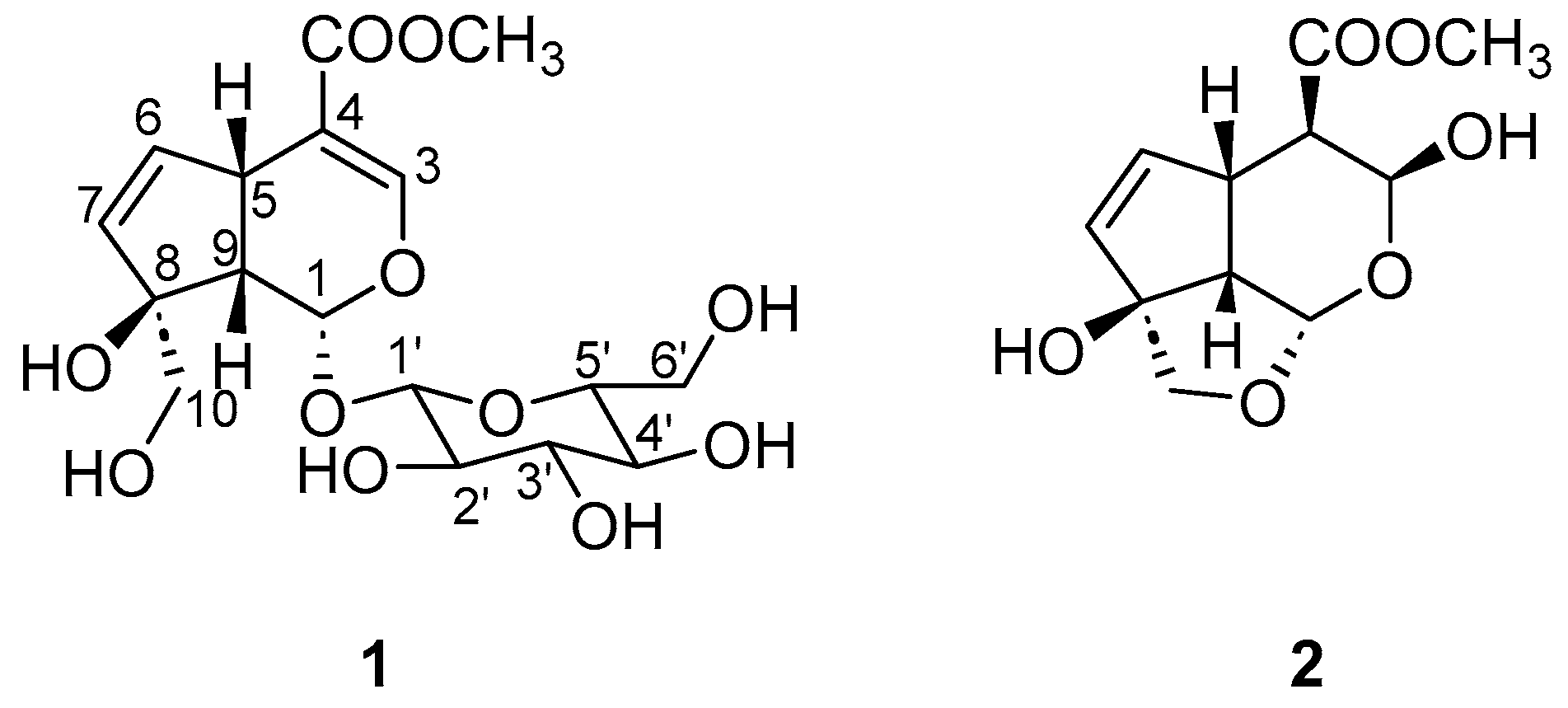
Figure 1. Structures of gardenoside (1) and β-gardiol (2).
Chemproc 18 00094 g001
Disclaimer/Publisher’s Note: The statements, opinions and data contained in all publications are solely those of the individual author(s) and contributor(s) and not of MDPI and/or the editor(s). MDPI and/or the editor(s) disclaim responsibility for any injury to people or property resulting from any ideas, methods, instructions or products referred to in the content.


