Proceeding Paper

Molecular Modelling of Withanolides Against EP2 Receptor for Treatment of Endometrial Cancer: A Pharmacokinetic and Molecular Docking Study †

Department of Pharmaceutical and Medicinal Chemistry, Ahmadu Bello University, Zaria 810107, Nigeria
*
Author to whom correspondence should be addressed.
Presented at the 29th International Electronic Conference on Synthetic Organic Chemistry, 14–28 November 2025; Available online: https://sciforum.net/event/ecsoc-29.
Chem. Proc. 2025, 18(1), 7; https://doi.org/10.3390/ecsoc-29-26724
Published: 11 November 2025

Abstract

Endometrial cancer is one of the most common gynecological cancers, with new global cases of approximately 420,000 new cases and 98,000 global deaths annually. Emerging evidence suggests that the EP2 receptor plays a critical role in tumor progression, angiogenesis, and immune evasion. Withanolides, a class of naturally occurring compounds, possess anticancer activity; however, the effect of the EP2 receptor in endometrial cancer remains largely unexplored. This study aims to explore the interaction between withanolides and the EP2 receptor using molecular docking techniques, with PF-04418948 as the reference antagonist. A select number of these ligands, with anticancer activity, were evaluated for their absorption, distribution, metabolism, excretion, and toxicity (ADMET) properties, and those with favorable drug-like properties, low toxicity profile, and no more than one Lipinski’s rule violation were docked to EP2 (PDB ID: 7CX2). Molecular docking studies revealed three ligands, (Pubchem 161671, 265237, and 21679027) with significantly higher binding affinity scores compared to that of the reference compound. Pubchem 161671 showed the highest binding affinity at −12.6 kcal/mol. Post-dock analysis revealed interactions with key amino acids, VAL89, LEU298, SER305, and MET31, which are essential for the antagonist activity of the EP2 receptor enzyme. Significant interaction with critical amino acid residues suggested potential inhibition of EP2 receptor activity, offering a potential therapeutic approach for treating endometrial cancer. Overall, this study proffers a deeper understanding of the potential of withanolides as leads for EP2 targeted therapy in endometrial cancer.

1. Introduction

Endometrial cancer (EC) is one of the leading gynecological malignancies worldwide, and its burden is steadily rising in both developed and developing regions. In Nigeria and across sub-Saharan Africa, challenges such as poor cancer registries, delayed presentation, and limited access to diagnostic and treatment facilities contribute to poorer outcomes compared with high-income settings [1,2,3]. Clinical reports from Nigerian tertiary hospitals describe EC as a common genital tract malignancy, often presenting with abnormal uterine bleeding, pelvic discomfort, or postmenopausal bleeding, with many patients already at advanced stages when diagnosed [4,5]. Biologically, EC is a heterogeneous disease driven by mutations in genes such as PTEN, PIK3CA, ARID1A, and TP53, and by activation of pathways including PI3K/AKT/mTOR and Wnt/β-catenin [2]. Increasing attention has also turned to PGE2 signaling, particularly via the EP2 receptor, a G-protein-coupled receptor that enhances tumor growth, angiogenesis, metastasis, and immune evasion [6,7,8]. While surgery, radiotherapy, chemotherapy, and immunotherapy remain standard treatments, recurrence, systemic toxicity, and limited responses in unselected patients highlight the need for novel targeted therapies [9,10,11,12]. Withanolides, naturally occurring steroidal lactones from Withania somnifera (ashwagandha) and other Solanaceae species, are well known in traditional medicine and widely distributed across South Asia, the Middle East, Africa, and the Mediterranean [13]. They have attracted growing interest for their broad pharmacological properties, including anti-inflammatory, immunomodulatory, and anticancer effects [14]. In experimental cancer models, withanolides such as withaferin A suppress tumor growth, trigger apoptosis, block angiogenesis, and interfere with oncogenic signaling pathways including NF-κB and PI3K/AKT [15,16,17]. Although their direct interaction with prostaglandin receptors has not been fully established, recent computational and biochemical studies suggest that withanolides may interact with G-protein-coupled receptors, making them promising candidates for further exploration against EP2 [16,18].
This study applies a structure-based in silico approach to evaluate natural and semi-synthetic withanolides as potential EP2 receptor antagonists in EC. By combining molecular docking, binding analysis, molecular dynamics refinement, and ADMET screening, we aim to identify withanolide scaffolds with high affinity and favorable drug-like properties. Computational methods such as molecular docking offer cost-effective and reliable ways to predict ligand–receptor interactions and guide early-stage drug discovery [19,20,21]. The identification of potent withanolides–EP2 interactions could pave the way for novel therapeutic options that inhibit PGE2-driven tumor progression, with particular relevance for resource-limited settings where EC outcomes remain poor.

2. Materials and Methods

2.1. Software, Hardware, and Databases

AutoDockVina version 1.5.6 [22], UCSF Chimera 1.13.1 [23], BioviaDiscovery Studio 2021 [24], Spartan 04, ChemSpider [25], SwissAdme [26], PROTOX III [27], and Intel processor, Corei7, 4gb RAM (HP Inc., Palo Alto, California, USA.) were used.
Protein Crystal Structure
A high-resolution, Cryo-EM structure of the Human prostaglandin E2 receptor was obtained from RCSB Protein Data Bank (https://www.rcsb.org/structure/7CX2, accessed on 28 July 2025.) PDB ID: 7CX2] [28].

2.2. In Silico Anticancer Studies

2.2.1. Evaluation of Theoretical Oral Bioavailability

The oral bioavailability of the selected compounds lig1, lig2, and lig3 was predicted theoretically based on Lipinski’s rule of five. The SWISSADME server [26] and PROTOX-III [27] servers were used for properties that defined the absorption, distribution, metabolism, excretion, and toxicity (ADMET) of the compounds, respectively. The servers also predicted various physicochemical properties, including lipophilicity, water solubility, drug-likeness, medicinal attributes, and compound toxicity, with high precision.

2.2.2. Protein Structure Preparation

In this study, the protein Cryo-EM structure of the PGE2-bound EP2-Gs complex with 7CX2 code and electron microscopy at 2.80 Å was downloaded from the Protein Data Bank and was used as the receptor starting structure. All crystallographic water molecules, ions, and bound ligands were removed from the 3D structures obtained from the Protein Data Bank using Chimera UCSF [23]. The crystallized ligands were separated and prepared using Chimera, then saved as lig.mol, while the receptors were isolated and prepared, then saved as rec.pdb. The output files from Chimera were then inputted into Autodock tools, where lig.mol and rec.pdb were edited by adding polar hydrogen and Gasteiger charges before being saved as pdbqt files [29]. AutoDock tools were applied in order to help set up docking runs and predict the binding free energy of the test ligands.

2.2.3. Ligand Structure Preparation

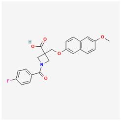
The 2D structures of the characterized compounds lig1, lig2, lig3, and the reference ligand, PF-04418948, were downloaded from Chemspider server, and Spartan 04 was used to convert the 2D structures to 3D. Using the AMI semi-empirical method, geometrical optimization was carried out on all the compounds using the Spartan software version 1.1.4, and the optimized structures were stored as mol2 files. AutoDock Tools was used to add hydrogen and Gasteiger charges and saved as mol2 files to pdbqt format [23].

2.2.4. Molecular Docking Analysis

Dock validation was carried out before the docking of the test compounds. The co-crystallized ligand was separated from the enzyme cryo structure and re-docked using the set up grid parameters. When a conformation that was superimposable with a geometrical conformation of the co-crystallized ligand in the active site was achieved, this grid parameter was used to dock the test compounds. Before molecular docking, the active sites were defined according to the coordinates of the crystallographic structures of both enzymes by defining the grid box, and the best pose was obtained, which was used for further studies. The UCSF Chimera was further used for post-docking visualization.

3. Results

3.1. Creation of Library

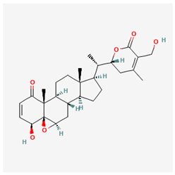
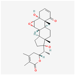
Table 1 shows the selected withanolides. Three compounds from withanolides class were downloaded from Chemspider.

3.2. Analysis of Theoretical Oral Bioavailability

Table 2 displays the calculated theoretical oral bioavailability parameters of all five selected compounds. The drug-likeness of these compounds was assessed based on Lipinski’s rule of five, i.e., their molecular weight (≤500 Da), hydrogen bond donors (≤5), hydrogen bond acceptors (≤10), and their MlogP values (≤5) [30]. Additional parameters such as the topological polar surface area (TPSA), number of rotatable bonds, and molar refractivity (MR), were also evaluated in the context of Veber’s rule, Egan’s rule, Ghose’s rule, and Muegge’s rule.

ADMET Profile

Table 3 shows the water solubility values, expressed as log Sw. The SwissADME server calculated values ranging from −3.20 to −4.00, indicating that the selected compounds possess good water solubility. The CytochromeP450 inhibitory potential of the compounds is also shown in the table. Lipophilicity values for the compounds, expressed as Consensus LogPo/w, ranged from 2.64 to 3.39. Table 4 shows the result of the toxicity profile test of the compounds.

3.3. Molecular Docking Studies

3.3.1. Grid Box

The gridbox parameter was used to generate the configuration file (config.txt). AutoDockVina produced results in pdbqt format, with the compounds saved in complexes alongside the reference enzymes. The specific gridbox parameter is detailed in Table 5 below.

3.3.2. Validation of Docking Procedures

The validation of the docking procedure is shown in Table 6. The protein structure was downloaded from the Protein Data Bank (PDB) with a co-crystallized molecule superimposed. In the dock validation procedure, the co-crystallized molecule was re-docked on the receptor.

3.3.3. Binding Affinity Ligands to EP2 Enzyme

Table 7 shows the binding energies of both the reference compound and the three selected compounds in their interactions with the EP2 enzyme.

3.3.4. Binding Poses and Binding Interaction Analysis of Isolated Compounds Against EP2 Enzyme

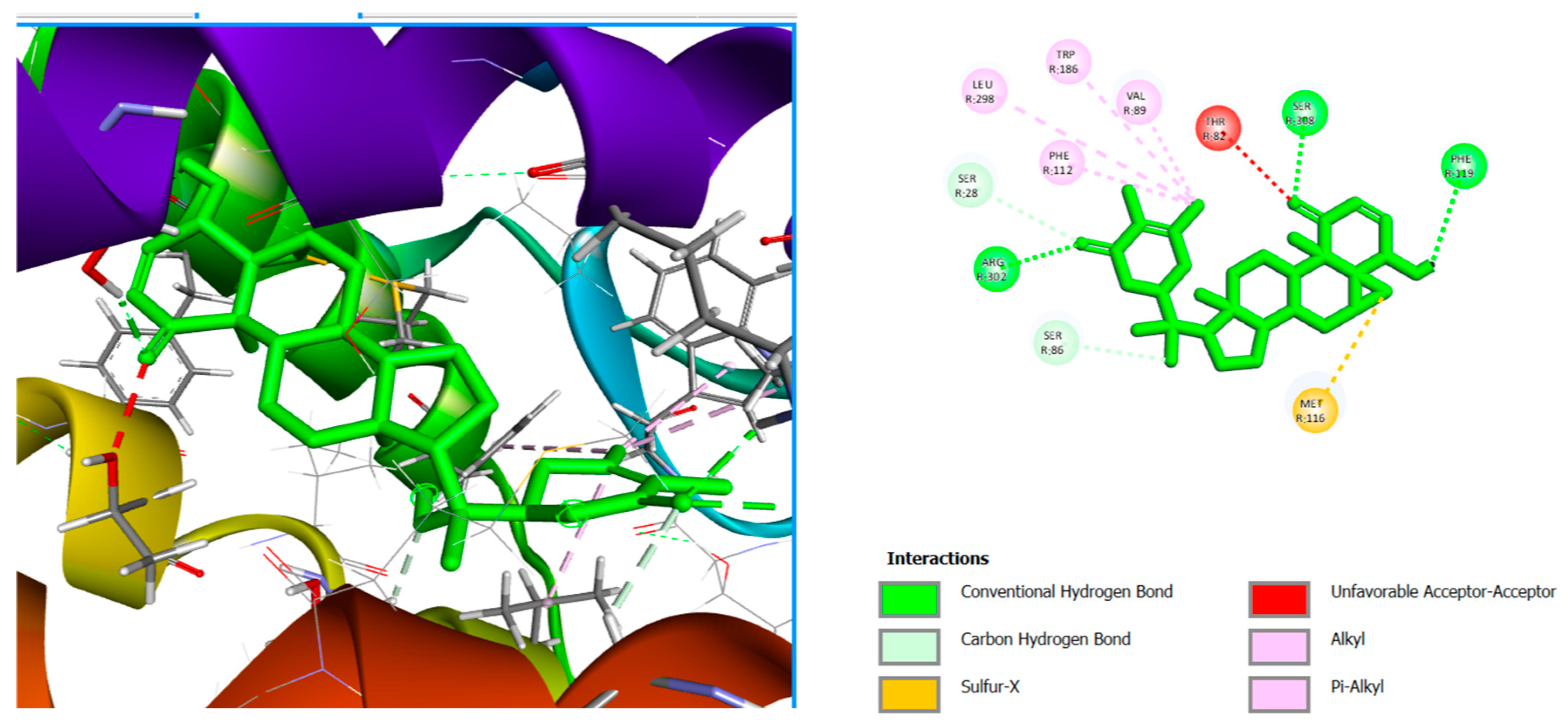
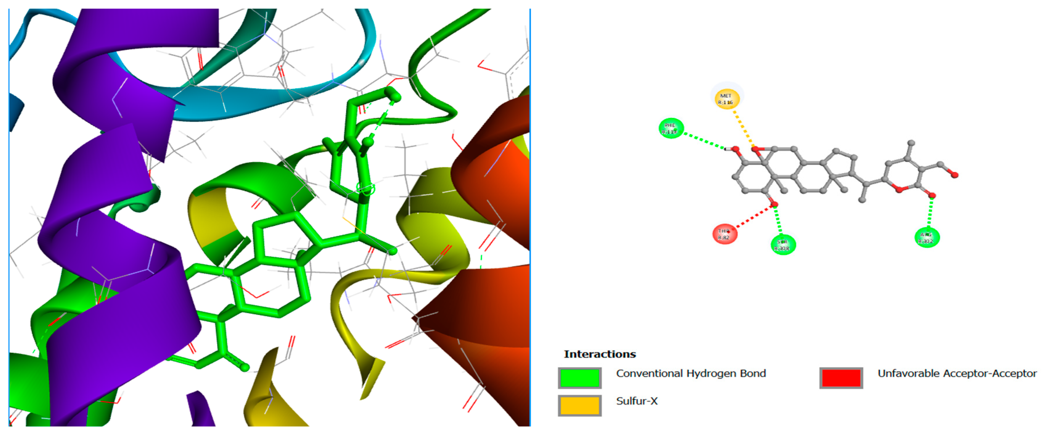
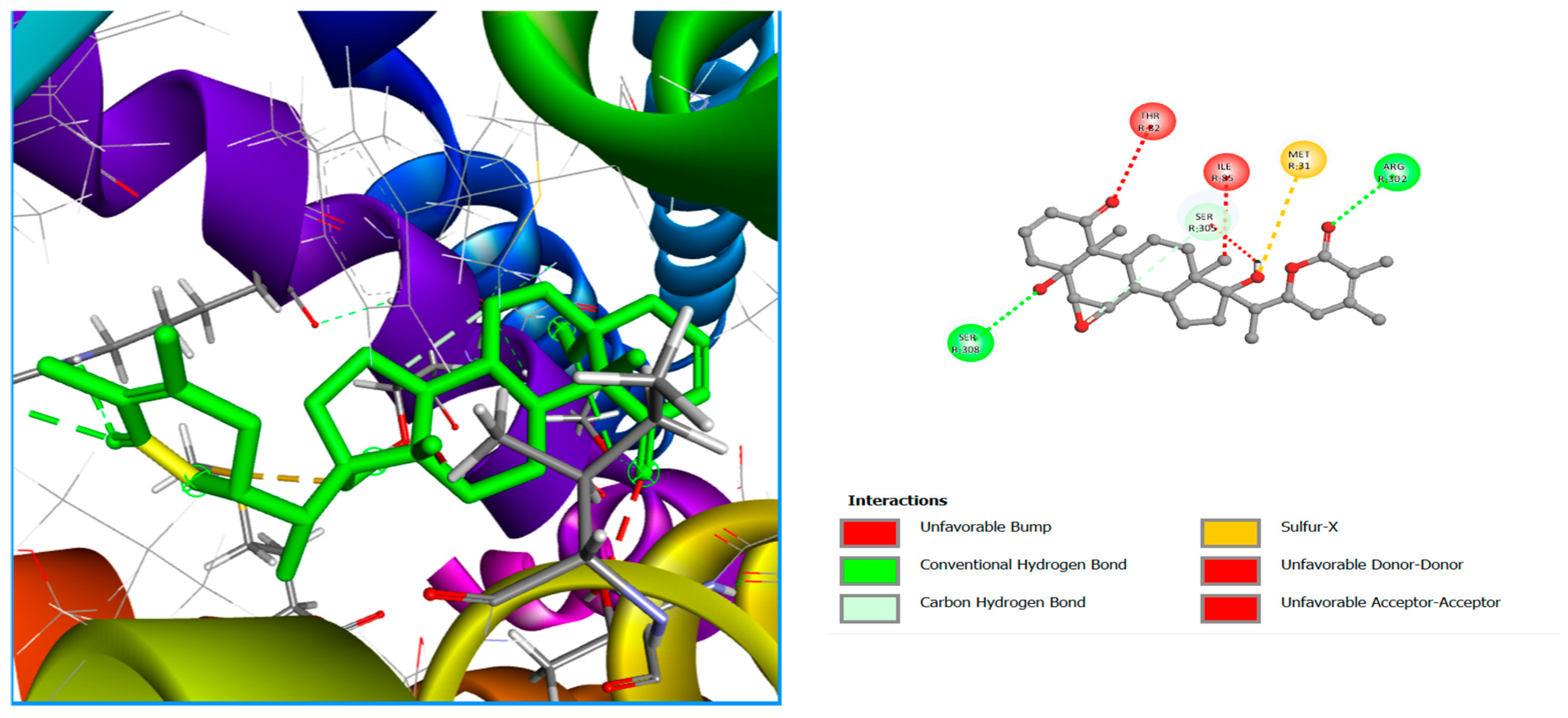
The binding conformation and interaction with residues on the active site of the enzymes studied using Chimera and Discovery Studio Visualizer [25] are shown in Figure 1, Figure 2, Figure 3 and Figure 4. Table 8, shows Molecular interactions of amino acid residues with EP2 enzyme, and the type of bonds with which they interact with these residues.

4. Discussion

The results of this study show that withanolides could be promising candidates for targeting the EP2 receptor in endometrial cancer. Looking at their drug-like properties, all three compounds, withanolide D (lig1), withaferin A (lig2), and withanone (lig3), performed well. Their molecular weights were within the drug-likeness threshold (470–486 g/mol) and they had acceptable numbers of hydrogen bond donors and acceptors. The lipophilicity values (MlogP 2.75, 1.95, and 2.75) suggest that these compounds are moderately hydrophobic, which is a sweet spot for oral absorption. Interestingly, lig2 was slightly more polar, which may reduce passive diffusion but could improve solubility. All three scored 0.55 for oral bioavailability, pointing to a good chance of achieving effective blood levels if developed as oral drugs. The ADMET screening gave more encouraging insights.
Their lipophilicity scores (LogP 2.64–3.39) fall into the optimal range for drug molecules. Each showed high gastrointestinal absorption, no penetration across the blood–brain barrier, and, importantly, no inhibition of cytochrome P450 enzymes—meaning fewer chances of drug–drug interactions. Solubility was moderate (LogS −3.20 to −4.00), which is not unusual for natural compounds and could be improved with formulation strategies. Overall, these pharmacokinetic features line up with what is typically seen in successful natural product-based drugs.
The toxicity analysis showed some disparities. Lig2 (withaferin A) was classified as toxicity Class II, meaning it can be fatal if swallowed at high doses, while lig1 and lig3 were in Class III, which is still toxic but less severe. While this indicates a red flag, it is important to note that many anticancer agents fall into similar categories; patient-dependent dose management is crucial. None of the compounds were predicted to be mutagenic, carcinogenic, or hormonally disruptive, which is a major strength for further development.
The docking studies revealed that the reference drug PF-04418948 bound to the EP2 receptor with an affinity of −9.6 kcal/mol, but all three withanolides bound much more strongly: −12.6 kcal/mol for lig1, −12.0 for lig2, and −11.3 for lig3. These higher scores suggest that withanolides may be even more effective inhibitors than the known antagonist. A closer look at the binding sites revealed that the compounds were found to interact with key residues, such as, VAL89, LEU298, SER305, and MET31, which are essential for the antagonist activity of the EP2 receptor enzyme—especially SER305 and MET31—[31]. that are critical for blocking EP2 activity [31]. Lig1, in particular, showed multiple stabilizing interactions, which may explain its best performance.

5. Conclusions

Altogether, these findings suggest that withanolides—especially withanolide D—strike a valuable balance of drug-likeness, manageable toxicity, and strong inhibitory potential against the EP2 receptor. Since EP2 is implicated in tumor progression, angiogenesis, and immune evasion, blocking it could open up new therapeutic avenues with regard to endometrial cancer. While laboratory and clinical studies are still needed to validate these results, our computational approach offers an early but strong case for withanolides as affordable and effective candidates for targeted cancer therapy.

Author Contributions

Conceptualization, G.A. and A.N.H.; methodology, G.A. and A.N.H.; software, G.A. and A.N.H.; validation, G.A. and A.N.H.; formal analysis, G.A. and A.N.H.; investigation, G.A.; resources, G.A. and A.N.H.; data curation, G.A.; writing—original draft preparation, G.A.; writing—review and editing, G.A. and A.N.H.; visualization, G.A. and A.N.H.; supervision, A.N.H.; project administration, A.N.H. All authors have read and agreed to the published version of the manuscript.

Funding

This research received no external funding.

Institutional Review Board Statement

Not applicable.

Informed Consent Statement

Not applicable.

Data Availability Statement

All data generated or analyzed during this study are included in this published article.

Conflicts of Interest

The authors declare no conflicts of interest.

Abbreviations

The following abbreviations are used in this manuscript:
EP2Prostaglandin E2 Receptor Subtype EP2
ADMETAbsorption, Distribution, Metabolism, Excretion, Toxicity.
PDB IDProtein Data Bank Identifier
ECEndometrial Cancer
PTENPhosphatase and TENsin Homolog
PIK3CAPhosphatidylinositol-4,5-Biphosphate 3-Kinase Catalytic Subunit Alpha
ARID1AAT-rich Interaction Domain-Containing Protein 1A
TP53Tumor Protein 53
PI3K/AKT/MtorPhosphatidylinositol-3-Kinase/Protein Kinase B/Mammalian Target of Rapamycin
NF-ΚbNuclear Factor Kappa-Light-Chain-Enhancer of Activated B Cells
UCSFUniversity of California, San Franscisco
PDBQTProtein Data Bank, Quaternion, Torsion

References

  1. Sung, H.; Ferlay, J.; Siegel, R.L.; Laversanne, M.; Soerjomataram, I.; Jemal, A.; Bray, F. Global Cancer Statistics 2020: GLOBOCAN Estimates of Incidence and Mortality Worldwide for 36 Cancers in 185 Countries. CA. Cancer J. Clin. 2021, 71, 209–249. [Google Scholar] [CrossRef]
  2. Bray, F.; Laversanne, M.; Sung, H.; Ferlay, J.; Siegel, R.L.; Soerjomataram, I.; Jemal, A. Global cancer statistics 2022: GLOBOCAN estimates of incidence and mortality worldwide for 36 cancers in 185 countries. CA. Cancer J. Clin. 2024, 74, 229–263. [Google Scholar] [CrossRef]
  3. Anakwenze, C.P.; Ewongwo, A.; Onyewadume, L.; Oyekan, A.; Chigbo, C.O.; Valle, L.; Geng, Y.; Olapade, P.; Okwunze, K.; Lasebikan, N.; et al. A systematic review of endometrial cancer clinical research in Africa. Infect. Agent. Cancer 2024, 19, 2. [Google Scholar] [CrossRef]
  4. Nzeribe, E.A.; Ododo, N.A.; Eteike, P.O. Profile of gynecological cancers in a tertiary hospital, Eastern Nigeria. Pan Afr. Med. J. 2023, 44, 139. [Google Scholar]
  5. Olatunde, O.A.; Samaila, M.O.; Imam, M.I.; Uchime, K.E.; Dauda, S.E. Histopathological patterns of endometrial carcinoma in a tertiary hospital in North-West Nigeria. Ecancermedicalscience 2024, 18, 1651. [Google Scholar] [CrossRef]
  6. Jabbour, H.N.; Sales, K.J. Prostaglandin receptor signalling and function in human endometrial pathology. Trends Endocrinol. Metab. 2004, 15, 398–404. [Google Scholar] [CrossRef] [PubMed]
  7. Sun, X.; Dongol, S.; Qiu, C.; Xu, Y.; Sun, C.; Zhang, Z.; Yang, X.; Zhang, Q.; Kong, B. MiR-652 promotes tumor proliferation and metastasis by targeting RORA in endometrial cancer. Mol. Cancer Res. 2018, 16, 1927–1939. [Google Scholar] [CrossRef] [PubMed]
  8. Finetti, F.; Travelli, C.; Ercoli, J.; Colombo, G.; Buoso, E.; Trabalzini, L. Prostaglandin E2 and Cancer: Insight into Tumor Progression and Immunity. Biology 2020, 9, 434. [Google Scholar] [CrossRef]
  9. Morice, P.; Leary, A.; Creutzberg, C.; Abu-Rustum, N.; Darai, E. Endometrial cancer. Lancet 2016, 387, 1094–1108. [Google Scholar] [CrossRef]
  10. Bestvina, C.M.; Fleming, G.F. Chemotherapy for Endometrial Cancer in Adjuvant and Advanced Disease Settings. Oncologist 2016, 21, 1250–1259. [Google Scholar] [CrossRef] [PubMed]
  11. Oaknin, A.; Bosse, T.J.; Creutzberg, C.L.; Giornelli, G.; Harter, P.; Joly, F.; Lorusso, D.; Marth, C.; Makker, V.; Mirza, M.R.; et al. Endometrial cancer: ESMO Clinical Practice Guideline for diagnosis, treatment and follow-up. Ann. Oncol. 2022, 33, 860–877. [Google Scholar] [CrossRef] [PubMed]
  12. Eskander, R.N.; Sill, M.W.; Beffa, L.; Moore, R.G.; Hope, J.M.; Musa, F.B.; Mannel, R.; Shahin, M.S.; Cantuaria, G.H.; Girda, E.; et al. Pembrolizumab plus Chemotherapy in Advanced Endometrial Cancer. N. Engl. J. Med. 2023, 388, 2159–2170. [Google Scholar] [CrossRef]
  13. Afewerky, H.K.; Ayodeji, A.E.; Tiamiyu, B.B.; Orege, J.I.; Okeke, E.S.; Oyejobi, A.O.; Bate, P.N.N.; Adeyemi, S.B. Critical review of the Withania somnifera (L.) Dunal: Ethnobotany, pharmacological efficacy, and commercialization significance in Africa. Bull. Natl. Res. Cent. 2021, 45, 176. [Google Scholar] [CrossRef]
  14. Palliyaguru, D.L.; Singh, S.V.; Kensler, T.W. Withania somnifera: From prevention to treatment of cancer. Mol. Nutr. Food Res. 2016, 60, 1342–1353. [Google Scholar] [CrossRef]
  15. Choudhary, M.I.; Hussain, S.; Yousuf, S.; Dar, A.; Mudassar; Atta-Ur-Rahman. Chlorinated and diepoxy withanolides from Withania somnifera and their cytotoxic effects against human lung cancer cell line. Phytochemistry 2010, 71, 2205–2209. [Google Scholar] [CrossRef]
  16. Atteeq, M. Evaluating anticancer properties of Withaferin A—A potent phytochemical. Front. Pharmacol. 2022, 13, 975320. [Google Scholar] [CrossRef]
  17. Silva, G.W.d.S.e.; Marques, A.M.; Sampaio, A.L.F. Anticancer Effects of Withanolides: In Silico Prediction of Pharmacological Properties. Molecules 2025, 30, 2457. [Google Scholar] [CrossRef]
  18. Saleem, S.; Muhammad, G.; Hussain, M.A.; Altaf, M.; Abbas Bukhari, S.N. Withania somnifera L.: Insights into the phytochemical profile, therapeutic potential, clinical trials, and future prospective. Iran. J. Basic Med. Sci. 2020, 23, 1501–1526. [Google Scholar] [CrossRef] [PubMed]
  19. Sliwoski, G.; Kothiwale, S.; Meiler, J.; Lowe, E.W. Computational methods in drug discovery. Pharmacol. Rev. 2013, 66, 334–395. [Google Scholar] [CrossRef]
  20. Sanches, R.C.O.; Tiwari, S.; Ferreira, L.C.G.; Oliveira, F.M.; Lopes, M.D.; Passos, M.J.F.; Maia, E.H.B.; Taranto, A.G.; Kato, R.; Azevedo, V.A.C.; et al. Immunoinformatics Design of Multi-Epitope Peptide-Based Vaccine Against Schistosoma mansoni Using Transmembrane Proteins as a Target. Front. Immunol. 2021, 12, 621706. [Google Scholar] [CrossRef] [PubMed]
  21. Pinzi, L.; Rastelli, G. Molecular Docking: Shifting Paradigms in Drug Discovery. Int. J. Mol. Sci. 2019, 20, 4331. [Google Scholar] [CrossRef]
  22. Trott, O.; Olson, A.J. AutoDock Vina: Improving the speed and accuracy of docking with a new scoring function, efficient optimization and multithreading. J. Comput. Chem. 2010, 31, 455. [Google Scholar] [CrossRef]
  23. Pettersen, E.F.; Goddard, T.D.; Huang, C.C.; Couch, G.S.; Greenblatt, D.M.; Meng, E.C.; Ferrin, T.E. UCSF Chimera—A visualization system for exploratory research and analysis. J. Comput. Chem. 2004, 25, 1605–1612. [Google Scholar] [CrossRef] [PubMed]
  24. Baroroh, U.; Biotek, M.; Muscifa, Z.S.; Destiarani, W.; Rohmatullah, F.G.; Yusuf, M. Molecular interaction analysis and visualization of protein-ligand docking using Biovia Discovery Studio Visualizer. Indones. J. Comput. Biol. 2023, 2, 22–30. [Google Scholar] [CrossRef]
  25. ChemSpider: Search and Share Chemistry—Homepage. Available online: https://www.chemspider.com/ (accessed on 25 September 2025).
  26. SwissADME. Available online: http://www.swissadme.ch/index.php (accessed on 25 September 2025).
  27. ProTox-3.0—Prediction of TOXicity of Chemicals. Available online: https://tox.charite.de/protox3/ (accessed on 25 September 2025).
  28. Qu, C.; Mao, C.; Xiao, P.; Shen, Q.; Zhong, Y.; Yang, F.; Shen, D.; Tao, X.; Zhang, H.; Yan, X.; et al. Cryo-EM Structure of the PGE2-Bound EP2-Gs Complex. In Full wwPDB EM Validation Report; wwPDB: Cambridgeshire, UK, 2021. [Google Scholar] [CrossRef]
  29. El-Hachem, N.; Haibe-Kains, B.; Khalil, A.; Kobeissy, F.H.; Nemer, G. Chapter 20 AutoDock and AutoDockTools for Protein-Ligand Docking: Beta-Site Amyloid Precursor Protein Cleaving Enzyme 1 (BACE1) as a Case Study. In Neuroproteomics. Methods in Molecular Biology; Humana Press: New York, NY, USA, 2017; Volume 1598. [Google Scholar] [CrossRef]
  30. Lipinski, C.A. Drug-like properties and the causes of poor solubility and poor permeability. J. Pharmacol. Toxicol. Methods 2000, 44, 235–249. [Google Scholar] [CrossRef] [PubMed]
  31. Yano, A.; Takahashi, Y.; Moriguchi, H.; Inazumi, T.; Koga, T.; Otaka, A.; Sugimoto, Y. An aromatic amino acid within intracellular loop 2 of the prostaglandin EP2 receptor is a prerequisite for selective association and activation of Gαs. Biochim. Biophys. Acta-Mol. Cell Biol. Lipids 2017, 1862, 615–622. [Google Scholar] [CrossRef] [PubMed]
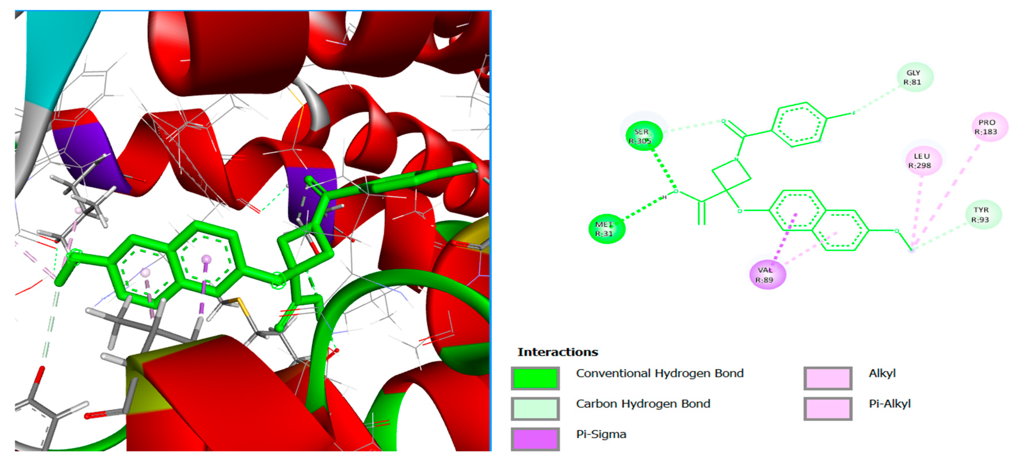
Figure 1. Three-dimensional molecular pose (left) and 2D (right) interactions of PF-04418948 on binding cavity of EP2 enzyme.
Chemproc 18 00007 g001
Figure 2. Three-dimensional molecular pose (left) and 2D (right) interactions of lig1 on binding cavity of EP2 enzyme.
Chemproc 18 00007 g002
Figure 3. Three-dimensional molecular pose (left) and 2D (right) interactions of lig2 on binding cavity of EP2 enzyme.
Chemproc 18 00007 g003
Figure 4. Three-dimensional molecular pose (left) and 2D (right) interactions of lig3 on binding cavity of EP2 enzyme.
Chemproc 18 00007 g004
Table 1. Library of withanolides’ derivatives.
Chemproc 18 00007 i001
Lig 1 (withanolide D)
Chemproc 18 00007 i002
Lig 2 (withaferin A)
Chemproc 18 00007 i003
Lig 3 (withanone)
Chemproc 18 00007 i004
PF-04418948
Table 2. Analysis of theoretical oral bioavailability of selected compounds based on Lipinski’s rule of five and pharmacokinetic parameters.
PropertiesLig1Lig2Lig3
CID16167126523721679027
Molecular FormulaC28H38O6C28H38O7C28H38O6
Mol. Wt (g/mol)470.60486.60470.60
Heavy atoms343534
Aromatic Heavy atoms000
Fraction Csp30.790.790.79
HbA676
HbD232
Nrb232
MR127.53128.69127.53
TPSA (Å2)96.36116.5996.36
MlogP2.751.952.75
Lipinski violationNoNoNo
InferencePassPassPass
Ghose violations121
Veber violations000
Egan violations000
Muegge violations000
Bioavailability score0.550.550.55
Synthetic accessibility6.856.886.38
Table 3. Pharmacokinetics’ prediction output and oral bioavailability of lig1–lig3 compounds.
PropertiesLig1Lig2Lig3
LogS (Silicos-IT)−3.78−3.20−4.00
Silicos-classModerately solubleModerately solubleModerately soluble
Consensus Log Po/w3.392.643.36
Log Kp (skin permeation) (cm/s)−6.96−7.55−7.01
GI AbsorptionHighHighHigh
BBB PermeantNoNoNo
Pgp substrateYesYesYes
CYP1A2 inhibitorNoNoNo
CYP2C19 inhibitorNoNoNo
CYP2C9 inhibitorNoNoNo
CYP2D6 inhibitorNoNoNo
CYP3A4 inhibitorNoNoNo
Table 4. Toxicity profile of the test compounds.
PropertiesLig1Lig2Lig3
Oral acute toxicityClass IIIClass IIIClass II
Ames Mutagenesis---
Carcinogenicity---
Hepatotoxicity---
Androgen receptor binding---
Thyroid receptor binding---
Estrogen receptor binding---
Aromatase binding---
-: Inactive; Class II: 5 mg/kg ≤ LD50 ≤ 50 mg/kg; Class III: 50 mg/kg ≤ LD50 ≤ 300 mg/kg.
Table 5. Enzyme grid box size center.
EnzymeGrid Box SizeCenter
XYZXYZ
7CX22216223.719114.893119.227
Table 6. Crystal structure of bio-target complex and re-docked ligand superimposed on the crystal structure for validation. The different colours show how the native ligand is super-imposed after docking.
Enzyme Code and NameCrystal Structure Complex
(Enzyme and Native Ligand)
Crystal Structure Complex (Enzyme, Native
Ligand, and Re-Docked Ligand) (Validation)
7CX2
PGE2-bound EP2-Gs complex
Chemproc 18 00007 i005Chemproc 18 00007 i006
Table 7. The binding energies of the reference compound and the three selected compounds against EP2 receptor.
Enzyme Affinity (kcal/mol)
PF-04418948Lig1Lig2Lig3
7CX2−9.6−12.6−12.0−11.3
Table 8. Molecular interactions of amino acid residues of withanolide compounds with EP2 enzyme.
CompoundsInteraction TypeInteracting Residues
PF-04418948Conventional Hydrogen BondSER305, MET31
AlkylLEU298, PRO183
Pi-AlkylVAL89
Carbon Hydrogen BondTYR93, GLY81
Lig 1Pi-AlkylPHE112, TRP186
AlkylVAL89, LEU298
Conventional Hydrogen BondARG302, SER308, PHE119
Sulfur-XMET116
Carbon Hydrogen BondSER28, SER86
Lig 2Conventional Hydrogen bondSER305, ARG302
Sulfur-XMET31
Carbon Hydrogen BondSER308
Lig3Conventional hydrogen bondARG302, SER302
Carbon Hydrogen BondSER305, SER308
Sulfur-XMet31
Disclaimer/Publisher’s Note: The statements, opinions and data contained in all publications are solely those of the individual author(s) and contributor(s) and not of MDPI and/or the editor(s). MDPI and/or the editor(s) disclaim responsibility for any injury to people or property resulting from any ideas, methods, instructions or products referred to in the content.