Metabolic Diversion from Geranylgeranoic Acid to 2,3-Dihydrogeranylgeranoic Acid in Hepatic Tumor Surveillance


Abstract
1. Introduction
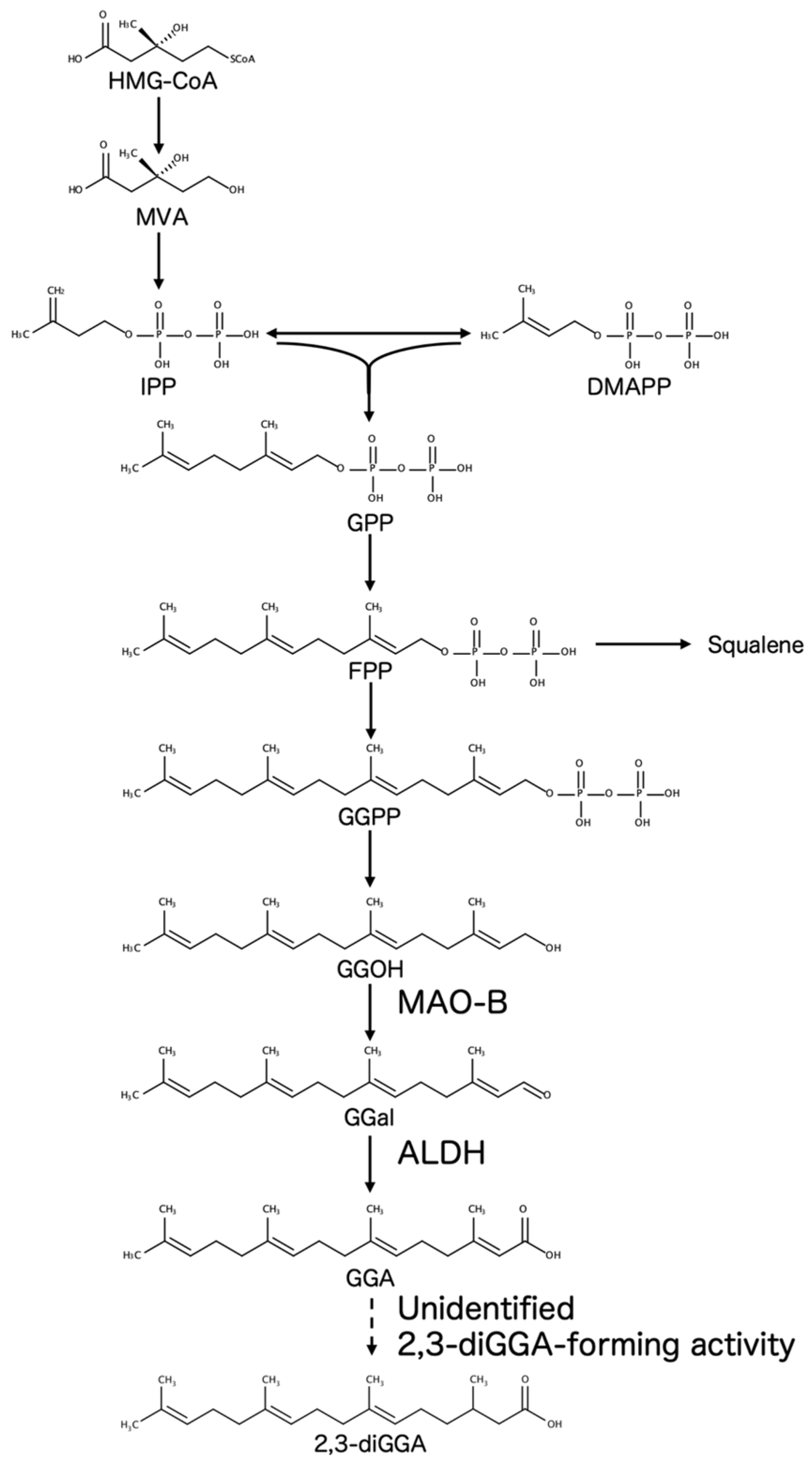
2. Mevalonate Pathway to GGA and 2,3-diGGA in Hepatic Metabolism
3. GGA as a Putative Hepatocellular Tumor-Suppressive Lipid Mediator
4. Formation and Tissue Distribution of 2,3-diGGA
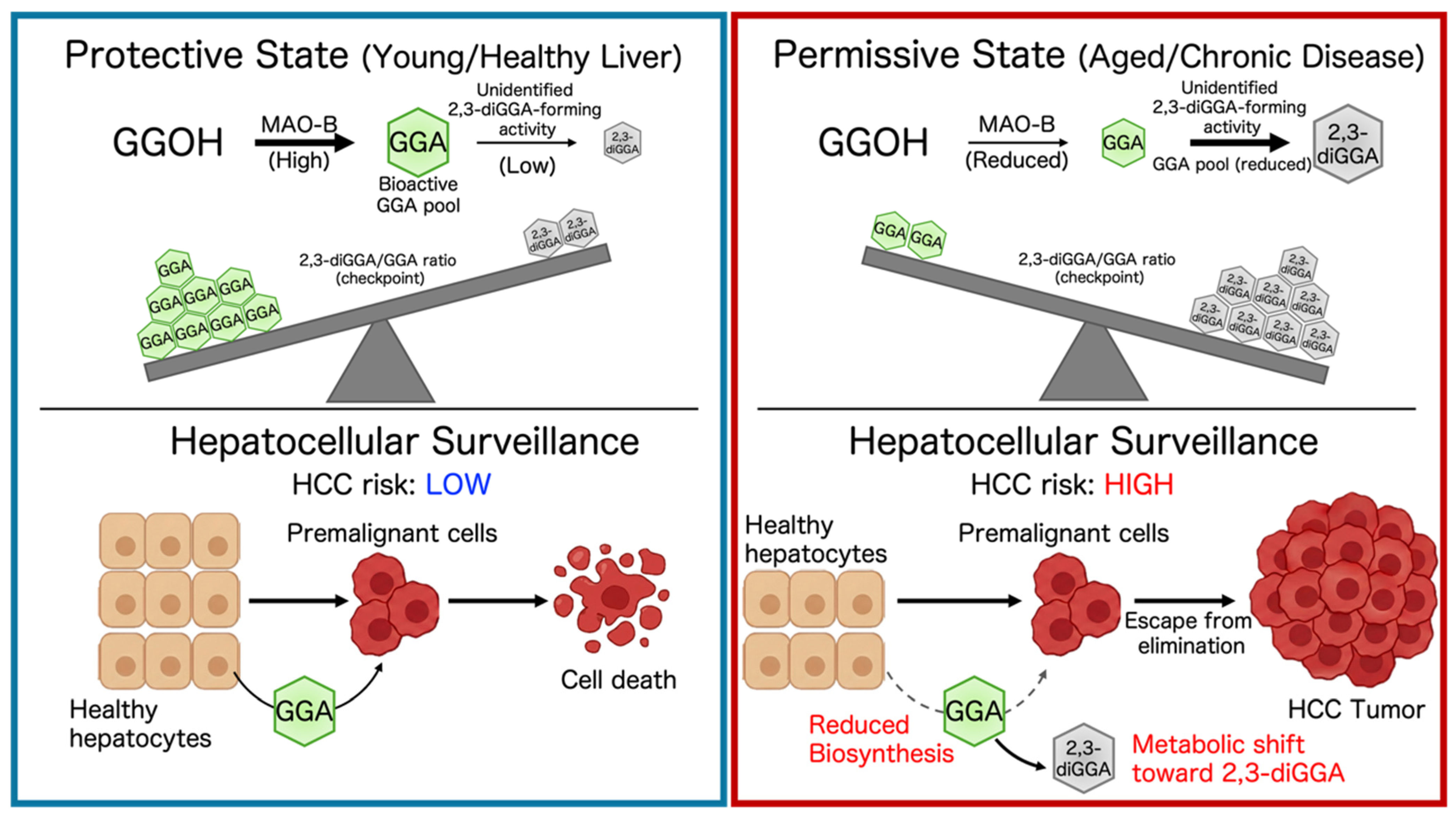
5. Proposed Metabolic Diversion from GGA to 2,3-diGGA and Implications for HCC Susceptibility
6. Future Directions and Experimental Strategies
7. Conclusions
Funding
Institutional Review Board Statement
Informed Consent Statement
Data Availability Statement
Conflicts of Interest
Abbreviations
| 2,3-diGGA | 2,3-dihydrogeranylgeranoic acid |
| ALDH | aldehyde dehydrogenase |
| CoA | coenzyme A |
| CYP3A4 | cytochrome P450 3A4 |
| DMAPP | dimethylallyl pyrophosphate |
| FPP | farnesyl pyrophosphate |
| GGA | geranylgeranoic acid |
| GGal | geranylgeranial |
| GGOH | geranylgeraniol |
| GGPP | geranylgeranyl pyrophosphate |
| GPP | geranyl pyrophosphate |
| HCC | hepatocellular carcinoma |
| HMG-CoA | 3-hydroxy-3-methylglutaryl coenzyme A |
| IPP | isopentenyl pyrophosphate |
| LC-MS/MS | liquid chromatography–tandem mass spectrometry |
| MAO-B | monoamine oxidase B |
| MVA | mevalonate |
References
- Woo, H.Y.; Yoo, S.Y.; Heo, J. Peretinoin, an Acyclic Retinoid, for the Secondary Prevention of Hepatocellular Carcinoma. Molecules 2021, 26, 295. [Google Scholar] [CrossRef] [Scilit] [PubMed]
- Araki, H.; Shidoji, Y.; Yamada, Y.; Moriwaki, H.; Muto, Y. Retinoid Agonist Activities of Synthetic Geranyl Geranoic Acid Derivatives. Biochem. Biophys. Res. Commun. 1995, 209, 66–72. [Google Scholar] [CrossRef] [Scilit] [PubMed]
- Muto, Y.; Moriwaki, H.; Ninomiya, M.; Adachi, S.; Saito, A.; Takasaki, K.T.; Tanaka, T.; Tsurumi, K.; Okuno, M.; Tomita, E.; et al. Prevention of second primary tumors by an acyclic retinoid, polyprenoic acid, in patients with hepatocellular carcinoma. Hepatoma Prevention Study Group. N. Engl. J. Med. 1996, 334, 1561–1567. [Google Scholar] [CrossRef] [Scilit] [PubMed]
- Muto, Y.; Moriwaki, H.; Saito, A. Prevention of second primary tumors by an acyclic retinoid in patients with hepatocellular carcinoma. N. Engl. J. Med. 1999, 340, 1046–1047. [Google Scholar] [CrossRef] [Scilit] [PubMed]
- Shidoji, Y.; Ogawa, H. Natural occurrence of cancer-preventive geranylgeranoic acid in medicinal herbs. J. Lipid Res. 2004, 45, 1092–1103. [Google Scholar] [CrossRef] [Scilit] [PubMed]
- Shidoji, Y.; Tabata, Y. Unequivocal evidence for endogenous geranylgeranoic acid biosynthesized from mevalonate in mammalian cells. J. Lipid Res. 2019, 60, 579–593. [Google Scholar] [CrossRef] [Scilit] [PubMed]
- Tabata, Y. Quantitative Screening of Geranylgeranoic Acid in Selected Plant-Based Foods Using LC/MS/MS. Front. Nutr. 2025, 12, 1652270. [Google Scholar] [CrossRef] [Scilit] [PubMed]
- Yasuda, I.; Shiratori, Y.; Adachi, S.; Obora, A.; Takemura, M.; Okuno, M.; Shidoji, Y.; Seishima, M.; Muto, Y.; Moriwaki, H. Acyclic retinoid induces partial differentiation, down-regulates telomerase reverse transcriptase mRNA expression and telomerase activity, and induces apoptosis in human hepatoma-derived cell lines. J. Hepatol. 2002, 36, 660–671. [Google Scholar] [CrossRef] [Scilit] [PubMed]
- Mitake, M.; Ogawa, H.; Uebaba, K.; Shidoji, Y. Increase in plasma concentrations of geranylgeranoic Acid after turmeric tablet intake by healthy volunteers. J. Clin. Biochem. Nutr. 2010, 46, 252–258. [Google Scholar] [CrossRef] [Scilit] [PubMed]
- Tabata, Y. Endogenous and dietary geranylgeranoic acid: A mini review on its role in hepatic tumor suppression. Eurasian J. Med. Oncol. 2025, 9, 356–361. [Google Scholar] [CrossRef] [Scilit]
- Faulkner, R.; Jo, Y. Synthesis, function, and regulation of sterol and nonsterol isoprenoids. Front. Mol. Biosci. 2022, 9, 1006822. [Google Scholar] [CrossRef] [Scilit] [PubMed]
- Shidoji, Y. Geranylgeranoic acid, a bioactive and endogenous fatty acid in mammals: A review. J. Lipid Res. 2023, 64, 100396. [Google Scholar] [CrossRef] [Scilit] [PubMed]
- Duttenhefner, J.N.; Reindl, K.M. Mevalonate pathway in pancreatic ductal adenocarcinoma: Mechanisms driving metabolic and cellular plasticity. Cancer Pathog. Ther. 2026, 4, 110–123. [Google Scholar] [CrossRef] [Scilit] [PubMed]
- Tabata, Y.; Shidoji, Y. Hepatic monoamine oxidase B is involved in endogenous geranylgeranoic acid synthesis in mammalian liver cells. J. Lipid Res. 2020, 61, 778–789. [Google Scholar] [CrossRef] [Scilit] [PubMed]
- Nakamura, N.; Shidoji, Y.; Moriwaki, H.; Muto, Y. Apoptosis in human hepatoma cell line induced by 4,5-didehydro geranylgeranoic acid (acyclic retinoid) via down-regulation of transforming growth factor-alpha. Biochem. Biophys. Res. Commun. 1996, 219, 100–104. [Google Scholar] [CrossRef] [Scilit] [PubMed]
- Nakamura, N.; Shidoji, Y.; Yamada, Y.; Hatakeyama, H.; Moriwaki, H.; Muto, Y. Induction of apoptosis by acyclic retinoid in the human hepatoma-derived cell line, HuH-7. Biochem. Biophys. Res. Commun. 1995, 207, 382–388. [Google Scholar] [CrossRef] [Scilit] [PubMed]
- Shidoji, Y.; Okamoto, K.; Muto, Y.; Komura, S.; Ohishi, N.; Yagi, K. Prevention of geranylgeranoic acid-induced apoptosis by phospholipid hydroperoxide glutathione peroxidase gene. J. Cell. Biochem. 2006, 97, 178–187. [Google Scholar] [CrossRef] [Scilit] [PubMed]
- Iwao, C.; Shidoji, Y. Induction of nuclear translocation of mutant cytoplasmic p53 by geranylgeranoic acid in a human hepatoma cell line. Sci. Rep. 2014, 4, 4419. [Google Scholar] [CrossRef] [Scilit] [PubMed]
- Iwao, C.; Shidoji, Y. Upregulation of energy metabolism-related, p53-target TIGAR and SCO2 in HuH-7 cells with p53 mutation by geranylgeranoic acid treatment. Biomed. Res. 2015, 36, 371–381. [Google Scholar] [CrossRef] [Scilit] [PubMed]
- Shidoji, Y.; Iwao, C. A rapid increase in lysophospholipids after geranylgeranoic acid treatment in human hepatoma-derived HuH-7 cells revealed by metabolomics analysis. Biochem. Biophys. Rep. 2021, 28, 101176. [Google Scholar] [CrossRef] [Scilit] [PubMed]
- Yabuta, S.; Shidoji, Y. TLR4-mediated pyroptosis in human hepatoma-derived HuH-7 cells induced by a branched-chain polyunsaturated fatty acid, geranylgeranoic acid. Biosci. Rep. 2020, 40, BSR20194118. [Google Scholar] [CrossRef] [Scilit] [PubMed]
- Tabata, Y.; Omori, M.; Shidoji, Y. Age-Dependent Decrease in Hepatic Geranylgeranoic Acid Content in C3H/HeN Mice and Its Oral Supplementation Prevents Spontaneous Hepatoma. Metabolites 2021, 11, 634. [Google Scholar] [CrossRef] [Scilit] [PubMed]
- Omori, M.; Shidoji, Y.; Moriwaki, H. Inhibition of Spontaneous Hepatocarcinogenesis by 4,5-Didehydrogeranylgeranoic Acid: Effects of Small-Dose and Infrequent Administration. Int. J. Transl. Med. 2023, 3, 487–495. [Google Scholar] [CrossRef] [Scilit]
- Nair, B.; Menon, A.; Khader, M.A.; Sethi, G.; Nath, L.R. Linking necroptosis with liver aging and chronic inflammation in hepatic pathology. Life Sci. 2025, 379, 123871. [Google Scholar] [CrossRef] [Scilit] [PubMed]
- Iwao, C.; Shidoji, Y. Polyunsaturated branched-chain fatty acid geranylgeranoic acid induces unfolded protein response in human hepatoma cells. PLoS ONE 2015, 10, e0132761. [Google Scholar] [CrossRef] [Scilit] [PubMed]
- Kodaira, Y.; Usui, K.; Kon, I.; Sagami, H. Formation of (R)-2,3-dihydrogeranylgeranoic acid from geranylgeraniol in rat thymocytes. J. Biochem. 2002, 132, 327–334. [Google Scholar] [CrossRef] [Scilit] [PubMed]
- Kodaira, Y.; Kusumoto, T.; Takahashi, T.; Matsumura, Y.; Miyagi, Y.; Okamoto, K.; Shidoji, Y.; Sagami, H. Formation of lipid droplets induced by 2,3-dihydrogeranylgeranoic acid distinct from geranylgeranoic acid. Acta Biochim. Pol. 2007, 54, 777–782. [Google Scholar] [CrossRef] [Scilit] [PubMed]
- Goldstein, J.L.; Brown, M.S. Regulation of the mevalonate pathway. Nature 1990, 343, 425–430. [Google Scholar] [CrossRef] [Scilit] [PubMed]
- Avalos, M.; Garbeva, P.; Vader, L.; van Wezel, G.P.; Dickschat, J.S.; Ulanova, D. Biosynthesis, evolution and ecology of microbial terpenoids. Nat. Prod. Rep. 2022, 39, 249–272. [Google Scholar] [CrossRef] [Scilit] [PubMed]
- Lange, B.M.; Rujan, T.; Martin, W.; Croteau, R. Isoprenoid biosynthesis: The evolution of two ancient and distinct pathways across genomes. Proc. Natl. Acad. Sci. USA 2000, 97, 13172–13177. [Google Scholar] [CrossRef] [Scilit] [PubMed]
- Kuzuyama, T.; Seto, H. Two distinct pathways for essential metabolic precursors for isoprenoid biosynthesis. Proc. Jpn. Acad. Ser. B 2012, 88, 41–52. [Google Scholar] [CrossRef] [Scilit] [PubMed]
- Holstein, S.A.; Hohl, R.J. Isoprenoids: Remarkable diversity of form and function. Lipids 2004, 39, 293–309. [Google Scholar] [CrossRef] [Scilit] [PubMed]
- Faylo, J.L.; Ronnebaum, T.A.; Christianson, D.W. Assembly-Line Catalysis in Bifunctional Terpene Synthases. Acc. Chem. Res. 2021, 54, 3780–3791. [Google Scholar] [CrossRef] [Scilit] [PubMed]
- Verdaguer, I.B.; Crispim, M.; Hernández, A.; Katzin, A.M. The Biomedical Importance of the Missing Pathway for Farnesol and Geranylgeraniol Salvage. Molecules 2022, 27, 8691. [Google Scholar] [CrossRef] [Scilit] [PubMed]
- Ericsson, J.; Greene, J.M.; Carter, K.C.; Shell, B.K.; Duan, D.R.; Florence, C.; Edwards, P.A. Human geranylgeranyl diphosphate synthase: Isolation of the cDNA, chromosomal mapping and tissue expression. J. Lipid Res. 1998, 39, 1731–1739. [Google Scholar] [CrossRef] [Scilit]
- Tong, H.; Wiemer, A.J.; Neighbors, J.D.; Hohl, R.J. Quantitative determination of farnesyl and geranylgeranyl diphosphate levels in mammalian tissue. Anal. Biochem. 2008, 378, 138–143. [Google Scholar] [CrossRef] [Scilit] [PubMed]
- Sagami, H.; Ishii, K.; Ogura, K. Geranylgeranylpyrophosphate synthetase of pig liver. Methods Enzymol. 1985, 110, 184–188. [Google Scholar] [CrossRef] [Scilit] [PubMed]
- Sagami, H.; Ishii, K.; Ogura, K. Occurrence and unusual properties of geranylgeranyl pyrophosphate synthetase of pig liver. Biochem. Int. 1981, 3, 669–675. [Google Scholar]
- Mitake, M.; Shidoji, Y. Geranylgeraniol oxidase activity involved in oxidative formation of geranylgeranoic acid in human hepatoma cells. Biomed. Res. 2012, 33, 15–24. [Google Scholar] [CrossRef] [Scilit] [PubMed]
- Muraguchi, T.; Okamoto, K.; Mitake, M.; Ogawa, H.; Shidoji, Y. Polished rice as natural sources of cancer-preventing geranylgeranoic acid. J. Clin. Biochem. Nutr. 2011, 49, 8–15. [Google Scholar] [CrossRef] [Scilit] [PubMed]
- Verdaguer, I.B.; Hernández, A.; Torres, S.; Rodríguez-Peiris, M.; Arenas, F.; Martinez, D.E.; López, M.B.; Crispim, M.; Tabata, Y.; Garcia-Ruiz, C.; et al. Prenols and prenoic acids in metabolism, disease, and aging. J. Lipid Res. 2026, 67, 101009. [Google Scholar] [CrossRef] [Scilit] [PubMed]
- Ramsay, R.R. Molecular aspects of monoamine oxidase B. Prog. Neuro-Psychopharmacol. Biol. Psychiatry 2016, 69, 81–89. [Google Scholar] [CrossRef] [Scilit] [PubMed]
- Gaweska, H.; Fitzpatrick, P.F. Structures and mechanism of the monoamine oxidase family. Biomol. Concepts 2011, 2, 365–377. [Google Scholar] [CrossRef] [Scilit] [PubMed]
- Tabata, Y.; Shidoji, Y. Hepatic CYP3A4 Enzyme Compensatively Maintains Endogenous Geranylgeranoic Acid Levels in MAOB-Knockout Human Hepatoma Cells. Metabolites 2022, 12, 140. [Google Scholar] [CrossRef] [Scilit] [PubMed]
- Tabata, Y. Geranylgeranoic acid and the MAOB–CYP3A4 axis: A metabolic shift underlying age-related liver cancer risk. Front. Aging 2025, 6, 1680031. [Google Scholar] [CrossRef] [Scilit] [PubMed]
- Ren, J.; Tong, Y.-M.; Cui, R.-X.; Wang, Z.; Li, Q.-L.; Liu, W.; Qu, K.; Zhang, J.-Y.; Liu, C.; Wan, Y. Comparison of survival between adolescent and young adult vs older patients with hepatocellular carcinoma. World J. Gastrointest. Oncol. 2020, 12, 1394–1406. [Google Scholar] [CrossRef] [Scilit] [PubMed]
- Zonfa, C.E.; Thyagarajan, A.; Sahu, R.P. Repurposing Itraconazole in Combination with Chemotherapy and Immune Checkpoint Inhibitor for Cancer. Med. Sci. 2026, 14, 55. [Google Scholar] [CrossRef] [Scilit] [PubMed]
- Olawale, M.E.; Lazarus, P. Inhibition of CYP2A6-mediated nicotine metabolism: A potential strategy for smoking cessation therapy. J. Pharmacol. Exp. Ther. 2026, 393, 103792. [Google Scholar] [CrossRef] [Scilit] [PubMed]
- Rached, G.; Campana, A.; Fiani, D.; Nguyen, C.; Eynde, V.V.D.; Gillman, P.K.; Barnett, B.S. Safety and Efficacy of Monoamine Oxidase Inhibitors in Patients Who Use Psychoactive Substances: Potential Drug Interactions and Substance Use Disorder Treatment Data. CNS Drugs 2026, 40, 359–417. [Google Scholar] [CrossRef] [Scilit] [PubMed]
- Eyal, S.; Alfasi, S.; Ben Zaken, K.; Sawaid, I.O.; Segev, L.; Mesfin, S.; Frankel, P.; Ezzy, R.; Saha-Detroja, T.; Madhavan, S.; et al. Consensus Molecules Associated with Parkinson’s Disease. Neurol. Int. 2026, 18, 23. [Google Scholar] [CrossRef] [Scilit] [PubMed]
- Shidoji, Y. Induction of Hepatoma Cell Pyroptosis by Endogenous Lipid Geranylgeranoic Acid-A Comparison with Palmitic Acid and Retinoic Acid. Cells 2024, 13, 809. [Google Scholar] [CrossRef] [Scilit] [PubMed]
- Shidoji, Y.; Nakamura, N.; Moriwaki, H.; Muto, Y. Rapid loss in the mitochondrial membrane potential during geranylgeranoic acid-induced apoptosis. Biochem. Biophys. Res. Commun. 1997, 230, 58–63. [Google Scholar] [CrossRef] [Scilit] [PubMed]
- Shimonishi, S.; Muraguchi, T.; Mitake, M.; Sakane, C.; Okamoto, K.; Shidoji, Y. Rapid downregulation of Cyclin D1 induced by geranylgeranoic acid in human hepatoma cells. Nutr. Cancer 2012, 64, 473–480. [Google Scholar] [CrossRef] [Scilit] [PubMed]
- Ajoolabady, A.; Tang, D.; Kroemer, G.; Ren, J. Ferroptosis in hepatocellular carcinoma: Mechanisms and targeted therapy. Br. J. Cancer 2023, 128, 190–205. [Google Scholar] [CrossRef] [Scilit] [PubMed]
- Wu, X.; Cao, J.; Wan, X.; Du, S. Programmed cell death in hepatocellular carcinoma: Mechanisms and therapeutic prospects. Cell Death Discov. 2024, 10, 356. [Google Scholar] [CrossRef] [Scilit] [PubMed]
- García-Pras, E.; Fernández-Iglesias, A.; Gracia-Sancho, J.; Pérez-del-Pulgar, S. Cell Death in Hepatocellular Carcinoma: Pathogenesis and Therapeutic Opportunities. Cancers 2021, 14, 48. [Google Scholar] [CrossRef] [Scilit] [PubMed]
- Okamoto, K.; Sakimoto, Y.; Imai, K.; Senoo, H.; Shidoji, Y. Induction of an incomplete autophagic response by cancer-preventive geranylgeranoic acid (GGA) in a human hepatoma-derived cell line. Biochem. J. 2011, 440, 63–71. [Google Scholar] [CrossRef] [Scilit] [PubMed]
- Liu, L.; Liao, J.-Z.; He, X.-X.; Li, P.-Y. The role of autophagy in hepatocellular carcinoma: Friend or foe. Oncotarget 2017, 8, 57707–57722. [Google Scholar] [CrossRef] [Scilit] [PubMed]
- Bialik, S.; Dasari, S.K.; Kimchi, A. Autophagy-dependent cell death—Where, how and why a cell eats itself to death. J. Cell Sci. 2018, 131, jcs215152. [Google Scholar] [CrossRef] [Scilit] [PubMed]
- Huang, X.; Yan, H.; Xu, Z.; Yang, B.; Luo, P.; He, Q. The inducible role of autophagy in cell death: Emerging evidence and future perspectives. Cell Commun. Signal. 2025, 23, 151. [Google Scholar] [CrossRef] [Scilit] [PubMed]
- Liu, Y.; Pan, R.; Ouyang, Y.; Gu, W.; Xiao, T.; Yang, H.; Tang, L.; Wang, H.; Xiang, B.; Chen, P. Pyroptosis in health and disease: Mechanisms, regulation and clinical perspective. Signal Transduct. Target. Ther. 2024, 9, 245. [Google Scholar] [CrossRef] [Scilit] [PubMed]
- Tan, Y.; Chen, Q.; Li, X.; Zeng, Z.; Xiong, W.; Li, G.; Li, X.; Yang, J.; Xiang, B.; Yi, M. Pyroptosis: A new paradigm of cell death for fighting against cancer. J. Exp. Clin. Cancer Res. 2021, 40, 153. [Google Scholar] [CrossRef] [Scilit] [PubMed]
- Koren, E.; Fuchs, Y. Modes of Regulated Cell Death in Cancer. Cancer Discov. 2021, 11, 245–265. [Google Scholar] [CrossRef] [Scilit] [PubMed]
- Sato, K.; Nakajima, T.; Choyke, P.L.; Kobayashi, H. Selective cell elimination in vitro and in vivo from tissues and tumors using antibodies conjugated with a near infrared phthalocyanine. RSC Adv. 2015, 5, 25105–25114. [Google Scholar] [CrossRef] [Scilit] [PubMed]
- Drapela, S.; Ilter, D.; Gomes, A.P. Metabolic reprogramming: A bridge between aging and tumorigenesis. Mol. Oncol. 2022, 16, 3295–3318. [Google Scholar] [CrossRef] [Scilit] [PubMed]
- Raskov, H.; Gaggar, S.; Tajik, A.; Orhan, A.; Gögenur, I. Metabolic switch in cancer—Survival of the fittest. Eur. J. Cancer 2023, 180, 30–51. [Google Scholar] [CrossRef] [Scilit] [PubMed]
- Yang, J.; Xiao, H.; Zhang, Z.; Zhang, L.; Zhang, X. Regulatory roles of bioactive lipids in vascular homeostasis and remodeling. Med. Rev. 2026, 6, 35–56. [Google Scholar] [CrossRef] [Scilit] [PubMed]
- Edmondson, D.E. Hydrogen peroxide produced by mitochondrial monoamine oxidase catalysis: Biological implications. Curr. Pharm. Des. 2014, 20, 155–160. [Google Scholar] [CrossRef] [Scilit] [PubMed]
- Fowler, J.; Volkow, N.; Wang, G.-J.; Logan, J.; Pappas, N.; Shea, C.; MacGregor, R. Age-related increases in brain monoamine oxidase B in living healthy human subjects. Neurobiol. Aging 1997, 18, 431–435. [Google Scholar] [CrossRef] [Scilit] [PubMed]
- Liu, Y.; Li, D.; Xie, C.; Pan, Y.; Shi, Q. Monoamine Oxidase B in Cancers: Implications for Therapeutics and Prognosis. J. Inflamm. Res. 2025, 18, 15555–15586. [Google Scholar] [CrossRef] [Scilit] [PubMed]
- Nicotra, A.; Pierucci, F.; Parvez, H.; Senatori, O. Monoamine oxidase expression during development and aging. Neurotoxicology 2004, 25, 155–165. [Google Scholar] [CrossRef] [Scilit] [PubMed]
- Tabata, Y. Monoamine oxidase B as a context-dependent metabolic switch in hepatocellular carcinoma. Eurasian J. Med. Oncol. 2026, 10, 025520546. [Google Scholar] [CrossRef] [Scilit]
- Tu, D.-Z.; Hu, X.-Y.; Lei, J.-X.; Liu, S.-Y.; Xiao, Z.-P.; Yang, L.; Ge, G.-B. A patent review of CYP3A4 inhibitors (2018–present). Expert Opin. Ther. Pat. 2025, 35, 503–513. [Google Scholar] [CrossRef] [Scilit] [PubMed]
- Mo, H.; Jeter, R.; Bachmann, A.; Yount, S.T.; Shen, C.-L.; Yeganehjoo, H. The Potential of Isoprenoids in Adjuvant Cancer Therapy to Reduce Adverse Effects of Statins. Front. Pharmacol. 2019, 9, 1515. [Google Scholar] [CrossRef] [Scilit] [PubMed]
- Riederer, P.; Müller, T. Monoamine oxidase-B inhibitors in the treatment of Parkinson’s disease: Clinical-pharmacological aspects. J. Neural Transm. 2018, 125, 1751–1757. [Google Scholar] [CrossRef] [Scilit] [PubMed]
- Ren, J.; Yan, G.; Yang, L.; Kong, L.; Guan, Y.; Sun, H.; Liu, C.; Liu, L.; Han, Y.; Wang, X. Cancer chemoprevention: Signaling pathways and strategic approaches. Signal Transduct. Target. Ther. 2025, 10, 113. [Google Scholar] [CrossRef] [Scilit] [PubMed]
- Chen, M.; Lan, H.; Yao, S.; Jin, K.; Chen, Y. Metabolic Interventions in Tumor Immunity: Focus on Dual Pathway Inhibitors. Cancers 2023, 15, 2043. [Google Scholar] [CrossRef] [Scilit] [PubMed]
- Punnasseril, J.M.J.; Auwal, A.; Gopalan, V.; Lam, A.K.; Islam, F. Metabolic Reprogramming of Cancer Cells and Therapeutics Targeting Cancer Metabolism. Cancer Med. 2025, 14, e71244. [Google Scholar] [CrossRef] [Scilit] [PubMed]
- Galli, S.; Pacilio, P.; Bianchini, E.; Alborghetti, M.; De Carolis, L.; Lombardo, P.; Garramone, F.; Salvetti, M.; Rinaldi, D. The Role of MAO-B Inhibitors in Fatigue in Parkinson’s Disease: A Narrative Review. J. Clin. Med. 2025, 14, 2598. [Google Scholar] [CrossRef] [Scilit] [PubMed]
- Diep, Y.N.; Park, H.J.; Kwon, J.-H.; Tran, M.; Ko, H.Y.; Jo, H.; Kim, J.; Chung, J.-I.; Kim, T.Y.; Kim, D.; et al. Astrocytic scar restricting glioblastoma via glutamate–MAO-B activity in glioblastoma-microglia assembloid. Biomater. Res. 2023, 27, 71. [Google Scholar] [CrossRef] [Scilit] [PubMed]
- Junkes, L.; Quagliato, L.A.; Appolinario, J.C.; Shader, R.I.; Nardi, A.E. MAO inhibitors for treatment-resistant depression: Bringing an updated perspective on pioneering drugs. Pharmacol. Res. 2025, 219, 107876. [Google Scholar] [CrossRef] [Scilit] [PubMed]
- Polpichai, N.; Maneenil, C.; Danpanichkul, P.; Rattananukrom, C.; Choudhury, A.; Wong, Y.J.; Sripongpun, P.; Liangpunsakul, S.; Kaewdech, A. Current and new strategies for hepatocellular carcinoma surveillance. Gastroenterol. Rep. 2025, 13, goaf045. [Google Scholar] [CrossRef] [Scilit] [PubMed]
- Beudeker, B.J.B.; Boonstra, A. Circulating biomarkers for early detection of hepatocellular carcinoma. Ther. Adv. Gastroenterol. 2020, 13, 1756284820931734. [Google Scholar] [CrossRef] [Scilit] [PubMed]
- Parikh, N.D.; Tayob, N.; Singal, A.G. Blood-based biomarkers for hepatocellular carcinoma screening: Approaching the end of the ultrasound era? J. Hepatol. 2023, 78, 207–216. [Google Scholar] [CrossRef] [Scilit] [PubMed]
- Singal, A.G.; Yang, J.D.; Parikh, N.D. Blood-based Biomarkers for HCC Surveillance: Ready for Center Stage? Am. J. Gastroenterol. 2024, 119, 2147–2150. [Google Scholar] [CrossRef] [Scilit] [PubMed]
- Kavanagh, K.L.; Jörnvall, H.; Persson, B.; Oppermann, U. Medium- and short-chain dehydrogenase/reductase gene and protein families. Cell. Mol. Life Sci. 2008, 65, 3895. [Google Scholar] [CrossRef] [Scilit] [PubMed]
- Rodriguez, S.B.; Leyh, T.S. An Enzymatic Platform for the Synthesis of Isoprenoid Precursors. PLoS ONE 2014, 9, e105594. [Google Scholar] [CrossRef] [Scilit] [PubMed]
- Pang, X.-Y.; Wang, S.; Jurczak, M.J.; Shulman, G.I.; Moise, A.R. Retinol saturase modulates lipid metabolism and the production of reactive oxygen species. Arch. Biochem. Biophys. 2017, 633, 93–102. [Google Scholar] [CrossRef] [Scilit] [PubMed]
- Zhang, H.E.; Henderson, J.M.; Gorrell, M.D. Animal models for hepatocellular carcinoma. Biochim. Biophys. Acta (BBA)—Mol. Basis Dis. 2019, 1865, 993–1002. [Google Scholar] [CrossRef] [Scilit] [PubMed]
- Zheng, H.; Xue, H.; Yun, W.-J. An overview of mouse models of hepatocellular carcinoma. Infect. Agent. Cancer 2023, 18, 49. [Google Scholar] [CrossRef] [Scilit] [PubMed]
- Guerra, B.; Recio, C.; Aranda-Tavío, H.; Guerra-Rodríguez, M.; García-Castellano, J.M.; Fernández-Pérez, L. The Mevalonate Pathway, a Metabolic Target in Cancer Therapy. Front. Oncol. 2021, 11, 626971. [Google Scholar] [CrossRef] [Scilit] [PubMed]
- Li, S.; Yuan, H.; Li, L.; Li, Q.; Lin, P.; Li, K. Oxidative Stress and Reprogramming of Lipid Metabolism in Cancers. Antioxidants 2025, 14, 201. [Google Scholar] [CrossRef] [Scilit] [PubMed]
- Salolin Vargas, V.P.; Gasbarra, M.; Calderon-Martinez, E.; Shah, Y.R.; Dahiya, D.S.; Saenz de Sicilia, M.G. Non-alcoholic fatty liver disease and drug induced liver injury: A metabolic storm waiting to happen. World J. Hepatol. 2025, 17, 105255. [Google Scholar] [CrossRef] [Scilit] [PubMed]
- Stolaki, E.V.; Psatha, K.; Aivaliotis, M. Metabolomics and Pharmacometabolomics: Advancing Precision Medicine in Drug Discovery and Development. Metabolites 2025, 15, 750. [Google Scholar] [CrossRef] [Scilit] [PubMed]
- Palatini, P.; De Martin, S. Pharmacokinetic drug interactions in liver disease: An update. World J. Gastroenterol. 2016, 22, 1260–1278. [Google Scholar] [CrossRef] [Scilit] [PubMed]
- Tanaka, Y. Metabolomics in liver diseases: A novel alternative for liver biopsy? World J. Hepatol. 2024, 16, 12–16. [Google Scholar] [CrossRef] [Scilit] [PubMed]
- Molendijk, J.; Robinson, H.; Djuric, Z.; Hill, M.M. Lipid mechanisms in hallmarks of cancer. Mol. Omics 2020, 16, 6. [Google Scholar] [CrossRef] [Scilit] [PubMed]
- Schwenk, S.; Kaplan, D.E. Chemoprevention of hepatocellular carcinoma. Hepatol. Commun. 2025, 9, e0836. [Google Scholar] [CrossRef] [Scilit] [PubMed]
- Park, J.; Choi, J.; Kim, D.-D.; Lee, S.; Lee, B.; Lee, Y.; Kim, S.; Kwon, S.; Noh, M.; Lee, M.-O.; et al. Bioactive Lipids and Their Derivatives in Biomedical Applications. Biomol. Ther. 2021, 29, 465–482. [Google Scholar] [CrossRef] [Scilit] [PubMed]
- Robichaud, P.P.; Surette, M.E. Polyunsaturated fatty acid-phospholipid remodeling and inflammation. Curr. Opin. Endocrinol. Diabetes Obes. 2015, 22, 112–118. [Google Scholar] [CrossRef] [Scilit] [PubMed]



| Property | GGA | 2,3-diGGA | Current Interpretation/Relevance |
|---|---|---|---|
| Chemical feature | Acyclic diterpenoid carboxylic acid with an unsaturated C2-C3 double bond | α-saturated derivative of GGA with a saturated C2-C3 bond | α-saturation preserves the acyclic diterpenoid backbone but alters biological activity [25,26,27] |
| Biosynthetic origin | Synthesized from the mevalonate pathway via GGPP and GGOH | Formed downstream of GGA or related polyprenyl intermediates | 2,3-diGGA formation may represent a downstream GGA-consuming branch [6,26,27] |
| Known or proposed enzymes | MAO-B is a major GGOH oxidase; CYP3A4 may provide a compensatory pathway | Responsible enzyme remains unidentified | Enzymological identification of the 2,3-diGGA-forming activity is a key unresolved issue [14,44] |
| Tissue distribution | Detected in liver and other tissues; hepatic GGA is relevant to tumor surveillance models | Relatively enriched in thymus compared with liver | Tissue distribution suggests organ-specific regulation of the GGA/2,3-diGGA balance [6,26] |
| Cell-death-inducing activity in hepatoma models | Induces apoptosis, autophagy-associated death, and pyroptosis-like cell death | Markedly reduced cell-death-inducing activity compared with GGA | Conversion from GGA to 2,3-diGGA may attenuate GGA-mediated cell elimination [17,20,21,25,26,27] |
| Association with aging | Hepatic GGA levels decline with age in C3H/HeN mice | Age-associated changes remain insufficiently characterized | Aging may reduce GGA availability, but age-related regulation of 2,3-diGGA requires further study [22,45] |
| Potential biological role | Putative tumor-suppressive lipid mediator involved in elimination of premalignant hepatocytes | Less active downstream metabolite; possible physiological roles in thymus or immune regulation remain unclear | The GGA/2,3-diGGA balance may influence hepatic tumor surveillance, but direct human evidence remains limited |
| Evidence level | Supported by cell, biochemical, metabolomic, and animal studies | Supported by biochemical and metabolomic studies, but mechanistic evidence remains limited | The proposed metabolic diversion model remains hypothesis-generating |
| Evidence Category | Model/System | Main Finding | Relevance to the GGA/2,3-diGGA Balance | Limitation | References |
|---|---|---|---|---|---|
| Endogenous GGA biosynthesis | Mammalian cells/isotope-tracing studies | GGA is synthesized from the mevalonate pathway; 2,3-diGGA appears downstream of GGA | Supports the existence of a late-stage mevalonate-derived GGA metabolic branch | Mainly cellular and biochemical evidence; direct human liver validation remains limited | [6] |
| MAO-B-dependent GGA formation | Human hepatoma-derived liver cells | MAO-B functions as a major GGOH oxidase involved in endogenous GGA formation | MAO-B activity may determine the input flux into the GGA pool | Does not directly test HCC outcome or 2,3-diGGA-forming activity | [14] |
| CYP3A4-associated compensation | MAOB-knockout human hepatoma cells | CYP3A4 can compensatively maintain endogenous GGA levels when MAO-B is disrupted | Drug- or disease-related CYP modulation may influence GGA availability | The impact on 2,3-diGGA formation and HCC susceptibility remains unresolved | [44] |
| GGA-induced cell death | Human hepatoma cell lines | GGA induces apoptosis, autophagy-associated cell death, and pyroptosis-like inflammatory cell death | Supports the concept of bioactive GGA as a cell-death-inducing lipid mediator | Mostly in vitro evidence at experimental concentrations | [17,20,21,25,51] |
| Reduced activity of 2,3-diGGA | Hepatoma cell models and biochemical studies | 2,3-diGGA shows markedly reduced cell-death-inducing activity compared with GGA | Supports the idea that conversion to 2,3-diGGA may attenuate GGA bioactivity | Direct causality between increased 2,3-diGGA and HCC development has not been established | [25,26,27] |
| Age-related decline in hepatic GGA | Aged C3H/HeN mice | Hepatic GGA levels decline with age in association with reduced MAO-B expression | Suggests that reduced GGA availability may be linked to age-associated hepatic tumor susceptibility | 2,3-diGGA/GGA ratio was not fully established as a risk marker | [22,45] |
| GGA supplementation and hepatoma prevention | Spontaneous hepatocarcinogenesis mouse models | Oral GGA or related analogs reduced spontaneous hepatoma incidence or multiplicity | Supports the protective potential of maintaining GGA availability | Does not directly test inhibition of 2,3-diGGA formation | [22,23] |
| Tissue-specific 2,3-diGGA formation | Rat thymus/tissue distribution studies | 2,3-diGGA formation is prominent in thymus-associated systems; liver shows lower relative 2,3-diGGA abundance under physiological conditions | Suggests that 2,3-diGGA formation is enzymatic and tissue-dependent | The responsible enzyme and relevance to liver disease remain unknown | [6,26,27] |
| Human HCC or chronic liver disease cohorts | Human liver tissue, serum, bile, or biobanked cohort samples | Direct evidence remains limited | Represents a major translational gap for validating the GGA/2,3-diGGA ratio as a candidate biomarker | Requires prospective or retrospective human cohort studies | [82,83,84,85] |
| Research Area | Key Scientific Question | Recommended Strategy | Expected Outcome/Significance | Priority |
|---|---|---|---|---|
| Enzymology | What enzyme is responsible for 2,3-diGGA formation? | Activity-guided purification from rat thymus or other high-activity tissues; subcellular fractionation; LC-MS/MS-based activity assay; proteomic identification of active fractions | Identification of the 2,3-diGGA-forming enzyme or enzyme complex | High |
| Cellular models | Does the 2,3-diGGA-forming pathway regulate intracellular GGA availability? | Knockdown, knockout, or overexpression of candidate enzymes in hepatoma cells and primary hepatocytes | Clarification of whether this pathway controls the GGA/2,3-diGGA balance and GGA-induced cell death | High |
| Animal models | Does modulation of this pathway affect hepatocarcinogenesis in vivo? | Liver-specific knockout or transgenic expression of candidate enzymes combined with spontaneous or chemically induced HCC models | Determination of causal links between GGA diversion, tumor incidence, multiplicity, or latency | High |
| Clinical translation | Is the GGA/2,3-diGGA ratio associated with chronic liver disease progression or HCC development? | Quantitative LC-MS/MS profiling of liver tissue, serum, and bile from chronic liver disease and HCC cohorts | Evaluation of the GGA/2,3-diGGA ratio as a candidate biomarker | High |
| Sex differences | Are there sex-dependent differences in GGA and 2,3-diGGA metabolism? | Sex-stratified metabolomic analyses in animal models and human liver disease cohorts | Clarification of whether sex influences GGA availability, 2,3-diGGA formation, and HCC susceptibility | Medium |
| Primary vs. metastatic liver tumors | Does the GGA/2,3-diGGA balance differ between primary HCC and metastatic liver tumors? | Comparative profiling of adjacent non-tumor liver, primary HCC tissues, and metastatic liver lesions | Determination of whether this pathway is specific to hepatocarcinogenesis or reflects broader hepatic tumor biology | Medium |
| Pharmacometabolomics | Do commonly used drugs alter GGA metabolism or 2,3-diGGA formation? | Analysis of drugs affecting MAO-B, CYP3A4, lipid metabolism, or redox pathways in cellular systems and clinical datasets | Identification of modifiable factors that may influence GGA homeostasis | Medium |
| Intervention development | Can the GGA/2,3-diGGA balance be shifted toward bioactive GGA? | Development of selective inhibitors of the 2,3-diGGA-forming enzyme or reduction-resistant GGA analogs | Basis for future HCC chemoprevention strategies | Long-term |
| Immunological relevance | Does 2,3-diGGA have physiological roles in thymus or immune regulation? | Profiling thymic 2,3-diGGA levels, immune cell development, and T-cell function in relevant models | Avoidance of unintended immunological effects when targeting 2,3-diGGA formation | Medium |
Disclaimer/Publisher’s Note: The statements, opinions and data contained in all publications are solely those of the individual author(s) and contributor(s) and not of MDPI and/or the editor(s). MDPI and/or the editor(s) disclaim responsibility for any injury to people or property resulting from any ideas, methods, instructions or products referred to in the content. |
© 2026 by the author. Licensee MDPI, Basel, Switzerland. This article is an open access article distributed under the terms and conditions of the Creative Commons Attribution (CC BY) license.
Share and Cite
Tabata, Y. Metabolic Diversion from Geranylgeranoic Acid to 2,3-Dihydrogeranylgeranoic Acid in Hepatic Tumor Surveillance. Livers 2026, 6, 82. https://doi.org/10.3390/livers6050082
Tabata Y. Metabolic Diversion from Geranylgeranoic Acid to 2,3-Dihydrogeranylgeranoic Acid in Hepatic Tumor Surveillance. Livers. 2026; 6(5):82. https://doi.org/10.3390/livers6050082
Chicago/Turabian StyleTabata, Yuki. 2026. "Metabolic Diversion from Geranylgeranoic Acid to 2,3-Dihydrogeranylgeranoic Acid in Hepatic Tumor Surveillance" Livers 6, no. 5: 82. https://doi.org/10.3390/livers6050082
APA StyleTabata, Y. (2026). Metabolic Diversion from Geranylgeranoic Acid to 2,3-Dihydrogeranylgeranoic Acid in Hepatic Tumor Surveillance. Livers, 6(5), 82. https://doi.org/10.3390/livers6050082