Review

Evidence-Based Bedside Management of Overt Hepatic Encephalopathy: From Guidelines to Clinical Practice


by
Eugenio Franceschini
,
Andrea De Sinno
,
Matteo Cappelli Aimone Chiorat
and
Dante Pio Pallotta
*
Department of Medical and Surgical Sciences, University of Bologna, Via Albertoni 15, 40138 Bologna, Italy
*
Author to whom correspondence should be addressed.
Livers 2026, 6(4), 65; https://doi.org/10.3390/livers6040065
Submission received: 16 May 2026 / Revised: 28 June 2026 / Accepted: 7 July 2026 / Published: 9 July 2026

Abstract

Overt hepatic encephalopathy (OHE) is a defining decompensation event in liver cirrhosis, associated with substantial morbidity, mortality, and high readmission rates. For the bedside clinician, managing OHE presents a complex diagnostic and investigative challenge that extends well beyond the simple administration of laxatives. This review synthesizes core recommendations from the 2022 EASL and the recent 2026 ACG guidelines to provide a structured, evidence-based framework for acute inpatient management. We emphasize that OHE diagnosis remains a clinical endeavor, where serum ammonia is valuable primarily for its high negative predictive value rather than as a confirmatory biomarker. Successful resolution hinges on a proactive ‘detective approach’ to identify and correct precipitating factors—such as infections, gastrointestinal bleeding, and iatrogenic dehydration—which drive clinical deterioration. Furthermore, we highlight the critical role of the ‘four pillars’ of management, including structured discharge planning, caregiver education, and nutritional support to ensure effective secondary prophylaxis. By bridging the gap between the latest international clinical consensus and bedside practice, this review provides essential strategies to optimize OHE management, minimize recurrence, and ultimately reduce the healthcare burden of this pleomorphic syndrome.

1. Introduction

Overt hepatic encephalopathy (OHE) is a symptomatic, generally transient neuropsychiatric syndrome caused by hepatic failure and/or portosystemic shunting [1]. As a frequent cause of hospitalization for patients with cirrhosis, the onset of OHE marks a major decompensating event that profoundly worsens both prognosis and quality of life [2]. Managing OHE frequently challenges front-line clinicians, as it requires a rapid differential diagnosis and the identification of subtle precipitating factors driving clinical deterioration [3]. The aim of this review is to bridge the gap between international guidelines and bedside practice by providing a structured, evidence-based framework for the clinical investigation and management of OHE. The objective of this article is to offer a comprehensive bedside guide that aligns the latest clinical consensus with the practical challenges of inpatient OHE management, from initial diagnostic investigation to secondary prevention.

2. Methodology of Guidelines Identification

To ensure that the clinical framework provided in this review is aligned with the most current evidence-based standards, a focused literature search was conducted. We queried the PubMed/MEDLINE database and the Scopus database using the following search string: “hepatic encephalopathy” or “portosystemic encephalopathy”. The search was restricted using the “Guidelines” filter to identify official consensus documents from major international societies, and to select articles published from 1 January 2021 to 31 March 2026. This strategy led to the identification and selection of the primary reference documents upon which this bedside approach is built:
-
The 2022 European Association for the Study of the Liver (EASL) Clinical Practice Guidelines on the management of hepatic encephalopathy [4]. The 2025 EASL Clinical Practice Guidelines on TIPS were considered, as a more up-to-date document, for the discussion of OHE in the context of TIPS (Section 7 “Hepatic Encephalopathy in Special Situations”) [5].
-
The 2026 American College of Gastroenterology (ACG) Clinical Guidelines are specifically dedicated to the diagnosis and management of hepatic encephalopathy [3].
These documents were analyzed and synthesized to bridge the gap between formal recommendations and the practical requirements of acute inpatient care. Based on this synthesis, we identified four pillars of OHE management: (1) diagnostic workup and severity assessment, (2) identification of precipitating factors, (3) acute therapeutic management, and (4) secondary prevention and discharge planning. For each domain, we developed a pragmatic bedside approach that integrates the core recommendations from the aforementioned guidelines. Table 1 provides a comparative summary of how the EASL and ACG guidelines address these specific clinical challenges and details their key recommendations for each point.
Although a rigorous multi-database search was performed, the fact that this review focuses predominantly on two major guideline documents constitutes an inherent limitation. Accordingly, we recognize the possibility that alternative guidance from regional societies, localized consensus statements, or specific institutional protocols might have been omitted.

3. Diagnosis and Severity Assessment of Overt Hepatic Encephalopathy

The diagnosis of OHE is entirely clinical and can be confounded by its pleomorphic presentation, which ranges from euphoria to apathy, lethargy, and ultimately coma [6]. OHE should be suspected in any patient with liver cirrhosis and new-onset confusion or neurologic symptoms, and the diagnosis of OHE heavily relies on excluding other causes of altered mental status, as no gold-standard confirmatory test has yet been identified [6]. For instance, asterixis—a negative myoclonus elicited by extending the arms, dorsiflexing the wrists, and spreading the fingers to observe for a wrist flap—is commonly seen in OHE but may also present in other neuropsychiatric conditions. Conversely, focal neurological signs, while atypical, can occasionally manifest in OHE [7,8].
The primary differential diagnoses for OHE include:
  • Other metabolic encephalopathies (e.g., uremia, Wernicke’s syndrome)
  • Delirium (particularly in older adults)
  • Medication effects (e.g., sedatives)
  • Central nervous system infections
  • Acute cerebrovascular events (e.g., ischemic stroke, intracranial hemorrhage)
  • Nonconvulsive status epilecticus [9,10]
The differential diagnosis should rely firstly on history and clinical manifestations, with laboratory tests being valuable for identifying precipitating factors and/or ruling out alternative causes of altered mental status [3]. Historically, serum ammonia was regarded as the hallmark biomarker for OHE due to its pathogenic role; however, its role has been increasingly questioned over time [11,12]. The diagnostic accuracy of serum ammonia relies substantially on its negative predictive value, as normal serum ammonia substantially rules out OHE, prompting the evaluation for other causes of altered mental status [13,14]. On the other hand, ammonia should not be used to identify patients with OHE, as elevated ammonia in the absence of neurological abnormalities is a common occurrence in patients with liver cirrhosis [4,13]. Lastly, despite some evidence of a correlation between ammonia level and OHE severity, this correlation appears to be non-linear, and the trend of serum ammonia does not appear to reliably reflect the clinical response to treatments [13,14,15]. Considering these limitations, both EASL 2022 and ACG 2026 guidelines recommend the use of serum ammonia only to rule out OHE in patients with normal serum ammonia and suspected other causes of mental status alteration [3,4], and ACG 2026 guidelines explicitly recommend against relying on serum ammonia alone for diagnosis or for management evaluation [3].
Neuroimaging is reserved for cases where a primary neurological condition (e.g., intracranial hemorrhage) is suspected based on patient history (e.g., prior trauma), specific clinical features (e.g., seizures, focal deficits), or failure to respond to standard OHE therapy [3,4,16]. Electroencephalogram (EEG), despite not being indicated routinely due to absence of an OHE-specific pattern, can be useful to rule out nonconvulsive status epilecticus, especially in patients non-responsive to standard of care treatments [9].
Upon suspecting OHE, clinicians must promptly assess its severity to identify patients requiring an escalated level of care. Firstly, vital signs should be evaluated to exclude hemodynamic instability. In all patients, OHE should be graded using standardized tools such as the West-Haven criteria, the Hepatic Encephalopathy Scoring Algorithm, or the Glasgow Coma Scale [17,18]. For patients with severe OHE (e.g., West Haven III or IV), a comprehensive evaluation to determine the correct setting of management is paramount, as patients with severe acute cognitive impairment could be deserving of management in an acute care setting or intensive care setting. These critical care interventions must always be carefully weighed against the patient’s overall prognosis and individual goals of care, as no single marker can identify patients who will benefit from this escalation. While both ACG and EASL guidelines acknowledge the cruciality of this evaluations in patients with severe OHE, they do not provide specifical indication for airway protection via oro-tracheal intubation [3,4,19]. The 2022 ACG guidelines for management of acute on chronic liver failure suggest to evaluate patients with grade III and IV for intubation, reporting it as a key concept, however their recommendation do not provide specifical criteria to identify patients who could benefit of intubation [20]. Intubation should be reserved for patients at high risk for aspiration pneumonia or in the presence of severe respiratory pump failure [21]. Previous studies demonstrated a relatively poor survival of intensive care managed OHE patients, especially patients who received intubation, outside of the transplant setting; and patients and their caregivers should be informed about these outcomes to assist their decision about life support and intensive care management [21,22,23].

4. Precipitating Factors: Identification and Management

Identifying precipitating factors is a critical step, as correcting these triggers is essential for the optimal management of OHE, and both EASL 2022 and ACG 2026 guidelines highlight this, explicitly recommending to actively search for precipitating factors and to correct them [3,4]. These factors drive OHE pathogenesis either by increasing ammonia production (e.g., gastrointestinal bleeding) or by impairing its clearance and exacerbating its neurotoxicity (e.g., infections, electrolyte imbalances) [24,25].
The most frequent precipitating factors include infections, gastrointestinal bleeding, constipation, electrolyte imbalances (e.g., hyponatremia, hypokalemia), and dehydration, which is often iatrogenic due to excessive diuretic dosing. In a minority of patients, no clear precipitating factor can be identified [7]. Notably, distinguishing between a differential diagnosis and a precipitating factor can be challenging, as many triggers (such as sepsis or hyponatremia) can independently impair mental status [2]. Nonetheless, correcting these underlying factors remains vital for reversing the encephalopathy. Table 2 summarizes the main precipitating factors for OHE.
A systematic, step-by-step approach is imperative. The clinical evaluation should begin with vital signs to detect hemodynamic instability and fever. A meticulous review of the patient’s medication history is mandatory to identify recent exposure to sedatives (particularly benzodiazepines) or excessive diuretic dosing [1]. The physical examination must extend beyond the neurological assessment to encompass all organ systems, screening for occult infections, ascites, and signs of bleeding; a digital rectal examination can be instrumental in detecting gastrointestinal bleeding [7].
The laboratory workup should include a complete blood count, renal and liver function tests, serum electrolytes, and inflammatory markers. Capillary blood glucose testing should always be incorporated into the initial assessment [3].
Infections are one the most common precipitating factor for OHE, and can be sometimes overlooked due to the possibility of OHE to represent their first manifestation, preceding the development of fever or systemic inflammation due alterations of the inflammatory system intrinsic to liver cirrhosis. On the other hand, infection can lead to a rapid clinical deterioration and development of liver failure [26,27]. Due to these considerations, ACG 2026 guidelines recommend the collection of blood and urine cultures from all OHE patients, even in the absence of clinical signs of infection, as well as diagnostic paracentesis if ascites is present and the procedure can be performed safely [3]. On the other hand, EASL guidelines simply report to systematically exclude precipitating factors, not providing any recommendation about the collection of cultures in patients without signs of infection [4]. Notably, and most importantly, neither the ACG nor the EASL guidelines suggest routine antimicrobial treatment for patients with OHE without evidence of infections, due to poor effectiveness and in order to avoid the development of resistance [3,4].
Finally, dehydration is a frequent yet under-recognized pitfall. It is often induced by the cornerstone treatments for cirrhosis: diuretics and lactulose. In patients already receiving lactulose, dehydration-induced OHE can easily be misdiagnosed as treatment failure, prompting an inappropriate increase in laxative dosing that further deteriorates the patient’s fluid and electrolyte balance [28].

5. Acute Management of Overt Hepatic Encephalopathy

The acute management of OHE is centered on two pillars: the administration of non-absorbable disaccharides (lactulose) and the systematic identification and treatment of precipitating factors.
Lactulose remains the cornerstone of OHE therapy, deriving its efficacy from a multimodal mechanism of action:
  • Acidification of the bowel lumen, which traps ammonia by converting it into the non-absorbable ammonium ion;
  • An osmotic laxative effect, accelerating the clearance of nitrogenous waste;
  • A prebiotic effect, promoting the growth of non-ammoniagenic bacteria (e.g., lactobacilli);
  • Inhibition of intestinal glutamine uptake, further reducing ammoniagenesis [29].
For acute episodes, lactulose is administered orally at a dose of 10–20 g (15–30 mL) up to every 2 h until two soft bowel movements occur; the dose is then titrated to a goal of two to three soft stools daily [3]. In patients with severe OHE who are unable to swallow or at risk of aspiration, lactulose should be administered via a nasogastric tube at doses equivalent to oral administration, or as a retention enema (200 g lactulose or 300 mL solution in 700 mL of water or normal saline, retained for 30–60 min and repeated every 4–6 h until mental status improves) [30]. Overtreatment must be strictly avoided to prevent dehydration, electrolyte disturbances (e.g., hypernatremia and hypokalemia), perianal skin irritation, and enema-related complications such as infection or bleeding, as these can act as secondary triggers for OHE recurrence [28].
Alternative and adjunctive strategies are increasingly recognized. Polyethylene glycol 3350 (PEG) is emerging as a possible treatment, especially for patients with poor lactulose tolerance. A meta-analysis by Li et al. based on 7 randomized controlled trials for a total of 434 patients confirmed the beneficial effect of PEG, which led to a faster resolution of OHE and shortened the duration of hospital stay [31].
Rifaximin, primarily established for secondary prophylaxis, was investigated as an adjunctive treatment to lactulose in a trial of 120 patients with OHE, demonstrating a significant decrease in mortality and length of hospital stay, as well as a higher rate of OHE resolution when compared to lactulose plus placebo [32]. Zacharias et al. performed a systematic review investigating the role of rifaximin across different clinical scenarios of hepatic encephalopathy, including covert HE, acute OHE management, and both primary and secondary prophylaxis. Their findings confirmed a potential beneficial effect of rifaximin combined with non-absorbable disaccharides in patients with acute OHE. However, the authors acknowledged that current evidence for key outcomes, such as mortality reduction, ranges from very low to moderate, and that rifaximin alone appears to have little to no impact on the clinical course of OHE [33]. Lastly, recent trials hinted towards a marginal beneficial effect in patients on treatment with broad-spectrum antibiotics; it appears reasonable to avoid this concomitant prescription to avoid the development of resistance and rise in health costs [34,35].
Micronutrient deficiency is a common occurrence in patients with liver cirrhosis, and it should be ruled out and treated. In patients were associated Wernicke encephalopathy is suspected, high-dose parenteral thiamine administration is mandatory, as acknowledge by both EASL and ACG guidelines [3,4].
Finally, continuous clinical vigilance is paramount. As most patients are expected to respond to standard treatments in the first 24–48 h, ACG guidelines recommend considering the presence of alternative diagnoses or undisclosed precipitant factors in patients with persistent OHE after 48 h [3].
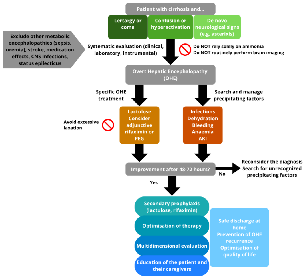
The key steps for managing OHE are summarized in Figure 1.

6. Secondary Prevention and Discharge

Following an initial episode of OHE, patients face a substantial risk of recurrence, with untreated rates reaching 40–60% within one year [36]. Consequently, upon resolution of the acute episode, clinical focus must shift toward long-term secondary prophylaxis. Pharmacological strategies center on titrated oral lactulose (targeting two to three soft stools daily) alongside rifaximin [37].
Rifaximin is a non-absorbable antibiotic that targets both aerobic and anaerobic bacteria while modulating the gut microbiota composition [38]. Although retrospective studies suggest rifaximin monotherapy is more effective than lactulose alone, dual therapy with rifaximin and lactulose is currently regarded as the gold standard, yielding the lowest rates of OHE recurrence and hospitalization [39].
Comprehensive discharge planning is critical to prevent relapse. Meticulous medication reconciliation is mandatory to identify and deprescribe potential triggers, notably benzodiazepines, zolpidem, and other GABAergic agents (e.g., gabapentin) [3]. If chronic opioids are necessary, their use must be strictly monitored to balance adequate analgesia against the risk of precipitating OHE; aggressively preventing opioid-induced constipation is vital in this setting [40].
Education for both the patient and their caregivers—who serve as the clinician’s eyes at home—is paramount. They should be instructed on using the Bristol Stool Scale to monitor bowel habits and trained to recognize early, covert signs of encephalopathy (e.g., sleep–wake reversal, irritability) before overt decompensation occurs [3]. Furthermore, emerging digital health tools and smartphone applications for home monitoring of cognitive function offer promising avenues for early intervention [41].
Finally, addressing the liver-muscle axis and the patient’s nutritional status is imperative. Sarcopenia and malnutrition are frequently overlooked risk factors for OHE. Therefore, all patients require standardized assessments of muscle health coupled with targeted nutritional counseling [42]. Ensuring adequate protein intake (1.2 g/kg/day) and minimizing fasting periods through late-evening snacks and early breakfasts—thereby preventing hypoglycemia driven by depleted hepatic glycogen reserves—have proven highly effective in mitigating the risk of cirrhotic decompensation [43,44].
Historically, branched-chain amino acids and ornithine L-aspartate supplementation have been associated with improved outcomes in patients with cirrhosis. However, the current quality of evidence regarding their efficacy in preventing OHE remains low, and recent ACG reccomend to consider them only in patients who are not able to achieve the target protein intake [3].

7. Hepatic Encephalopathy in Special Situations

OHE typically develops within the context of portosystemic shunts in patients with liver cirrhosis, classified as Type C according to the 1998 World Congress of Gastroenterology classification [45]. However, this classification also recognizes two other, less frequent forms of OHE: Type A, which occurs in the setting of acute liver failure (ALF), and Type B, which is associated with portosystemic shunts in patients without underlying cirrhosis [46]. Despite key clinical overlaps in the acute management of OHE across these etiologies, specific alternative considerations must be addressed.
Hepatic encephalopathy is a hallmark feature in the definition of ALF; here, the severity of OHE serves as a critical prognostic factor and a key determinant in selecting the appropriate management setting [30].In this specific clinical scenario, robust evidence supporting standard ammonia-reducing therapies (such as lactulose and rifaximin) is lacking, whereas promising results have been reported with the use of continuous renal replacement therapy [47,48,49]. Furthermore, because ALF patients are frequently managed in the Intensive Care Unit (ICU), the utility of rifaximin may be limited by concomitant broad-spectrum antibiotic therapy; indeed, data show negligible beneficial effects of rifaximin in this subpopulation [34,50]. Finally, urgent evaluation for liver transplantation remains mandatory for any patient presenting with ALF [47].
Portosystemic shunts, while common in portal hypertension and cirrhosis, can occasionally occur in individuals without a history of liver disease. Congenital portosystemic shunts are rare vascular malformations that can present in isolation or be associated with other congenital defects, such as cardiac malformations [51]. If spontaneous closure does not occur, these shunts can lead to hepatopulmonary syndrome, portopulmonary hypertension, and either overt or covert hepatic encephalopathy [52]. Less frequently, portosystemic shunts may spontaneously develop after abdominal surgery, leading to unexpected clinical manifestations up to OHE [53]. Patients with large, symptomatic shunts should be evaluated for radiological or surgical closure, which often resolves the clinical symptoms [54]. Consistent with this, the ACG guidelines recommend excluding the presence of large portosystemic shunts in any patient with OHE who fails to respond to standard medical therapy [3].
Recurrent hepatic encephalopathy represents a well-documented complication of transjugular intrahepatic portosystemic shunt (TIPS) placement, typically resulting from acute or chronic over-shunting. Established risk factors for post-TIPS OHE include sarcopenia, impaired baseline liver function, age over 65 years, and shunt diameter, with larger TIPS devices being inherently associated with a higher risk [55]. To this date, the optimal regimen for the prophylaxis of hepatic encephalopathy in recipients of TIPS is unclear. A pivotal trial failed to demonstrate a protective effect of lactulose against placebo, and a following trial did not demonstrate a clinically beneficial effect of treatment with L-ornithine L-aspartate [56,57]. The use of rifaximin (starting from two weeks before the procedure in elective TIPS, and continuing for at least 6 months) is supported by a trial by Bureau et al., which demonstrated the effectiveness of rifaximin in reducing the incidence of post-TIPS OHE; however, the trial subanalysis did not observe a significant beneficial effect in patients without a previous history of OHE [55]. On the other hand, a more recent meta-analysis reported poor effectiveness of any single prophylactic treatment, with a potential beneficial effect of the combination of lactulose and rifaximin, which is, however, only based on observational studies [58]. ACG guidelines recommend prophylaxis with rifaximin in patients receiving elective TIPS, adopting the treatment schedule used in the aforementioned trial (starting 14 days before the TIPS and continuing for at least 6 months) [3]. EASL 2025 guidelines on TIPS, on the other hand, acknowledging the limited evidence, provide a strong recommendation to consider rifaximin prophylaxis only in patients with a previous history of OHE, providing only a weak recommendation for patients without a previous history of OHE, where prophylaxis may be considered [5]. Notably, the EASL 2025 clinical practice guidelines on TIPS recommend considering shunt reduction or occlusion in patients with clinically significant over-shunting, including those experiencing recurrent OHE (defined as at least three episodes) [5].
OHE associated with renal dysfunction represents a complex and clinically challenging scenario, driven by limited nitrogen excretion in the setting of kidney impairment. Because ammonia is a small, water-soluble, and non-protein-bound molecule, it is highly dialyzable and can be efficiently cleared from the bloodstream via hemodialysis; however, this intervention has historically been restricted to patients with urea cycle disorders or inborn errors of metabolism [59]. Consequently, due to the lack of robust clinical data, the ACG guidelines do not support its implementation solely for ammonia clearance in patients with liver cirrhosis [3]. On the other hand, in the presence of standard nephrological indications—such as electrolyte imbalances, uremia, or volume overload, which can actively precipitate OHE—hemodialysis can effectively lead to an improvement in the mental status of patients with concomitant OHE and impaired kidney function [3,4]. Similarly, extracorporeal albumin dialysis systems have been investigated as artificial liver support treatments in the context of acute and acute-on-chronic liver failure, with recent data demonstrating a potential beneficial effect of the Molecular Adsorbent Recycling System (MARS) on the clinical course of OHE [60]. Although promising, their high cost and the overall limited evidence regarding survival benefits have prevented them from becoming the standard of care for OHE to date, with international guidelines only conditionally recommending their use in selected cases of liver failure [3,4].

8. Future Perspective: Fecal Microbiota Transplant and Hepatic Encephalopathy

Within the complex and multifactorial pathogenesis of hepatic encephalopathy, the role of gut microbiota alterations in chronic liver disease and portal hypertension has gained increasing recognition [61]. Fecal microbiota transplantation (FMT) was first reported as a potential strategy for OHE by Kao et al. in 2016, who described a beneficial yet transient effect in a patient with persistent grade II hepatic encephalopathy [62]. Subsequently, in a pivotal trial by Bajaj et al., patients treated with enema-delivered FMT combined with standard-of-care medications experienced fewer recurrent OHE episodes, thereby underscoring the potential role of microbiota modulation and FMT in secondary prevention [63]. Further trials demonstrated comparable beneficial effects in patients receiving capsule-delivered FMT [64,65]. Notably, the largest trial on FMT in OHE to date—the phase 2, randomized, placebo-controlled, double-blind THEMATIC trial—consistently demonstrated that FMT is a safe and effective treatment for patients with recurrent OHE [66]. Based on these findings, FMT appears to be a potentially transformative strategy for preventing OHE recurrence; however, to date, its clinical use should remain restricted to clinical trials, as emphasized by the ACG guidelines [3].

9. Guidelines Discrepancies and Practical Considerations

Despite some key elements of convergence, such as the centrality of lactulose as initial treatment in acute OHE, ACG, and EASL guidelines also present some points of divergence, as reported in Table 1. Despite some differences being due to manuscript design and the four-year gap between the guidelines—explaining why the 2025 EASL specific documents were preferred for the TIPS discussion over the 2022 guidelines on hepatic encephalopathy—analyzing these discrepancies serves a vital clinical purpose by highlighting how management strategies can be optimized at the patient’s bedside.
The first point to dissect is the conflicting role of ammonia, with the EASL guidelines recommending routine testing to leverage its high negative predictive value, and the ACG guidelines conditionally recommending against its routine use to guide treatment decisions. As reported in Section 3, while serum ammonia can be an informative test in cirrhotic patients with impaired mental status, clinicians should not consider hyperammonemia alone as definitive or supportive for a diagnosis of OHE, which must instead rely on a comprehensive clinical assessment. Over-relying on ammonia levels can easily lead to misdiagnosis—for instance, treating a cirrhotic patient suffering from sepsis-related encephalopathy with high doses of laxatives—thereby causing dehydration or electrolyte imbalances that could paradoxically worsen the patient’s mental status. Similarly, treatment response should not be based on variation in serum ammonia, but on clinical evolution of mental status. Similarly, assessing treatment response should not rely on fluctuations in serum ammonia levels, but rather on the clinical evolution of the patient’s neuropsychiatric status.
PEG and rifaximin are conditionally recommended by the ACG as alternative and adjunctive therapies to lactulose for OHE management, a stance not explicitly shared by the EASL framework. While both strategies are effective in reversing acute OHE, the underlying certainty of evidence is low. However, due to their favorable safety profiles, using adjunctive rifaximin (provided the patient is not on broad-spectrum antibiotics) and prescribing PEG as a substitute for lactulose-intolerant patients or those with drug-induced abdominal distension appear clinically reasonable.
Both guidelines recognize the screening and prompt management of precipitating factors as a cornerstone of OHE therapy. While the EASL framework limits its scope to more generalized recommendations, the ACG guideline introduces two highly pragmatic operational insights—primarily framed as ‘Key Concepts’ or embedded within the core narrative: (1) the mandate to routinely obtain blood, urine, and ascites cultures upon admission, and (2) a specific 48-to-72 h timeframe to evaluate treatment response. Given that OHE and altered mental status in general can be the sole presenting sign of an occult infection, performing microbiological cultures in all patients presenting with OHE is highly reasonable, even in the absence of typical infectious symptoms. Crucially, however, it must be underscored that neither guideline recommends the routine use of empirical broad-spectrum antibiotics without diagnostic justification. Because standard OHE therapies typically yield clinical improvement within the first 24 to 48 h, investigating alternative neurological diagnoses or unrecognized precipitants becomes mandatory when delirium persists beyond the 48-to-72 h threshold.

10. Conclusions

Overt hepatic encephalopathy (OHE) remains a common and clinically relevant complication of cirrhosis that requires more than just adherence to theoretical protocols. As clinical guidelines evolve—most recently with the 2026 ACG update—the challenge for the frontline physician is to translate these evidence-based recommendations into a precise and personalized bedside investigative process.
Effective management hinges on a dual focus: establishing a rigorous clinical diagnosis by excluding OHE mimickers and adopting a proactive and systematic evaluation to identify and correct precipitating factors. While lactulose remains the therapeutic cornerstone, clinicians must be aware of the risk of overtreatment, where iatrogenic dehydration and electrolyte imbalances can paradoxically drive OHE recurrence.
Finally, bridging the gap from acute care to long-term stability requires a multimodal secondary prevention strategy. By combining titrated pharmacological therapy with multidisciplinary interventions—including caregiver education and structured discharge planning—clinicians can break the cycle of readmissions. This systematic framework ensures that the latest clinical consensus is not just understood but effectively implemented to improve the quality of life for patients with cirrhosis.

Author Contributions

Conceptualization, E.F. and D.P.P.; methodology, D.P.P.; writing—original draft preparation, E.F., A.D.S. and M.C.A.C.; writing—review and editing, D.P.P.; supervision, D.P.P. All authors have read and agreed to the published version of the manuscript.

Funding

This research received no external funding.

Institutional Review Board Statement

Not applicable.

Informed Consent Statement

Not applicable.

Data Availability Statement

No new data were created or analyzed in this study.

Conflicts of Interest

The authors declare no conflicts of interest.

Abbreviations

The following abbreviations are used in this manuscript:
Overt hepatic encephalopathyOHE
American College of GastroenterologyACG
European Association for the Study of the LiverEASL
ElectroencephalogramEEG

References

  1. Rahimi, R.S.; Brown, K.A.; Flamm, S.L.; Brown, R.S. Overt Hepatic Encephalopathy: Current Pharmacologic Treatments and Improving Clinical Outcomes. Am. J. Med. 2021, 134, 1330–1338. [Google Scholar] [CrossRef] [PubMed]
  2. Tapper, E.B.; Aberasturi, D.; Zhao, Z.; Hsu, C.-Y.; Parikh, N.D. Outcomes after Hepatic Encephalopathy in Population-Based Cohorts of Patients with Cirrhosis. Aliment. Pharmacol. Ther. 2020, 51, 1397–1405. [Google Scholar] [CrossRef] [PubMed]
  3. Bajaj, J.S.; Jakab, S.S.; Jesudian, A.B.; Rahimi, R.S.; Duarte-Rojo, A.; Chen, P.-H.; Wong, R.J.; Tapper, E.B.; Tandon, P. ACG Clinical Guideline: Hepatic Encephalopathy. Am. J. Gastroenterol. 2026, 121, 588–618. [Google Scholar] [CrossRef] [PubMed]
  4. European Association for the Study of the Liver. EASL Clinical Practice Guidelines on the Management of Hepatic Encephalopathy. J. Hepatol. 2022, 77, 807–824. [Google Scholar] [CrossRef] [PubMed]
  5. Bureau, C.; Larrue, H.; Cortes-Cerisuleo, M.; Miraglia, R.; Procopet, B.; Rudler, M.; Trebicka, J.; VanWagner, L.B.; Hernandez-Gea, V. EASL Clinical Practice Guidelines on TIPS. J. Hepatol. 2025, 83, 177–210. [Google Scholar] [CrossRef] [PubMed]
  6. Dellatore, P.; Cheung, M.; Mahpour, N.Y.; Tawadros, A.; Rustgi, V.K. Clinical Manifestations of Hepatic Encephalopathy. Clin. Liver Dis. 2020. [Google Scholar] [CrossRef] [PubMed]
  7. Amodio, P. Hepatic Encephalopathy: Diagnosis and Management. Liver Int. Off. J. Int. Assoc. Study Liver 2018, 38, 966–975. [Google Scholar] [CrossRef] [PubMed]
  8. Agarwal, R.; Baid, R. Asterixis. J. Postgrad. Med. 2016, 62, 115–117. [Google Scholar] [CrossRef] [PubMed]
  9. Nguyen, A.; Butt, M.A.; Upadhyay, S.; Sheikh, A.B.; Shekhar, R. Non-Convulsive Status Epilepticus in Hepatic Encephalopathy: A Case Series and Review of the Literature. Eur. J. Case Rep. Intern. Med. 2022, 9, 003179. [Google Scholar] [CrossRef] [PubMed]
  10. Jo, Y.M.; Lee, S.W.; Han, S.Y.; Baek, Y.H.; Ahn, J.H.; Choi, W.J.; Lee, J.Y.; Kim, S.H.; Yoon, B.A. Nonconvulsive Status Epilepticus Disguising as Hepatic Encephalopathy. World J. Gastroenterol. WJG 2015, 21, 5105–5109. [Google Scholar] [CrossRef] [PubMed]
  11. Tapper, E.B.; Jiang, Z.G.; Patwardhan, V.R. Refining the Ammonia Hypothesis: A Physiology-Driven Approach to the Treatment of Hepatic Encephalopathy. Mayo Clin. Proc. 2015, 90, 646–658. [Google Scholar] [CrossRef] [PubMed]
  12. Ong, J.P.; Aggarwal, A.; Krieger, D.; Easley, K.A.; Karafa, M.T.; Van Lente, F.; Arroliga, A.C.; Mullen, K.D. Correlation between Ammonia Levels and the Severity of Hepatic Encephalopathy. Am. J. Med. 2003, 114, 188–193. [Google Scholar] [CrossRef] [PubMed]
  13. Ballester, M.P.; Durmazer, E.N.; Qi, T.; Jalan, R. The Value of Ammonia as a Biomarker in Patients with Cirrhosis. Semin. Liver Dis. 2024, 44, 356–368. [Google Scholar] [CrossRef] [PubMed]
  14. Nicolao, F.; Efrati, C.; Masini, A.; Merli, M.; Attili, A.F.; Riggio, O. Role of Determination of Partial Pressure of Ammonia in Cirrhotic Patients with and without Hepatic Encephalopathy. J. Hepatol. 2003, 38, 441–446. [Google Scholar] [CrossRef] [PubMed]
  15. Bajaj, J.S.; Pyrsopoulos, N.T.; Rahimi, R.S.; Heimanson, Z.; Allen, C.; Rockey, D.C. Serum Ammonia Levels Do Not Correlate With Overt Hepatic Encephalopathy Severity in Hospitalized Patients With Cirrhosis. Clin. Gastroenterol. Hepatol. Off. Clin. Pract. J. Am. Gastroenterol. Assoc. 2024, 22, 1950–1952.e1. [Google Scholar] [CrossRef] [PubMed]
  16. Rahimi, R.S.; Rockey, D.C. Overuse of Head Computed Tomography in Cirrhosis With Altered Mental Status. Am. J. Med. Sci. 2016, 351, 459–466. [Google Scholar] [CrossRef] [PubMed]
  17. Hassanein, T.I.; Hilsabeck, R.C.; Perry, W. Introduction to the Hepatic Encephalopathy Scoring Algorithm (HESA). Dig. Dis. Sci. 2008, 53, 529–538. [Google Scholar] [CrossRef] [PubMed]
  18. Weissenborn, K. Hepatic Encephalopathy: Definition, Clinical Grading and Diagnostic Principles. Drugs 2019, 79, 5–9. [Google Scholar] [CrossRef] [PubMed]
  19. Scarpati, G.; De Robertis, E.; Esposito, C.; Piazza, O. Hepatic Encephalopathy and Cirrhotic Cardiomyopathy in Intensive Care Unit. Minerva Anestesiol. 2018, 84, 970–979. [Google Scholar] [CrossRef] [PubMed]
  20. Bajaj, J.S.; O’Leary, J.G.; Lai, J.C.; Wong, F.; Long, M.D.; Wong, R.J.; Kamath, P.S. Acute-on-Chronic Liver Failure Clinical Guidelines. Off. J. Am. Coll. Gastroenterol. ACG 2022, 117, 225. [Google Scholar] [CrossRef] [PubMed]
  21. Gibbs, J.T.; Louissaint, J.; Tapper, E.B. Rate of Successful Extubation in Mechanically Ventilated Patients with Cirrhosis and Hepatic Coma. Dig. Dis. Sci. 2022, 67, 5336–5344. [Google Scholar] [CrossRef] [PubMed]
  22. Saffo, S.; Garcia-Tsao, G. Early Mechanical Ventilation for Grade IV Hepatic Encephalopathy Is Associated with Increased Mortality among Patients with Cirrhosis: An Exploratory Study. Acute Crit. Care 2022, 37, 355–362. [Google Scholar] [CrossRef] [PubMed]
  23. Juneja, D.; Gopal, P.B.; Kapoor, D.; Raya, R.; Sathyanarayanan, M. Profile and Outcome of Patients with Liver Cirrhosis Requiring Mechanical Ventilation. J. Intensive Care Med. 2012, 27, 373–378. [Google Scholar] [CrossRef] [PubMed]
  24. Zhao, L.; Gao, Y.; Guo, S.; Lu, X.; Yu, S.; Ge, Z.Z.; Zhu, H.D.; Li, Y. Sepsis-Associated Encephalopathy: Insight into Injury and Pathogenesis. CNS Neurol. Disord. Drug Targets 2021, 20, 112–124. [Google Scholar] [CrossRef] [PubMed]
  25. Asaki, Y.; Murofushi, Y.; Yasukawa, K.; Hara, M.; Takanashi, J.-I. Neurochemistry of Hyponatremic Encephalopathy Evaluated by MR Spectroscopy. Brain Dev. 2020, 42, 767–770. [Google Scholar] [CrossRef] [PubMed]
  26. Wang, Q.-M.; Ji, Q.; Duan, Z.-J.; Zhang, M.; Chang, Q.-Y. A Study on the Position and Etiology of Infection in Cirrhotic Patients: A Potential Precipitating Factor Contributing to Hepatic Encephalopathy. Exp. Ther. Med. 2013, 6, 584–590. [Google Scholar] [CrossRef] [PubMed]
  27. Guimarães, L.; Piedade, J.; Duarte, J.; Baldin, C.; Victor, L.; Costa, B.; Veiga, Z.; Alcântara, C.; Fernandes, F.; Pereira, G. Hepatic Encephalopathy in Cirrhotic Patients With Bacterial Infections: Frequency, Clinical Characteristics, and Prognostic Relevance. J. Clin. Exp. Hepatol. 2023, 13, 559–567. [Google Scholar] [CrossRef] [PubMed]
  28. Bajaj, J.S.; Sanyal, A.J.; Bell, D.; Gilles, H.; Heuman, D.M. Predictors of the Recurrence of Hepatic Encephalopathy in Lactulose-Treated Patients. Aliment. Pharmacol. Ther. 2010, 31, 1012–1017. [Google Scholar] [CrossRef] [PubMed]
  29. Bloom, P.P.; Tapper, E.B. Lactulose in Cirrhosis: Current Understanding of Efficacy, Mechanism, and Practical Considerations. Hepatol. Commun. 2023, 7, e0295. [Google Scholar] [CrossRef] [PubMed]
  30. Gluud, L.L.; Vilstrup, H.; Morgan, M.Y. Non-absorbable Disaccharides versus Placebo/No Intervention and Lactulose versus Lactitol for the Prevention and Treatment of Hepatic Encephalopathy in People with Cirrhosis. Cochrane Database Syst. Rev. 2016, 4, CD00304. [Google Scholar] [CrossRef] [PubMed]
  31. Li, M.; Zhang, Z.; Chen, Q.; Zhou, X.; Shui, D.; Huang, J. Comparative Effectiveness and Safety of Polyethylene Glycol Electrolyte Solution Versus Lactulose for Treatment of Hepatic Encephalopathy. J. Clin. Gastroenterol. 2022, 56, 41–48. [Google Scholar] [CrossRef] [PubMed]
  32. Shehata, H.H.; Elfert, A.A.; Abdin, A.A.; Soliman, S.M.; Elkhouly, R.A.; Hawash, N.I.; Soliman, H.H. Randomized Controlled Trial of Polyethylene Glycol versus Lactulose for the Treatment of Overt Hepatic Encephalopathy. Eur. J. Gastroenterol. Hepatol. 2018, 30, 1476. [Google Scholar] [CrossRef] [PubMed]
  33. Zacharias, H.D.; Kamel, F.; Tan, J.; Kimer, N.; Gluud, L.L.; Morgan, M.Y. Rifaximin for Prevention and Treatment of Hepatic Encephalopathy in People with Cirrhosis. Cochrane Database Syst. Rev. 2023, 2023, CD011585. [Google Scholar] [CrossRef] [PubMed]
  34. Ward, J.A.; Yerke, J.; Lumpkin, M.; Kapoor, A.; Lindenmeyer, C.C.; Bass, S. Evaluation of a Protocol for Rifaximin Discontinuation in Critically Ill Patients with Liver Disease Receiving Broad-Spectrum Antibiotic Therapy. World J. Hepatol. 2023, 15, 1226–1236. [Google Scholar] [CrossRef] [PubMed]
  35. Kulkarni, A.V.; Avadhanam, M.; Karandikar, P.; Rakam, K.; Gupta, A.; Simhadri, V.; Premkumar, M.; Zuberi, A.A.; Gujjarlapudi, D.; Narendran, R.; et al. Antibiotics With or Without Rifaximin for Acute Hepatic Encephalopathy in Critically Ill Patients With Cirrhosis: A Double-Blind, Randomized Controlled (ARiE) Trial. Off. J. Am. Coll. Gastroenterol. ACG 2024, 119, 864. [Google Scholar] [CrossRef] [PubMed]
  36. Sharma, B.C.; Sharma, P.; Agrawal, A.; Sarin, S.K. Secondary Prophylaxis of Hepatic Encephalopathy: An Open-Label Randomized Controlled Trial of Lactulose Versus Placebo. Gastroenterology 2009, 137, 885–891.e1. [Google Scholar] [CrossRef] [PubMed]
  37. Sharma, B.C.; Sharma, P.; Lunia, M.K.; Srivastava, S.; Goyal, R.; Sarin, S.K. A Randomized, Double-Blind, Controlled Trial Comparing Rifaximin plus Lactulose with Lactulose Alone in Treatment of Overt Hepatic Encephalopathy. Am. J. Gastroenterol. 2013, 108, 1458–1463. [Google Scholar] [CrossRef] [PubMed]
  38. Bajaj, J.S. Review Article: Potential Mechanisms of Action of Rifaximin in the Management of Hepatic Encephalopathy and Other Complications of Cirrhosis. Aliment. Pharmacol. Ther. 2016, 43, 11–26. [Google Scholar] [CrossRef] [PubMed]
  39. Moon, A.M.; Kim, H.P.; Jiang, Y.; Lupu, G.; Bissram, J.S.; Barritt, A.S.; Tapper, E.B. Systematic Review and Meta-Analysis on the Effects of Lactulose and Rifaximin on Patient Reported Outcomes in Hepatic Encephalopathy. Am. J. Gastroenterol. 2023, 118, 284–293. [Google Scholar] [CrossRef] [PubMed]
  40. Acharya, C.; Betrapally, N.S.; Gillevet, P.M.; Sterling, R.K.; Akbarali, H.; White, M.B.; Ganapathy, D.; Fagan, A.; Sikaroodi, M.; Bajaj, J.S. Chronic Opioid Use Is Associated with Altered Gut Microbiota and Predicts Readmissions in Patients with Cirrhosis. Aliment. Pharmacol. Ther. 2017, 45, 319–331. [Google Scholar] [CrossRef] [PubMed]
  41. Louissaint, J.; Grzyb, K.; Bashaw, L.; Mohammad, R.A.; Parikh, N.D.; Tapper, E.B. An Electronic Decision Support Intervention Reduces Readmissions for Patients With Cirrhosis. Off. J. Am. Coll. Gastroenterol. ACG 2022, 117, 491. [Google Scholar] [CrossRef] [PubMed]
  42. Siramolpiwat, S.; Kiattikunrat, K.; Soontararatpong, R.; Pornthisarn, B.; Vilaichone, R.; Chonprasertsuk, S.; Bhanthumkomol, P.; Nunanun, P.; Issariyakulkarn, N. Frailty as Tested by the Liver Frailty Index Is Associated with Decompensation and Unplanned Hospitalization in Patients with Compensated Cirrhosis. Scand. J. Gastroenterol. 2021, 56, 1210–1219. [Google Scholar] [CrossRef] [PubMed]
  43. Guo, Y.; Tian, Z.; Jiang, N.; Ding, X.; Mao, T.; Jing, X. Effects of Late Evening Snack on Cirrhotic Patients: A Systematic Review and Meta-Analysis. Gastroenterol. Res. Pract. 2018, 2018, 9189062. [Google Scholar] [CrossRef] [PubMed]
  44. Daftari, G.; Tehrani, A.N.; Pashayee-Khamene, F.; Karimi, S.; Ahmadzadeh, S.; Hekmatdoost, A.; Salehpour, A.; Saber-Firoozi, M.; Hatami, B.; Yari, Z. Dietary Protein Intake and Mortality among Survivors of Liver Cirrhosis: A Prospective Cohort Study. BMC Gastroenterol. 2023, 23, 227. [Google Scholar] [CrossRef] [PubMed]
  45. Zhang, J.-Q.; Cheng, D.-L.; Zhou, C.-Z.; Xu, X.-J. Risk Factors for Hepatic Encephalopathy after Transjugular Intrahepatic Portosystemic Shunt in Cirrhotic Patients: A Comprehensive Minireview. World J. Hepatol. 2025, 17, 109678. [Google Scholar] [CrossRef] [PubMed]
  46. Ferenci, P.; Lockwood, A.; Mullen, K.; Tarter, R.; Weissenborn, K.; Blei, A.T. Hepatic Encephalopathy—Definition, Nomenclature, Diagnosis, and Quantification: Final Report of the Working Party at the 11th World Congresses of Gastroenterology, Vienna, 1998. Hepatology 2002, 35, 716. [Google Scholar] [CrossRef] [PubMed]
  47. Maiwall, R.; Kulkarni, A.V.; Arab, J.P.; Piano, S. Acute Liver Failure. Lancet 2024, 404, 789–802. [Google Scholar] [CrossRef] [PubMed]
  48. Warrillow, S.; Fisher, C.; Bellomo, R. Correction and Control of Hyperammonemia in Acute Liver Failure: The Impact of Continuous Renal Replacement Timing, Intensity, and Duration. Crit. Care Med. 2020, 48, 218–224. [Google Scholar] [CrossRef] [PubMed]
  49. Slack, A.J.; Auzinger, G.; Willars, C.; Dew, T.; Musto, R.; Corsilli, D.; Sherwood, R.; Wendon, J.A.; Bernal, W. Ammonia Clearance with Haemofiltration in Adults with Liver Disease. Liver Int. Off. J. Int. Assoc. Study Liver 2014, 34, 42–48. [Google Scholar] [CrossRef] [PubMed]
  50. Bai, Z.; Li, C.; Lai, Y.; Hu, X.; Shi, L.; Guan, X.; Xu, Y. Role of Rifaximin in the Prognosis of Critically Ill Patients with Liver Cirrhosis. Antibiotics 2025, 14, 287. [Google Scholar] [CrossRef] [PubMed]
  51. Franchi-Abella, S.; Gonzales, E.; Ackermann, O.; Branchereau, S.; Pariente, D.; Guérin, F. the International Registry of Congenital Portosystemic Shunt members. Congenital Portosystemic Shunts: Diagnosis and Treatment. Abdom. Radiol. 2018, 43, 2023–2036. [Google Scholar] [CrossRef] [PubMed]
  52. Wu, G.Y. Congenital Portosystemic Shunts: A Review. J. Clin. Transl. Hepatol. 2026, 14, 478–486. [Google Scholar] [CrossRef] [PubMed]
  53. Ziola, S.; Cittadini, G.; Pasta, A.; Labanca, S.; Pieri, G.; Marenco, S.; Giannini, E.G. Recurrent Hepatic Encephalopathy After Abdominal Surgery in a Non-Cirrhotic Patient: A Case Report. Reports 2026, 9, 112. [Google Scholar] [CrossRef] [PubMed]
  54. Friis, K.H.; Thomsen, K.L.; Laleman, W.; Montagnese, S.; Vilstrup, H.; Lauridsen, M.M. Post-Transjugular Intrahepatic Portosystemic Shunt (TIPS) Hepatic Encephalopathy—A Review of the Past Decade’s Literature Focusing on Incidence, Risk Factors, and Prophylaxis. J. Clin. Med. 2023, 13, 14. [Google Scholar] [CrossRef] [PubMed]
  55. Bureau, C.; Thabut, D.; Jezequel, C.; Archambeaud, I.; D’Alteroche, L.; Dharancy, S.; Borentain, P.; Oberti, F.; Plessier, A.; De Ledinghen, V.; et al. The Use of Rifaximin in the Prevention of Overt Hepatic Encephalopathy After Transjugular Intrahepatic Portosystemic Shunt: A Randomized Controlled Trial. Ann. Intern. Med. 2021, 174, 633–640. [Google Scholar] [CrossRef] [PubMed]
  56. Riggio, O.; Masini, A.; Efrati, C.; Nicolao, F.; Angeloni, S.; Salvatori, F.M.; Bezzi, M.; Attili, A.F.; Merli, M. Pharmacological Prophylaxis of Hepatic Encephalopathy after Transjugular Intrahepatic Portosystemic Shunt: A Randomized Controlled Study. J. Hepatol. 2005, 42, 674–679. [Google Scholar] [CrossRef] [PubMed]
  57. Bai, M.; He, C.; Yin, Z.; Niu, J.; Wang, Z.; Qi, X.; Liu, L.; Yang, Z.; Guo, W.; Tie, J.; et al. Randomised Clinical Trial: L-Ornithine-l-Aspartate Reduces Significantly the Increase of Venous Ammonia Concentration after TIPSS. Aliment. Pharmacol. Ther. 2014, 40, 63–71. [Google Scholar] [CrossRef] [PubMed]
  58. Liang, A.; Brar, S.; Almaghrabi, M.; Khan, M.Q.; Qumosani, K.; Teriaky, A. Primary Prevention of Hepatic Encephalopathy Post-TIPS: A Systematic Review and Meta-Analysis. Medicine 2023, 102, e35266. [Google Scholar] [CrossRef] [PubMed]
  59. Wong, K.Y.; Wong, S.N.; Lam, S.Y.; Tam, S.; Tsoi, N.S. Ammonia Clearance by Peritoneal Dialysis and Continuous Arteriovenous Hemodiafiltration. Pediatr. Nephrol. 1998, 12, 589–591. [Google Scholar] [CrossRef] [PubMed]
  60. Gadour, E.; Kaballo, M.A.; Shrwani, K.; Hassan, Z.; Kotb, A.; Aljuraysan, A.; Miuţescu, B.; Sherwani, N.; Mahallawi, W. Safety and Efficacy of Single-Pass Albumin Dialysis (SPAD), Prometheus, and Molecular Adsorbent Recycling System (MARS) Liver Haemodialysis vs. Standard Medical Therapy (SMT): Meta-Analysis and Systematic Review. Gastroenterol. Rev./Prz. Gastroenterol. 2024, 19, 101–111. [Google Scholar] [CrossRef] [PubMed]
  61. Afecto, E.; Ponte, A.; Fernandes, S.; Silva, J.; Gomes, C.; Correia, J.; Carvalho, J. Fecal Microbiota Transplantation in Hepatic Encephalopathy: A Review of the Current Evidence and Future Perspectives. Acta Gastro-Enterol. Belg. 2021, 84, 87–90. [Google Scholar] [CrossRef] [PubMed]
  62. Kao, D.; Roach, B.; Park, H.; Hotte, N.; Madsen, K.; Bain, V.; Tandon, P. Fecal Microbiota Transplantation in the Management of Hepatic Encephalopathy. Hepatology 2015, 63, 339–340. [Google Scholar] [CrossRef] [PubMed]
  63. Bajaj, J.S.; Salzman, N.H.; Acharya, C.; Sterling, R.K.; White, M.B.; Gavis, E.A.; Fagan, A.; Hayward, M.; Holtz, M.L.; Matherly, S.; et al. Fecal Microbial Transplant Capsules Are Safe in Hepatic Encephalopathy: A Phase 1, Randomized, Placebo-Controlled Trial. Hepatology 2019, 70, 1690–1703. [Google Scholar] [CrossRef] [PubMed]
  64. Bajaj, J.S.; Fagan, A.; Gavis, E.A.; Kassam, Z.; Sikaroodi, M.; Gillevet, P.M. Long-Term Outcomes of Fecal Microbiota Transplantation in Patients With Cirrhosis. Gastroenterology 2019, 156, 1921–1923.e3. [Google Scholar] [CrossRef] [PubMed]
  65. Bloom, P.P.; Donlan, J.; Torres Soto, M.; Daidone, M.; Hohmann, E.; Chung, R.T. Fecal Microbiota Transplant Improves Cognition in Hepatic Encephalopathy and Its Effect Varies by Donor and Recipient. Hepatol. Commun. 2022, 6, 2079–2089. [Google Scholar] [CrossRef] [PubMed]
  66. Bajaj, J.S.; Fagan, A.; Gavis, E.A.; Sterling, R.K.; Gallagher, M.L.; Lee, H.; Matherly, S.C.; Siddiqui, M.S.; Bartels, A.; Mousel, T.; et al. Microbiota Transplant for Hepatic Encephalopathy in Cirrhosis: The THEMATIC Trial. J. Hepatol. 2025, 83, 81–91. [Google Scholar] [CrossRef] [PubMed]
Figure 1. Key steps for the diagnosis and management of overt hepatic encephalopathy. OHE: overt hepatic encephalopathy.
Livers 06 00065 g001
Table 1. Management of Overt Hepatic Encephalopathy: Synthesis of EASL 2022 and ACG 2026 Guidelines with Bedside Application Pearls. Abbreviations: ACG, American College of Gastroenterology; EASL, European Association for the Study of the Liver; OHE, Overt Hepatic Encephalopathy.
Clinical DomainEASL 2022 RecommendationsACG 2026 RecommendationsPractical Bedside Application
1. Diagnosis & SeverityAmmonia: Should be measured; a normal value brings the diagnosis into question (Strong).
Imaging: Perform CT/MRI in case of doubt or non-response (Strong).
ICU: Grade 3–4 requires ICU (Strong).
Ammonia: Suggests against routine testing to guide treatment decisions (Conditional).
Imaging: Suggests against routine imaging without focal neurological deficits (Conditional).
ICU: Use clinical scores (West-Haven, GCS) for triage.
  • Diagnosis is clinical. Use ammonia only for its high negative predictive value (to rule out OHE).
  • Avoid routine brain imaging unless focal signs are present.
  • Use standardized tools (West-Haven) to evaluate the need for ICU escalation.
2. Precipitating FactorsGeneral: Factors should be sought and managed (Strong).General: Thorough evaluation for bleeding, infection, medications, and metabolic abnormalities is necessary.
  • Systematic approach: systematic review of medication, physical exam, and laboratory.
  • Infection screening: Blood/urine cultures and diagnostic paracentesis (if feasible) due to high infection frequency.
3. Acute Phase TreatmentMedical: Supports the use of non-absorbable disaccharides.Lactulose: Recommended to improve outcomes (Strong).
PEG: Suggested as an alternative to lactulose (Conditional).
Rifaximin: Suggested as an add-on to lactulose (Conditional).
  • Cornerstone: Oral/rectal lactulose.
  • Alternatives: Consider high-volume PEG for lactulose-intolerant patients.
  • Early Synergy: Consider adding Rifaximin in the acute phase.
  • Warning: Avoid laxative overtreatment to prevent iatrogenic dehydration.
4. Secondary Prevention & DischargeLactulose: Recommended as first-line (Strong).
Rifaximin: Add-on after >1 episode within 6 months (Strong).
Diet: consider substituting animal proteins with vegetal protein, without protein restriction
Lactulose: Recommended as first-line (Strong).
Rifaximin: Suggested as an outpatient add-on even after the first episode (Conditional).
Diet: protein intake target of 1.2–1.5 g/kg/d to avoid sarcopenia (strong)
  • Maintenance: Titrate lactulose to 2–3 soft stools/day.
  • Prevention: Rifaximin + Lactulose is the cornerstone.
  • Holistic care: Multidisciplinary (including dietary) evaluation involving the caregiver to address frailty and social determinants.
Table 2. Precipitating factors for overt hepatic encephalopathy.
Precipitating Factors for Overt Hepatic Encephalopathy
Infections (including spontaneous bacterial peritonitis)
Bleeding (especially gastrointestinal)
Dehydration (including iatrogenic due to excessive doses of diuretics or laxatives)
Hypo- and hypernatraemia
Hypokalaemia
Hypo- and hyperglycemia
Constipation and bowel obstruction
Disclaimer/Publisher’s Note: The statements, opinions and data contained in all publications are solely those of the individual author(s) and contributor(s) and not of MDPI and/or the editor(s). MDPI and/or the editor(s) disclaim responsibility for any injury to people or property resulting from any ideas, methods, instructions or products referred to in the content.