Article

Development and Internal Validation of a Novel Pediatric-Adapted Liver (PAL) Score for Predicting Advanced Fibrosis: Comparison with Transient Elastography

1
2nd Pediatric Discipline, Iuliu Hațieganu University of Medicine and Pharmacy, 400012 Cluj-Napoca, Romania
2
2nd Pediatric Clinic, Center of Expertise in Pediatric Liver Rare Diseases, Emergency Clinical Hospital for Children, 400177 Cluj-Napoca, Romania
*
Author to whom correspondence should be addressed.
Livers 2026, 6(4), 58; https://doi.org/10.3390/livers6040058
Submission received: 26 April 2026 / Revised: 11 June 2026 / Accepted: 22 June 2026 / Published: 26 June 2026

Abstract

Background & Aims: Accurate assessment of liver fibrosis is important for the management of pediatric chronic liver disease (CLD). Transient Elastography (TE) has emerged as a validated non-invasive method for accurately assessing hepatic fibrosis, yet it remains available only in specialized centers and requires specialized equipment. We aimed to develop and internally validate a novel, simple, blood-based scoring system—the pediatric-adapted liver score (PAL score)—to predict advanced fibrosis as defined by liver stiffness, measured using TE across diverse etiologies. Methods: A retrospective study was conducted on 107 pediatric patients with CLD who underwent liver stiffness measurement through TE. Advanced fibrosis was defined as a liver stiffness measurement corresponding to the F3 METAVIR stage or above. Independent predictors of advanced fibrosis were identified using multivariable logistic regression with manual backward elimination. To facilitate bedside utility, the regression model was simplified into a ratio-based index. Performance was assessed via the area under the receiver operating characteristic curve (AUROC) and validated using bootstrap resampling (10,000 iterations). Results: Gamma-glutamyl transferase (GGT), platelets, and albumin were identified as independent predictors of fibrosis. The simplified PAL score demonstrated good discrimination with an AUROC of 0.901 (95% CI: 0.84–0.95). While statistically equivalent to the adult-derived GGT-to-platelet ratio (GPR) and S-Index, the PAL score incorporates parameters of hepatic synthesis and portal hypertension that are absent from other ratios and is easier to calculate at the patient’s bedside. At a clinically practical integer cut-off of 5.0, the score achieved a sensitivity of 95.5% and a negative likelihood ratio of 0.06, effectively ruling out advanced fibrosis. Bootstrap validation confirmed the stability of the model (bootstrap-corrected AUC 0.901). Conclusions: The PAL score is the first simple fibrosis index derived for a diverse pediatric population. Highlighting its primary strength as a highly effective screening tool, the score achieves a sensitivity of 95.5% and a negative likelihood ratio of 0.06 at a user-friendly cut-off of 5. These robust metrics allow clinicians to confidently rule out advanced fibrosis, offering an accessible triage alternative in primary care settings where transient elastography is unavailable.

1. Introduction

Chronic liver disease (CLD) represents a significant global health challenge and is a leading cause of productivity loss among individuals [1]. Pediatric CLD has different causes, including biliary atresia, metabolic disorders and infectious or autoimmune hepatitis, affecting children across different age ranges and involving differing progression patterns. Despite its clinical importance, pediatric CLD remains understudied compared to adult cohorts, leading to a reliance on adult-derived diagnostic frameworks that may not accurately reflect the unique pathophysiology of a growing child.
CLD is characterized by the development of liver fibrosis, with different degrees of severity along a spectrum. The end-stage of liver fibrosis is cirrhosis, but multisystem involvement due to progressing fibrosis, even at earlier stages, is well-documented [2,3,4,5]. Moreover, fibrosis progression depends on the underlying etiology of CLD, with different diseases manifesting different rates of progression towards advanced fibrosis. Inherently, disease progression can be influenced through medical therapy in different ways [6].
An accurate assessment of liver fibrosis serves as an important indicator of disease progression and dictates the need for clinical intervention and proactive patient management. Liver biopsy is considered the gold standard for grading and staging, both for establishing the presence of disease and for assessing the severity of fibrotic changes that come with it [7]. However, there are substantial obstacles to performing this procedure in children: the need for sedation for most pediatric patients, the associated risks of bleeding or biliary leakage, small tissue samples that may not accurately represent the diverse nature of histological changes in the larger liver parenchyma and ethical concerns regarding the appropriateness of this procedure when non-invasive alternatives are available.
In response to these challenges, non-invasive alternatives have gained traction. Transient elastography (TE), an ultrasound-based technique, has become accepted over the past two decades as a reliable measure of liver stiffness and therefore a surrogate for liver fibrosis in both adults and children with CLD [8,9,10,11]. While TE and other imaging techniques are highly effective, they are limited by their higher equipment costs, the need for specialized operators and attendant training, and their availability only in high-resource settings. Consequently, there has been an ongoing need for simple, serum-based tests that can be derived from inexpensive routine laboratory data. Scores such as AST-to-platelet ratio index (APRI), gamma-glutamyl transferase to platelet ratios (GPR), Forns index and others have been validated in adults, mostly with chronic viral hepatitis [12,13,14]. The performance of these scores in pediatric cohorts has not been evaluated. Additionally, there have not been any scores developed specifically for pediatric CLD populations, with the notable exception of testing and validating scores in pediatric cohorts of metabolic dysfunction-associated liver disease (MASLD, formerly known as non-alcoholic liver disease) [15,16,17].
Therefore, there is a clear clinical need for a simple tool that addresses a wide variety of disease etiologies and is tailored for use in primary care or general pediatric wards. The objective of this study is to develop and internally validate a new pediatric-adapted liver fibrosis (PAL) score that aims to predict the presence of advanced liver fibrosis as measured by TE. The score combines markers or liver parenchymal injury, portal hypertension and hepatic synthetic functions to provide an accurate assessment of the presence or absence of advanced liver fibrosis in children, as measured via transient elastography of the liver.

2. Materials and Methods

We retrospectively collected data on pediatric patients with CLD from our tertiary referral center through chart review. Patients exhibiting acute febrile illness or presumed acute liver decompensation were not included in the study. Data on age, sex, CLD etiology, liver function tests (aspartate and alanine aminotransferases (AST and ALT)), gamma-glutamyl transpeptidase (GGT), alkaline phosphatase (ALP), total and conjugated bilirubin (tB and cB), peripheral blood cell counts, albumin and results of TE performed within the same day as the other tests were gathered. Liver stiffness measurements by TE were performed using the FibroScan Expert 630 system (Echosens, Paris, France). Fibrosis staging was quantified by an experienced operator from F0 (patient with CLD, but liver fibrosis is not present) to F4 (liver fibrosis consistent with cirrhosis) according to the METAVIR scoring system [18,19]. Patients with F0, F1, and F2 were grouped into the “no or mild fibrosis” group, and those with F3 and F4 were grouped into the “advanced fibrosis” group. For reference purposes, participants with stage F0 and a valid kPa measurement recorded in their chart were compared to the published reference values [20]. Missing data were handled using pairwise deletion: participants with missing values for a specific variable (e.g., conjugated bilirubin) were excluded only from statistical calculations involving that specific variable, rather than being completely excluded from the cohort, to maximize the statistical power of our sample.
To build our predictive model, we employed a deliberate two-step approach. First, potential predictors were evaluated through univariate mean comparisons between the two groups, and effect sizes were computed for each variable. To prevent overburdening the multivariable model with non-contributory variables, only parameters that demonstrated both statistical significance (p < 0.05) and substantial effect sizes were considered for inclusion. (For the above-mentioned liver function tests, we computed normalized values by dividing each participant’s lab value by the corresponding upper limit of normal; however, because the effect sizes for the normalized values were similar to the raw values, we opted to maximize bedside utility by using only raw values).
Second, a multivariate binary logistic regression model was constructed to identify the independent predictors of advanced fibrosis (F3 or F4 staging). To achieve the most parsimonious model, a manual stepwise backward elimination approach was utilized. This method was explicitly chosen because it allows for the initial assessment of the joint predictive capability of all promising variables within a saturated model. Variables were then systematically removed based on their weakest contribution to the model fit (highest p-values via the Wald statistic) until a final model containing only significant independent predictors (p < 0.05).
Following the multivariate analysis, a simplified clinical index was derived to facilitate bedside application. This was achieved by examining the regression coefficients to determine the direction of effect for each significant predictor. Variables associated with advanced fibrosis risk were placed in the numerator, while protective variables were placed in the denominator. To ensure practical usability, the final index was constructed as a ratio without using logarithmic or exponential functions.
The GPR, Albumin-Platelet product (AP Product) and the S-index, which were derived and validated in adults with CLD, were also computed and compared to our score [14,21,22,23].
A receiver operating characteristic (ROC) analysis of the new score was conducted, along with a comparison with the existing scores validated in adults. Internal validation using bootstrapping with 10,000 iterations was used to estimate 95% confidence interval and the optimal cut-off point for ruling in advanced fibrosis was established using Youden’s J index [24]. Calibration of the score was assessed using the Brier score.
All statistical operations were conducted using Jamovi 2.6, which is based on the R environment for statistical computing and MedCalc 23.5.2 [25,26,27].

3. Results

107 individuals with CLD and valid TE measurements were retrospectively included for further analysis. We included 49 males and 58 females. According to TE measurements of liver stiffness 54 patients did not have fibrosis (F0 staging), 40 patients presented with liver stiffness corresponding to varying stages of fibrosis (F1 to F3) and 13 patients presented liver stiffness measurements corresponding to cirrhosis (stage F4). Stratification of patients according to age, diagnostic category and fibrosis staging via TE is provided in Supplementary Material S1. Of the 54 patients recorded with F0 staging, 45 also has a kPa liver stiffness value recorded in their charts next to the staging. Because the sample data did not meet the assumption of normality (Shapiro–Wilk p < 0.05), a one-sample Wilcoxon-rank test was used to compare the liver stiffness values of our F0 subgroup to the published reference mean (3.797 +/− 0.4859 kPa). There were no differences between patients with F0 staging in our cohort and the published reference values (n = 45, mean 3.91 kPa, median 4.2 kPa, standard deviation 0.912, standard error 0.136, p = 0.309).
There was no significant age difference between patients with no or mild fibrosis (median age 113, range 1–215) compared to those with advanced fibrosis (median age 111 months, range 4–191, U statistic 796, p = 0.29, Mann–Whitney U test). There were significant differences across groups in liver function tests and peripheral blood cell counts. Relevant variables for inclusion in the multivariate logistic regression model were identified by comparing the two groups’ means. Table 1 presents the results of mean comparisons across groups and the effect sizes for these variables. Boxplots of these comparisons are available in Supplementary Material S2.

3.1. Multivariate Logistic Regression Analysis

Evaluation of independent predictors for advanced fibrosis was performed using a multivariable binary logistic model. To ensure clinical relevance, parsimony, and statistical robustness, we only included into the initial saturated model the variables that demonstrated: (1) statistical significance in the between-group comparisons; (2) substantial effect sizes; and (3) clinical relevance to the progression of CLD. First, we required statistical significance (p < 0.05) and moderate-to-large effect sizes. Second, we evaluated these mathematically significant variables for direct pathophysiological relevance to liver disease. Consequently, parameters like red blood cells and hemoglobin were excluded be-because they are pathophysiologically collinear with platelets (all reflecting cytopenia secondary to portal hypertension-induced hypersplenism). Platelets were retained as the single, most established parameter that accurately reflects hypersplenism. Based on these combined clinical and statistical criteria, the initial saturated model included GGT, platelets, albumin, AST, tB and cB.
We proceeded with a manual backward elimination procedure to refine the model. Variables contributing the least to model fit (highest p-values by the Wald statistic) were systematically removed in a stepwise fashion. AST was excluded first, followed by cB and ultimately by tB.
Following a manual backward elimination process to prevent overfitting GGT, platelets, and albumin were retained as significant independent predictors of advanced fibrosis. The regression coefficients (betas), standard errors and odds ratios (ORs) for these variables are summarized in Table 2.

3.2. Derivation of a Simplified Score

To enhance clinical utility and bedside utilization, we derived a simpler index score based on the direction of the regression coefficients. GGT was a positive predictor (risk factor), whereas platelets and albumin were negative predictors (protective factors). Thus, our pediatric-adjusted liver score (PAL score) was computed as a simple ratio (a multiplier of 100 was applied to convert the ratio into an integer-based scale for easier interpretation):
P A L S c o r e = 100 × G G T P l a t e l e t s × A l b u m i n
GGT—gamma glutamyl transferase, U/L; Platelets, thousands/cubic mm; Albumin, grams/dL.

3.3. Comparison of the Simplified PAL Score with the Logistic Regression Model

We computed the PAL score for all the participants in our cohort and compared it to the original logistic regression model as well as other scores that use similar inputs for calculation.
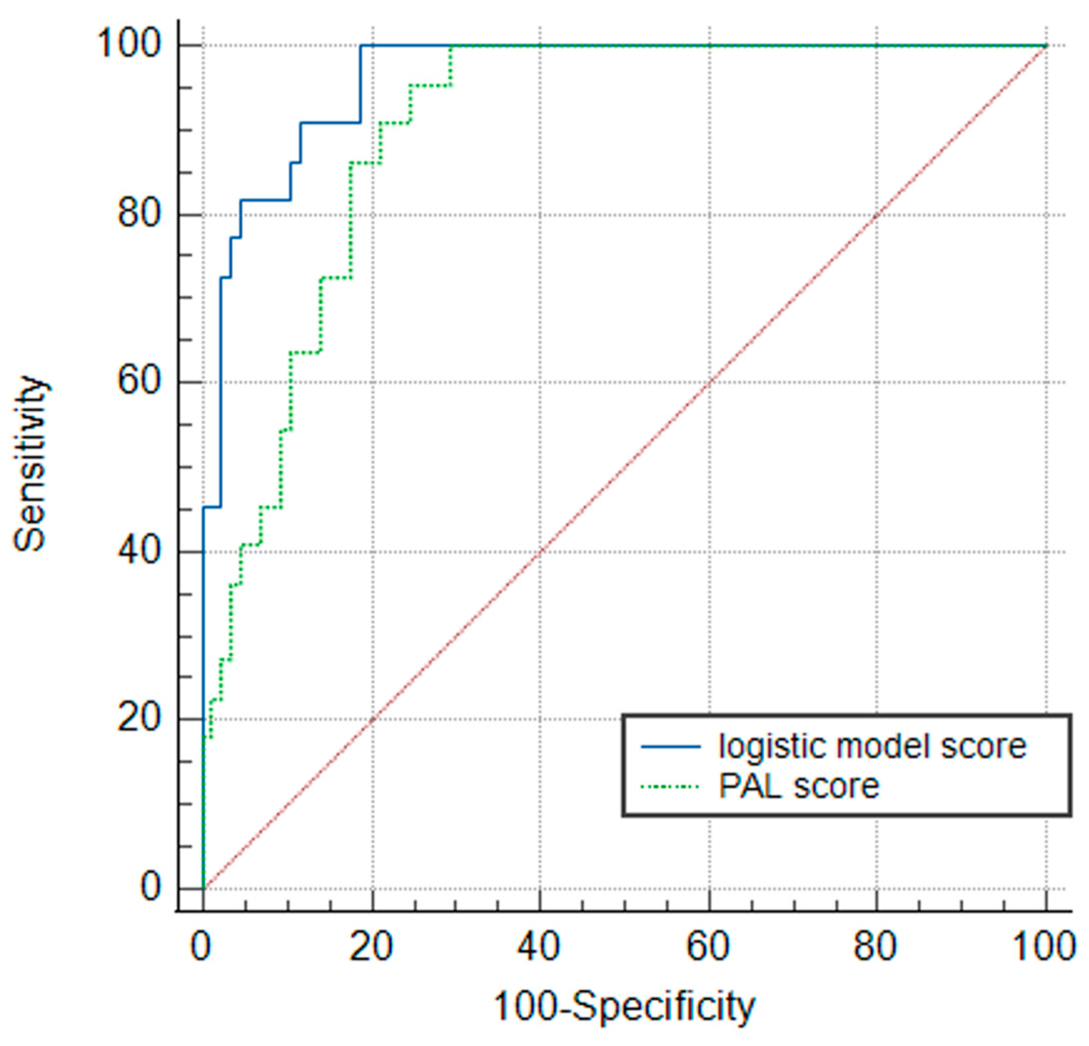
The logistic regression model demonstrated the highest diagnostic accuracy, with an area under the curve (AUC) of 0.963 (95% CI 0.93–0.995, p < 0.001) and a Brier score of 0.064. The simplified PAL score achieved an AUC 0.901 (95% CI 0.844–0.957, p < 0.001) and a Brier score of 0.119. A pairwise comparison using DeLong’s test indicated that the complex logistic regression model was statistically superior to the simplified PAL Score (AUC difference 0.062, z = −2.4, p < 0.016). However, the PAL score retained excellent accuracy (AUC > 0.90, Figure 1). This suggests that the simplified ratio successfully captures the core predictive signal of the logistic equation without requiring the complex exponential calculation of the regression model.
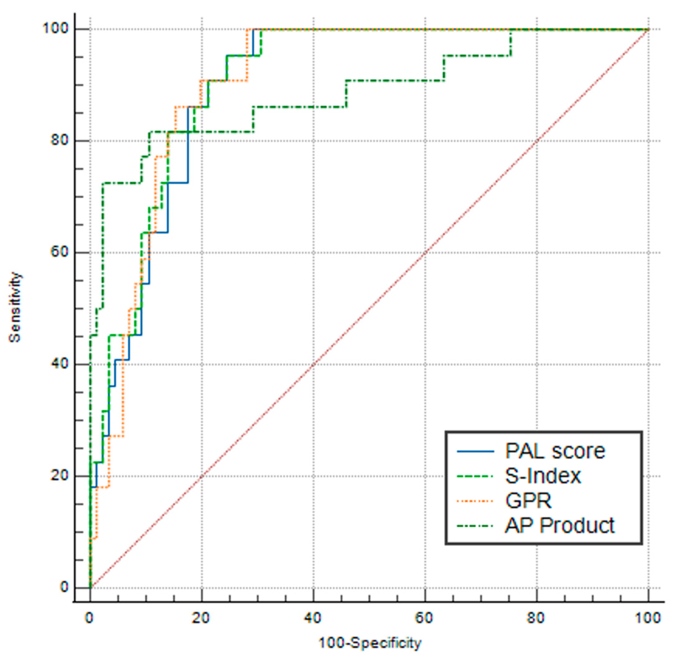
Table 3 presents the results of pairwise comparisons of the PAL score to GPR, AP Product, and the S-Index using DeLong’s test. The analysis revealed no statistically significant differences in diagnostic accuracy between the new PAL score (AUC 0.901) and the established indices. Specifically, the PAL Score demonstrated performance statistically equivalent to the S-Index (p = 0.071), GPR (p = 0.771), and AP Product (p = 0.818), indicating that the simplified ratio maintains robust diagnostic power comparable to these existing markers. Figure 2 presents the combined ROC curves of these scores.

3.4. Internal Validation and Optimal Cut-Off Selection

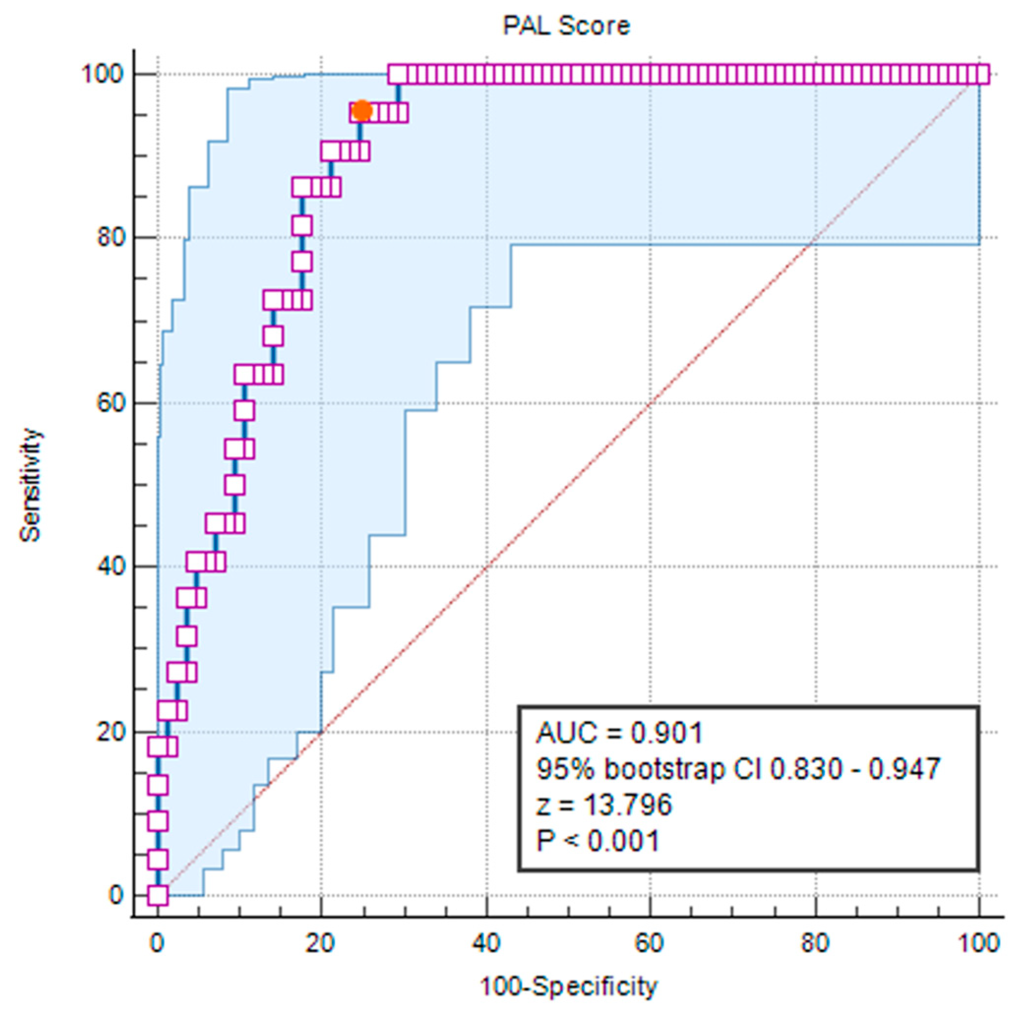
We performed an internal validation on our cohort, using a bootstrap resampling procedure (10,000 iterations) to assess the stability of our model. This analysis confirmed the robustness of our score’s diagnostic accuracy: a bootstrap-corrected AUC of 0.901 and a 95% bootstrap confidence interval of 0.830–0.947 (z statistic = 13.796, p < 0.0001, Figure 3).
The optimal diagnostic threshold for the PAL score to assess the presence of advanced fibrosis was determined in the same bootstrapping procedure with 10,000 iterations using Youden’s J index to maximize the combined sensitivity and specificity. The maximum Youden index (J = 0.7075) determined by the bootstrapping procedure was observed at a PAL Score of 5.058. At this cut-off, the score demonstrated a sensitivity of 95.5% (correctly identifying 21/22 cases within the advanced fibrosis group) and a specificity of 75.3%. The 95% bootstrap confidence interval for the optimal criterion ranged from 3.33 to 6.83. This interval encompasses the derived cut-off of 5.058, suggesting that while the precise mathematical optimum may fluctuate, the score consistently discriminates well within this range for the presence or absence of advanced fibrosis as established by TE.
For clinical ease of use, we evaluated the score’s performance at the nearest-integer threshold (criterion ≥ 5). At this cut-off, the sensitivity remained virtually unchanged at 95.45% and the specificity dropped only slightly to 74.12%. At this cut-off, the score has an extremely low negative likelihood ratio of 0.06, indicating that a score below 5 provides strong evidence against the presence of advanced fibrosis. Table 4 is the contingency table for the cut-off of value ≥5 in our cohort and Table 5 presents the diagnostic accuracy parameters for this cut-off in our cohort and their attendant 95% confidence intervals.

4. Discussion

This study presents the derivation and internal validation of the PAL score, a novel, non-invasive index for estimating the presence of advanced fibrosis in pediatric CLD, compared to TE. To our knowledge, this is the first simple ratio-based scoring system derived specifically from a pediatric cohort with diverse etiologies, filling a critical gap in the current diagnostic landscape.
The accurate assessment of liver fibrosis in children is a clinical imperative. It is a fundamental step in evaluating patients for multi-organ morbidity and total patient health. Cirrhosis is associated with numerous adverse effects in various organ systems [2,28,29,30]. In the pediatric population, there are profound systemic consequences of unrecognized liver fibrosis. Advancing fibrosis and the subsequent impaired blood flow through the liver parenchyma impede metabolic exchange between hepatocytes and plasma, leading to significant growth impairment and nutritional compromise [31]. Furthermore, recent evidence has highlighted that children with CLD exhibit significant structural and functional cardiac alterations, including increased left ventricular mass and diastolic dysfunction, sometimes even before fibrosis progresses to cirrhosis [32]. Therefore, the early identification of advanced fibrosis is not merely useful for managing liver disease, but a requisite for preventing and managing additional systemic morbidity.
The PAL score addresses this need by offering a triage tool. By identifying at-risk patients (PAL ≥ 5) with high sensitivity (95.5%), clinicians can confidently target resource-intensive monitoring (e.g., endoscopy for varices, cardiac surveillance) and treatment toward the high-risk group, as well as potentially avoid unnecessary invasive procedures, for lower-risk patients. The NLR of 0.06 offers a justification for patients with scores below 5 to be conservatively managed with confidence by their primary care provider or general pediatrician, avoiding potentially unnecessary visits or intervention for this lower-risk group. This stratification offers a clear pathway to reduce healthcare costs and minimize procedure-related trauma in children.
Unlike existing scores that often rely on a single pathophysiological axis, the PAL score incorporates three distinct parameters that mirror the progression of liver disease: liver injury (GGT), as GGT serves as a marker of active hepatobiliary damage; portal hypertension (platelets) as thrombocytopenia is the most validated surrogate for hypersplenism and portal pressure; synthetic function (albumin), as hypoalbuminemia reflects the loss of functioning hepatic parenchyma. Because pediatric CLD frequently involves structural or cholestatic biliary etiologies (such as biliary atresia or sclerosing cholangitis), GGT is often a reliable marker of ongoing liver injury. Platelet count serves as a robust surrogate for portal hypertension; as fibrosis increases hepatic vascular resistance, the resulting splenomegaly leads to platelet sequestration, which is further compounded by reduced hepatic synthesis of thrombopoietin. Finally, albumin reflects the progressive loss of functioning hepatic parenchyma. Because albumin is exclusively synthesized by hepatocytes, its decline correlates with the replacement of healthy tissue by fibrotic scarring. This “three-pronged” approach provides a more holistic snapshot of the liver than scores relying on injury markers alone (e.g., AST/ALT ratios). Some scores used for assessing the presence of fibrosis or cirrhosis, like the Göteborg University Cirrhosis Index (GUCI) [33] and King’s score [34] include the international normalized ratios (INR), which is a sensible option, since the INR provides crucial clinical information about bleeding risk in patients with liver damage and serves as a proxy for the assessment of the liver’s functional protein-producing parenchyma. We decided not to add coagulation parameters to this score. Although coagulation studies are routine for patients with CLD, they require additional, separate blood draws on specialized blood containers. Thus, by using albumin as a proxy for the liver’s functional parenchyma, all the parameters needed to compute our score can be found within the complete blood count and biochemistry assessment of serum, making it a viable option even in the most resource-limited settings.
Although numerous non-invasive tests exist, they are almost exclusively derived from adult cohorts with chronic viral hepatitis or MASLD, limiting their applicability to the heterogeneous etiology of pediatric CLD. Our comparisons revealed that while the PAL Score is statistically equivalent to established markers that use similar parameters for their calculation (GPR, S-Index, AP Product), it possesses distinct methodological advantages. The GPR requires standardizing GGT to the “Upper Limit of Normal” (ULN). Because GGT reference ranges may vary by age and laboratory, calculating the GPR is often cumbersome in clinical practice. The PAL score uses raw GGT values, eliminating this variability. Like GPR, APRI relies on ULN values of AST and was primarily validated in adults with chronic hepatitis C. Furthermore, AST is less specific to liver injury than GGT and can be elevated in hemolysis or muscle injury, both of which are common confounders in pediatrics. Other scores, like the AST-to-ALT ratios would be inappropriate for this same reason, as well as the fact that the diverse etiologies in our cohort likely exhibit overlapping patterns of biochemical abnormalities due to different pathophysiologies of disease progression. Other scores used in adults, like the FIB-4, include age as a parameter [12], as a surrogate for disease duration. But, given the different rates of CLD progression associated with different etiologies, using age as a proxy of disease duration is inappropriate in a cohort with diverse etiologies, since progression rates are not the same in patients with different causes of CLD. In the original publication about FIB4, Sterling et al. noted that increasing age is associated with more advanced fibrosis, which would be fine in a homogenous cohort, like the one researched by Sterling et al., which entirely comprise adults with viral hepatitis C and HIV co-infection. It is, however, in our view, not an appropriate choice when dealing with pediatric cohorts or in cohort with diverse CLD etiologies.
While the S-Index also utilizes GGT, platelets, and albumin, it employs a squared albumin term and a multiplier of 1000. This more complex formula, designed to be used in adults with chronic hepatitis B, is difficult to calculate at the bedside. Our analysis suggests that the simpler linear interaction in the PAL Score performs equally well in children without adding complexity to the calculation. Lastly, the AP Product assesses only the synthetic function and portal hypertension. By excluding a marker of active injury (such as GGT or AST), the AP Product risks classifying patients with active liver parenchymal damage but preserved synthetic function as “low risk.” The inclusion of GGT in the PAL score safeguards against this “silent” progression.
The fact that the PAL Score achieved statistical equivalence (AUC 0.901) to established adult scores is a significant finding. It demonstrates that a pediatric-specific, simplified model can perform as well as, or better than, complex adult algorithms, without the burden of age-specific normalizations or calculators.
Strengths and Limitations. We acknowledge the limitations of this study, primarily the relatively small sample size (n = 107, 22 of which were included in the Advanced Fibrosis group). However, this is consistent with the lower prevalence of pediatric CLD compared to adult populations. Additionally, as this is a single-center study, the results require external validation in independent cohorts to confirm the stability of the cut-off value. Given the relatively small number of patients in our single-center cohort, it is not implausible that our established cut-off value of 5 might be different when the score is evaluated in larger cohorts. However, our bootstrapping method predicts the 95% confidence interval of the score between 3.33 to 6.83. Because of the retrospective nature of our study and the fact that our center is a tertiary referral center, there is also the possibility of referral bias to be considered. The cohort in our study likely contains a higher prevalence of advanced fibrosis or patients with more complex etiologies, that would otherwise be expected. This means that our cohort might not perfectly mirror the entire population of children with CLD within our region. These issues therefore further underscore the need for external validation of the present findings.
Although in our cohort we have made methodological efforts to exclude patients with acute intercurrent illness that might cause routine laboratory testing to be altered, we emphasize that caution must be exerted when considering the calculation of this score, as with any other of the more established scores, for that matter. Any intercurrent illness could potentially be associated with altered lab values not due to CLD, but due to acute illness, and this might lead to a misclassification of patients when altered parameters are used for the score (e.g., altered platelet numbers due to infection or inflammation).
It is important to note that unlike adult studies in the past two decades that have led to development of well-known scores, we did not use liver biopsy as the reference standard. Liver biopsy is the gold standard for assessing fibrosis and cirrhosis of the liver and it allows for the direct visualization of the parenchyma under the microscope and direct quantification of necro-inflammatory activity that defines severe CLDs [35]. However, in recent decades TE has become an accepted method to assess the stage of liver fibrosis, by measuring liver stiffness and reference values for pediatric patients have been published [20,36]. TE has proven to be a viable option when evaluating children, with good concordance with biopsy results [36,37,38]. There are some advantages to performing TE in children as opposed to biopsy, like the lack of pain, no risk of organ injury, no requirement for sedation and the ability to evaluate a much larger portion of the liver parenchyma, as opposed to biopsy which can only sample a locally limited portion of the tissue. Recently, guidelines have endorsed the use of TE for assessing liver fibrosis in adults [39]. Moreover, our PAL score is not designed or intended to represent a replacement of biopsy or TE altogether, but rather as a tool to be used to stratify patients in primary care or general pediatric practice, and to guide advance those patients with high likelihood of advanced liver fibrosis to specialized tertiary centers sooner, where TE, biopsy or other diagnostic and therapeutic interventions might be warranted. At the same time, patients with likely less advanced CLD could be more confidently managed by their local pediatric practice with comparatively rarer visits and fewer referrals to specialized pediatric hepatology centers.
However, reliance on TE is subject to certain limitations. TE measures liver stiffness, which, while strongly correlated with fibrosis, can occasionally be confounded by acute hepatic inflammation, venous congestion, or biliary obstruction, potentially leading to overestimations of the true fibrotic burden. While TE is an increasingly accepted and validated surrogate for fibrosis in pediatric hepatology, the PAL score’s diagnostic thresholds ultimately predict liver stiffness of TE rather than directly observed histological fibrosis.
The primary strength of the PAL score is its simplicity and specificity for the pediatric context. Its derivation from a cohort with diverse diagnoses—ranging from biliary atresia to metabolic defects—supports its generalizability across the spectrum of pediatric CLD, unlike scores limited to MASLD [17]. Although patients are unlikely to present with more than one or, very rarely two etiologies contributing to their CLD, the fact that this score was developed on a diverse cohort lends weight to its’ use a general screening tool for all sorts of patients with CLD. Limiting development only to cohorts with a certain narrow diagnosis (for example, only biliary atresia or only chronic viral hepatitis), might lead to an overfitted score that is poorly adapted to evaluating pathologies other than that on which it has been developed.
For this cohort, all TE measurements were performed by a single highly skilled operator, reducing inter-operator bias. The observation that our F0 cohort measurements were statistically congruent with population-level reference values (p > 0.05) provides an important external validation of our methodology. This alignment confirms that measurements accurately reflect baseline liver stiffness values without systemic bias.
The identification of an integer cut-off in our study (criterion ≥ 5.0, with a bootstrapped confidence interval between 3.33 to 6.83) and the negative likelihood ratio of 0.06 make the PAL score a user-friendly “rule-out” tool for clinicians. These results, of course, should be externally validated and given the simplicity of the parameters and the score, this should be an amenable task for other groups who can easily retrospectively collect such data. Future steps in research should also focus on validating this score in cohorts of patients who have also had liver biopsy in close relation to their standard bloodwork, as this would be an easily available step for research groups who have already collected this data in patients.

5. Conclusions

The PAL Score is the first simple, non-invasive fibrosis index specifically derived for a diverse pediatric population. By combining markers of liver injury, synthetic function, and portal hypertension, it provides accurate information at the patient’s bedside. Demonstrating a high sensitivity of 95.5% and an exceptional negative likelihood ratio of 0.06 at a user-friendly cut-off of 5.0, the score serves as an effective screening tool to rule out advanced fibrosis. Its implementation can help reduce unnecessary invasive procedures and streamline the clinical management of children with CLD, particularly as an accessible alternative in settings without access to transient elastography. Moving forward, multi-center studies—ideally incorporating biopsy-proven cohorts—are necessary to provide robust external validation of these findings and confirm external generalizability.

Supplementary Materials

The following supporting information can be downloaded at: https://www.mdpi.com/article/10.3390/livers6040058/s1, Supplementary Material S1: Patient classification; Supplementary Material S2: Boxplots of parameters compared between groups.

Author Contributions

Conceptualization, A.-Ș.N.; methodology, A.-Ș.N., A.G. and T.L.P.; formal analysis, A.-Ș.N.; investigation, A.G., T.L.P., G.B., A.M. and S.A.; data curation, A.G., T.L.P., G.B., A.M. and S.A.; writing—original draft preparation, A.-Ș.N.; writing—review and editing, A.G., T.L.P., G.B., A.M. and S.A.; visualization, A.-Ș.N.; supervision, T.L.P. All authors have read and agreed to the published version of the manuscript.

Funding

This research received no external funding.

Institutional Review Board Statement

The study was conducted in accordance with the Declaration of Helsinki and approved by the Ethics Committee of the Iuliu Hațieganu University of Medicine and Pharmacy (AVZ100/20 June 2023).

Informed Consent Statement

Patient consent was waived due to the retrospective nature of the current study and anonymous data collection.

Data Availability Statement

Curated data and Jamovi files are available upon reasonable request from the authors after an appropriate amount of time after publication.

Conflicts of Interest

The authors declare no conflicts of interest.

Abbreviations

The following abbreviations are used in this manuscript:
PALPediatric adapted liver (score)
CLDChronic liver disease
TETransient elastography
kPakiloPascal
ALTAlanine aminotransferase
ASTAspartate aminotransferase
ALPAlkaline phosphatase
GGTGamma glutamyl transferase
tBTotal bilirubin
cBConjugated bilirubin
GPRGGT to platelet ratios
ROCReceiver operator characteristics
MASLDMetabolic associated liver disease

References

  1. Karlsen, T.H.; Sheron, N.; Zelber-Sagi, S.; Carrieri, P.; Dusheiko, G.; Bugianesi, E.; Pryke, R.; Hutchinson, S.J.; Sangro, B.; Martin, N.K.; et al. The EASL–Lancet Liver Commission: Protecting the next Generation of Europeans against Liver Disease Complications and Premature Mortality. Lancet 2022, 399, 61–116. [Google Scholar] [CrossRef] [PubMed]
  2. Carvalho, M.V.H.; Kroll, P.C.; Kroll, R.T.M.; Carvalho, V.N. Cirrhotic Cardiomyopathy: The Liver Affects the Heart. Braz. J. Med. Biol. Res. 2019, 52, e7809. [Google Scholar] [CrossRef] [PubMed]
  3. Desai, M.S.; Zainuer, S.; Kennedy, C.; Kearney, D.; Goss, J.; Karpen, S.J. Cardiac Structural and Functional Alterations in Infants and Children With Biliary Atresia, Listed for Liver Transplantation. Gastroenterology 2011, 141, 1264–1272.e4. [Google Scholar] [CrossRef] [PubMed]
  4. Fierro-Angulo, O.M.; González-Regueiro, J.A.; Pereira-García, A.; Ruiz-Margáin, A.; Solis-Huerta, F.; Macías-Rodríguez, R.U. Hematological Abnormalities in Liver Cirrhosis. World J. Hepatol. 2024, 16, 1229–1244. [Google Scholar] [CrossRef] [PubMed]
  5. Meena, B.L.; Narayan, S.J.A.; Sarin, S.K. Hepatic Encephalopathy in Non-Cirrhotic Portal Hypertension. Metab. Brain Dis. 2025, 40, 103. [Google Scholar] [CrossRef] [PubMed]
  6. Poynard, T.; Mathurin, P.; Lai, C.-L.; Guyader, D.; Poupon, R.; Tainturier, M.-H.; Myers, R.P.; Muntenau, M.; Ratziu, V.; Manns, M.; et al. A Comparison of Fibrosis Progression in Chronic Liver Diseases. J. Hepatol. 2003, 38, 257–265. [Google Scholar] [CrossRef] [PubMed]
  7. Goodman, Z.D. Grading and Staging Systems for Inflammation and Fibrosis in Chronic Liver Diseases. J. Hepatol. 2007, 47, 598–607. [Google Scholar] [CrossRef] [PubMed]
  8. Ziol, M.; Handra-Luca, A.; Kettaneh, A.; Christidis, C.; Mal, F.; Kazemi, F.; de Lédinghen, V.; Marcellin, P.; Dhumeaux, D.; Trinchet, J.-C.; et al. Noninvasive Assessment of Liver Fibrosis by Measurement of Stiffness in Patients with Chronic Hepatitis C. Hepatology 2005, 41, 48–54. [Google Scholar] [CrossRef] [PubMed]
  9. Marcellin, P.; Ziol, M.; Bedossa, P.; Douvin, C.; Poupon, R.; Lédinghen, V.D.; Beaugrand, M. Non-Invasive Assessment of Liver Fibrosis by Stiffness Measurement in Patients with Chronic Hepatitis B. Liver Int. 2009, 29, 242–247. [Google Scholar] [CrossRef] [PubMed]
  10. El-Guindi, M.A.; Allam, A.A.; Abdel-Razek, A.A.; Sobhy, G.A.; Salem, M.E.; Abd-Allah, M.A.; Sira, M.M. Transient Elastography and Diffusion-Weighted Magnetic Resonance Imaging for Assessment of Liver Fibrosis in Children with Chronic Hepatitis C. World J. Virol. 2024, 13, 96369. [Google Scholar] [CrossRef] [PubMed]
  11. Kehler, T.; Grothues, D.; Evert, K.; Wahlenmayer, J.; Knoppke, B.; Melter, M. Elastography—The New Standard in the Assessment of Fibrosis After Pediatric Liver Transplantation? Pediatr. Transplant. 2024, 28, e14832. [Google Scholar] [CrossRef] [PubMed]
  12. Sterling, R.K.; Lissen, E.; Clumeck, N.; Sola, R.; Correa, M.C.; Montaner, J.; Sulkowski, M.S.; Torriani, F.J.; Dieterich, D.T.; Thomas, D.L.; et al. Development of a Simple Noninvasive Index to Predict Significant Fibrosis in Patients with HIV/HCV Coinfection. Hepatology 2006, 43, 1317–1325. [Google Scholar] [CrossRef] [PubMed]
  13. Forns, X.; Ampurdanès, S.; Llovet, J.M.; Aponte, J.; Quintó, L.; Martínez-Bauer, E.; Bruguera, M.; Sánchez-Tapias, J.M.; Rodés, J. Identification of Chronic Hepatitis C Patients without Hepatic Fibrosis by a Simple Predictive Model: Identification of Chronic Hepatitis C Patients without Hepatic Fibrosis by a Simple Predictive Model. Hepatology 2002, 36, 986–992. [Google Scholar] [CrossRef] [PubMed]
  14. Lemoine, M.; Shimakawa, Y.; Nayagam, S.; Khalil, M.; Suso, P.; Lloyd, J.; Goldin, R.; Njai, H.-F.; Ndow, G.; Taal, M.; et al. The Gamma-Glutamyl Transpeptidase to Platelet Ratio (GPR) Predicts Significant Liver Fibrosis and Cirrhosis in Patients with Chronic HBV Infection in West Africa. Gut 2016, 65, 1369–1376. [Google Scholar] [CrossRef] [PubMed]
  15. Furdela, V.; Pavlyshyn, H.; Shulhai, A.-M.; Kozak, K.; Furdela, M. Triglyceride Glucose Index, Pediatric NAFLD Fibrosis Index, and Triglyceride-to-High-Density Lipoprotein Cholesterol Ratio Are the Most Predictive Markers of the Metabolically Unhealthy Phenotype in Overweight/Obese Adolescent Boys. Front. Endocrinol. 2023, 14, 1124019. [Google Scholar] [CrossRef] [PubMed]
  16. Jayasekera, D.; Hartmann, P. Noninvasive Biomarkers in Pediatric Nonalcoholic Fatty Liver Disease. World J. Hepatol. 2023, 15, 609–640. [Google Scholar] [CrossRef] [PubMed]
  17. Alkhouri, N.; Mansoor, S.; Giammaria, P.; Liccardo, D.; Lopez, R.; Nobili, V. The Development of the Pediatric NAFLD Fibrosis Score (PNFS) to Predict the Presence of Advanced Fibrosis in Children with Nonalcoholic Fatty Liver Disease. PLoS ONE 2014, 9, e104558. [Google Scholar] [CrossRef] [PubMed]
  18. Rozario, R.; Ramakrishna, B. Histopathological Study of Chronic Hepatitis B and C: A Comparison of Two Scoring Systems. J. Hepatol. 2003, 38, 223–229. [Google Scholar] [CrossRef] [PubMed]
  19. Staub, F.; Tournoux-Facon, C.; Roumy, J.; Chaigneau, C.; Morichaut-Beauchant, M.; Levillain, P.; Prevost, C.; Aubé, C.; Lebigot, J.; Oberti, F.; et al. Liver Fibrosis Staging with Contrast-Enhanced Ultrasonography: Prospective Multicenter Study Compared with METAVIR Scoring. Eur. Radiol. 2009, 19, 1991–1997. [Google Scholar] [CrossRef] [PubMed]
  20. Mărginean, C.O.; Meliţ, L.E.; Ghiga, D.V.; Săsăran, M.O. Reference Values of Normal Liver Stiffness in Healthy Children by Two Methods: 2D Shear Wave and Transient Elastography. Sci. Rep. 2020, 10, 7213. [Google Scholar] [CrossRef] [PubMed]
  21. Zhou, K.; Gao, C.-F.; Zhao, Y.-P.; Liu, H.-L.; Zheng, R.-D.; Xian, J.-C.; Xu, H.-T.; Mao, Y.-M.; Zeng, M.-D.; Lu, L.-G. Simpler Score of Routine Laboratory Tests Predicts Liver Fibrosis in Patients with Chronic Hepatitis B. J. Gastroenterol. Hepatol. 2010, 25, 1569–1577. [Google Scholar] [CrossRef] [PubMed]
  22. Fujita, K.; Yamasaki, K.; Morishita, A.; Shi, T.; Tani, J.; Nishiyama, N.; Kobara, H.; Himoto, T.; Yatsuhashi, H.; Masaki, T. Albumin Platelet Product as a Novel Score for Liver Fibrosis Stage and Prognosis. Sci. Rep. 2021, 11, 5345. [Google Scholar] [CrossRef] [PubMed]
  23. Elsabaawy, M.; Eissa, M.; Shaban, A.; Naguib, M. Validation of Albumin Platelet Product as a Non-Invasive Fibrosis Staging Tool in Patients with Chronic HCV-Related Liver Disease. Sci. Rep. 2025, 15, 20265. [Google Scholar] [CrossRef] [PubMed]
  24. Youden, W.J. Index for Rating Diagnostic Tests. Cancer 1950, 3, 32–35. [Google Scholar] [CrossRef]
  25. MedCalc® Statistical Software, version 23.5.2; MedCalc Software Ltd.: Ostend, Belgium, 2025. Available online: https://www.Medcalc.org (accessed on 9 June 2026).
  26. R Core Team. R: A Language and Environment for Statistical Computing; R Foundation for Statistical Computing: Vienna, Austria, 2024; Available online: https://www.R-Project.org/ (accessed on 9 June 2026).
  27. Jamovi, version 2.6; Computer Software; The Jamovi Project: Sidney, Australia, 2025. Available online: https://www.Jamovi.org (accessed on 9 June 2026).
  28. Hasa, E.; Hartmann, P.; Schnabl, B. Liver Cirrhosis and Immune Dysfunction. Int. Immunol. 2022, 34, 455–466. [Google Scholar] [CrossRef] [PubMed]
  29. Hadengue, A.; Benhayoun, M.K.; Lebrec, D.; Benhamou, J.P. Pulmonary Hypertension Complicating Portal Hypertension: Prevalence and Relation to Splanchnic Hemodynamics. Gastroenterology 1991, 100, 520–528. [Google Scholar] [CrossRef] [PubMed]
  30. Nadim, M.K.; Kellum, J.A.; Forni, L.; Francoz, C.; Asrani, S.K.; Ostermann, M.; Allegretti, A.S.; Neyra, J.A.; Olson, J.C.; Piano, S.; et al. Acute Kidney Injury in Patients with Cirrhosis: Acute Disease Quality Initiative (ADQI) and International Club of Ascites (ICA) Joint Multidisciplinary Consensus Meeting. J. Hepatol. 2024, 81, 163–183. [Google Scholar] [CrossRef] [PubMed]
  31. Cameron, R.; Kogan-Liberman, D. Nutritional Considerations in Pediatric Liver Disease. Pediatr. Rev. 2014, 35, 493–496. [Google Scholar] [CrossRef] [PubMed][Green Version]
  32. Niculae, A.-Ștefan; Căinap, S.S.; Grama, A.; Pop, T.L. Pediatric Cirrhotic Cardiomyopathy: Literature Review and Effect Size Estimations of Selected Parameters. Eur. J. Pediatr. 2024, 183, 4789–4797. [Google Scholar] [CrossRef] [PubMed]
  33. Islam, S.; Antonsson, L.; Westin, J.; Lagging, M. Cirrhosis in Hepatitis C Virus-Infected Patients Can Be Excluded Using an Index of Standard Biochemical Serum Markers. Scand. J. Gastroenterol. 2005, 40, 867–872. [Google Scholar] [CrossRef] [PubMed]
  34. Cross, T.J.S.; Rizzi, P.; Berry, P.A.; Bruce, M.; Portmann, B.; Harrison, P.M. King’s Score: An Accurate Marker of Cirrhosis in Chronic Hepatitis C. Eur. J. Gastroenterol. Hepatol. 2009, 21, 730–738. [Google Scholar] [CrossRef] [PubMed]
  35. Chowdhury, A.B.; Mehta, K.J. Liver Biopsy for Assessment of Chronic Liver Diseases: A Synopsis. Clin. Exp. Med. 2023, 23, 273–285. [Google Scholar] [CrossRef] [PubMed]
  36. Castera, L.; Rinella, M.E.; Tsochatzis, E.A. Noninvasive Assessment of Liver Fibrosis. N. Engl. J. Med. 2025, 393, 1715–1729. [Google Scholar] [CrossRef] [PubMed]
  37. Galina, P.; Alexopoulou, E.; Mentessidou, A.; Mirilas, P.; Zellos, A.; Lykopoulou, L.; Patereli, A.; Salpasaranis, K.; Kelekis, N.L.; Zarifi, M. Diagnostic Accuracy of Two-Dimensional Shear Wave Elastography in Detecting Hepatic Fibrosis in Children with Autoimmune Hepatitis, Biliary Atresia and Other Chronic Liver Diseases. Pediatr. Radiol. 2021, 51, 1358–1368. [Google Scholar] [CrossRef] [PubMed]
  38. Nobili, V.; Vizzutti, F.; Arena, U.; Abraldes, J.G.; Marra, F.; Pietrobattista, A.; Fruhwirth, R.; Marcellini, M.; Pinzani, M. Accuracy and Reproducibility of Transient Elastography for the Diagnosis of Fibrosis in Pediatric Nonalcoholic Steatohepatitis. Hepatology 2008, 48, 442–448. [Google Scholar] [CrossRef] [PubMed]
  39. Sterling, R.K.; Patel, K.; Duarte-Rojo, A.; Asrani, S.K.; Alsawas, M.; Dranoff, J.A.; Fiel, M.I.; Murad, M.H.; Leung, D.H.; Levine, D.; et al. AASLD Practice Guideline on Blood-Based Noninvasive Liver Disease Assessment of Hepatic Fibrosis and Steatosis. Hepatology 2025, 81, 321–357. [Google Scholar] [CrossRef] [PubMed]
Figure 1. Receiver Operating Characteristic (ROC) curve comparing the full logistic regression model (solid curve) to the simplified PAL Score (dotted curve).
Livers 06 00058 g001
Figure 2. Combined ROC curves illustrating the diagnostic performance of the PAL score, S-Index, GPR, and AP Product. All scores demonstrated comparable area under the curve (AUC) values for predicting advanced fibrosis in the pediatric cohort (see Table 3 also).
Livers 06 00058 g002
Figure 3. Internal validation of the PAL Score using bootstrap resampling (AUC = 0.901, z = 13.796, p < 0.001). Shaded area represents the 95% CI (0.830–0.947) of the ROC curve; red marker represents the Youden index location.
Livers 06 00058 g003
Table 1. Baseline clinical and biochemical characteristics and univariate comparisons across fibrosis groups.
VariableNo or Mild Fibrosis (n = 85) * Advanced Fibrosis (n = 22) p-ValueEffect Size
Mean ± SDMedian (IQR)Mean ± SDMedian (IQR)
Age (months)111.72 ± 56.41113.00 (85)93.77 ± 67.4111 (136.25)0.280.14
WBC (‘000/cubic mm)8.13 ± 3.247.43 (3.43)9.1 ± 4.517.68 (6.7)0.61−0.07
RBC (millions/cubic mm)4.72 ± 0.614.72 (0.59)4.3 ± 0.674.24 (0.74)0.0020.43
Hb (g/dL)13.07 ± 1.7412.90 (2)11.91 ± 1.9912.30 (2.8)0.030.3
Platelets (‘000/cubic mm)305.07 ± 118.86267 (110)160.5 ± 103.33130 (91)<0.0010.72
AST (U/L)81.70 ± 104.9141.00 (50.8)179.67 ± 257.389.00 (147.15)0.005−0.38
ALT (U/L)97.60 ± 139.9341.00 (87)126.76 ± 105.1283.50 (167.25)0.018−0.32
ALP (U/L)283.85 ± 162.39255 (184.3)432.28 ± 257.51416.5 (397.3)0.014−0.34
GGT (U/L)82.03 ± 163.2727.00 (51)195.30 ± 265.8397.50 (105.4)<0.001−0.51
tB (mg/dL)0.71 ± 0.730.54 (0.33)5.09 ± 8.51.21 (3.39)<0.001−0.58
cB * (mg/dL)0.28 ± 0.520.18 (0.1)3.57 ± 6.680.64 (1.39)<0.001−0.74
Albumin (g/dL)4.65 ± 0.314.70 (0.37)3.9 ± 0.593.85 (0.89)<0.0010.74
Data are presented as Mean ± Standard Deviation and Median (Interquartile Range) for 107 pediatric patients with chronic liver disease. Statistical significance and effect sizes were calculated to identify potential predictors of advanced fibrosis (F3–F4 staging). * conjugated bilirubin in the No or mild fibrosis group has n = 83 due to two missing values; WBC—white blood cells; RBC—red blood cells; Hb—hemoglobin; ALT—alanine aminotransferase; AST—aspartate aminotransferase; ALP—alkaline phosphatase; GGT—gamma glutamyl transferase; tB—total bilirubin; cB—conjugated bilirubin. Significant p-values are shows in bold print.
Table 2. Final multivariable binary logistic regression model for predicting advanced fibrosis.
Model Coefficients—No or Mild Fibrosis Group vs. Advanced Fibrosis Group
95% Confidence Interval
PredictorEstimateSEZpOdds ratioLowerUpper
Intercept17.9185.2653.40<0.0016.05 × 1071992.297571.84 × 1012
Platelets−0.0170.004−3.79<0.0010.98310.974410.992
GGT0.0050.0022.660.0081.00551.001451.01
Albumin−3.6661.14−3.220.0010.02560.002740.239
Table 3. ROC summary and pairwise comparisons of the PAL Score against established adult-derived indices.
Model/ComparisonAUC/AUC Difference95% CI Lower95% CI Upperp-Value
ROC Curve Summary
PAL score0.9010.8440.957<0.001
S-index0.910.8550.964<0.001
GPR0.9050.8490.961<0.001
AP Product0.8880.7930.982<0.001
Pairwise AUC Comparisons
PAL score vs. S-index−0.00909−0.01890.000760.071
PAL score vs. GPR−0.00428−0.03310.02460.771
PAL score vs. AP Product0.01283−0.09680.12240.818
Table 4. Contingency table for the PAL score at the optimal clinical threshold (≥5.0). Cross-tabulation of the simplified score against the transient elastography reference standard, maximizing Youden’s J index for clinical utility.
Contingency Table
No or Mild vs. Advanced
Advanced FibrosisNo or Mild FibrosisTotal
PAL score≥5212243
<516364
Total2285107
Table 5. Diagnostic performance metrics for the PAL score at the ≥5.0 threshold.
95% Confidence Interval
ResultLowerUpper
Sensitivity95.45%77.16%99.88%
Specificity74.12%63.48%83.01%
Positive Likelihood Ratio3.68802.544585.345
Negative Likelihood Ratio0.06130.009000.418
Positive Predictive Value48.84%39.71%58.04%
Negative Predictive Value98.44%90.24%99.77%
Accuracy78.50%69.51%85.86%
Disclaimer/Publisher’s Note: The statements, opinions and data contained in all publications are solely those of the individual author(s) and contributor(s) and not of MDPI and/or the editor(s). MDPI and/or the editor(s) disclaim responsibility for any injury to people or property resulting from any ideas, methods, instructions or products referred to in the content.