1. Introduction
Ventral hernias are a common and often debilitating sequela of abdominal surgery, occurring in up to 20% of patients following a traditional midline laparotomy [
1]. This risk increases substantially in patients who undergo orthotopic liver transplantation (OLT), where the incidence can reach as high as 43% [
1,
2,
3,
4,
5,
6]. For many patients, a hernia causes discomfort and functional limitation while also posing life-threatening risks such as intestinal incarceration and strangulation that may complicate long-term transplant success.
The gradual enlargement of the hernia over time results in a relative loss of abdominal domain, with adverse effects on posture, respiration, micturition, defecation, and overall biomechanical function. Progressive enlargement also causes significant cosmetic deformity, and stretching of the skin with atrophy of subcutaneous fat may lead to areas of ischemia susceptible to ulceration and infection [
6].
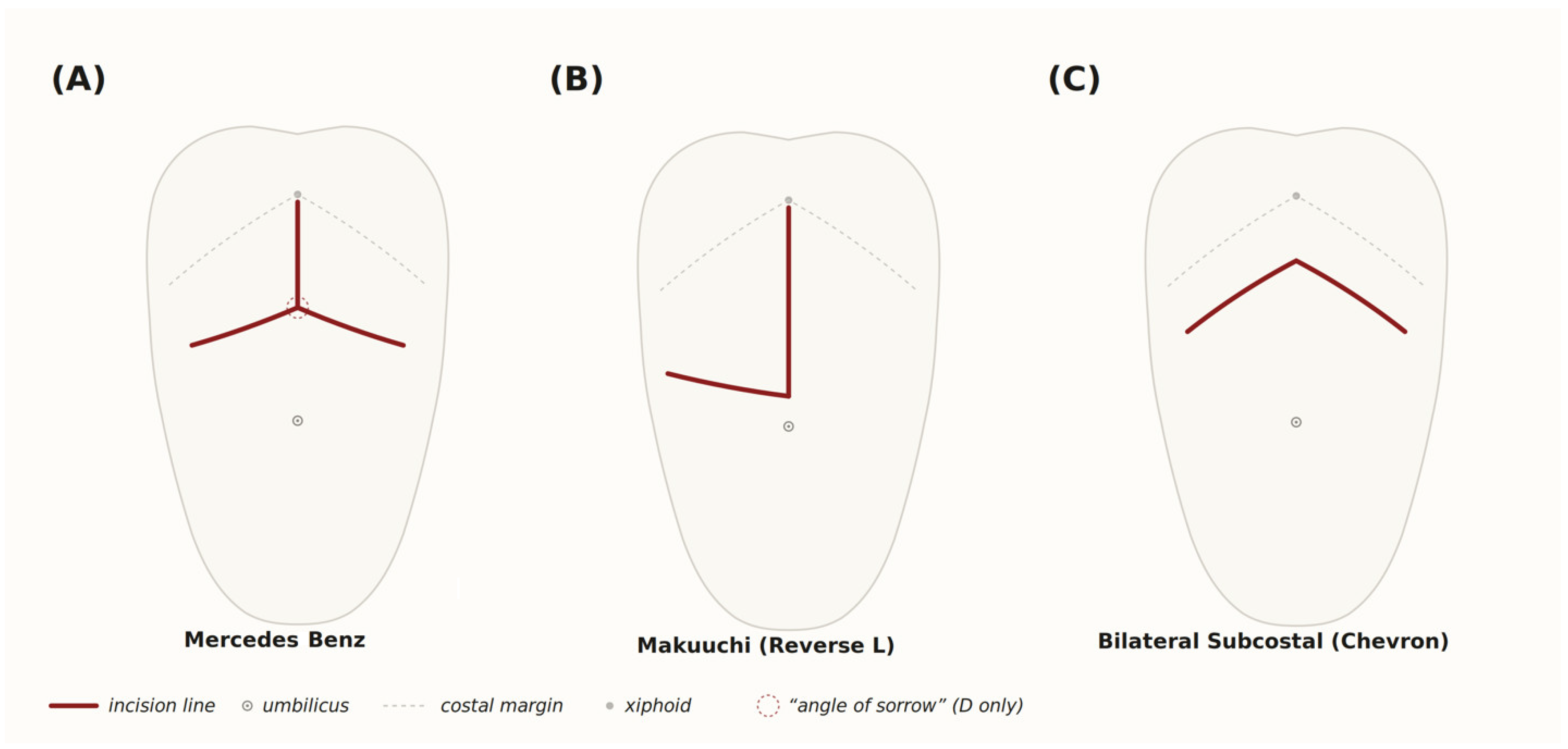
The transplant population is biologically distinct from the average surgical patient. Chronic immunosuppression, altered wound healing, and commonly associated comorbid conditions all contribute to increased complexity. The subcostal incision used for liver transplantation, including the “Mercedes Benz”, the “Chevron” and “Reverse L” configurations [see
Figure 1], transects the right Rectus Abdominis muscle, disrupting the abdominal wall’s continuity and compromising dynamic support. As survival after OLT continues to improve, the number of patients requiring repair of incisional hernias has increased substantially.
Given these challenges, abdominal wall reconstruction in OLT patients must emphasize not only structural closure but also biologic integration, preservation of vascular and neural supply, and restoration of dynamic function. The objective of this paper is to present our institutional approach to ventral hernia repair following OLT, supported by a retrospective analysis of clinical outcomes in 40 consecutive patients treated over an 11.8-year period.
2. Pathophysiology and Risk Factors in the Post-OLT Population
Incisional hernia formation after liver transplantation is multifactorial, arising from the interplay of surgical technique, altered physiology, and chronic systemic influences. The most obvious anatomic factor is the incision itself. The subcostal “Chevron” incision crosses multiple muscle groups and disrupts the continuity of the rectus abdominis and oblique musculature. When extended vertically into a “Mercedes-Benz” configuration, the disruption is even more extensive. Of particular significance is the junction where the bilateral subcostal incisions meet the upper midline incision—a region transplant surgeons have termed the “angle of sorrow”—where fascial integrity is most severely compromised, and hernia formation is most likely and especially problematic.
Beyond mechanical disruption, the OLT population experiences a confluence of systemic factors that impair wound healing. Chronic immunosuppression dampens the inflammatory and cellular responses required for collagen deposition and tensile strength development. Many patients present with nutritional deficits, loss of muscle mass, or ongoing hepatic synthetic dysfunction. Recurrent ascites or intra-abdominal infections can place persistent tension and contamination on a repair site. Pulmonary disease, chronic cough, and obesity further increase intra-abdominal pressure. Together, these factors make standard hernia repair strategies poorly suited to the OLT population.
3. Conceptual Framework of Repair
The abdominal wall is a complex, layered structure that relies on coordinated interaction between its muscular, fascial, and neurovascular components. Disruption of this system during liver transplantation results in a significant loss of balance across the abdomen. Restoring this balance begins with recreating myofascial continuity through component separation, which allows for medial advancement of the rectus abdominis via release of the external oblique aponeurosis. Preserving the abdominal wall’s neurovascular structures promotes tissue perfusion, healing, and restoration of dynamic function [
7].
The critical element is re-establishing myofascial continuity. In some cases, this will involve advancement of the two sections of the transected Left Rectus Adominis muscle. In all cases, it involves advancement of some combination of the External Obliques and Rectus Abdominis muscles to create a dynamic muscular repair. Reinforcement of this reconstructed myofascial layer is achieved through placement of a biologic mesh in an onlay fashion, secured with a multipoint suture fixation technique in a progressive tension pattern. This technique distributes tension evenly across the repair while minimizing shear forces and reducing seroma formation. The mesh integrates naturally with host tissue rather than acting as a static barrier [
8].
The choice of biologic rather than synthetic mesh is critical in this population. Acellular dermal matrices (ADM) provide a scaffold that supports neovascularization and fibroblast ingrowth, ultimately allowing incorporation and remodeling into host tissue. This biologic integration minimizes foreign body response and makes the material more resistant to infection—a key advantage for patients on immunosuppressive therapy. In contrast, traditional synthetic meshes can harbor bacteria, provoke chronic inflammation, and may require removal in the setting of infection. In our series, human acellular dermal matrix (HADM) was used in 83.7% of encounters, with Cortiva (Integra LifeSciences Corp., Princeton, NJ, USA) selected over AlloDerm (Allergan Aesthetics, an AbbVie Company, Irvine, CA, USA) in the majority of cases given equivalent perioperative outcomes and a more favorable cost profile, consistent with our prior comparative study [
9,
10,
11,
12].
More recently, we have incorporated a poly-4-hydroxybutyrate (P4HB) mesh, Phasix™ (Becton, Dickinson and Company [BD], Franklin Lakes, NJ, USA) as an adjunct in select cases. This resorbable biosynthetic mesh provides initial tensile strength while gradually resorbing over 12 to 18 months, allowing native tissue to assume load-bearing function. In this series, P4HB was reserved primarily as a supplement in cases with very large defects (≥300 cm2) when additional mechanical support is desired in combination with onlay HADM. The decision on biologic mesh use is individualized based on defect size, contamination risk, and tissue quality.
4. Methods
4.1. Study Design and Population
This is a single-institution, single-surgeon retrospective review of all patients with a history of orthotopic liver transplantation who underwent abdominal wall reconstruction at the University of Alabama at Birmingham between June 2014 and April 2026. No patients were excluded; this represents a consecutive series. The study was approved by the UAB Institutional Review Board (IRB-300012379, 10 February 2024) and conducted in accordance with the Declaration of Helsinki. Patient consent was waived due to the retrospective and de-identified nature of the data.
4.2. Data Collection
Clinical data were abstracted from the electronic medical record and maintained in a secure institutional database. Variables collected included patient demographics, comorbidities, hernia characteristics (location, size, recurrence status, and prior mesh), operative details (technique, mesh type and size, estimated blood loss, and operative time), postoperative complications, length of stay, discharge disposition, and follow-up data, including last clinical appointment and vital status.
4.3. Outcomes and Definitions
The primary outcome was hernia recurrence, defined as any clinical or radiographic evidence of fascial defect at the prior repair site documented in the medical record. Secondary outcomes included surgical site occurrences (SSO: seroma, hematoma, wound dehiscence, skin necrosis, surgical site infection, and need for debridement), return to the operating room, interventional radiology drainage, ICU transfer, thromboembolic events, and 30-day mortality. Follow-up was assessed from the date of surgery to the last documented appointment in the health system.
4.4. Preoperative Optimization
In our practice, abdominal wall reconstruction is typically performed well after the transplant recovery period, once patients have achieved long-term stability on their immunosuppressive regimens. All patients in this series underwent routine nutritional screening as part of their preoperative evaluation, and only those determined to be in an adequate nutritional state were considered candidates for elective repair. Parameters assessed include serum albumin, prealbumin, body composition, and overall functional status, with referral for nutritional optimization when indicated. Close coordination with the transplant hepatology team regarding immunosuppression management and metabolic status is an integral component of preoperative planning. Emergent repair for incarceration or strangulation is performed regardless of nutritional or metabolic status.
4.5. Statistical Analysis
Given the descriptive nature of this single-arm case series, data are presented as means ± standard deviation, medians with ranges, and frequencies with percentages. No inferential statistical comparisons were performed. All analyses were performed using Python (version 3.12; Python Software Foundation, Wilmington, DE, USA) [
13] with the pandas [
14] and NumPy [
15] libraries.
5. Operative Technique and Technical Considerations
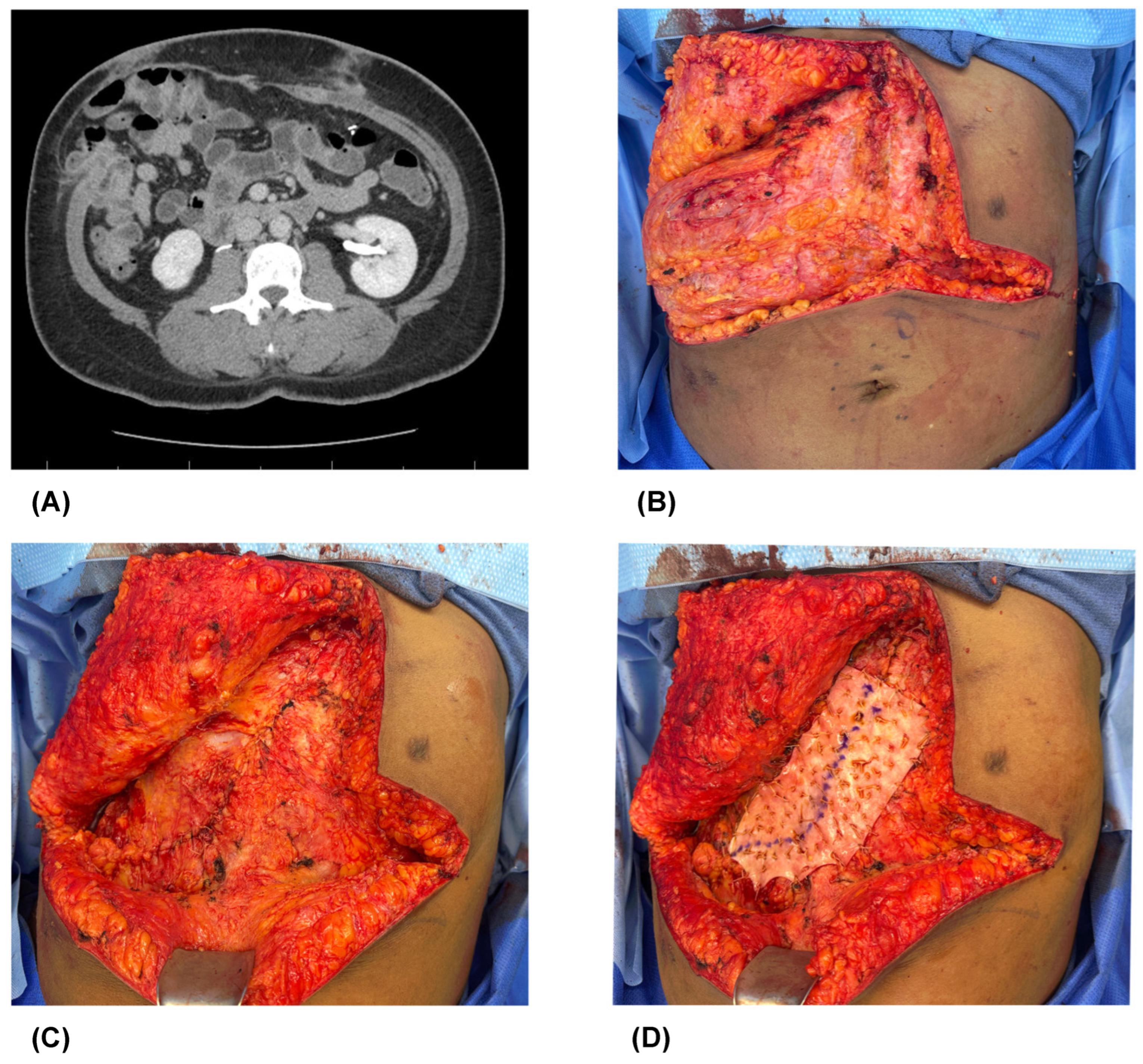
Although each repair must be tailored to the patient’s specific anatomy and surgical history, our approach follows a structured sequence. Preoperative CT scan demonstrates the extent of a typical hernia prior to reduction (
Figure 2A). A midline or subcostal incision is used to access the hernia defect, taking care to preserve viable skin flaps and minimize disruption of the subcutaneous vascular network anprovide adequate exposure of the defect (
Figure 2B). Dense adhesions from prior transplantation are released under direct visualization, and the hernia sac is circumferentially mobilized to define the true fascial edges. Meticulous hemostasis and preservation of perforating vessels are critical.
Incisional planning is a critical and often underappreciated element of abdominal wall reconstruction in the post-transplant population. Preservation of the periumbilical perforators—branches of the deep inferior epigastric artery that supply the central abdominal skin and soft tissue—is paramount, as disruption of these vessels significantly increases the risk of superficial wound complications, including skin flap necrosis, wound dehiscence, and surgical site infection [
16,
17]. In patients presenting with a vertical midline scar from prior surgical access in addition to the chevron incision, our approach utilizes the existing chevron incision to excise as much of the vertical scar and associated supra-umbilical skin excess as possible. This serves a dual purpose: it removes scarred, poorly vascularized tissue that would otherwise overlie the repair, and it reduces the distance between the periumbilical perforators and the inferior edge of the resulting wound closure. By shortening this critical distance, the skin flaps remain within the reliable perfusion territory of the periumbilical perforating vessels, minimizing the risk of distal flap ischemia and wound breakdown. This approach to incisional planning—prioritizing perforator preservation while managing scar burden and skin redundancy—is especially important in the immunosuppressed transplant population, where wound healing capacity is already compromised.
Once the fascial margins are delineated, component separation is performed to reestablish myofascial continuity (
Figure 2). The external oblique is released from the underlying internal oblique just lateral to the semilunar line, allowing for medial advancement of the rectus complex (
Figure 2C). In cases where further mobilization is required, posterior rectus sheath release or transversus abdominis release (TAR) is incorporated to achieve tension-free midline closure.
For subcostal hernias—including defects at the “angle of sorrow” where bilateral subcostal incisions meet the upper midline—several technical modifications are employed. The external oblique release is extended superolaterally along the costal margin. Mesh fixation near the costal margin and xiphoid requires suture placement through or around the costal cartilage and periosteum, as the fascial envelope becomes attenuated at the bony-fascial junction. Intraoperative Botox injection was used in 18.6% of cases to reduce postoperative muscle spasms and to reduce strain along the muscle inset.
After approximation of the native fascia, HADM is placed as an onlay reinforcement and secured with multipoint suture fixation in a progressive tension pattern, beginning centrally and extending peripherally (
Figure 2D). This technique distributes forces evenly across the repair and minimizes potential spaces where fluid could accumulate. Closed-suction drains are placed in the subcutaneous plane, and layered closure is completed to restore contour and reinforce vascularity. Drains were placed in 97.7% of encounters.
Throughout the repair, careful attention is given to maintaining perfusion and avoiding excessive tissue undermining, as these patients have limited capacity to tolerate wound complications. In select cases, limited use of adjuncts such as negative pressure wound therapy can aid in optimizing perfusion and controlling postoperative edema.
6. Results
6.1. Patient Demographics and Comorbidities
Forty patients underwent 43 abdominal wall reconstruction procedures during the study period. Three patients required two separate repairs. Patient demographics and comorbidities are presented in
Table 1. The mean age was 55.7 ± 10.2 years (median 57.5, range 31–73), and the cohort was 55.0% female. The majority were White (87.5%). Mean BMI was 31.2 ± 4.9 kg/m
2, with 60.0% classified as obese (≥30 kg/m
2). The comorbidity burden was substantial: 60.0% had pulmonary disease, 57.5% hypertension, 40.0% diabetes mellitus, 32.5% cardiovascular disease, 25.0% active cirrhosis, and 22.5% chronic kidney disease. All patients had a confirmed history of OLT and were maintained on immunosuppressive therapy.
6.2. Hernia Characteristics
Hernia characteristics are summarized in
Table 2. The majority of encounters (67.4%) involved recurrent or secondary hernias, and 41.9% had prior mesh in situ. Hernia locations included midline ventral (48.8%), subcostal region (41.9%, encompassing RUQ, LUQ, and flank defects), and combined or multi-site defects (18.6%). Incarceration or non-reducibility was documented in 34.9%. Among cases with recorded defect measurements (
n = 34), mean hernia size was 203.3 ± 184.7 cm
2 (median 158.5, range 4–700). Mesh explantation was required in 20.9% of encounters.
6.3. Operative Details
Operative details are presented in
Table 3. Mean operative time was 3.8 ± 1.2 h (median 3.6). Mean estimated blood loss was 153 ± 58 mL. Mean length of stay was 4.7 ± 1.9 days (median 4.2). Component separation was performed in all cases (100%), with bilateral rectus advancement in 67.4%, bilateral external oblique release in 58.1%, posterior release in 34.9%, and intraoperative Botox injection in 18.6%. HADM was used in 83.7% of encounters (Cortiva 72.1%, AlloDerm 11.6%). The majority of patients (88.4%) were discharged home without home health services.
6.4. Postoperative Complications
Postoperative complications are detailed in
Table 4. The overall surgical site occurrence rate was 14.0% (6/43). Seroma was the most common SSO, occurring in 9.3% of encounters and typically managed conservatively. Surgical site infection or abscess occurred in 4.7%, wound dehiscence in 2.3%, and there were no hematomas or cases of skin necrosis. Two patients (4.7%) required return to the operating room, and one (2.3%) required interventional radiology drainage. Three patients (7.0%) required ICU transfer or had a MET call activation. There were no thromboembolic events and no 30-day mortalities.
One hernia recurrence (2.3%) was identified at 37 months postoperatively. This occurred in a 64-year-old male with a BMI of 27.2 who presented to us with a recurrent hernia, prior mesh, and a 300 cm2 defect. He underwent re-repair and has had no subsequent recurrence.
The overall complication rate was 18.6% (8/43). Four patients (10.0%) died during the total follow-up period; however, no deaths occurred within 30 days of surgery, and the cause of death was not captured in this dataset.
6.5. Subgroup Analysis: Initial vs. Recurrent Hernias
A descriptive comparison of initial and recurrent hernia repairs is presented in
Table 5. Demographics, operative parameters, and blood loss were similar between groups. Patients with initial repairs had a higher SSO rate (28.6% vs. 6.9%), while the single recurrence occurred in the recurrent hernia group (3.4%). These observations are descriptive only and should be interpreted with caution, given the small subgroup sizes.
6.6. Follow-Up
Mean follow-up was 34.0 ± 27.9 months (median 31.0, range 0–108 months). Of 43 encounters, 32 (74.4%) had at least 12 months of follow-up, and 24 (55.8%) had at least 24 months. Surveillance consisted of scheduled postoperative visits in the plastic surgery clinic with physical examination; imaging was obtained when clinically indicated.
7. Discussion
Ventral hernia repair after liver transplantation represents a distinct surgical challenge. Our experience with 40 patients (43 encounters) over an 11.8-year period demonstrates that a framework integrating component separation for anatomic restoration, biologic mesh for integration and infection resistance, and multipoint fixation for tension distribution provides a reproducible and durable solution for this population. Of the 40 patients, one had a recurrence, and two had an additional hernia inferior to the umbilicus. In those cases, the additional hernias were reconstructed secondarily.
The 2.3% recurrence rate observed in our series compares favorably to published outcomes in the transplant population. Zolper et al. reported a 5.7% recurrence rate at 3 years in 35 solid organ transplant patients undergoing modified CST with biologic mesh [
10]. Black et al. reported complications in 31.6% of 19 SOT patients undergoing CST with biologic underlay [
2]. In the broader AWR literature, overall complication rates average 31–45% [
8]. Our 14.0% SSO rate and 18.6% overall complication rate fall within the favorable range of these benchmarks, though differences in cohort composition, technique, follow-up duration, and outcome definitions limit direct comparison.
Several features of our cohort underscore the complexity of these reconstructions. Nearly two-thirds of the patients presented with recurrent hernias, over 40% had prior mesh requiring management, and more than one-third were incarcerated or non-reducible. The comorbidity burden was substantial, with 60% having pulmonary disease and 40% having diabetes. Despite this complexity, zero mesh explantations were required—an important finding in an immunosuppressed population where mesh-related infections can be devastating.
7.1. Subcostal and Combined Defects
An important consideration specific to the OLT population is the high prevalence of subcostal hernias. In our series, 41.9% of defects involved the subcostal region. The junction of bilateral subcostal incisions with the upper midline—the “angle of sorrow”—presents unique challenges due to proximity to the costal margin and xiphoid process, where fascial structures become attenuated and traditional suture-based fixation is limited. Posterior release was employed in 34.9% of cases, predominantly those with subcostal or combined defects, to achieve tension-free closure when anterior release alone was insufficient. The external oblique release is extended superolaterally along the costal margin in these cases, and mesh fixation near the bony-fascial junction requires suture placement through or around the costal cartilage and periosteum.
7.2. Preoperative Considerations
Optimal outcomes in the OLT population depend on close coordination between transplant surgeons, plastic surgeons, hepatologists, and nutritionists. Abdominal wall reconstruction in this series was performed well after the transplant recovery period, once patients had achieved long-term immunosuppressive stability. All patients underwent routine nutritional screening, and only those in an adequate nutritional state proceeded to elective repair. This approach to preoperative optimization—ensuring metabolic and nutritional readiness before undertaking complex abdominal wall reconstruction—is a critical component of the multidisciplinary care model that supports durable outcomes in this population.
7.3. Limitations
This study has several limitations inherent to its retrospective, single-surgeon design. The absence of a comparator group precludes definitive conclusions about the superiority of this technique over alternative approaches such as posterior component separation with synthetic mesh or laparoscopic repair. Immunosuppression regimen details and the exact interval between transplantation and hernia repair were not consistently captured. While 74.4% of encounters had at least 12 months of follow-up, the remaining 25.6% had shorter surveillance, and patients continue to be monitored for late recurrences. The overall mortality rate of 10% during the follow-up period reflects the medical complexity of this population, though the cause of death was not related to AWR. A prospective, multicenter comparative trial would be needed to establish the optimal reconstructive strategy for this population.
8. Conclusions
Abdominal wall reconstruction after liver transplantation requires a strategy that addresses both the biological and mechanical challenges unique to this population. Our experience with 40 patients (43 encounters) demonstrates that component separation with onlay biologic mesh reinforcement and multipoint progressive tension suture fixation provides durable reconstruction, with a 2.3% recurrence rate and a 14.0% surgical site occurrence rate at a mean follow-up of 34 months. These results support the continued use of this approach in the post-OLT population and highlight the importance of multidisciplinary care and preoperative optimization in achieving favorable outcomes. Future prospective, comparative studies are needed to further define the optimal reconstructive strategy for this challenging patient population.
Author Contributions
Conceptualization, L.A., J.A. and J.I.d.l.T.; investigation, L.A., J.A. and J.I.d.l.T.; data curation, L.A.; formal analysis, L.A.; writing—original draft preparation, L.A.; writing—review and editing, L.A., J.A. and J.I.d.l.T.; visualization, L.A.; supervision, J.A. and J.I.d.l.T. All authors have read and agreed to the published version of the manuscript.
Funding
This research received no external funding.
Institutional Review Board Statement
The study was conducted in accordance with the Declaration of Helsinki and approved by the Institutional Review Board of The University of Alabama at Birmingham (IRB-300012379, 1 February 2024).
Informed Consent Statement
Patient consent was waived due to the retrospective and de-identified nature of the data. Written permission from patients was obtained for the photographs used.
Data Availability Statement
De-identified data supporting the findings of this study are available from the corresponding author upon reasonable request. Due to patient privacy considerations, individual-level data are not publicly deposited.
Conflicts of Interest
The authors declare no conflicts of interest.
Abbreviations
OLT—Orthotopic Liver Transplantation; ADM—Acellular Dermal Matrix/Matrices; HADM—Human Acellular Dermal Matrix; AWR—Abdominal Wall Reconstruction; VHR—Ventral Hernia Repair; CS—Component Separation; SSO—Surgical Site Occurrence; P4HB—Poly-4-hydroxybutyrate; TAR—Transversus Abdominis Release; EBL—Estimated Blood Loss; LOS—Length of Stay
References
- Garmpis, N.; Spartalis, E.; Schizas, D.; Patsouras, D.; Damaskos, C.; Spartalis, M.; Garmpi, A.; Nikiteas, N.I.; Dimitroulis, D. Incisional Hernias Post Liver Transplantation: Current Evidence of Epidemiology, Risk Factors and Laparoscopic Versus Open Repair. In Vivo 2019, 33, 1059–1066. [Google Scholar] [CrossRef] [PubMed]
- Black, C.K.; Zolper, E.G.; Walters, E.T.; Wang, J.; Martinez, J.; Tran, A.; Naz, I.; Kotha, V.; Kim, P.J.; Sher, S.R.; et al. Utility of a modified components separation for abdominal wall reconstruction in the liver and kidney transplant population. Arch. Plast. Surg. 2019, 46, 462–469. [Google Scholar] [CrossRef] [PubMed]
- Brunson, M.E.; Alexander, J.W. Mechanisms of transfusion-induced immunosuppression. Transfusion 1990, 30, 651–658. [Google Scholar] [CrossRef] [PubMed]
- Butler, J.R.; O’Brien, D.C.; Kays, J.K.; Ridlen, K.; Kubal, C.A.; Ekser, B.; Timsina, L.; Fridell, J.A.; Mangus, R.S.; Powelson, J.A. Postoperative renal impairment is associated with increased length of stay for incisional hernia repair after liver transplantation. Surg. Open Sci. 2019, 1, 69–73. [Google Scholar] [CrossRef] [PubMed]
- Robertson, J.D.; de la Torre, J.I.; Gardner, P.M.; Grant, J.H., III; Fix, R.J.; Vásconez, L.O. Abdominoplasty repair for abdominal wall hernias. Ann. Plast. Surg. 2003, 51, 10–16. [Google Scholar] [CrossRef] [PubMed]
- Shell, D.H., IV; de La Torre, J.; Andrades, P.; Vasconez, L.O. Open repair of ventral incisional hernias. Surg. Clin. N. Am. 2008, 88, 61–83. [Google Scholar] [CrossRef] [PubMed]
- Balla, A.; Alarcón, I.; Morales-Conde, S. Minimally invasive component separation technique for large ventral hernia: Which is the best choice? Surg. Endosc. 2020, 34, 14–30. [Google Scholar] [PubMed]
- Soto, E.; Zoog, E.; Nolte, M.D.; Fang, H.A.; de la Torre, J.I. Outcomes of Ventral Hernia Repair After Orthotopic Liver Transplant Using Component Separation and Onlay Biologic Mesh. Ann. Plast. Surg. 2023, 90, S387–S390. [Google Scholar] [CrossRef] [PubMed]
- Lindsey, J.T., Jr.; Boyd, C.J.; Davis, C.B.; Wilson, J.T.; Kurapati, S.; de la Torre, J.I. Alloderm and Cortiva Have Similar Perioperative Wound Complications in Abdominal Wall Reconstruction. J. Surg. Res. 2020, 255, 255–260. [Google Scholar] [CrossRef] [PubMed]
- Zolper, E.G.; Black, C.K.; Devulapalli, C.; Wang, J.; Mahan, M.; Kroemer, A.H.; Hawksworth, J.S.; Matsumoto, C.S.; Girlanda, R.; Fishbein, T.M.; et al. Long-term outcomes of abdominal wall reconstruction using open component separation and biologic mesh in the liver, kidney, and small bowel transplant population. Hernia 2020, 24, 469–479. [Google Scholar] [PubMed]
- Lindsey, J.T., Jr.; Boyd, C.J.; Davis, C.; Wilson, J.; Kurapati, S.; de la Torre, J.I. The Case for Onlay Biologic Mesh in Abdominal Wall Reconstruction Using Progressive Tension Suture Fixation. Ann. Plast. Surg. 2021, 86, S498–S502. [Google Scholar] [CrossRef] [PubMed]
- Denney, B.; de la Torre, J.I. Multipoint Suture Fixation Technique for Abdominal Wall Reconstruction with Component Separation and Onlay Biological Mesh Placement. Am. Surg. 2017, 83, 515–521. [Google Scholar] [CrossRef] [PubMed]
- Python Software Foundation. Python Language Reference, Version 3.12; Python Software Foundation: Wilmington, DE, USA, 2023. Available online: https://www.python.org (accessed on 9 June 2026).
- The Pandas Development Team. Pandas-Dev/Pandas: Pandas. 2024. Available online: https://zenodo.org/records/20127038 (accessed on 9 June 2026).
- Harris, C.R.; Millman, K.J.; van der Walt, S.J.; Gommers, R.; Virtanen, P.; Cournapeau, D.; Wieser, E.; Taylor, J.; Berg, S.; Smith, N.J.; et al. Array programming with NumPy. Nature 2020, 585, 357–362. [Google Scholar] [CrossRef] [PubMed]
- Saulis, A.S.; Dumanian, G.A. Periumbilical rectus abdominis perforator preservation significantly reduces superficial wound complications in “separation of parts” hernia repairs. Plast. Reconstr. Surg. 2002, 109, 2275–2280. [Google Scholar] [CrossRef] [PubMed]
- Elhage, S.A.; Marturano, M.N.; Prasad, T.; Colavita, P.D.; Kercher, K.W.; Augenstein, V.A.; Heniford, B.T. Impact of perforator sparing on anterior component separation outcomes in open abdominal wall reconstruction. Surg. Endosc. 2021, 35, 4624–4631. [Google Scholar] [PubMed]
| Disclaimer/Publisher’s Note: The statements, opinions and data contained in all publications are solely those of the individual author(s) and contributor(s) and not of MDPI and/or the editor(s). MDPI and/or the editor(s) disclaim responsibility for any injury to people or property resulting from any ideas, methods, instructions or products referred to in the content. |