Article

Predictors of Cirrhosis and Cholangiocarcinoma in Patients with Primary Sclerosing Cholangitis: An Academic Center Experience


by
Ahmad Hassan Ali
1,
Alhareth Al-Juboori
1,
Deepthi S. Rao
2,
Jamal A. Ibdah
1,
Nanda Deepa Thimmappa
3,
Ayman H. Gaballah
1 and
Ghassan M. Hammoud
1,*
1
Division of Gastroenterology and Hepatology, School of Medicine, University of Missouri, One Hospital Drive, Columbia, MO 65212, USA
2
Division of Gastrointestinal Pathology, School of Medicine, University of Missouri, Columbia, MO 65212, USA
3
Division of Diagnostic Radiology, School of Medicine, University of Missouri, Columbia, MO 65212, USA
*
Author to whom correspondence should be addressed.
Livers 2026, 6(2), 30; https://doi.org/10.3390/livers6020030
Submission received: 3 February 2026 / Revised: 30 March 2026 / Accepted: 10 April 2026 / Published: 15 April 2026

Abstract

Background and goals: The outcomes of patients with primary sclerosing cholangitis (PSC) in central Missouri are unknown. The University of Missouri–Columbia services 600,000 individuals in central Missouri. Our aims were (a) to examine the outcomes of PSC patients receiving care at our academic institution, and (b) to identify the predictors of PSC-related serious adverse events. Methods: A retrospective study of patients with PSC in a non-transplant center. The primary outcome was the development of ≥1 of PSC-related serious adverse event for (1) progression to cirrhosis, or (2) development of cholangiocarcinoma. Results: From 2000 to 2018, 42 patients fulfilled the criteria for the diagnosis of PSC. A total of 55% of the patients were male, and 79% had associated inflammatory bowel disease (IBD). The median follow-up from time of diagnosis of PSC until the last follow-up or death was 5.5 years. A total of 57% of the patients developed ≥ 1 PSC-related adverse event; 36% (8/22) of those who progressed to decompensation underwent liver transplantation. The median time from diagnosis of PSC until progression to decompensation was 6.3 years; the median time from decompensation to transplantation was 10.8 years. A total of 12% of the patients developed ≥ 1 cancer (cholangiocarcinoma = 2; gallbladder cancer = 2; colon cancer = 1; and hepatocellular carcinoma = 1). The overall mortality was 9.5%. The median time from PSC diagnosis until death was 10.2 years. A Cox hazards regression analysis showed only age (HR = 1.16; p = 0.032; 95% CI, 1.01–1.13) and serum bilirubin (HR = 1.42; p = 0.036; 95% CI, 1.03–2.69) at the time of PSC diagnosis were independently associated with PSC-related serious events. Conclusions: Age and bilirubin are important predictors of PSC-related outcomes.

1. Introduction

Primary sclerosing cholangitis (PSC) is a chronic biliary disease characterized by cholestasis and ongoing destruction of the intra- and/or extrahepatic biliary ducts, often leading to cirrhosis and its consequent complications [1]. PSC is often associated with inflammatory bowel disease (IBD), namely ulcerative colitis (UC) [2]. PSC is one of the most important risk factors for the development of cholangiocarcinoma, a lethal bile duct cancer, with a lifetime risk up to 30% [3]. Moreover, PSC is the fifth leading indication for liver transplantation in the Western population, and the leading indication for liver transplantation in some Nordic countries [4]. Furthermore, patients with PSC–IBD are at significantly higher risk for colorectal cancer, compared with patients with IBD alone. Studies have shown that colorectal cancer occurs at a much younger age in patients with PSC–IBD compared to patients with IBD alone [5].
Epidemiological studies have found the highest prevalence rates of PSC in Northern European countries and only a very few parts of North America, ranging between 3.85 and 16.2 per 100,000 persons [5]. By contrast, the reported prevalence of PSC in Southern Europe [6] and South East Asia [7]. is nearly 70 times lower.
Very little is known about the epidemiology of PSC in North America. The reported incidence and prevalence of PSC show significant variation, depending on the criteria used for the ascertainment of PSC cases, the population under assessment, and the geographical area(s) studied. The highest reported prevalence rate for PSC of 13.6 per 100,000 for age- and sex-adjusted persons was in the year 2000 in Olmsted County, Minnesota, United States [8]. Toy et al. reported a lower age-adjusted PSC prevalence of 4.15 per 100,000 in Northern California in the year 2005 [9]. In Alaska, no PSC patients were identified between the years 1984 and 2000 [10]. Only two Canadian studies have reported the incidence and prevalence of PSC in Canada [11,12]. These studies support the notion that PSC is a rare disease. Based on the studies reported, we estimate a PSC prevalence of 50,000 in the United States.
The University of Missouri–Columbia Healthcare System services 18 counties in central Missouri, with an estimated population of more than 600,000 as of the 2017 census. In this paper, we describe the clinical features, natural history, morbidity, mortality, and outcomes of patients with PSC diagnosed and seen at the University of Missouri in Columbia.

2. Materials and Methods

2.1. Study Design and Case Ascertainment

This is a retrospective chart review of all patients suspected of having PSC between the years 2000 and 2018, which was performed at our non-liver transplant academic medical center. This study was approved by the University of Missouri–Columbia Institutional Review Board (IRB #2012024). All methods related to this study were performed in accordance with the relevant guidelines and regulations. Informed consent was waived by the University of Missouri IRB because this is a retrospective chart review study. The University of Missouri i2b2 searching database was queried for a search word “sclerosing cholangitis” between the years 2000 and 2018. Patients suspected of having PSC are referred to our University liver clinic from primary care providers and local gastroenterologists. Patients with suspected/established diagnosis of PSC with high-risk features, such as those with decompensated liver disease, malignant-appearing biliary strictures, are occasionally referred by their local gastroenterologists to liver transplant centers instead of to our university clinics to facilitate their clinical care; thus, a potential for selection bias exists in our study. A list of all potentially eligible patients was created using the i2b2 database search, and all charts were manually reviewed by a hepatology fellow (A.H.A.) to confirm diagnosis of PSC and to collect all study-related data, which were stored electronically in an Excel spreadsheet. The final list was re-reviewed with the lead investigator (G.M.H.). For the purposes of this study, all abdominal MRI/MRCP exams were re-interpreted by two experienced abdominal radiologists (N.D.T. and A.H.G.). All ERCPs at our institution were performed and reviewed by one advanced interventional therapeutic endoscopist (G.M.H.). All liver biopsies were reviewed by an expert gastrointestinal pathologist (D.S.R) for diagnostic accuracy and fibrosis staging.
The diagnosis of large-duct PSC was made based on the following established criteria: (a) presence of cholestasis as evidenced by elevated serum alkaline phosphatase (ALP) of ≥6 months in the absence of obstruction of the biliary tree, and (b) cholangiographic findings consistent with PSC, or (c) histological findings upon a liver biopsy compatible with PSC [13]. Small-duct PSC was diagnosed when there was evidence of chronic cholestasis (elevated ALP > 6 months) and histological evidence of PSC but a normal cholangiogram [13]. For each subject, the following data were collected: age at time of diagnosis of PSC; sex; presence or absence of IBD (UC, Crohn’s disease (CD), or indeterminate colitis); PSC overlap with autoimmune hepatitis (AIH); magnetic resonance cholangiopancreatography (MRCP) findings; endoscopic retrograde cholangiopancreatography (ERCP) findings; laboratory parameters at the time of diagnosis of PSC (ALP; aspartate aminotransferase (AST); alanine aminotransferase (ALT); total bilirubin; albumin; hemoglobin; platelet count; prothrombin time (PT); carbohydrate antigen 19-9 (CA 19-9); carcinoembryonic antigen (CEA); perinuclear antineutrophil cytoplasmic (pANCA); total immunoglobulin G (IgG); and immunoglobulin G4 (IgG4)); endoscopic treatment (if applicable); treatment with ursodeoxycholic acid (UDCA); treatment with immunosuppressive agents (if applicable for PSC–AIH overlap); progression to cirrhosis; development of varices and ascites; liver transplantation; development of cholangiocarcinoma, hepatocellular carcinoma, colorectal cancer, and/or gallbladder cancer; and vital status at the time of the chart review.

2.2. Inclusion and Exclusion Criteria

Patients were included if they met all the following criteria: male or female age ≥ 18 years at the time of diagnosis of PSC; had large- or small-duct PSC; and had at least 6 months follow-up after the diagnosis of PSC. Patients were excluded if they met any of the following criteria: diagnosis of IgG4-related sclerosing cholangitis; concomitant liver disease (metabolic dysfunction-associated steatotic liver disease, viral hepatitis, alcoholic hepatitis, hemochromatosis, Wilson’s disease, alpha-1 antitrypsin deficiency-related liver disease, and primary biliary cholangitis); acute bacterial cholangitis; secondary sclerosing cholangitis; and those with clinical follow-up < 6 months. Patients with PSC with overlap AIH were included in the study.

2.3. Primary and Secondary Outcomes

The primary outcome of this study was the development of ≥1 of the PSC-related serious events: progression to cirrhosis (clinical signs of cirrhosis “spider angioma, gynecomastia distended abdominal wall veins, ascites, and muscle wasting”; parenchymal changes on imaging consistent with cirrhosis, presence of splenomegaly, thrombocytopenia, and/or histological findings consistent with cirrhosis) and consequent portal hypertension (manifested as development of ascites, varices, and/or hepatic encephalopathy), and the development of cholangiocarcinoma. Diagnosis of cirrhosis on histology was established using Ludwig’s histological system [14]. Secondary outcomes included the development of gallbladder cancer, colorectal cancer, hepatocellular carcinoma, and whether patients experienced either normalization or stabilization of serum ALP within the first year after the diagnosis of PSC.

2.4. Statistical Analysis

Continuous data were expressed as median with range. Categorical data were expressed as frequency and percentage. Chi-squared and Fisher’s exact tests were used to compare patients with PSC who did and did not develop ≥ 1 PSC-related serious event (i.e., progression to cirrhosis or development of cholangiocarcinoma). The Wilcoxon rank sum test was used to compare means between PSC patients who did and did not experience ≥ 1 PSC-related serious events. The Kaplan–Meier survival method was used to compare PSC patients who did and did not experience normalization of serum ALP within the first year of the diagnosis of PSC using the logrank test. The primary endpoint was the time from diagnosis of PSC to the first occurrence of any of the defined PSC-related serious events (i.e., progression to cirrhosis and liver decompensation, or development of cholangiocarcinoma), and those were treated as a failures; whereas, patients with PSC who were alive at the last known clinic follow-up, died, or underwent liver transplant during follow-up were censored. Cox proportional hazards regression was used to determine the independent predictors of PSC-related serious adverse events (i.e., progression to cirrhosis/liver decompensation, development of cholangiocarcinoma, or both). The Schoenfeld and scaled Schoenfeld residuals method was used to examine the assumption of proportionality of each predictor included in the Cox model. A non-significant p value (>0.05) indicated no violation of the assumption of the proportionality. Statistical analyses were conducted using STATA v12.1 (Stata-Corp LP, College Station, TX, USA), and graphs were created using GraphPad Prism version 9. A p-value of < 0.05 was considered statistically significant.

3. Results

3.1. Clinical and Laboratory Features of the Study Cohort

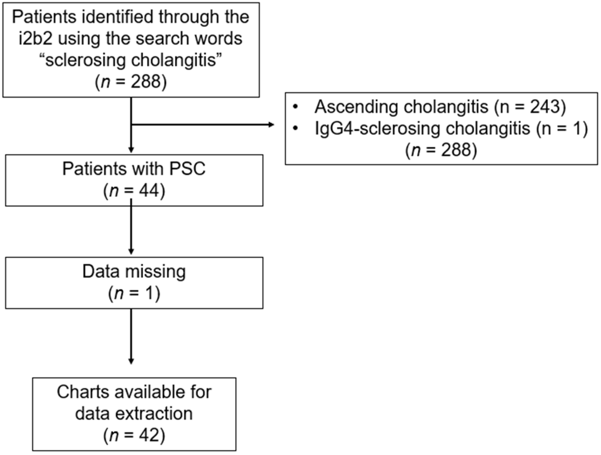
The case-finding strategy using the University of Missouri–Columbia i2b2 yielded 288 patients (Figure 1). Table 1 features the clinical and laboratory characteristics of the study cohort.
Between the years 2000 and 2018, 42 patients (39 Caucasians and 3 African Americans) fulfilled the diagnostic criteria for PSC and followed up at the University of Missouri–Columbia. The median age at the time of the PSC diagnosis was 36 years (range: 7–84 years), and 55% (23/42) were male. Moreover, 79% (33/42) had concomitant IBD; UC was the most commonly-associated IBD in this cohort (50% (21/42) had UC, 19% (8/42) had CD, and 10% (4/42) had indeterminate colitis). The diagnosis of IBD preceded the diagnosis of PSC in the majority of patients; 85% (28/33) of patients were diagnosed with IBD before PSC, and IBD was detected by screening in five patients. Only one patient presented with abdominal pain and bloody diarrhea in conjunction with a mixed pattern of elevated liver chemistries, and diagnosis of PSC and IBD had been made simultaneously. The median follow-up of this cohort from the time of diagnosis of PSC until the last known clinic follow-up or death was 5.5 years (range: 0.4–24.3 years).
With respect to the diagnosis of PSC, 88% (37/42) of the subjects had MRCPs, of whom 95% (35/37) had MR findings consistent with PSC; the other five patients were diagnosed with PSC on ERCP. A total of 62% (26/42) of patients underwent ERCPs, and evidence of PSC was found in all ERCPs, except in one patient. Indications for ERCP included cholestasis of unclear etiology in 74% (31/42), clinical features suspicious for acute cholangitis in 7% (3/42), and/or biliary stricture and/or dilatation or dominant stricture on MRCP in 19% (8/42) of patients. Intrahepatic PSC was more frequent than extrahepatic PSC, 93% (39/42) vs. 50% (21/42), respectively. No case of small-duct PSC in this cohort was found. A total of 43% (18/42) of the subjects had a liver biopsy, and 89% (16/18) had histological findings compatible with PSC; no patient had cirrhotic-stage PSC on histopathology. Upon a microscopic examination of liver biopsies from patients with PSC, the classic lesion is well-established periductal fibrosis, which was seen in a subset of cases, while others demonstrated rather subtle features of bile duct injury and/or loss of bile ducts (i.e., ductopenia), given that PSC can be a focal liver disease. These histological findings, in conjunction with the radiological features noted on the cholangiographic studies, were supportive of the diagnosis of PSC.

3.2. Clinical and Laboratory Features of Patients with PSC–AIH Overlap

PSC–AIH overlap syndrome was not uncommon. Of those who had undergone evaluation by liver biopsy, 33% (6/18) had histological findings compatible with PSC–AIH overlap syndrome; thus, the prevalence of PSC–AIH overlap in this cohort was 14% (6/42). As for the examination of liver biopsies from patients with PSC–AIH overlap syndrome, portal inflammation with numerous plasma cells and expanded fibrous tissue, portal edema, prominent interface hepatitis, and foci of lobular necroinflammatory activity, along with varying degrees of bile duct injury were noted. These histological findings are typical of liver biopsies from patients with PSC–AIH overlap syndrome. All patients presented with non-cirrhotic stage PSC, except for three patients.
Table 2 illustrates a comparison between the PSC only and the PSC–AIH groups. Compared to patients with PSC only, those with PSC–AIH were younger (median age: 41 vs. 30 years, respectively, p = 0.04). Further, far more patients with PSC–AIH were treated with UDCA compared to those with PSC only (83% (5/6) vs. 22% (8/36), respectively; p = 0.007). Moreover, the liver biochemistry was worse in the PSC–AIH group compared to the PSC only group. Specifically, the median ALP, AST, and ALT were found to be significantly higher in patients with PSC–AIH vs. those with PSC only (481 vs. 246 U/L, respectively, p = 0.01; 132 vs. 51 U/L, respectively, p = 0.04; and 151 vs. 49 U/L, respectively, p = 0.029).
Regarding progression to cirrhosis, the PSC–AIH overlap group had a worse clinical course compared to the PSC only group. The rate of PSC-related serious adverse events tended to be higher in the PSC–AIH overlap group compared to the PSC only group (83% (5/6) vs. 53% (19/36), respectively; p = 0.17). The rate of progression to cirrhosis and the development of esophageal varices tended to be higher in the PSC–AIH overlap group compared to the PSC only group (83% (5/6) vs. 47% (17/36), respectively, p = 0.12; and 83% (5/6) vs. 39% (14/36), respectively, p = 0.056). The rate of liver transplantation tended to be higher in the PSC–AIH overlap group compared to the PSC only group (33% (2/6) vs. 17% (6/36), respectively, p = 0.32). All malignancies occurred exclusively in the PSC only group.

3.3. Treatment

A total of 31% (13/42) of patients had exposure to ursodeoxycholic acid (UDCA; median duration of treatment 2 years) at a median dose of 20 mg/kg/body weight. Subjects with PSC–AIH overlap syndrome were treated with tapering courses of prednisone during acute flares, usually 40–60 mg per day for 1 week, followed by reduction by 10 mg every week until 10 mg daily as maintenance, and with azathioprine at a dose of 50–100 mg per day as maintenance therapy. A total of 36% (15/42) of patients developed biliary strictures with a worsening of serum hepatic biochemistries requiring endoscopic intervention and biliary stenting.

3.4. Biochemical Course

Of the entire cohort, 21% (9/42) experienced either normalization (n = 8) or stabilization (n = 1) of serum ALP within the first year of a diagnosis of PSC. Of those who experienced normalization of serum ALP, only two patients were on UDCA for a median of 8 months at a dose of 20 mg/kg/day.

3.5. Outcomes

Table 3 illustrates a comparison between patients who did and did not experience a PSC-related serious adverse event during the follow-up period. A total of 57% (24/42) of patients developed ≥ 1 serious PSC-related event, 52% (22/42) progressed to cirrhotic-stage PSC, and 12% (5/42) developed hepatobiliary and/or colorectal cancer. The median time from diagnosis of PSC until development of a PSC-related serious adverse event was 5 years (range: 1–21 years). Moreover, 86% (19/22) of those who progressed to cirrhosis developed clinically significant portal hypertension manifested as esophageal, gastric varices and ascites in 59% (13/22). No patient developed spontaneous bacterial peritonitis. The time from PSC diagnosis until progression to cirrhosis and liver decompensation was 6.3 years (range: 0.9–21.2 years). Of those who progressed to cirrhosis, 36% (8/22) underwent liver transplantation due to end-stage liver disease; the time from progression to cirrhosis and liver decompensation until undergoing liver transplantation was 10.8 years (range: 1.8–15.4 years), and the time from undergoing liver transplantation until the last known clinic follow-up was 1.7 years (range: 0.5–13 years). Of those who underwent liver transplantation, 12% (1/8) developed recurrent PSC in the liver allograft 3.2 years after liver transplantation. No patient had cholangiocarcinoma or hepatocellular carcinoma in their liver explant. With respect to IBD-related events, 9% (3/33) of patients with IBD underwent colectomy, two patients for IBD refractory to medical treatment, and one due to detection of low-grade dysplasia on surveillance colonoscopy. None of the patients who underwent colectomy had colorectal cancer on surgical specimen.

3.6. Hepatobiliary and Colorectal Malignancy

A total of 12% (5/42) of patients developed hepatobiliary and/or colorectal cancers during the follow-up (median time from PSC diagnosis until development of malignancy was 6 years; range: 1–21 years; Table 4). Two patients developed intrahepatic cholangiocarcinoma (4 and 8 years after diagnosis of PSC, respectively). Both underwent partial hepatectomy and had no locoregional metastasis. Both patients had no evidence of cholangiocarcinoma recurrence until their last clinic visit (8 and 11 years after diagnosis of cholangiocarcinoma, respectively). One patient developed perihilar cholangiocarcinoma 2.6 years after PSC diagnosis and underwent neoadjuvant chemotherapy and radiation, followed by orthotopic liver transplantation. This patient developed recurrent cholangiocarcinoma in the transplanted liver 7 months after liver transplantation. This patient was alive at the time of the last clinic visit (4 months after diagnosis of recurrent cholangiocarcinoma).
Two patients developed gallbladder adenocarcinoma; both patients were undergoing routine cancer surveillance, and had gallbladder polyps that increased in size over a 1-year period. Both patients underwent open cholecystectomy and histology confirmed gallbladder adenocarcinoma. No patient had gallbladder cancer metastases to regional organs/lymph nodes, and none received chemotherapy. Both patients had no evidence of gallbladder cancer recurrence at the time of their last clinic follow-up (8 and 10 years, respectively). One patient progressed to cirrhosis and developed hepatocellular carcinoma, and underwent liver transplantation for cirrhosis complicated by hepatocellular carcinoma; he had no metastases intraoperatively, and had no evidence of recurrent at the time of his last clinic visit (4.6 years after diagnosis of hepatocellular carcinoma, and 3.7 years after liver transplantation).
Two patients developed colon cancer in the setting of long-standing UC (time from diagnosis of PSC until diagnosis of colorectal cancer was 10 and 14 years, respectively). Of these two patients, one underwent routine surveillance for colorectal cancer; he had high-grade dysplasia on a colonoscopy-obtained colonic mucosa biopsy. He underwent subtotal colectomy and had stage I colorectal cancer; there was no evidence of tumor in the 20 lymph nodes resected during colectomy. This patient had no evidence of recurrent colorectal cancer 21 years after diagnosis of colorectal cancer. The other patient had not undergone surveillance for colorectal cancer in the setting of PSC–UC. He was diagnosed with metastatic colon cancer 8 years after diagnosis of PSC, and died 6 months after diagnosis of colon cancer.

3.7. Survival Modeling and Predictors of PSC-Related Serious Adverse Events

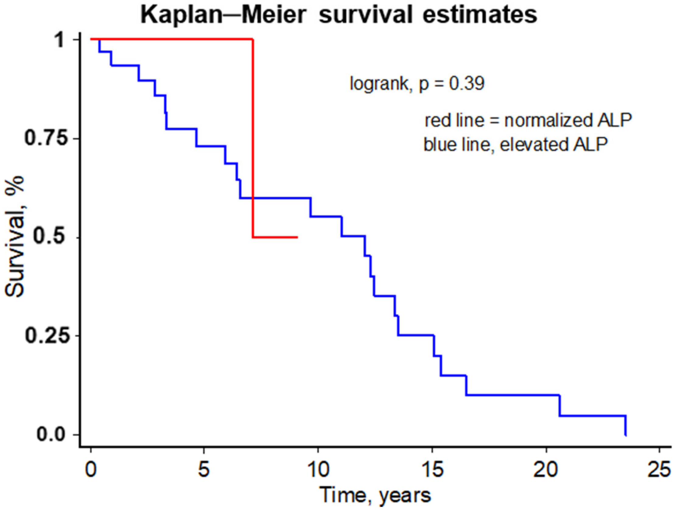
We examined whether patients who experienced normalization of serum ALP had better PSC-related event-free survival (i.e., progression to cirrhosis or development of cholangiocarcinoma). Using Kaplan–Meier survival modeling (Figure 2), those patients with PSC who experienced normalization of serum ALP within one year after diagnosis of PSC tended to have better 2- and 5-year PSC-related event-free survival compared with those who did not experience normalization of serum ALP within 1 year after the diagnosis of PSC; this difference was not statistically significant (100% vs. 93.5%, and 100% vs. 73%, respectively; p = 0.39). Using Cox hazards regression analysis (age at the time of diagnosis of PSC; presence/absence of IBD; serum ALP, AST, ALT, and total bilirubin at the time of diagnosis of PSC), only age at the time of diagnosis of PSC (hazards ratio (HR) = 1.16; p = 0.032; 95% CI, 1.01–1.13) and serum bilirubin at baseline (HR = 1.42; p = 0.036; 95% CI, 1.03–2.69) were independently associated with a high risk for the development of a PSC-related serious event (i.e., progression to cirrhosis and development of cholangiocarcinoma).

4. Discussion

In this retrospective study, we report on the clinical features and outcomes of patients with PSC seen at the University of Missouri–Columbia between the years 2000 and 2018. The University of Missouri health system services many residents living in central Missouri, with an estimated population of more than 600,000 as of the 2017 census. This study highlights the devastating outcomes of patients with PSC [15]. In a North American, non-transplant academic cohort, and as reported in previous studies, our study shows that advanced age at the time of diagnosis of PSC and serum bilirubin at the time of diagnosis of PSC are important predictors of PSC-related serious adverse outcomes [16,17,18,19]. Furthermore, 19% of patients experienced normalization of serum ALP within the first year of diagnosis of PSC. Moreover, although the difference was not statistically significant, patients with PSC who experienced normalization of serum ALP within the first year of diagnosis of PSC tended to have a better 5- and 10-year survival, compared to those who had persistently elevated ALP; this finding is in agreement with previous studies [20,21,22,23,24] and has important implications with regards to prognosis, patient counseling and, more importantly, in designing therapeutic clinical trials in PSC [25].
PSC continues to be an important cause of morbidity and mortality. More than one-half of our PSC cohort developed a serious, life-altering PSC-related event. The majority of patients progressed to end-stage liver disease and developed significant portal hypertension manifested as varices and/or ascites. Moreover, one-third of those who progressed to end-stage liver disease underwent liver transplantation. The recurrence rate of PSC in the liver allograft was 12% in our cohort, which is comparable to prior reports [26].
In addition to the burden of progression to end-stage liver disease, PSC is an important risk factor for hepatobiliary and colorectal cancer [27]. Twelve percent of this cohort developed hepatobiliary and/or colorectal cancers. These findings highlight the impact of PSC on patients’ quality of lives and outcomes. Surveillance for colorectal cancer and hepatobiliary cancers in patients with PSC has been shown to be associated with better survival rates [28,29]. Currently, the leading societies recommend screening for colorectal cancer in patients with PSC–IBD and screening for gallbladder cancer in all PSC patients on an annual basis [13,30,31]. Moreover, the leading societies recommend surveillance for hepatocellular carcinoma once diagnosis of cirrhosis is established. However, the surveillance for cholangiocarcinoma remains a subject of debate [32,33].
Determining the incidence and prevalence of PSC in the geographical area served by our academic institution is challenging. Although our university health system services residents of 18 counties in central Missouri, we see a small fraction of all patients at our institution, and many patients have access to other tertiary and academic centers in other surrounding states. To provide incidence and prevalence rates for a disease, the sample size should be large enough to include many (if not all) cases to be representative of the population under study. With rare diseases, such as PSC, this is even more challenging, because cases are more difficult to find. For the current study, we used only one research database for case finding; thus, it is possible that we could have missed many other cases of PSC, which would preclude examining the epidemiology of PSC in the geographical area served by our institution.
There are limitations to our study worth noting. The sample size is small; therefore, overfitting of the survival model is a limitation, and therefore, caution should be exercised when interpreting our results. Our study included almost exclusively Caucasians (mirroring the racial make-up of central Missouri, United States); thus, our study data may not apply to non-Caucasians. Dividing the adverse events related to PSC to decompensation-related vs. malignancy-related events in terms of survival models and cumulative incidence function analysis is the appropriate/ideal statistical method; however, in our study, the number of adverse events is too small to categorize the adverse events. Further, due to the nature of our academic center (non-transplant), capturing all data related to outcomes of PSC in all patients is not feasible, since the care of the majority of patients with PSC at our institute is transferred to liver transplant institutes once they progress to advanced liver disease stages, develop cholangiocarcinoma, recurrent bacterial cholangitis, etc., and therefore, missing outcomes of interest is possible. Moreover, since our center is a non-liver transplant institute, referral bias is a limitation, and our data/results may not necessarily apply to patients with PSC who are candidates for liver transplant. Finally, the analysis of survival difference between those who experienced ALP normalization vs. those who had persistently increased ALP was underpowered due to the small number of patients in the former group (n = 8).

5. Conclusions

In conclusion, we report the clinical outcomes of patients with PSC between the years 2000 and 2018 at our institution that serves a large population of central Missouri. The natural history of PSC in central Missouri is similar to the natural history studies reported in other regions. More than one-half of the PSC cohort suffered from serious PSC-related events (progression to cirrhosis or development of cholangiocarcinoma). Similar to prior studies, age and serum bilirubin at the time of diagnosis of PSC were found to be important predictors of PSC-related adverse events. Larger studies are needed to better define the epidemiology and outcomes of patients with PSC. Even though it extends the survival of patients with PSC, liver transplantation is fraught by its costs, invasive nature, shortage of organs and, more importantly, recurrence of PSC in the liver allograft. Effective therapies are urgently needed for the treatment of PSC.

Author Contributions

A.H.A. presented and conceptualized the research paper and performed the statistical analysis; A.A.-J. assisted in drafting the manuscript; D.S.R. interpreted liver biopsies and contributed in drafting the manuscript; J.A.I. reviewed the manuscript and provided critical points; N.D.T. and A.H.G. interpreted cross-sectional imaging and contributed to drafting the final manuscript; G.M.H. conceptualized the research paper, provided critical points in the statistical analysis, re-interpreted all available endoscopic retrograde cholangiopancreatography (when applicable), and contributed to drafting the initial and final manuscript. All authors have read and agreed to the published version of the manuscript.

Funding

This research received no external funding.

Institutional Review Board Statement

This study was conducted in accordance with the Declaration of Helsinki, and approved by the Institutional Review Board (or Ethics Committee) of the University of Missouri School of Medicine (IRB protocol # 2012024; date of approval: 25 July 2018).

Informed Consent Statement

Informed consent for this study was waived by the Institutional Review Board of the University of Missouri-Columbia (IRB protocol # 2012024; date of approval: 25 July 2018).

Data Availability Statement

The original contributions presented in this study are included in the article, and further inquiries can be directed to the corresponding author.

Conflicts of Interest

The authors declare no conflicts of interest.

References

  1. Ali, A.H.; Carey, E.J.; Lindor, K.D. Current research on the treatment of primary sclerosing cholangitis. Intractable Rare Dis. Res. 2015, 4, 1–6. [Google Scholar] [CrossRef] [Scilit]
  2. Schrumpf, E.; Elgjo, K.; Fausa, O.; Gjone, E.; Kolmannskog, F.; Ritland, S. Sclerosing cholangitis in ulcerative colitis. Scand. J. Gastroenterol. 1980, 15, 689–697. [Google Scholar] [CrossRef] [Scilit] [PubMed]
  3. Razumilava, N.; Gores, G.J. Cholangiocarcinoma. Lancet 2014, 383, 2168–2179. [Google Scholar] [CrossRef] [Scilit] [PubMed]
  4. Brandsaeter, B.; Broome, U.; Isoniemi, H.; Friman, S.; Hansen, B.; Schrumpf, E.; Oksanen, A.; Ericzon, B.-G.; Höckerstedt, K.; Mäkisalo, H.; et al. Liver transplantation for primary sclerosing cholangitis in the Nordic countries: Outcome after acceptance to the waiting list. Liver Transplant. 2003, 9, 961–969. [Google Scholar] [CrossRef] [Scilit] [PubMed]
  5. Boonstra, K.; Beuers, U.; Ponsioen, C.Y. Epidemiology of primary sclerosing cholangitis and primary biliary cirrhosis: A systematic review. J. Hepatol. 2012, 56, 1181–1188. [Google Scholar] [CrossRef] [Scilit]
  6. Escorsell, A.; Pares, A.; Rodes, J.; Solis-Herruzo, J.A.; Miras, M.; de la Morena, E.; Spanish Association for the Study of the Liver. Epidemiology of primary sclerosing cholangitis in Spain. J. Hepatol. 1994, 21, 787–791. [Google Scholar]
  7. Ang, T.L.; Fock, K.M.; Ng, T.M.; Teo, E.K.; Chua, T.S.; Tan, J.Y. Clinical profile of primary sclerosing cholangitis in Singapore. J. Gastroenterol. Hepatol. 2002, 17, 908–913. [Google Scholar] [CrossRef] [Scilit]
  8. Bambha, K.; Kim, W.R.; Talwalkar, J.; Torgerson, H.; Benson, J.T.; Therneau, T.M.; Loftus, E.V., Jr.; Yawn, B.P.; Dickson, E.R.; Melton, L.J., 3rd. Incidence, clinical spectrum, and outcomes of primary sclerosing cholangitis in a United States community. Gastroenterology 2003, 125, 1364–1369. [Google Scholar] [CrossRef] [Scilit]
  9. Toy, E.; Balasubramanian, S.; Selmi, C.; Li, C.S.; Bowlus, C.L. The prevalence, incidence and natural history of primary sclerosing cholangitis in an ethnically diverse population. BMC Gastroenterol. 2011, 11, 83. [Google Scholar] [CrossRef] [Scilit]
  10. Hurlburt, K.J.; McMahon, B.J.; Deubner, H.; Hsu-Trawinski, B.; Williams, J.L.; Kowdley, K.V. Prevalence of autoimmune liver disease in Alaska Natives. Am. J. Gastroenterol. 2002, 97, 2402–2407. [Google Scholar] [CrossRef] [Scilit]
  11. Byron, D.; Minuk, G.Y. Clinical hepatology: Profile of an urban, hospital-based practice. Hepatology 1996, 24, 813–815. [Google Scholar] [CrossRef] [Scilit] [PubMed]
  12. Kaplan, G.G.; Laupland, K.B.; Butzner, D.; Urbanski, S.J.; Lee, S.S. The burden of large and small duct primary sclerosing cholangitis in adults and children: A population-based analysis. Am. J. Gastroenterol. 2007, 102, 1042–1049. [Google Scholar] [CrossRef] [Scilit] [PubMed]
  13. Chapman, R.; Fevery, J.; Kalloo, A.; Nagorney, D.M.; Boberg, K.M.; Shneider, B.; Gores, G.J.; American Association for the Study of Liver Diseases. Diagnosis and management of primary sclerosing cholangitis. Hepatology 2010, 51, 660–678. [Google Scholar] [CrossRef] [Scilit] [PubMed]
  14. Ludwig, J.; Dickson, E.R.; McDonald, G.S. Staging of chronic nonsuppurative destructive cholangitis (syndrome of primary biliary cirrhosis). Virchows Arch. A Pathol. Anat. Histol. 1978, 379, 103–112. [Google Scholar] [CrossRef] [Scilit]
  15. Karlsen, T.H.; Boberg, K.M. Update on primary sclerosing cholangitis. J. Hepatol. 2013, 59, 571–582. [Google Scholar] [CrossRef] [Scilit]
  16. Broome, U.; Olsson, R.; Loof, L.; Bodemar, G.; Hultcrantz, R.; Danielsson, A.; Prytz, H.; Sandberg-Gertzén, H.; Wallerstedt, S.; Lindberg, G. Natural history and prognostic factors in 305 Swedish patients with primary sclerosing cholangitis. Gut 1996, 38, 610–615. [Google Scholar] [CrossRef] [Scilit]
  17. Wiesner, R.H.; Grambsch, P.M.; Dickson, E.R.; Ludwig, J.; MacCarty, R.L.; Hunter, E.B.; Fleming, T.R.; Fisher, L.D.; Beaver, S.J.; LaRusso, N.F. Primary sclerosing cholangitis: Natural history, prognostic factors and survival analysis. Hepatology 1989, 10, 430–436. [Google Scholar] [CrossRef] [Scilit]
  18. Weismuller, T.J.; Trivedi, P.J.; Bergquist, A.; Imam, M.; Lenzen, H.; Ponsioen, C.Y.; Holm, K.; Gotthardt, D.; Färkkilä, M.A.; Marschall, H.-U.; et al. Patient Age, Sex, and Inflammatory Bowel Disease Phenotype Associate with Course of Primary Sclerosing Cholangitis. Gastroenterology 2017, 152, 1975–1984e8. [Google Scholar] [CrossRef] [Scilit]
  19. Goode, E.C.; Clark, A.B.; Mells, G.F.; Srivastava, B.; Spiess, K.; Gelson, W.T.H.; Trivedi, P.J.; Lynch, K.D.; Castren, E.; Vesterhus, M.N.; et al. Factors Associated with Outcomes of Patients with Primary Sclerosing Cholangitis and Development and Validation of a Risk Scoring System. Hepatology 2019, 69, 2120–2135. [Google Scholar] [CrossRef] [Scilit]
  20. Stanich, P.P.; Bjornsson, E.; Gossard, A.A.; Enders, F.; Jorgensen, R.; Lindor, K.D. Alkaline phosphatase normalization is associated with better prognosis in primary sclerosing cholangitis. Dig. Liver Dis. 2011, 43, 309–313. [Google Scholar] [CrossRef] [Scilit]
  21. Lindstrom, L.; Hultcrantz, R.; Boberg, K.M.; Friis-Liby, I.; Bergquist, A. Association between reduced levels of alkaline phosphatase and survival times of patients with primary sclerosing cholangitis. Clin. Gastroenterol. Hepatol. 2013, 11, 841–846. [Google Scholar] [CrossRef] [Scilit] [PubMed]
  22. Al Mamari, S.; Djordjevic, J.; Halliday, J.S.; Chapman, R.W. Improvement of serum alkaline phosphatase to <1.5 upper limit of normal predicts better outcome and reduced risk of cholangiocarcinoma in primary sclerosing cholangitis. J. Hepatol. 2013, 58, 329–334. [Google Scholar] [CrossRef] [Scilit] [PubMed]
  23. Ali, A.H.; Hilscher, M.; Stanich, P.P.; Carey, E.; Lindor, K.; Tabibian, J.H. Association Between Baseline Serum Alkaline Phosphatase, Normalization of Alkaline Phosphatase, and Colorectal Cancer Risk in Primary Sclerosing Cholangitis. In Digestive Disease Week 2016; Gastroenterology American Gastroenterology Association: San Diego, CA, USA, 2016; p. S1057. [Google Scholar]
  24. Rupp, C.; Rossler, A.; Halibasic, E.; Sauer, P.; Weiss, K.H.; Friedrich, K.; Wannhoff, A.; Stiehl, A.; Stremmel, W.; Trauner, M.; et al. Reduction in alkaline phosphatase is associated with longer survival in primary sclerosing cholangitis, independent of dominant stenosis. Aliment. Pharmacol. Ther. 2014, 40, 1292–1301. [Google Scholar] [CrossRef] [Scilit] [PubMed]
  25. Ali, A.H.; Tabibian, J.H.; Lindor, K.D. Update on pharmacotherapies for cholestatic liver disease. Hepatol. Commun. 2017, 1, 7–17. [Google Scholar] [CrossRef] [Scilit]
  26. Gautam, M.; Cheruvattath, R.; Balan, V. Recurrence of autoimmune liver disease after liver transplantation: A systematic review. Liver Transplant. 2006, 12, 1813–1824. [Google Scholar] [CrossRef] [Scilit]
  27. Tabibian, J.H.; Ali, A.H.; Lindor, K.D. Primary Sclerosing Cholangitis, Part 2: Cancer Risk, Prevention, and Surveillance. Gastroenterol. Hepatol. 2018, 14, 427–432. [Google Scholar]
  28. Boonstra, K.; Weersma, R.K.; van Erpecum, K.J.; Rauws, E.A.; Spanier, B.W.; Poen, A.C.; van Nieuwkerk, K.M.; Drenth, J.P.; Witteman, B.J.; Tuynman, H.A.; et al. Population-based epidemiology, malignancy risk, and outcome of primary sclerosing cholangitis. Hepatology 2013, 58, 2045–2055. [Google Scholar] [CrossRef] [Scilit]
  29. Ali, A.H.; Tabibian, J.H.; Nasser-Ghodsi, N.; Lennon, R.J.; DeLeon, T.; Borad, M.J.; Hilscher, M.; Silveira, M.G.; Carey, E.J.; Lindor, K.D. Surveillance for hepatobiliary cancers in patients with primary sclerosing cholangitis. Hepatology 2018, 67, 2338–2351. [Google Scholar] [CrossRef] [Scilit]
  30. Lindor, K.D.; Kowdley, K.V.; Harrison, M.E.; American College of Gastroenterology. ACG Clinical Guideline: Primary Sclerosing Cholangitis. Am. J. Gastroenterol. 2015, 110, 646–659; quiz 60. [Google Scholar] [CrossRef] [Scilit]
  31. European Association for the Study of the Liver. EASL Clinical Practice Guidelines: Management of cholestatic liver diseases. J. Hepatol. 2009, 51, 237–267. [Google Scholar] [CrossRef] [Scilit]
  32. Folseraas, T.; Boberg, K.M. Cancer Risk and Surveillance in Primary Sclerosing Cholangitis. Clin. Liver Dis. 2016, 20, 79–98. [Google Scholar] [CrossRef] [Scilit]
  33. Razumilava, N.; Gores, G.J. Surveillance for Cholangiocarcinoma in Patients with Primary Sclerosing Cholangitis: Effective and Justified? Clin. Liver Dis. 2016, 8, 43–47. [Google Scholar] [CrossRef] [Scilit]
Figure 1. Flow chart of case ascertainment at the University of Missouri.
Livers 06 00030 g001
Figure 2. Kaplan–Meier survival free of a PSC-related serious adverse event, defined as progression to cirrhosis or diagnosis of cholangiocarcinoma, in patients who experienced ALP normalization (red line) compared to those who had persistent elevation of ALP (blue line).
Livers 06 00030 g002
Table 1. Clinical and laboratory features of the study population (n = 42) at the time of diagnosis of PSC (between the years 2000 to 2018).
Clinical and Laboratory Variables Descriptive Statistics;
Median (Range)
Age at the time of diagnosis of PSC, years 36 (7–84)
Male sex, n (%)23 (55%)
IBD, yes, n (%)33 (79%)
Follow-up until last known clinic follow-up or death, years5.5 (0.4–24.3)
PSC–AIH overlap, n (%)6 (14%)
Treated with UDCA, n (%)13 (31%)
Alkaline phosphatase (40–129 U/L)264 (51–2574)
Aspartate aminotransferase (0–30 U/L)54 (15–276)
Alanine aminotransferase (10–32 U/L)55 (14–691)
Total bilirubin (0.1–1 mg/dL)0.9 (0.1–5.9)
Total immunoglobulin (760–1590 mg/dL)1388 (709–2998)
Prothrombin time (13.0–15.3 s) 14.1 (11.9–17.4)
Hemoglobin (12–15.5 g/dL)11.9 (7.2–16.1)
Platelet count (150–450 × 109/L)225 (50–691)
Abbreviations: AIH, autoimmune hepatitis; IBD, inflammatory bowel disease.
Table 2. Clinical and laboratory features of the study population (n = 42) comparing those with PSC and those with PSC–AIH overlap.
Clinical and Laboratory Variables PSC
(n = 36)
PSC–AIH
(n = 6)
p Value
Age at the time of diagnosis of PSC, years 41 (15–71)30 (7–84)0.04
Male sex, n (%)20 (55%)3 (50%)0.57
IBD, yes, n (%)30 (83%)3 (50%)0.1
Follow-up until last known clinic follow-up or death, years4.8 (0.2–24.3)5.3 (0.8–20.6)0.83
Treated with UDCA, n (%)8 (22%)5 (83%)0.007
Alkaline phosphatase (40–129 U/L)246 (51–2574)481 (387–795)0.01
Aspartate aminotransferase (0–30 U/L)51 (15–276)132 (30–172)0.04
Alanine aminotransferase (10–32 U/L)49 (14–691)151 (79–216)0.029
Total bilirubin (0.1–1 mg/dL)0.9 (0.1–5.2)1.8 (0.3–5.9)0.24
Total immunoglobulin (760–1590 mg/dL)1385 (709–2670)1805 (1196–2998)0.31
Prothrombin time (13.0–15.3 s) 12 (12.1–13)13 (15–17.4)0.46
Hemoglobin (12–15.5 g/dL)12.8 (11.5–15.2)15 (12.6–17.4)0.08
Platelet count (150–450 × 109/L)260 (185–382)250 (50–691)0.14
PSC-related serious adverse events, yes, n (%)19 (53%)5 (83%)0.17
Cirrhosis, yes, n (%)17 (47%)5 (83%)0.12
Varices, yes, n (%)14 (39%)5 (83%)0.056
Cholangiocarcinoma, yes, n (%)3 (8.3%)00.62
Gallbladder cancer, yes, n (%)2 (5.6%)00.73
Hepatocellular carcinoma, yes, n (%)1 (3%)00.86
Colon cancer, yes, n (%)2 (5.6%)00.73
Liver transplant, yes, n (%)6 (17%)2 (33%)0.32
Abbreviations: AIH, autoimmune hepatitis; IBD, inflammatory bowel disease.
Table 3. Clinical and laboratory features of the study population (n = 42) at the time of diagnosis of PSC (between the years 2000 and 2018) categorized based on experiencing a PSC-related serious adverse event.
Clinical and Laboratory Variables No PSC-Related Serious
Adverse
Event (n = 18)
Experienced a PSC-Related Serious Adverse
Event (n = 24)
p Value
Age at the time of diagnosis of PSC, years 24 (12–84)46 (7–72)0.001
Male sex, n (%)11 (61%)12 (50%)0.34
IBD, yes, n (%)15 (83%)18 (75%)0.39
Follow-up until last known clinic follow-up or death, years3.1 (0.2–10.6)10.3 (0.3–24.3)0.001
PSC–AIH overlap, n (%)1 (6%)5 (21%)0.17
Treated with UDCA, n (%)7 (39%)6 (25%)0.27
Alkaline phosphatase (40–129 U/L)270 (74–846)264 (51–2574)0.86
Aspartate aminotransferase (0–30 U/L)64 (15–276)51 (19–172)0.95
Alanine aminotransferase (10–32 U/L)109 (14–691)49 (15–216)0.18
Total bilirubin (0.1–1 mg/dL)0.7 (0.1–1.9)1.2 (0.3–5.9)0.01
Total immunoglobulin (760–1590 mg/dL)1311 (709–2670)1484 (1090–2998)0.28
Prothrombin time (13.0–15.3 s) 11.9 (11.9–16.8)12.3 (13–17.4)0.36
Hemoglobin (12–15.5 g/dL)14.2 (13.1–15.5)13.1 (12.5–14.1)0.58
Platelet count (150–450 × 109/L)256 (312–460)200 (50–691)0.75
Abbreviations: AIH, autoimmune hepatitis; IBD, inflammatory bowel disease
Table 4. Patients with PSC who developed hepatobiliary and/or colorectal cancer during the follow-up period.
Case Type of CancerTiming (from PSC Diagnosis to Cancer Diagnosis)StageTreatmentOutcome
1Intrahepatic cholangiocarcinoma4 yearsT1b, N0, M0Partial hepatectomyNo evidence of recurrence at 8 years
2 *Intrahepatic cholangiocarcinoma8 yearsT1a, N0, M0Partial hepatectomyNo evidence of recurrence at 11 years
2 *Gallbladder cancer 3.5 yearsT1b, N0, M0Open cholecystectomy No recurrence at 10 years
2 *Colon cancer10 yearsT2, N0, M0Colectomy No evidence of recurrence at 21 years
3 Perihilar cholangiocarcinoma2.6 years Early stageLiver transplant §Recurrence in liver allograft at 7 months
3 Gallbladder cancer6 years T1b, N0, M0Open cholecystectomyNo recurrence at 8 years
4Colon cancer14 years Metastatic Palliative Died 6 months after diagnosis of colon cancer
5Hepatocellular carcinoma11 years Within Milan criteria Liver transplant No recurrence 3.7 years after liver transplantation
* Same patient. Same patient. § Patient underwent liver transplant per the Mayo Clinic protocol for liver transplant for perihilar cholangiocarcinoma. Liver transplant was performed at a liver transplant center; no access to the records to determine TNM staging.
Disclaimer/Publisher’s Note: The statements, opinions and data contained in all publications are solely those of the individual author(s) and contributor(s) and not of MDPI and/or the editor(s). MDPI and/or the editor(s) disclaim responsibility for any injury to people or property resulting from any ideas, methods, instructions or products referred to in the content.