Review

Liver Cirrhosis: Evolving Definitions, and Recent Advances in Diagnosis, Prevention and Management

1
Academy Unit, Nguma-Bada Campus, College of Medicine and Dentistry, James Cook University, Smithfield, QLD 4878, Australia
2
Faculty of Medicine, Northern Clinical School, The University of Queensland, The Prince Charles Hospital, Chermside, QLD 4032, Australia
3
Blacktown Mt Druitt Clinical School and Research Centre, School of Medicine, Western Sydney University, Blacktown, NSW 2148, Australia
4
Department of Gastroenterology, Blacktown Mount Druitt Hospital, Western Sydney Local Health District, Blacktown, NSW 2148, Australia
5
Storr Liver Centre, The Westmead Institute for Medical Research, Westmead, NSW 2145, Australia
*
Author to whom correspondence should be addressed.
Livers 2025, 5(3), 28; https://doi.org/10.3390/livers5030028
Submission received: 22 April 2025 / Revised: 3 June 2025 / Accepted: 20 June 2025 / Published: 25 June 2025

Abstract

Liver cirrhosis poses major challenges for both individual health and global healthcare systems. Recent studies have challenged the traditional and predictable linear course of cirrhosis, demonstrating marked heterogeneity in the patterns of the first decompensating events. This review presents an updated epidemiology of cirrhosis and its main causes, outlines an overview of the clinical features, and explores the evolving concepts of the spectrum of decompensation. It further delineates recent advancements in the diagnosis, prognostic scoring, and management of decompensated cirrhosis and the subsequent clinically challenging complications of portal hypertension. Emerging innovations in non-invasive imaging, diagnostic serum biomarkers, and etiology-specific therapies, together with the development of novel liver support systems, underscore a paradigm shift toward a multimodal approach for cirrhosis care. Furthermore, the integration of precision medicine into clinical practice holds promise for reshaping the future of liver cirrhosis management in the coming decades.

Graphical Abstract

1. Liver Cirrhosis—Causes, Pathogenesis, Diagnosis, Natural History, Clinical Complications, and Prognostic Scores

Liver disease and its sequelae, such as liver cirrhosis, liver failure, and hepatocellular carcinoma (HCC), continue to pose a significant global public health problem, causing over two million deaths annually. This represents one in every 25 deaths [1], with liver cirrhosis remaining the predominant contributor to liver-related mortality globally [1,2,3]. Cirrhosis, first described by Hippocrates in the fifth century BCE, is the result of several etiologies causing chronic necroinflammation, followed by diffuse hepatic fibrosis [4,5]. Histologically, it is characterized by the disruption of the normal hepatic architecture, replaced by widespread nodular regeneration surrounded by fibrous dense septa with ensuing parenchymal extinction and distortion of hepatic architecture, collectively leading to pronounced distortion of the hepatic vascular architecture. Subsequently, this collapse leads to elevated resistance to portal blood flow and, therefore, to hepatic failure and portal hypertension. Although chronic liver inflammation does not invariably culminate in cirrhosis, when it does, the interval to progression can span from weeks to several decades [6].
Traditionally, cirrhosis has been dichotomized into a compensated and decompensated stage. In its early stages, cirrhosis is typically compensated, with most patients remaining asymptomatic [7]. The initial asymptomatic phase of cirrhosis is typically followed by a relatively brief symptomatic period, which may last for several months to a few years. The symptomatic (initial) phase—commonly designated as decompensated cirrhosis—is characterized by the onset of multiple complications, which result in a substantial increase of morbidity and mortality, resulting in frequent hospitalization, as well as impaired quality of life of patients and caregivers alike [8,9]. Complications such as the first occurrence of ascites, gastroesophageal variceal bleeding, hepatic encephalopathy (HE), or, in some individuals, non-obstructive jaundice (increased bilirubin concentration) herald the onset of decompensated cirrhosis. Acute decompensation (AD) of cirrhosis is often defined as the acute development of gastrointestinal hemorrhage, ascites, HE, susceptibility to bacterial and fungal infections, or any combination thereof, requiring hospitalization [10].
Decompensation represents a prognostic watershed, resulting in high short-term mortality and a median survival of 2 years, compared to 12 years in the compensated phase [11].
Relative to the general population, patients with compensated cirrhosis have a fivefold increase in mortality risk, whereas those with decompensated cirrhosis exhibit a tenfold increased risk [11,12]. Acute-on-chronic liver failure (ACLF) is the most severe form of acute decompensation, characterized by organ failure, and is associated with high short-term mortality [13]. In patients with decompensated cirrhosis, most deaths can be attributed to hepatic and extrahepatic organ failure [6,14]. Conversely, during the compensated stage, mortality is primarily associated with cardiovascular disease, renal disease, and malignant conditions [6].
Although cirrhosis is associated with significant morbidity and mortality, it remains under-recognized and garners less public awareness than other chronic conditions, including heart failure, chronic kidney disease, diabetes mellitus, and chronic obstructive pulmonary disease. This disparity can be attributed, in part, to the stigmatization surrounding cirrhosis and the perception that it is primarily a consequence of alcohol use [15,16].

1.1. Novel Concepts of Decompensation of Liver Cirrhosis

For decades, it has been widely accepted that the occurrence of clinical complications related to portal hypertension and impairment of hepatic function, such as ascites, HE, jaundice, and gastrointestinal bleeding, demarcates the transition from the compensated to the decompensated stage of cirrhosis. This transition represents a stratification variable with a deep negative impact on the prognosis of patients with cirrhosis [17]. However, this binary concept has been widely challenged. Recent findings suggest that patients with AD are quite heterogeneous, and the traditional definition of AD oversimplifies the clinical course of the disease, which comprises various heterogeneous phenotypic–prognostic subgroups (Figure 1) [18]. One of the most recent landmark studies is the CANONIC trial [19]. This study identified a specific subgroup of patients with AD characterized by severe systemic inflammation, organ failure, and mitochondrial dysfunction and proposed a redefinition of AD as a clinical state that may predispose to the development of acute-on-chronic liver failure (ACLF), in accordance with the EASL-CLIF criteria. Consequently, these novel concepts placed emphasis on the acute nature and dynamics of the decompensated state rather than on the nature of the decompensating event itself. AD was later reaffirmed in the PREDICT and ACLARA studies [10,20]. The heterogeneity of the AD definition remains a heated doctrinal debate in hepatology. The novel element of this debate is the focus on the significance of the pathway by which decompensation takes place and the rate of complication onset. Recently, experts have proposed distinctive trajectories of decompensation, such as non-acute decompensation (NAD) and AD, mainly by shifting the emphasis to the contexts precipitating the first episode of decompensation and the heterogeneous presentation patterns in decompensated cirrhosis [18]. A recent study by Verma et al. has elucidated that NAD is clinically, prognostically, and pathophysiologically distinct from compensated cirrhosis and AD [21]. NADs have a 12-month mortality rate of 18.3% when compared with a mortality rate of 68.8% in those with AD. NAD, who demonstrated similar markers of systemic inflammation with compensated cirrhosis and lower than those observed in AD. Furthermore, NAD exhibits both apoptotic and non-apoptotic cell death, with levels significantly higher than those observed in compensated cirrhosis and markedly lower than in AD. Predictors of progression from NAD to AD include severe ascites, elevated bilirubin, Gasdermin D, and receptor-interacting serine/threonine protein kinase 3, along with low insulin-like growth factor-1. Effective management of AD or prophylactic therapies to prevent progression from NAD to AD can result in clinical stabilization and, in some cases, may even lead to recompensation and reversal of ACLF.

1.2. Causes of Cirrhosis

The etiological factors of cirrhosis can be found in Table 1. The presence of multiple causative factors in a single patient can accelerate the progression to cirrhosis. In addition, the underlying etiology may impact the comorbidities associated with cirrhosis. For instance, metabolic syndrome is more common in patients with MASLD. Patients with cryptogenic cirrhosis typically have evidence of more active fibrosis and a higher risk of liver-related clinical events, while having similar demographics to the MASH spectrum.

1.3. Burden of Liver Cirrhosis

Annually, liver disease accounts for approximately two million deaths globally—about one million from cirrhosis and one million from HCC and viral hepatitis. Females account for approximately one-third of all liver-related deaths [1]. The estimated mortality associated with cirrhosis worldwide is 1,472,000 in 2019 [22].
As of 2023, cirrhosis ranks as the tenth leading cause of mortality in Africa, ninth in both Southeast Asia and Europe, fifth in the Eastern Mediterranean region, and twelfth in North America [1]. In Australia, cirrhosis and other liver diseases ranked as the 20th leading cause of death according to the Australian Bureau of Statistics in 2023 [23].
The impact of cirrhosis varies significantly across different populations, including geographic regions, genders, races and ethnicities, and socioeconomic strata. Moreover, the burden of cirrhosis has evolved considerably over time [2,22,24]. Cirrhosis prevalence is difficult to assess and is possibly underestimated because the initial stages are asymptomatic and the disorder is often undiagnosed. The global age-standardized death rate in 2019 was 18 deaths per 100,000 population [22]. The estimated ASDR varies substantially across countries, ranging from 3.3 deaths per 100,000 population in Singapore to 126.7 per 100,000 in Egypt, as reported in 2019 [25].
Liver cirrhosis ranks as the 15th most common cause of death globally, contributing significantly to mortalities and disability-adjusted life years (DALYs) [26]. Cirrhosis ranks as the seventh leading cause of DALYs among individuals aged 50–74, the twelfth among those aged 25–49, and the fifteenth across all age groups [22].
Age-standardized cirrhosis deaths peak in Eastern Sub-Saharan Africa, reflecting the region’s high hepatitis B and C prevalence (44.15 [38.47–51.91] per 100,000 population) and are the lowest in Australasia (5.48 [5.05–5.93] deaths per 100,000 population in 2019) [27]. According to the WHO, viral hepatitis accounted for approximately 1.3 million deaths in 2024, a toll comparable to tuberculosis and malaria combined [28,29].
Expanded HBV vaccination programs and enhanced access to efficacious antivirals contributed to the reduction in global age-standardized mortality rates associated with HBV-induced cirrhosis [30,31]. Likewise, the advent of safe and effective DAAs in 2015 has marked a paradigm shift in HCV treatment. However, the full extent of the impact of direct-acting antivirals (DAAs) on the global prevalence of HCV-related cirrhosis remains to be fully elucidated [32,33].
Globally, 2.3 billion people are active drinkers of alcohol [34,35,36]. Total alcohol per capita consumption in the world’s population over 15 years of age rose from 5.5 liters of pure alcohol in 2005 to 6.4 liters in 2016 [34]. Alcohol is now the leading global cause of cirrhosis, where 60% of cases in high-income regions such as Europe, North America, and Latin America can be attributed to alcohol [37,38]. Approximately one-third of patients with alcohol use disorder (AUD) will develop ALD [38]. Of note, AUD is reported to be more prevalent in high-income countries, while it is likely underdiagnosed and underreported in low-income countries [34]. ALD can co-exist with other causes of liver disease, including viral hepatitis and metabolic dysfunction-associated steatotic liver disease (MASLD). MASLD, also known as metabolic-associated fatty liver disease (MAFLD), and formerly known as non-alcoholic fatty liver disease (NAFLD), is the second leading cause of end-stage liver disease, impacting a quarter of the world’s population [39,40]. It also ranks as the second most common cause for liver transplantation overall and is the leading cause among females [1]. However, the prevalence of MASLD around the world varies widely due to numerous differences in ethnicity, geographic regions, genetic factors, and lifestyle factors [41]. Worldwide, the percentage of total deaths from all causes attributable to MASLD has increased from (0.8–0.14%) to 0.17% (0.13–0.23%) [1].
In 2022, approximately 2.5 billion adults globally were overweight, and 537 million were living with diabetes—populations that are all at increased risk for MASLD [42,43]. Due to the global rise in metabolic risk factors and an aging population, it is anticipated that the prevalence of MASLD will more than double between 2016 and 2030 [44]. Importantly, metabolic risk factors among children and adolescents represent a significant and growing threat to global health over the next decades [44]. The prevalence of overweight and obesity among the Indigenous adult population in Australia is higher across all age groups, placing them at greater risk of cardiometabolic conditions and fatty liver disease [45,46].
MASLD includes a spectrum of conditions ranging from isolated hepatic steatosis (metabolic dysfunction-associated steatotic liver, MASL) and metabolic dysfunction-associated steatohepatitis (MASH) to more advanced stages such as fibrosis and cirrhosis. In addition to MASLD, the category of steatotic liver disease (SLD) also encompasses MASLD cases with moderate alcohol intake (MetALD), alcohol-related liver disease (ALD), specific etiologies such as drug-induced liver injury (DILI) and monogenic disorders, and cryptogenic SLD [47].
The prevalence of primary sclerosing cholangitis (PSC) and inflammatory bowel disease (IBD) is notably higher in high-income, industrialized countries [48,49]. Consequently, most epidemiological studies on PSC are predominantly derived from Western nations. PSC is a risk factor for gallbladder, cholangiocarcinoma, and colorectal cancers, contributing to premature death [50]. Overall, magnetic resonance cholangiopancreatography (MRCP)–based studies estimate the prevalence of PSC among patients with IBD to be approximately 8%. Notably, around 70% of all PSC cases are associated with underlying IBD, with ulcerative colitis representing nearly 80% of these cases [49,51].
Like PSC, primary biliary cholangitis appears to be more common in the Western population. The incidence rates of primary biliary cholangitis (PBC) vary from 0.33 to 5.8 cases per 100,000 population annually, while the prevalence rates range from 1.91 to 40.2 cases per 100,000 population [52].
Autoimmune hepatitis occurs in all races, ages, and ethnic groups [53]. Females are more affected than males (4:1). Compared to age- and sex-matched controls, affected patients have approximately a 1.5-fold higher risk of developing various malignancies, including HCC, colorectal cancer, lymphoma, and non-melanoma skin cancers [54,55].
In 2017, an estimated 112 million patients worldwide were living with compensated cirrhosis, while 10.6 million had progressed to decompensated cirrhosis [22]. Compared to the general population, patients with compensated cirrhosis have a mortality risk approximately five times higher than that of the general population, whereas those with decompensated cirrhosis have a tenfold increase in mortality risk [56]. The reported overall survival rates for patients with compensated and decompensated cirrhosis are 87% versus 75% at one year and 67% versus 45% at five years, respectively [56].
Epidemiological data reveal an increasing prevalence of decompensated cirrhosis. Prior studies have demonstrated that transition to decompensated cirrhosis from compensated cirrhosis was at an annual rate ranging from 5% to 12%, but this rate varies by the underlying disease etiology [11,57,58,59]. According to the Global Burden of Disease (GBD) Study 2017, the global prevalence of decompensated cirrhosis rose from 5.2 million in 1990 to 10.6 million in 2017. This increase was accompanied by an increase in the age-standardized prevalence rate, from 110.6 to 132.5 per 100,000 population over the same period. In 2017, the etiology of decompensated cirrhosis was attributed to the hepatitis B virus (28%), hepatitis C virus (25%), alcohol-related liver disease (23%), non-alcoholic fatty liver disease (9%), and other less common etiologies (16%) [22].
Acute-on-chronic liver failure (ACLF) is a frequent and serious complication among hospitalized patients with liver cirrhosis, which constitutes the most severe clinical manifestation of AD [13]. Over the past few decades, various definitions of ACLF have emerged from different regions, including the European Association for the Study of the Liver—Chronic Liver Failure (EASL-CLIF) criteria, the NASCELD definition in North America, and the APASL criteria in East Asia. As a result, epidemiological data on ACLF are inconsistent and challenging to compare [60]. A meta-analysis of 30 studies reported that the global prevalence of ACLF—based on the EASL-CLIF criteria—among patients hospitalized for decompensated cirrhosis is approximately 35% [61]. Alcohol-related liver disease was identified as the leading underlying cause in 45% of ACLF cases worldwide. Additionally, the global 90-day mortality rate for ACLF was estimated at 58% [61].
The Australia and New Zealand Liver Transplant Registry (ANZLTR) data highlight important trends in the etiologies of chronic liver disease (CLD) leading to liver transplantation (LT) [62,63]. Notably, the proportion of LT cases due to alcohol-related liver disease has stabilized and has recently surpassed that for the hepatitis C virus (HCV), which has declined significantly due to the introduction of direct-acting antivirals. Similarly, cases of LT related to the hepatitis B virus (HBV) have also decreased, which is attributed to improved antiviral therapies and universal neonatal vaccination programs in Australia. In contrast, the incidence of LT for NAFLD has risen considerably, making it the third most common cause of CLD requiring transplantation [62,63]. This trend is mirrored in the rising cases of hepatocellular carcinoma (HCC) associated with MASLD, reflecting the broader public health issues of obesity and metabolic syndrome in Australasia. The increase in HCC cases underscores a significant shift in the landscape of liver disease in the region.

1.4. Pathophysiology of Liver Cirrhosis

The progression from chronic liver disease to cirrhosis involves a series of maladaptive responses to chronic liver injury: inflammation, activation of hepatic stellate cells, and subsequent fibrogenesis and angiogenesis, alongside parenchymal extinction lesions resulting from microthrombi and vascular occlusion [64,65,66]. This sequence of events leads to significant microvascular changes in the liver, marked by sinusoidal remodeling, which includes extracellular matrix deposition from proliferating activated hepatic stellate cells, resulting in the loss of fenestrae, leading to the capillarization of hepatic sinusoids [67]. Moreover, this process promotes the formation of intrahepatic shunts, nodules of fibrosis, and hepatic endothelial dysfunction.
These histological abnormalities associated with cirrhosis disrupt the hepatic angioarchitecture, resulting in increased resistance to portal blood flow, which is a primum movens in the development of portal hypertension [68,69,70,71]. Moreover, disruption in the balance between intrahepatic vasoconstrictors and vasodilators results in predominant vasoconstriction, contributing to a dynamic, functional component of hepatic resistance that may lead to rapid changes in portal pressure [72,73]. Nitric oxide is the most extensively studied vasoactive agent in the context of hepatic endothelial dysfunction. In cirrhotic livers, sinusoidal endothelial cells exhibit impaired nitric oxide production, primarily due to decreased activity of endothelial nitric oxide synthase. This reduction is attributed to insufficient protein kinase B-dependent phosphorylation, a lack of essential cofactors, elevated oxidative stress leading to increased nitric oxide scavenging, and elevated levels of endogenous nitric oxide inhibitors [74]. Acute events, such as infections, may further suppress nitric oxide levels. This reduction in nitric oxide exacerbates hepatic resistance, thereby contributing to elevated portal pressure [75]. Concomitantly, the production of vasoconstrictors—primarily driven by androgenic stimulation and thromboxane A2—is elevated, alongside activation of the renin–angiotensin system, antidiuretic hormone, and endothelin-1, all of which contribute to a further restriction of sinusoidal blood flow.
The initial elevation in portal pressure, driven by increased intrahepatic vascular resistance, leads to circulatory disturbances, most notably the development of splanchnic arterial vasodilation [75]. In contrast to what happens in hepatic circulation, the production of nitric oxide by endothelial cells is amplified in splanchnic circulation as a result of vascular shear stress initially and later by disease exacerbation caused by bacterial translocation and sustained inflammatory response typical for advanced cirrhosis [75,76,77,78]. Vasodilation within the splanchnic capillaries and arterioles increases portal venous inflow. When combined with elevated intrahepatic vascular resistance, this leads to a rise in portal pressure, culminating in the development of portal hypertension. Since the splanchnic vascular bed comprises about 25% of the total systemic vascular resistance, persistent splanchnic vasodilation decreases the effective arterial blood volume, leading to systemic hypotension and arterial underfilling. This, in turn, triggers the activation of neurohumoral vasoconstrictor systems, including the renin–angiotensin–aldosterone system, the sympathetic nervous system, and non-osmotic vasopressin secretion. These systems aim to counteract vasodilation, leading to sodium and water retention and an increase in plasma volume, predisposing patients to ascites, hyponatremia, kidney injury, infection, or hemorrhages [79]. Some of this excess plasma volume accumulates in the peritoneal cavity as ascites due to portal hypertension. Increased sinusoidal pressure induces ascites from increased lymph production, which extravasates into the peritoneum when the lymphatic drainage capacity is exceeded. As cirrhosis progresses, vasodilation intensifies, and systemic blood pressure continues to decline, with a maximal activation of vasoconstrictors. This cascade leads to marked vasoconstriction within the renal circulation, which can progress to hepatorenal syndrome, a form of acute kidney injury.
Systemic vasodilation can also contribute to pulmonary ventilation/perfusion mismatch, which, in severe cases, may result in hepatopulmonary syndrome (HPS) with arterial hypoxemia or portopulmonary hypertension due to increased pulmonary vasoconstriction. The expansion of plasma volume increases cardiac output, contributing to a hyperdynamic circulatory state [80]. This, combined with splanchnic vasodilation, augments portal venous inflow and exacerbates portal hypertension. The elevated portal pressure leads to a reversal in blood flow and, subsequently, the dilation of existing collateral channels at anatomical sites where the systemic and portal circulations intersect, such as the gastro-esophageal junction, and activates angiogenesis, which facilitates the formation of new collateral vessels, for which vascular endothelial growth factor (VEGF)-driven angiogenesis plays an important role [66].The most clinically relevant portosystemic collaterals are gastroesophageal varices. Variceal bleeding occurs when the intravariceal pressure surpasses the elastic capacity of the vessel wall. The risk of variceal bleeding is directly related to increased wall tension, which is influenced by portal pressure, variceal diameter, and the thinness of the variceal wall. Dilatation of the gastric mucosal vessels contributes to the development of portal hypertensive gastropathy. Moreover, the presence of portosystemic shunts, in conjunction with progressive hepatic dysfunction, plays a central role in the pathogenesis of HE by declining the first-pass metabolism of orally administered drugs, impairing endothelial function, and reducing the hepatic clearance of gut-derived ammonia [81]. While the mechanisms are not fully understood, the presence of hepatic fibrosis alongside liver injury from inflammation contributes to genetic and epigenetic alterations that can lead to a progression into malignancy and the development of HCC.
Pathophysiological evidence has long delineated portal hypertension with splanchnic and systemic vasodilation and a hyperdynamic circulatory state, as a central mechanism in the development of AD [82,83]. Portal hypertension (PH) is defined as increased pressure within the portal vein. It is due to a rise in the hepatic venous pressure gradient (HVPG) due to increased intrahepatic vascular resistance and impaired hepatic sinusoidal circulation. PH, most frequently arising from CLD, is an important determinant of its disease course and prognosis. Clinically significant portal hypertension (CSPH) is a major milestone in the natural history of CLD. It is defined as an increase in HVPG to ≥10 mmHg. Above this threshold, the complications of portal hypertension might emerge [84].
Recent findings have integrated the concept of systemic inflammation—evidenced by the translocation of gut microbiota components, elevated oxidative stress levels, and increased circulating pro-inflammatory cytokines and chemokines—into the classical paradigms of AD [85,86,87]. The bidirectional interaction between the gut and liver highlights the critical role of the gastrointestinal microbiome in the pathogenesis and progression of chronic liver disease, as well as in triggering decompensation events [83,88,89]. Bile acids and liver-derived antimicrobial peptides play a critical role in regulating and shaping the composition and function of the gastrointestinal microbiota. Conversely, the portal vein serves as the primary conduit for the transport of gut-derived metabolites and microbial products to the liver. Among the principal etiological factors of chronic liver disease, alcohol consumption and dietary patterns not only induce direct local hepatic injury, triggering the release of damage-associated molecular patterns (DAMPs), but also contribute to the gut’s microbial dysbiosis and increased gut permeability. This disruption facilitates the translocation of pathogen-associated molecular patterns (PAMPs) into the portal venous circulation, further perpetuating hepatic inflammation. In end-stage liver disease, these factors are often exacerbated by increased bacterial translocation and a diminished hepatic capacity to clear microbial products [90]. Bacterial translocation is facilitated by delayed intestinal transit, bacterial overgrowth, and increased gut permeability in the context of altered gut microbiota function and composition [91,92,93].
A growing body of evidence has emerged suggesting that cirrhosis is associated with alterations in gut microbiota composition, most notably marked by a loss of genetic diversity, a decline in autochthonous species, and an overrepresentation of potentially pathogenic and uncommon taxa like Enterococcus species [92]. These alterations worsen as cirrhosis progresses. Though mechanisms linking microbiota changes to disease progression are not fully understood, one hypothesis proposes that these alterations may compromise microbiota function, resulting in intestinal inflammation, disruption of the epithelial barrier, and increased intestinal permeability, thereby further aggravating bacterial translocation. The enrichment of pathogenic species may also lead to elevated endotoxemia, causing an increased systemic inflammation. This cascade of events can be attributed to the onset of circulatory dysfunction and directly promotes the progression of multi-organ dysfunction and failure.

1.5. Diagnosing Cirrhosis

Chronic liver disease is asymptomatic until the onset of cirrhosis accompanied by clinical decompensation [94]. Clinical decompensation events include ascites, variceal bleeding, HE, sepsis, and non-obstructive jaundice. Consequently, the diagnostic evaluation of patients suspected of having cirrhosis is contingent upon the disease phase. In individuals with the compensated phase of cirrhosis, the objectives are to quantify the extent of hepatic fibrosis, evaluate the presence and severity of portal hypertension, and elucidate the underlying etiology of the liver disease. These factors are linked to potential complications of cirrhosis and help guide the necessary follow-up care.
A thorough medical history and physical examination remain essential for identifying patients with, or at risk of, cirrhosis. The prevalence of muscle cramps can reach 64% in all patients with cirrhosis, pruritus (32%), poor-quality of sleep (63%), or sexual dysfunction (53%) [95]. Risk factors such as alcohol use or diabetes and symptoms experienced in cirrhosis are neither specific nor sensitive, but some may offer a specificity greater than 90%, including Terry’s nails (highly specific but insensitive marker—white discoloration, absent lunula, dark pink at tip), palmar erythema (a symmetrical, reddish discoloration of the palms, particularly over the thenar and hypothenar eminences), caput medusa (distended and engorged paraumbilical veins radiating from the umbilicus across the abdomen), gynecomastia, facial telangiectasia, spider angiomas, decreased body hair, jaundice, and testicular atrophy [96]. Clubbing of fingers can be observed in the case of concomitant HPS. Dupuytren’s contracture, which affects primarily the fourth and fifth fingers and may present in males over 60 years of age of Northern European ancestry, is more commonly a consequence of chronic excessive alcohol consumption rather than an indicator of cirrhosis [97]. Alcoholism and alcohol-related liver disease are frequently cited as predisposing factors for sialadenitis and parotid enlargement [98].
An evaluation of liver fibrosis is essential for identifying patients at risk of developing cirrhosis. Liver fibrosis is typically categorized into four stages, each representing a progressive increase in severity. Stages 3 and 4 fibrosis, the latter being classified as cirrhosis, are strongly correlated with subsequent liver-related morbidity and mortality [99,100,101,102]. Therefore, these stages represent critical points for timely intervention aimed at preventing further disease progression.
Demonstrating irregular nodular liver through imaging by ultrasonography, CT, or MRI in conjunction with impaired hepatic synthetic function is sufficient for the diagnosis of cirrhosis. Other findings can be detected, such as shrunken liver, evidence of portosystemic collaterals, and splenomegaly. Differential diagnoses include nodular regenerative hyperplasia, nodules but no fibrosis, congenital hepatic fibrosis, and non-cirrhotic portal hypertension. However, conventional imaging may yield a false-negative diagnosis of cirrhosis. Non-invasive liver disease assessments (NILDA) [also known as non-invasive testing (NIT)] are increasingly utilized. Several indices combining various markers are now available to assess the degree of fibrosis. They include direct and indirect serum blood markers (panels) and imaging modalities or a combination of these testing modalities (Table 2 and Table 3) [103,104,105]. These tests are inexpensive and simpler to follow longitudinally than liver biopsy. The most commonly utilized serologic tests to detect direct signs of liver fibrosis and dysfunction include thrombocytopenia, indicating reduced platelet production and splenic sequestration, and an elevated ratio of AST to ALT. Indices such as FIB-4 are widely accepted for risk stratification in patients with either MASLD or ALD, classifying the score as low (<1.30), intermediate (1.30–2.67), and high (>2.67) [106]. Age influences the FIB-4 score, with the values increasing as patients age. For individuals over 65 years, the low-risk threshold is adjusted to ≤2.0, while the high-risk threshold (>2.67) remains unchanged [107]. Cut-offs of less than 1.45 and greater than 3.25 have been developed for HCV. Risk stratification using the FIB-4 index is recommended by societal guidelines for individuals with diagnosed MASLD or those with risk factors such as obesity or diabetes mellitus. A FIB-4 score below 1.3 is associated with a negative likelihood ratio of 0.4 for advanced fibrosis, effectively identifying patients at low risk [108]. Suggested cut-off values to rule out or in liver fibrosis for FIB-4, NAFLD fibrosis score, ELF, transient elastography (TE) and AST/ALT ratio can be found in Table 3 Combination strategies may include the combination of FibroScan and ultrasonography conducted simultaneously, fibrotest and FibroScan also conducted simultaneously (liver biopsy can be obtained if discordant on fibrosis classification), and FibroScan and fibrometer also conducted simultaneously and whereby the results are introduced into computer for assessment of fibrosis severity. APRI and fibrotest can be conducted sequentially, whereas MEFIB combines magnetic resonance elastography (MRE) with the FIB-4 index undertaken separately [109]. Likewise, the FibroScan-AST (FAST) and ADAPT is a composite score that includes age, diabetes status, PRO-C3 (a marker of collagen formation), and platelet count to assess liver fibrosis [110].
The Baveno VII consensus, convened in October 2021, reinforced the central role of non-invasive tools—particularly transient elastography (TE)—in the assessment of clinically significant portal hypertension (CSPH) [9]. The term “compensated advanced chronic liver disease” (cACLD) was coined to represent the spectrum of severe fibrosis and cirrhosis in individuals with chronic liver disease.
A liver biopsy is the gold standard for the assessment of liver fibrosis. However, it is seldom needed, and the current indication is to determine the etiology of liver disease in cases of uncertainty and not to stage fibrosis. The transjugular approach yields samples of similar quality to percutaneous ones but further provides diagnostic information by measurement of hepatic-vein pressure gradient (HVPG) [111].
Table 2. Commonly used tests for diagnosis of cirrhosis.
(A) Imaging-based non-invasive liver disease assessment of hepatic fibrosis, steatosis, and portal hypertension
Imaging ModalityComponentsAetiology of Liver DiseaseEvidenceComments
UltrasonographyHepatic nodularity, signs of portal hypertensionAllWell-validated
  • Sensitivity is low in early stages of cirrhosis
CT/MRIHepatic nodularity, signs of portal hypertensionAllWell-validated
  • Sensitivity is low in early stages of cirrhosis
Transient elastography (FibroScan)Liver stiffness measurementAllWell-validated but exact cut-offs for specific fibrosis stages and causes not established.
  • Failure rate, <5–15%, reason: High BMI (M probe) ascites
  • Operator-dependent; variability can occur based on technician experience
Acoustic radiation force impulse imagingLiver stiffness measurementAllModerate validation in single etiology CLD with histology as reference standard
  • Failure rate, <5–15%, reason: High BMI (M probe) ascites
Magnetic resonance elastography (MRE)Liver stiffness measurementAllLimited validation in single etiology CLD with histology as reference standard
  • High cost, limited availability
  • Failure rate < 5% mainly due to liver iron deposition, large ascites, very high BMI, 3T (for 2D gradient recalled echo)
  • Expensive and may not be available in all institutions
  • Time-consuming procedure compared to other tests
  • Requires specialized equipment and trained personnel
(B) Blood-based biomarker algorithms for fibrosis
Indirect serum non-invasive fibrosis testsComponentEtiology of liver diseaseModel algorithmyearComments
Fibroindex [112]Indirect markers:
AST, platelets, gamma globulin
HCV1.738 − 0.064 (platelet [×104/mm3]) + 0/005 (AST IU/L) + 0.463 (gamma globulin [g/Dl])2007
  • Population Specific: Primarily validated in limited patient groups, reducing generalizability.
  • Sensitivity Issues: Poor detection of early fibrosis (F0–F1).
  • Comorbidity Influence: Affected by conditions like obesity and diabetes.
  • Laboratory Variability: Results can differ based on measurement methods.
  • Limited Scope: Assesses fibrosis only, not liver function.
King’s Score [113]Indirect markers: AST, INR, platelets. Clinical variable: AgeHCVAge × AST × INR/[platelet count (109/L)]2009
  • Population Specific: Primarily validated in specific patient populations, limiting broader applicability
  • Sensitivity Issues: May not accurately detect mild fibrosis (F0–F1)
  • Influence of Comorbidities: Conditions such as obesity and diabetes can affect the score
  • Biochemical Variability: Results may vary based on different laboratory practices
  • Limited Insight: Focuses solely on fibrosis assessment, not liver function, or other pathologies
APRI [114]Indirect markers: AST, plateletsHBV, HCV[(AST level/ULN)/platelet count (109/L)] × 1002003
  • Muscle injury leading to inaccurate results (elevated AST)
  • May not effectively detect mild fibrosis (F0–F1)
Fibrosis-4 Index
(FIB-4) [106]
Indirect markers: AST, ALT, platelets; clinical variable: ageHBV, HCV, MASLDAge (y) × AST (U/L)
Platelet count (109/L) × √ALT (U/L)
2006
  • Validated mainly in hepatitis C, less accurate for other liver diseases
  • Older patients may have higher scores
  • Less effective in differentiating between significant fibrosis (F3) and cirrhosis (F4)
  • Comorbid conditions can impact the score result
NAFLD Fibrosis Score (NFS) [115]Indirect markers: AST, ALT, platelets, albumin, Clinical variables: Age, BMI, IFG/diabetesMASLD−1.675 + (0.037 × age) + (0.094 × BMI) + 1.13 × IFG/diabetes (yes = 1, no = 0) + 0.99 × (AST/ALT ratio) − (0.013 × platelets) − (0.66 × albumin)2007
  • Primarily validated in populations with non-alcoholic fatty liver disease (NAFLD), limiting generalizability
  • May not perform well in individuals with other liver diseases or significant metabolic disorders
  • Relies on patient self-reported data, which can introduce bias
Easy Liver Fibrosis Test (eLIFT) [116]Indirect markers: GGT, AST, platelets, prothrombin indexHBV, HCV, MASLD, ALDComponent weighted scores (0–4)2017
  • Population-Specific: Validated mainly in hepatitis C, limiting accuracy in other liver diseases
  • Sensitivity Issues: May not effectively detect mild fibrosis (F0–F1)
  • Influence of Comorbidities: Conditions like diabetes and obesity can skew results
  • Biochemical Variability: Results may vary based on laboratory measurement methods
  • Not Comprehensive: Does not assess liver function or other liver pathologies
AST/ALT [117,118,119]Indirect markers: AST, ALTHBV, HCV, MASLD, ALD(AST/AST upper limit of normal)/Platelet count × 100-
  • Can give false positives in conditions with elevated AST unrelated to fibrosis (e.g., muscle injury)
  • Platelet count may be affected by other non-liver-related conditions, leading to misleading results
  • Not very reliable in patients with liver disease other than hepatitis C
Forns Index [120]Indirect markers: GGT, Cholesterol, plateletsHBV, HCV7.811 − 3.131 × ln [platelets (mm3)/1000] + 0.781 × ln [GGT (IU/L)] + 3.467 × ln [age] − 0.014 × [cholesterol (mg/dL)]2002
  • Population Specific: Validated mainly in hepatitis C, limiting accuracy in other liver diseases
  • Sensitivity Issues: May not effectively detect mild fibrosis (F0–F1)
  • Influence of Comorbidities: Conditions like diabetes and obesity can skew results
  • Biochemical Variability: Results may vary based on laboratory measurement methods
  • Not Comprehensive: Does not assess liver function or other liver pathologies
(C) Proprietary blood-based NILDA
FibroSureTM/FibroTest® [121]Indirect markers: α2M, GGT, total bilirubin, haptoglobin, ApoA-IHBV, HCV, MASLD, ALDProprietary
Biopredictive, France
2001
  • Relatively expensive
  • Results can be affected by acute inflammation, hemolysis, and Gilbert’s syndrome
ELFTM [122]Direct markers: HA, PIIINP, TIMP-1, Clinical variable: AgeHBV, HCV, MASLDProprietary
Siemens, UK
2004
  • Higher costs and limited availability
  • Limited data on performance in patients with alcohol-related liver disease
HepascoreTM [123,124]Direct marker: HA, TIMP-1, Clinical variable: α2MHCV, MASLDProprietary
Pathwest, Australia
2005
  • Population Specific: Validated primarily in hepatitis C patients, limiting its accuracy in other liver diseases like NAFLD
  • Influence of Inflammation: Acute liver inflammation can lead to overestimation of fibrosis severity
  • Cutoff Value Limitations: Predefined cutoffs may not apply universally across different ethnic or demographic groups
Fibrospect IITM [124,125]Direct marker: HA, Indirect markers: Total bilirubin, α2M, GGT, Clinical variable: Age, SexHCVProprietary
Prometheus, USA
2004
  • Requires biochemical markers, which may not be available in all settings
  • Less effective in distinguishing early stages of fibrosis (F0–F1)
FibroMeterTM [124]Direct marker: HA, Indirect marker: Platelets, prothrombin index, urea, AST, α2M, Clinical variable: AgeHBV, HCV, MASLD, ALDProprietary
BioLiveScale, France
2005
  • Population Specific: Validated mainly in select populations, limiting accuracy in others
  • Complex Calculation: Requires multiple blood tests, complicating routine use
  • Influence of Comorbidities: Conditions like obesity and diabetes can skew results
  • Not Liver Function Specific: Assesses fibrosis but does not evaluate liver function
  • Variability in Results: Results can differ based on laboratory measurement methods
  • Limited Sensitivity: May struggle to distinguish early stages of fibrosis (F0–F1)
Abbreviations: ALT, Alanine aminotransferase; AST, Aspartate aminotransferase; APoA-1, apolipoprotein A-1; eLIFT, easy liver fibrosis; FIB-4, Fibrosis-4 index; HA, hyaluronic acid; IFG, impaired fasting glucose; INR, international normalized ratio (also known as prothrombin time); NFS, NAFLD fibrosis score; PIIINP, amino-terminal propeptide of type III procollagen; TIMP-1, tissue inhibitor matrix metalloproteinase 1; α2M, α2-macroglobulin.
Table 3. Proposed cutoff values to rule out/in liver fibrosis for most-used fibrosis non-invasive tests/assessments.
TestRule Out FibrosisRule in Fibrosis Stage 2Rule in Fibrosis Stage 3 or 4
FIB-4<1.32.67–3.25>3.25
NAFLD cirrhosis score (NFS)<1.455Not established>0.676
Fibro test<0.310.48–0.72>0.72
ELF<7.79.8–10.510.5
Transient elastography<6 kPa8 kPa–12 kPa>12 kPa
AST/ALT ratio<0.5 (F0–F1)0.5–1.0>1.0

1.6. Special Considerations: Impact of COVID-19 Pandemic and Cirrhosis

Although the liver is not the primary organ affected by COVID-19, mild elevations in serum aminotransferases are frequently observed, occurring in up to two-thirds of infected patients [126]. In contrast, elevations in serum bilirubin or the international normalized ratio (INR) are relatively uncommon [127,128]. Patients with cirrhosis have a 1.7-fold higher risk of COVID-19-related mortality compared to those without cirrhosis, predominantly driven by respiratory complications [129,130]. The COVID-19 pandemic significantly disrupted hepatology services, particularly for hepatitis B and C, as reported by the WHO [131]. Many healthcare facilities prioritised COVID-19 responses, leading to reduced access to care, delayed diagnoses, and interruptions in ongoing treatment programs. The COVID-19 pandemic has limited the progress of HBV and HCV elimination strategies. A one-year disruption in the delivery of birth doses and routine childhood hepatitis B vaccinations is projected to result in 5.3 million additional chronic HBV infections among those born between 2020 and 2030, potentially leading to an estimated 1 million additional HBV-related deaths later in life [1,132,133]. Meanwhile, a global one-year delay in hepatitis C elimination efforts is estimated to lead to 44,800 additional cases of HCC and an additional 72,300 deaths by 2030 [134]. Finally, alcohol-related liver disease has increased during the COVID-19 pandemic [135,136]. Future decompensation and missed diagnosis of HCC are expected in the coming years [137].

1.7. Diagnosing Clinically Significant Portal Hypertension

It is widely accepted that portal pressure rises in a relatively linear manner with increasing degree of fibrosis over time. However, the evidence supporting this relationship remains limited. Nonetheless, measurement of the hepatic venous pressure gradient (HVPG) is widely recognized as the reference standard technique for the diagnosis of portal hypertension. An HVPG value exceeding 5 mmHg is indicative of sinusoidal portal hypertension. A multitude of studies have delineated an HVPG threshold greater than 10 mmHg as clinically significant portal hypertension (CSPH), while an HVPG surpassing 12 mmHg markedly increased the risk of variceal hemorrhage [138]. In a study of 213 patients with HVPG less than 10 mmHg, approximately 90% remained decompensation-free for at least 4 years [139]. The clinical manifestations associated with portal hypertension include ascites, varices (gastroesophageal, ectopic, and intra-abdominal), hepatorenal syndrome (HRS), hepatic encephalopathy (HE), HPS, porto-pulmonary hypertension, hepatic hydrothorax, and cirrhotic cardiomyopathy [9].
However, HVPG measurement, though safe, is expensive and can be obtained in specialized units and has a high inter-individual variance of 26% [140]. Prognostic performance of non-invasive testing for portal hypertension can be comparable to that of HPVG [141]. The optimal non-invasive alternative for identifying patients with CSPH involves a combination of VCTE and platelet count [142]. In patients with viral, alcohol, and/or non-obese MASH-related advanced chronic liver disease, an LSM by TE of ≥25 kPa, irrespective of platelet count, is sufficient to rule in CSPH. It can be also ruled in if an LSM of 15–20 kPa with platelet count of <150 × 109/L or an LSM of 15–20 kPa with platelet count of <110 × 109/L [9,142,143] is detected. CSPH can be ruled out if there is an LSM by TE of ≤15 kPa and a platelet count of ≥150 × 109/L [9]. Moreover, CSPH can also be diagnosed based on clinical findings, including the presence of decompensation events, gastroesophageal varices observed on endoscopy, portosystemic collaterals, or hepatofugal blood flow identified through imaging [143].
Patients are considered not to have compensated advanced chronic liver disease (cACLD) when the LSM via transient elastography (TE) is below 10 kPa. Values between 10 and 15 kPa are suggestive of cACLD; values > 15 kPa are highly suggestive of cACLD [9].
Evidence suggests that esophageal varices develop only after HVPG is elevated (typically above 10–12 mm Hg). As portal pressure measurement does not take place routinely, it is recommended to screen for varices every year if a patient with cirrhosis has decompensation, or every 2–3 years if the patient is compensated [144]. The results of 7387 patients pooled from 26 studies demonstrated that the need for endoscopy can be obviated if the LSM < 20 kPa and the platelet count ≥ 150 × 109 L, as the likelihood ratio to develop high-risk varices is as low as 0.09 [145].
In patients with portal hypertension, the abnormal hemodynamic pressure in the splenic circulation leads to significant splenic remodeling, characterized by enhanced angiogenesis and fibrogenesis, as well as lymphoid hyperplasia [146]. These pathophysiological changes ultimately lead to splenomegaly and increased spleen stiffness. As a result, the spleen stiffness measurement (SSM) has emerged as a particularly appealing non-invasive tool for assessing portal hypertension. Moreover, SSM offers the added advantage of detecting portal hypertension due to presinusoidal or prehepatic causes, which may not be detected in liver stiffness measurements (LSM) [147,148]. Despite its potential, SSM has not achieved widespread clinical adoption outside research settings, primarily due to several practical limitations. These include a high rate of invalid measurements—particularly in cases where the spleen is poorly visualized with certain probes—and a general lack of operator expertise and standardization across clinical centers. Despite this, SSM is believed to enhance the prediction accuracy for varices needing treatment and can offer valuable clinical assistance in avoiding unnecessary endoscopy [149]. A recent study also found that the addition of SSM to LSM, BMI, and platelet count outperformed the ANTICIPATE ± NASH model for CSPH risk stratification in a cohort of contemporary patients with cACLD, supporting its wider implementation into clinical practice [104,142,150,151].

1.8. Screening for HCC

Globally, liver cancer ranks as the fourth leading cause of cancer-related mortality after lung, breast, and colorectal cancer, but it remains the second leading cause of cancer-related death in men [1].
Hepatocellular carcinoma (HCC) accounts for roughly 90% of all primary liver cancers. Annually, it is estimated that 2–4% of individuals with cirrhosis are likely to progress and develop HCC, underscoring the elevated risk associated with this advanced liver disease [152].
Despite the lack of randomized trials for the screening of hepatocellular carcinoma, surveillance is recommended every 6 months for patients with cirrhosis through ultrasonography and serum α-fetoprotein (cutoff > 20 ng/mL) [153,154].
A metanalysis of 32 observational studies that included 13,367 patients has shown that HCC screening was associated with early-stage detection (58.8% vs. 27.0%) and an increased rate of curative therapies (58.2% vs. 34.0%) when compared with no screening [155].
If lesions measuring 1 cm or larger are detected on ultrasonography, further evaluation is performed using either quadruple-phase computed tomography (CT) or dynamic contrast-enhanced magnetic resonance imaging (MRI). A solid lesion exhibiting specific features (arterial phase hyperenhancement and portal venous phase washout) is highly diagnostic of HCC in patients with cirrhosis.
The detection of hepatocellular carcinoma (HCC) may be enhanced by emerging biomarkers such as the GALAD score, which integrates patient age, sex, alpha-fetoprotein (AFP), AFP-L3%, and des-gamma-carboxy prothrombin (DCP) [156]. Liquid biopsy techniques, particularly those analyzing circulating tumor DNA, hold promise for improving early detection of HCC in the future [157].

1.9. Prognostic Scores

Cirrhosis is among the severe conditions where survival is the primary endpoint. Consequently, the main objective of the prognostic scores for patients with cirrhosis is to estimate the likelihood of mortality within a specified timeframe [158].
However, the development of reliable predictive tools for cirrhosis patient outcomes has posed a considerable challenge for clinicians. The main objective is to develop a single score generated by aggregating a subset of individual predictive variables, each of which is expected to weigh on the progression of the disease.
Prognostic scores further serve as a quantitative measure of hepatic functional reserve, as well as the patient’s ability to withstand surgical interventions or other aggressive therapeutic interventions.
Factors associated with reduced survival have traditionally been used for the development of these scores. The Child–Turcotte score, first proposed in 1964 and modified as the Child–Pugh score thereafter, has been widely used to address these basic issues (Table 4) [159]. The Child–Turcotte–Pugh (CTP) score included known factors associated with decreased survival, such as lower serum levels of albumin, higher INRs, and elevated bilirubin levels, and incorporated clinical variables, including ascites and hepatic encephalopathy. It is widely used as a simple at-the-bedside descriptive or prognostic indicator. The CTP ranges from 5 (75% 5-year survival) to 15 (20% 5-year survival if >12) [160].
The challenges surrounding optimal indications for transplantation and the prioritization of liver graft allocation have driven the development and widespread adoption of the MELD score. However, it is important to recognize that the MELD score was initially designed to predict survival following transjugular intrahepatic portosystemic shunt (TIPS) procedures [161].
The MELD score, which is calculated using serum bilirubin, serum creatinine, and the international normalized ratio (INR), ranges from 6 to 40, with higher scores indicating more severe liver disease [162]. The calculation follows a specific formula: MELD(i) = round1 (0.378 × loge(bilirubin)) + (1.120 × loge(INR)) + (0.957 × loge(creatinine)) + 0.643, rounded to the tenth decimal place. The standard MELD score was further adjusted to incorporate sodium soon after, and the sodium (Na) level was found to be an independent predictor of mortality in cirrhosis [163]. MELD-Na = MELD(i) + 1.32 × (137 − Na) − (0.033 × MELD(i) × (137 − Na)). Serum sodium (Na) is capped at 137, and serum creatinine is limited to a maximum of four. If a patient has undergone dialysis at least twice in the past week, serum creatinine is automatically adjusted to 4.0. The highest achievable MELD score is 40. While the MELD score effectively predicts mortality in patients with liver cirrhosis, it may have prognostic inaccuracies when predicting death resulting from extrahepatic organ dysfunction [164]. Furthermore, other scoring systems such as the Acute Physiology and Chronic Health Evaluation (APACHE) II and III, the Sequential Organ Failure Assessment (SOFA), the Multiple Organ Dysfunction Score (MODS), and RIFLE (Risk, Injury, Failure, Loss, and End-Stage Renal Failure) can also be employed for hospitalized cirrhosis patients.
Despite being the most widely utilized prognostic tool in liver transplantation, the MELD-Na score is associated with several intrinsic limitations. The MELD-Na score is dynamic. A study examining the predictive accuracy of MELD over time from data on 120,156 patients has demonstrated that its predictive ability is decreasing due to shifts in the epidemiology of liver diseases. Originally, the MELD-Na score was designed when hepatitis C was the primary reason for transplantation. As hepatitis C prevalence declines and cases of MASLD and ALD rise, the score’s ability to effectively predict mortality has diminished [165]. Incorporating serum creatinine into the score does not accurately represent true renal function [166]. Individuals with lower muscle mass (such as those with sarcopenia) may have reduced serum creatinine levels, which can lead to an inaccurate assessment of their actual renal function [167]. Furthermore, the threshold for serum creatinine levels in the MELD-Na score has been scrutinized, as it caps at 4 mg/dL (68.42 µmol/L). This implies that individuals with higher creatinine levels may exhibit comparable mortality rates, irrespective of their dialysis status [168].
In contrast to CPS, which is best suited for long-term prognostication, the MELD-Na score is designed to predict short-term prognostication in patients with decompensated cirrhosis. However, it does not reliably assess risk in individuals with acute-on-chronic liver failure (ACLF) [169]. The MELD-Na ranges from 6 (1.9% 90-day mortality) to 40 (71.3% 90-day mortality).
In 2021, Kim et al. introduced the MELD 3.0 score to enhance the predictive accuracy of the MELD-Na score using contemporary data [170]. This revised model incorporated variables such as female sex and serum albumin, showing improved discrimination (C-statistic of 0.869 compared to 0.862, p < 0.01). It reclassified 8.8% of patients to a higher MELD score, thereby increasing their chances of transplantation, especially among women, and reducing waitlist mortality compared to the MELD-Na score. The MELD 3.0 reliably addresses the sex disparities present in the current MELD-Na score.

1.10. ACLF and ACLF Scores and FIPS

The concept of acute-on-chronic liver failure (ACLF) has been broadly utilized in the critical care of hepatology to investigate patients who underwent artificial support therapies as a bridge to liver transplantation (LT) [171]. This syndrome is a specific, but rather complex and multifactorial, form of AD of cirrhosis and is characterized by a unique dynamic natural course and rapid evolution of organ failure, following a precipitating event in a patient with previously well- or reasonably well-compensated cirrhosis [172]. These precipitating events include either an indirect (e.g., variceal hemorrhage, sepsis) or a direct (e.g., drug-induced) hepatotoxic factor. The short-term mortality for this condition is more than 50% [19,173,174]. A scoring system composed of three scores (CLIF-C OFs, CLIF-C AD, and CLIF-C ACLFs) was specifically developed for patients with AD, both with and without ACLF, supporting a stepwise, clinically rational approach to therapeutic decision-making. Designed for bedside application, it enables dynamic, daily updates to facilitate continuous risk stratification. Such adaptability assists in guiding critical care decisions, including escalation to intensive care, prioritization for liver transplantation, early discharge planning, or, when appropriate, recognition of futility in further intensive intervention.
Numerous studies have emphasized the significance of organ failure in determining the prognosis of severely ill cirrhotic patients, and the role of systemic inflammation in this context [175]. Organ failure includes hepatic encephalopathy (severe), mechanical ventilation use, shock, and renal failure requiring dialysis. Thirty-day survival for patients with decompensated cirrhosis and no organ failure is 95%, while this can be reduced to two organ failures without infection (84%) and with infection (62%), respectively. For those with four organ failures, survival can be reduced to 0–24%.
The Chronic Liver Failure Consortium Acute Decompensation score (CLIF-C ADs) was developed to assess hospitalized patients with cirrhosis who are experiencing AD but have not yet developed ACLF, utilizing data from the CANONIC study [176]. This scoring system was built on the hypothesis that, within this population, there is a low-risk subgroup that could be safely discharged from the hospital, as well as a high-risk group with a significant likelihood of progressing to ACLF, which is associated with increased mortality.
The analysis identified five independent prognostic variables—age, serum sodium, white cell count, creatinine, and international normalized ratio (INR). These parameters were weighted accordingly and incorporated into a composite scoring system ranging from 0 to 100. The CLIF-C AD score integrates a series of clinical and laboratory parameters that reflect the severity of underlying liver disease and the patient’s overall health status. It can be calculated using the following formula: CLIF-C AD score = 10 × (0.03 × age [years] + 0.66 × ln (creatinine [mg/dL]) + 1.71 × ln (INR) + 0.88 × ln (white blood cell count [×109 cells/L]) − 0.05 × (sodium [mmol/L]) + 8). The CLIF-C AD score demonstrated significantly better predictive accuracy for both 3-month and 12-month mortality compared to the MELD, MELD-Na, and Child–Pugh scores [176]. Specifically, the CLIF-C AD score improved the prediction of death by 10–20% over these other models. Furthermore, specific cut-off points were established, with a score of ≤45 indicating a very low-risk group with a 3-month mortality rate of 1.8%. Conversely, a score of ≥60 identified a high-risk group with approximately 31% 3-month mortality. Patients with scores between 45 and 60 were classified as an intermediate-risk group.
Patients with a CLIF-C ADs of less than or equal to 45 can be discharged from the hospital early. Those with scores exceeding 60 are at high risk of progression to ACLF and warrant management in enhanced or intensive care settings. Patients with scores between 46 and 60 require ongoing inpatient care [176].
Recently, the Freiburg Index of Post-TIPS Survival (FIPS) was developed, improving the risk classification of patients with decompensated cirrhosis allocated to transjugular intrahepatic portosystemic shunt (TIPS) implantation. However, the prognostic value of the FIPS was further validated in patients hospitalized with AD, outside the setting of TIPS implantation, demonstrating better or similar prognostication for long-term mortality in comparison with other scores [177]. FIPS has been particularly shown to be superior for patients presenting with variceal bleeding. A recent study by Sturm et al. found that FIPS can remarkably identify patients at risk of further decompensation and ACLF after TIPS [178].

2. Management of Liver Cirrhosis

2.1. General Considerations

Once cirrhosis is diagnosed, the primary goals of management are to address the underlying cause whenever feasible, prevent or delay hepatic decompensation, monitor for hepatocellular carcinoma and esophageal varices, manage complications, evaluate the prognosis, and assess the patient’s eligibility for liver transplantation. Reversing the cause of the disease is associated with a lower risk of hepatic decompensation and increases the chances of re-compensation.
Evidence from cohort and case-control studies suggests that lifestyle modifications should not be overlooked in patients with cirrhosis. Lifestyle advice should be provided to all patients, as these interventions are easy to implement, carry minimal risk of side effects, and incur little or no cost.
Irrespective of liver disease etiology, insulin resistance, obesity and metabolic syndrome have deleterious effects and are linked pathophysiologically to alcohol liver disease through mechanisms such as increased steatosis, chronic inflammation, oxidative stress, and insulin resistance and are independently associated with liver-related mortality, as they increase the risk of complications such as advanced fibrosis and cirrhosis, regardless of alcohol intake [179,180].
In patients with decompensated cirrhosis, maintaining adequate nutritional intake is critical to prevent the progressive loss of skeletal muscle mass, which may contribute to sarcopenia and frailty. All patients with decompensated liver disease should be provided with dietary counselling and access to appropriate educational resources [181]. In non-obese patients, the recommended daily energy intake is 35 kcal/kg, with a protein intake of 1.2–1.5 g/kg per day, supplemented with vitamins and zinc as needed. To achieve these nutritional goals, it is advisable to encourage small, frequent, high-calorie meals, including a bedtime snack [181]. Aerobic and resistance exercises, with a focus on balance and flexibility, should be part of the management plan [182]. All patients with cirrhosis, regardless of etiology, must abstain from alcohol and receive counselling on smoking cessation.
Foods and beverages rich in antioxidants may have a prophylactic role in cirrhosis. Regular coffee intake, in particular, has been linked with reduced liver fibrosis, lower risk of HCC, and improved overall survival [183,184,185]. Evidence suggests that consuming at least two cups of coffee daily is required to achieve these benefits. Short-term administration of ascorbic acid and, likewise, dark chocolate have been reported to attenuate the post-prandial increase in HVPG observed in patients with cirrhosis [186,187].
Vaccination against both hepatitis A and B, influenza, and pneumococcal pneumonia is recommended for all patients with cirrhosis. If analgesia is necessary, paracetamol can be safely administered in doses up to 2 g daily in cirrhotic patients. However, non-steroidal anti-inflammatory drugs (NSAIDs) should be avoided, particularly in those with decompensated cirrhosis, due to the risk of precipitating acute kidney injury [188]. Drugs such as angiotensin-converting enzyme inhibitors and angiotensin receptor blockers are typically avoided in patients with ascites, as they can induce significant hypotension and deterioration of renal function [189]. While statins are generally safe for use in compensated cirrhosis, they should be prescribed cautiously at low doses due to the potential risk of rhabdomyolysis.

2.2. Portal Hypertension Complications and Management

An optimal approach to managing patients with decompensated cirrhosis should prioritize the prevention of disease progression, focusing on averting further decompensation rather than solely addressing complications. Currently, the standard management focuses on suppressing the underlying etiological factors driving liver inflammation and cirrhosis, while also targeting the key pathogenic mechanisms involved in decompensation and disease development (Table 5).
To date, no therapies have been shown to directly alter the overall progression of decompensated cirrhosis. The development of disease-modifying treatments remains an active area of ongoing research. Recently, resmetirom (Rezdiffra), an oral, once-daily, liver-targeted thyroid hormone receptor beta selective agonist (THR-β), was approved by the U.S. Food and Drug Administration for adult patients with MASH and moderate to advanced fibrosis (stage F2–F3). Clinical trials have shown that resmetirom significantly reduces hepatic steatosis compared to placebo, with improvements in liver histology visible within a few months of treatment initiation. Evidence suggests that resmetirom may also facilitate a reduction in liver fibrosis by at least one stage.
Future treatment strategies for portal hypertension (PH) are expected to involve a comprehensive approach that targets various mechanisms contributing to the synergistic progression of cirrhosis. Promising new therapies that have shown efficacy include statins, glucagon-like peptide 1 (GLP-1) agonists, peroxisome proliferator-activated receptor (PPAR) agonists (e.g., lanifibranor), sodium–glucose cotransporter 2 (SGLT2) inhibitors, dual or pan-FXR receptor agonists, soluble guanylate cyclase (sGC) activators and stimulators, enoxaparin, and rivaroxaban. Numerous randomized controlled trials (RCTs) are currently underway, making the fields of PH and cirrhosis particularly dynamic, with significant advancements in management and prognosis anticipated in the near future.
The therapeutic efficacy of nonselective beta-blockers (NSBBs) in managing portal hypertension is attributed to their capacity to reduce portal pressure by diminishing both portal and collateral blood flow (Table 6). These hemodynamic effects are mediated by Beta 1-adrenergic blockade, which lowers cardiac output, and Beta 2-adrenergic blockade, which induces vasoconstriction in the splanchnic arterial circulation. Carvedilol, a third-generation NSBB, confers additional advantages due to its intrinsic alpha-1 adrenergic antagonism, which, along with facilitating nitric oxide-mediated vasodilation, induces intrahepatic vasodilation and attenuates portal pressure [190]. Notably, carvedilol achieves a more pronounced reduction in hepatic venous pressure gradient (HVPG) compared to traditional NSBBs, such as propranolol and nadolol, demonstrating superior efficacy in decreasing portal hypertension. Titration is not required based on resting heart rate for Carvedilol [143].
Due to its hepatic metabolism, carvedilol is prescribed at lower doses in cirrhosis compared to its use in heart failure. An initial daily dose of 6.25 mg is recommended, with escalation to 12.5 mg per day after 2–3 days in patients who tolerate the treatment. The total daily dose may be administered either once or as two 6.25 mg doses. In patients with compensated cirrhosis who experience poor tolerability or a systolic blood pressure below 90 mmHg, the dose should be reduced to 6.25 mg daily, given either as a single dose or in divided doses. For individuals with Child–Turcotte–Pugh (CTP) class B or C cirrhosis, lower starting doses may be more appropriate [191]. About one-third of patients with compensated cirrhosis present with arterial hypertension, and in these cases, carvedilol can be titrated up to 25 mg/day to effectively manage blood pressure. Given its greater reduction of portal pressure in direct comparisons with traditional NSBBs, improved tolerance, simpler dosing regimen, potential to prevent ascites, and possible survival benefit, carvedilol has become the NSBB of choice for managing portal hypertension [190,192].
Evidence suggests that carvedilol may confer a survival benefit in patients with compensated cirrhosis and CSPH [193,194]. Based on current data, both the Baveno VII consensus and AASLD practice guidelines recommend considering the use of nonselective beta-blockers (NSBBs), with carvedilol (12.5 mg/day) as the preferred agent, for patients with compensated cirrhosis and CSPH, provided there are no contraindications [9,143]. NSBBs should be discontinued in patients who develop marked systemic arterial hypotension—defined as a systolic blood pressure below 90 mmHg—or who experience serious adverse reactions. Absolute contraindications to NSBB therapy include asthma, second- or third-degree atrioventricular block (without an implanted pacemaker), sick sinus syndrome, and clinically significant bradycardia (heart rate < 50 bpm). Relative contraindications encompass psoriasis, peripheral artery disease, chronic obstructive pulmonary disease, pulmonary arterial hypertension (though this remains controversial), insulin-dependent diabetes mellitus (due to interference with hypoglycemia symptom recognition), and Raynaud syndrome [143]. The critical determinant of the therapeutic window for NSBBs in decompensated cirrhosis is maintaining arterial perfusion to prevent the onset of renal hypoperfusion [143,195].

3. The Transjugular Intrahepatic Portosystemic Shunt

3.1. TIPS

Transjugular intrahepatic portosystemic shunt (TIPS) has become widely accepted as a minimally invasive therapeutic option to treat the complications of PH, such as variceal hemorrhage, refractory ascites, and hydrothorax [196,197,198,199,200].
The TIPS procedure involves an interventional radiologist percutaneously creating a portosystemic shunt using a self-expandable, polytetrafluoroethylene (PTFE)-covered stent graft, such as the Viatorr® stent graft (W. L. Gore and Associates), through the liver.
When a TIPS stent is established, a reduction in portal pressures can provide symptomatic improvement and reduce the risk of complications associated with PH. The procedure is generally performed under general anesthetic and takes 90–120 min for uncomplicated cases. A shunt may also be established between the inferior vena cava and the portal vein, referred to as a direct intrahepatic portosystemic shunt (DIPS). DIPS is typically indicated when the hepatic veins are occluded or unsuitable, as is often the case in Budd–Chiari syndrome.

3.2. Physiological Effects of TIPS

A TIPS is an endovascular shunt created under radiographic guidance, aiming to alleviate portal hypertension. The placement of TIPS diverts portal flow to the systemic circulation, leading to a transient increase in cardiac index and central blood volume, as well as the deactivation of the renin–angiotensin–aldosterone system. It is estimated that TIPS can result in approximately a 15% rise in cardiac output due to improved cardiac inotropy [200]. Following TIPS creation, systemic vascular resistance and, consequently, cardiac afterload decrease. This is accompanied by increases in right ventricular pressure, pulmonary arterial pressure, and pulmonary capillary wedge pressure. While the heart rate typically remains stable initially, it may rise over subsequent months. Post-TIPS, plasma levels of copeptin, aldosterone, and renin decline, whereas norepinephrine levels, urinary sodium excretion, and renal blood flow are elevated. Renal function may improve after TIPS through improved end-organ perfusion and alleviation of maladaptive vasoconstriction [201]. TIPS is also associated with increased portosystemic shunting, which can result in new onset or worsening of hepatic encephalopathy [202].

4. Cirrhosis Complications and Management

4.1. Ascites

Ascites represents the initial and most prevalent decompensating event in cirrhosis, occurring in 5–10% of patients with compensated disease annually [12,166]. Ascites manifests as an increase in abdominal circumference with abdominal discomfort. The development of ascites profoundly impacts a patient’s occupational and social life, often resulting in hospitalization, necessitating long-term management, and directly leading to complications such as SBP, restrictive ventilatory dysfunction, and abdominal hernias. The presence of ascites heralds a significantly worse prognosis, with five-year survival rates decreasing from approximately 80% in patients with compensated cirrhosis to approximately 30% in those with decompensated cirrhosis and ascites [11]. Among patients with ascites, 37.1% develop dilutional hyponatremia, while 11.4% experience refractory ascites or progress to hepatorenal syndrome within five years [203].
In cirrhosis, elevated lymph production and lymphangiogenesis create an imbalance between the lymphatic volume produced and the amount that can be reabsorbed into systemic circulation [201]. This imbalance results in the accumulation of fluid within the peritoneal cavity. Ascites is graded according to the volume of fluid present. Grade 1 denotes mild ascites, detectable only by ultrasonography, and is generally manageable with dietary sodium restriction or diuretic therapy, although invasive intervention is often unnecessary. Grade 2 describes moderate, recurrent ascites, characterized by noticeable abdominal distension, discomfort, and shifting dullness. Grade 3, or severe (large/gross) ascites, presents as tense abdominal distension with a palpable fluid wave and is typically refractory to medical management, necessitating paracentesis [94].
Ascites is subclassified as either uncomplicated or complicated (recurrent or refractory), with complicated ascites imparting a poor prognosis—median survival post-diagnosis is approximately six months. Recurrent ascites is defined as the recurrence of ascitic fluid three or more times within a 12-month period, notwithstanding adherence to appropriate diuretic therapy and sodium restriction [204]. Patients with recurrent or refractory ascites are treated with large volume paracentesis with albumin replacement and should be evaluated for TIPS and/or liver transplantation, given the poor prognosis. Refractory ascites (RA) includes two distinct subgroups. The first is diuretic-resistant ascites, which refers to fluid accumulation that does not improve despite dietary sodium restriction and intensive diuretic treatment. The second is diuretic-intractable ascites, characterized by a failure to manage ascites due to complications induced by diuretics, which prevent the use of an effective dosage.
Diagnostic paracentesis is recommended in all patients with a de novo onset of grade 2 or 3 ascites, as well as for any patients hospitalized with complications of cirrhosis [205,206].
The neutrophil count, total protein, albumin concentration, and microbial cultures should consistently be evaluated in the analysis of ascitic fluid [205,206,207,208,209,210,211]. A neutrophil count exceeding 250 cells/μL is diagnostic for SBP. A total protein concentration of <1.5 g/dL is often considered a predisposing factor for SBP, although evidence remains inconclusive [207,208]. For ascitic fluid cultures, the inoculation of at least 10 mL into blood culture bottles at the bedside is recommended to improve diagnostic sensitivity [210]. The serum-ascites albumin gradient (SAAG) is a valuable diagnostic tool when the etiology of ascites is unclear (Figure 2). A SAAG value of ≥1.1 g/dL strongly suggests the presence of portal hypertension with an accuracy rate of approximately 97% [210]. Additional diagnostic tests, including amylase levels, cytological analysis, and cultures for mycobacteria, should be selected based on the patient’s clinical presentation. In cases where the ascitic cholesterol concentration exceeds 45 mg/dL, further testing with cytology and carcinoembryonic antigen (CEA) measurements is a cost-effective approach for differentiating between malignant and non-malignant causes of ascites [211].
For patients with cirrhosis and grade 2 ascites, sodium intake is limited to under 2 g per day (90 mmol/day) to induce a negative sodium balance and facilitate net fluid reduction [204]. Sodium restriction does not appear to enhance diuretic efficacy. In a randomized trial involving 115 hospitalized patients, daily sodium intakes of 2760 mg and 920 mg yielded comparable rates of refractory ascites (5.7% vs. 4.8%, respectively) [212]. Fluid restriction is not indicated unless hyponatremia is present. In most cirrhotic patients with ascites, dietary sodium restriction alone is insufficient to achieve adequate fluid balance, necessitating diuretic treatment. Aldosterone antagonists (e.g., spironolactone) and loop diuretics (e.g., furosemide, torsemide, and bumetanide) constitute the cornerstone of diuretic therapy in the management of cirrhotic ascites [204]. Aldosterone antagonists, particularly spironolactone at dosages of 100–400 mg/day, form the cornerstone of diuretic management [204,213]. In cases of persistent ascites, loop diuretics such as furosemide (40–160 mg/day) may be added to optimize fluid balance. For refractory ascites, large-volume paracentesis (LVP) is the first-line intervention, with or without albumin [204].
Albumin infusion is recommended at the time of LVP when more than 5 liters of ascitic fluid are removed to mitigate post-paracentesis circulatory dysfunction (PPCD) [204]. The risk of PPCD rises when more than 8 L of ascitic fluid are removed in a single session. Based on expert consensus, 6–8 g of albumin should be administered per liter of ascitic fluid evacuated. PPCD results from splanchnic vasodilation post-paracentesis, leading to increased plasma renin activity, with subsequent water and sodium retention [214]. This underlying pathophysiology may lead to renal impairment, hepatic encephalopathy (HE), and increased mortality.

4.1.1. Albumin in Refractory Ascites

The well-established indications for albumin in decompensated cirrhosis include SBP, PPCD, and HRS [215]. Recent evidence suggests that long-term albumin therapy could offer a survival advantage in patients with refractory ascites. Several studies have explored albumin’s role in decompensated cirrhosis with ascites [215].
The ANSWER trial randomized 440 patients with cirrhosis and medically controlled ascites to receive either standard medical therapy alone or standard therapy supplemented with albumin infusions (40 g twice weekly for two weeks, then 40 g once weekly) [216]. At 18 months, survival was significantly higher in the albumin group compared to the control group (77% vs. 66%, p = 0.028) [216].
A study by Di Pascoli et al. [217] of 70 patients with cirrhosis and refractory ascites non-randomly assigned 45 patients to receive 20 g of albumin twice weekly in addition to standard care. At 24 months, mortality was significantly lower in the albumin group (41.6% vs. 65.5%, p = 0.032), and patients had fewer emergent hospitalizations (p = 0.008) [218]. Those receiving albumin also had lower rates of overt hepatic encephalopathy, ascites recurrence, SBP, and non-SBP infections, with a trend toward reduced HRS incidence [217,218]. The dose of albumin used may be critical to achieving positive results [219].
While long-term albumin therapy may offer benefits in refractory ascites, further data are required before routine clinical use can be recommended.

4.1.2. TIPS and LT in Refractory Ascites

Numerous metanalyses have demonstrated that TIPS is more effective than paracentesis in controlling refractory ascites [218,220,221,222,223]. Data from a meta-analysis of 305 patients in RCTs using covered TIPS stents (polytetrafluoroethylene) showed that TIPS has improved transplant-free survival compared to paracentesis [224]. TIPS reduced the two-year mortality (51% vs. 65%) and reduced the risk of recurrent ascites (42% vs. 89%) [225]. Moreover, TIPS did not impact survival in patients with sarcopenia [225,226,227]. On the contrary, TIPS improved muscle mass in patients with decompensated cirrhosis. Patient selection and timing for TIPS are crucial for achieving successful outcomes [228]. Generally, patients with high MELD scores ≥ 18 are considered poor candidates for the procedure. Additionally, certain risk factors, such as advanced age, cardiopulmonary insufficiency, and sarcopenia, increase the likelihood of complications post-TIPS and the risk of hepatic encephalopathy [227,229]. TIPS stents with smaller diameters (8–10 mm) have been associated with a reduced incidence of post-TIPS hepatic encephalopathy while maintaining effective control of ascites [230].
For patients who are not suitable candidates for TIPS, the safety and efficacy of permanent indwelling peritoneal catheters remain inadequately established [231]. The alfapump (Sequana Medical NV; Ghent, Belgium) is an implantable, battery-powered device designed to transport ascites from the peritoneal cavity to the bladder, enabling elimination through urination in patients with refractory or recurrent ascites (that is poorly controlled by diuretics and dietary measures (>3 paracenteses per year)) and is contraindicated for TIPS. Studies indicate that inserting an alfapump can decrease the need for paracentesis and improve both quality of life and nutritional status [232,233,234,235,236,237].
Patients with RA who exhibit considerable liver dysfunction that precludes TIPS implantation should be evaluated for liver transplantation (LT). Those with RA but preserved hepatic function may be at a disadvantage under the current MELD-based organ allocation system, as the presence of ascites contributes an additional mortality risk equivalent to 4.5 MELD points, specifically in individuals with a MELD score below 21 [238,239]. Furthermore, many patients with RA present with hyponatremia, which is incorporated into the MELD-sodium score [240]. Post-LT, the hemodynamic abnormalities associated with decompensated cirrhosis may require weeks to months for resolution, and patients may continue to experience ascites in the early post-transplant period, requiring adherence to a sodium-restricted diet until the ascites resolves.
Hepatic hydrothorax presents as a pleural effusion in the absence of cardiac, pulmonary, or pleural disease. Response to diuretic therapy is typically poor, necessitating therapeutic thoracocentesis for symptom relief. In selected patients, TIPS may provide sustained, long-term benefit.

4.2. Muscle Cramps

Muscle cramps are common in patients with liver cirrhosis, especially among those receiving diuretic therapy for ascites, and can adversely influence the quality of life [241]. The exact mechanisms behind muscle cramps in patients with cirrhosis are still not fully understood. However, in addition to correcting electrolyte imbalances (such as hypokalaemia and hypomagnesemia), muscle cramps may respond to treatments like baclofen (starting at 10 mg/day with a weekly increase of 10 mg/day, up to a maximum of 30 mg/day) and albumin (20–40 g/week) [242]. Other medications, such as orphenadrine and methocarbamol, have also been suggested for managing muscle cramps in these patients [243,244]. Additionally, quinidine at a dosage of 400 mg/day for 4 weeks has shown greater effectiveness than placebo in alleviating painful muscle cramps in patients with cirrhosis. However, its use may be limited due to side effects, such as diarrhea, which is noticed in about one-third of cases and results in treatment discontinuation [245].
In a recent randomized controlled trial (RCT) involving 80 patients, it was found that one sip of pickle brine at the onset of cramps significantly decreased cramp severity compared to tap water, with a reduction of 2.3 points versus 0.4 points on a 10-point visual analogue scale at the 28-day follow-up [246]. Similarly, in a 2-week randomized, double-blind, crossover trial of 30 patients, the administration of 1000 mg of taurine twice daily significantly reduced leg cramping, resulting in seven fewer cramps compared to the placebo group [247].

4.3. Hyponatraemia

Hyponatremia, defined as a serum sodium concentration of ≤135 mEq/L, is observed in almost half (49%) of patients with cirrhosis and ascites, with over 22% having serum sodium levels of ≤130 mEq/L. Most patients with cirrhosis, ascites, and hyponatremia present with hypervolemic hyponatremia [248]. Nonetheless, hypovolemic and euvolemic forms should also be taken into account during assessment. Hyponatremia is associated with an increased incidence of refractory ascites, hepatic encephalopathy, SBP, HRS, and overall mortality [248,249,250].
Hyponatremia in cirrhosis is classified by severity as mild (126–135 mEq/L), moderate (120–125 mEq/L), and severe (<120 mEq/L). Mild hyponatremia typically does not require specific treatment other than monitoring and water restriction. However, patients with symptomatic hyponatremia, moderate or severe cases, or those awaiting liver transplantation may require more targeted management. Treatment of hypervolemic hyponatremia includes fluid restriction, adjustment or cessation of diuretics and laxatives, administration of hyperoncotic albumin, and/or the use of vasopressin receptor antagonists (vaptans) [250].
In patients with acute hyponatremia (onset within 48 h), rapid correction is generally recommended to prevent cerebral edema, as there is little risk of developing osmotic demyelination syndrome (ODS). In contrast, those with chronic hyponatremia require a slower and more controlled correction (the goal rate of increase of serum (Na) is 4–6 mEq/L per 24 h period, not to exceed 8 mEq/L per 24 h period) to avoid overcorrection and reduce the risk of ODS [204].

4.4. Bacterial Infections and Spontaneous Bacterial Peritonitis

Bacterial infections are found in approximately one-third of hospitalized patients with cirrhosis, representing a significantly higher prevalence compared to individuals without cirrhosis. The sequelae of infections and sepsis among patients with cirrhosis include the development of acute-on-chronic liver failure and other system failures [251].
Patients with cirrhosis have a 2.6-fold higher risk of developing sepsis compared to individuals without underlying liver disease. Bacterial infections are present in approximately 25% to 46% of patients hospitalized with cirrhosis [252,253]. Their onset is closely associated with the development of cirrhosis-related complications and is linked to a fourfold increase in mortality [254].
Spontaneous bacterial peritonitis, along with urinary tract infection, represents the most frequent infection in patients with cirrhosis, followed by pneumonia, skin and soft tissue infections, and spontaneous bacteremia [253,255,256]. Fungal infections may occur in approximately 10–13% of cases [257].
Fever, hypothermia, chills, or localized symptoms should prompt suspicion of a bacterial infection. However, in cirrhotic patients, these typical signs may be absent. A bacterial infection should be considered when a cirrhotic patient shows signs of clinical deterioration, particularly in the presence of acute kidney injury (AKI), encephalopathy, and/or jaundice.
Spontaneous bacterial peritonitis (SBP) is defined as the infection of ascitic fluid without an identifiable intra-abdominal source amenable to surgical intervention. Its clinical presentation is heterogeneous. The clinical presentation of SBP includes abdominal pain, abdominal tenderness on palpation (with or without rebound tenderness), and signs of ileus. However, it is important to note that up to one-third of patients with SBP may remain entirely asymptomatic or present solely with encephalopathy and/or AKI, complicating early recognition of the condition.
Diagnostic paracentesis should be routinely performed for all hospitalized individuals with ascites to exclude SBP. This should include both an ascitic neutrophil count (greater than 250/mm3) and an ascitic fluid culture, ideally inoculated into blood culture bottles to enhance sensitivity [207,258]. Although advances in the diagnosis and treatment of SBP have improved outcomes, the in-hospital mortality rate remains significant, at approximately 20% [94].
Spontaneous bacterial infections are predominantly mono-bacterial, with approximately 60% caused by Gram-negative organisms, while fungal pathogens account for less than 5% of cases. The primary causative microorganisms are enteric bacteria, with Escherichia coli being the most frequent, followed by Klebsiella pneumoniae, Staphylococcus aureus, Enterococcus faecalis, and Enterococcus faecium [253]. In recent years, there has been an increasing shift toward Gram-positive and multidrug-resistant organisms (MDRO), particularly in nosocomial and healthcare-associated SBP [255,259,260]. MDROs now account for around 35% of infections in cirrhotic patients, resulting in a reduced efficacy of the standard empirical antibiotic regimens [255].
Patients with suspected SBP should be treated empirically with IV antibiotics (before obtaining culture results) and, traditionally, third-generation cephalosporins such as ceftriaxone, 2 g daily, or IV cefotaxime (2 g every 12 h) and intravenous albumin [204]. Acute kidney injury is the strongest risk factor for the prediction of mortality in patients with spontaneous bacterial peritonitis. Therefore, albumin should be administered on days 1 and 3. In a randomized controlled trial involving 126 patients, the addition of 25% albumin (1.5 g/kg on day 1 and 1 g/kg on day 2) to antibiotic therapy significantly reduced the 3-month mortality from 41% to 22%. Following an initial episode of SBP, patients should receive secondary prophylaxis with antibiotics, such as trimethoprim–sulfamethoxazole or ciprofloxacin. In contrast, primary prophylaxis is often less effective due to the increasing prevalence of antibiotic-resistant organisms in the community. Antibiotic use is also linked to adverse effects, such as trimethoprim-related hyperkalemia, antibiotic-associated diarrhea, and Clostridioides difficile infection [204].
Primary prophylaxis may be warranted in patients at high risk for infection or SBP, particularly in those with cirrhosis and acute upper gastrointestinal bleeding who have not had a prior episode of SBP [260,261]. For patients considered to have a high risk of developing SBP, prophylactic antibiotic therapy may be appropriate after careful patient selection, for instance, in patients with low protein ascites and advanced liver failure (Child–Turcotte–Pugh score > 9 points with serum bilirubin level > 51 µmol/L [3 mg/dL]) or impaired kidney function (serum creatinine level > 1.2 mg/dL, blood urea nitrogen level > 25 mg/dL, or serum sodium level < 130 mEq/L), low (<1.5 g/dL) ascitic fluid protein concentration [262,263].

4.5. Portal Hypertension-Related Bleeding

Gastrointestinal bleeding ranks as the second most frequent complication in cirrhosis, with acute variceal bleeding (AVB) being the predominant cause. AVB constitutes a medical emergency and requires timely and effective management to prevent short-term mortality. Even with therapeutic advancements for AVB, 6-week mortality still ranges from 10% to 15%; mortality is even higher in the presence of infection [144]. Bleeding occurs due to the rupture of the variceal wall, which is a consequence of increased wall tension. This elevated tension is directly related to increased variceal transluminal pressure, enlargement of variceal diameter, and reduced wall thickness [264]. AVB is closely correlated with the degree of portal hypertension, the severity of liver dysfunction, and specific variceal characteristics, such as size and the presence of red wale marks [265,266,267,268,269]. It is important to note that esophageal varices develop and enlarge at an annual rate of approximately 7%, with the incidence of a first variceal bleed estimated at 12% per year. [270]. Both primary prophylaxis (to prevent initial bleeding) and secondary prophylaxis (to prevent rebleeding) are essential strategies for improving clinical outcomes in patients with cirrhosis [9].
Portal hypertensive bleeding risk stratification defines high-risk varices as moderate/large varices, varices exhibiting red wale signs, or any varices present in patients with CTP class C. For patients who are intolerant of or have contraindications to NSBBs, annual endoscopic screening for varices is recommended. Primary prophylactic strategies for esophageal variceal bleeding in individuals with decompensated cirrhosis include the use of NSBBs and EVL.
Beta-blockers are the standard of care for individuals with large varices or prior bleeding [143,153,271]. The Baveno VII consensus recommends carvedilol (optimally dosed at 12.5 mg daily) as the preferred β-blocker if large varices are encountered on endoscopy, compared to other β-blockers (grade B evidence, strong recommendation) [9]. In a randomized controlled trial (RCT) of 152 patients, carvedilol (without banding) resulted in lower rates of variceal bleeding (10% vs. 23%) over 20 months, compared with band ligation every two weeks until variceal eradication [272]. Esophageal ulcers from band ligation caused bleeding in approximately 8% of patients in the banding group [273].
In patients with high-risk varices without NSBB use, EVL may be performed and repeated every 2–4 weeks until the eradication of the varices [143]. Endoscopic band ligation consists of placing rubber elastic bands on medium or large varices, and it is repeated until the lesions are eradicated.
A 2019 meta-analysis of 32 randomized controlled trials including 3362 patients with cirrhosis and high-risk varices found that endoscopic variceal ligation (EVL) monotherapy was associated with reduced overall mortality compared to placebo (OR 0.48, 95% CI 0.28–0.80) [274].
Conversely, EVL monotherapy was linked to a higher, though not statistically significant, mortality risk compared to non-selective beta-blocker (NSBB) monotherapy (OR 1.35, 95% CI 0.98–1.86).

Approach to Variceal Bleeding

In the context of variceal bleeding, implementing a restrictive transfusion strategy (transfusion threshold 7 g/dL) in stable patients can be safe and effective, particularly when carefully monitored and combined with appropriate volume resuscitation. RCTs have demonstrated decreased mortality with restrictive transfusion treatment for upper gastrointestinal bleeding [275].
Acute variceal bleeding should be managed with band ligation during prompt endoscopy (<12–24 h after presentation), vasoactive medications (terlipressin, somatostatin, or octreotide), and prophylactic antibiotics (e.g., ceftriaxone) [276]. Octreotide, compared with placebo, improved hemostasis rates at 5 days (77% vs. 58%) in a meta-analysis of randomized trials [277]. Prophylactic antibiotics were shown to reduce short-term mortality (18.5% vs. 22.2% with placebo) in another meta-analysis [278]. In a randomized trial of 63 patients with acute variceal bleeding who achieved initial hemostasis, transjugular intrahepatic portosystemic shunt (TIPS) performed within 72 h significantly improved 1-year survival (86% vs. 61%) compared with no TIPS implantation [279].
Currently, pre-emptive TIPS is recommended for patients presenting with an acute variceal hemorrhage (AVH) who are CTP class C score 10–13 or CTP class B > 7 with active bleeding on endoscopy. If the patient is not a candidate for TIPS, it is recommended to initiate therapy with NSBB and ongoing EVL with the goal of variceal eradication [9,143,280].
Variceal embolization or obliteration via retrograde transvenous obliteration (RTO) or antegrade transvenous obliteration (ATO) may be indicated in cases of variceal bleeding where a transjugular intrahepatic portosystemic shunt (TIPS) is contraindicated [143,281]. Endoscopic therapeutic options for managing gastric variceal bleeding remain limited, with available modalities including band ligation, cyanoacrylate injection, and endoscopic coiling. While cyanoacrylate has been suggested to be more effective than band ligation in preventing gastric variceal rebleeding, current studies are limited and carry a potential risk of bias [282]. A recent randomized controlled trial found that cyanoacrylate was associated with a significantly higher risk of gastric variceal rebleeding compared to balloon-occluded retrograde transvenous obliteration (BRTO) (p  =  0.024) [283].
The RTO and ATO techniques have evolved since the introduction of the first balloon-occluded retrograde transvenous obliteration (BRTO) in 1996 [284]. In the original BRTO procedure, a balloon was inflated within the gastro-renal shunt, followed by the injection of a sclerosant into the shunt [281]. Novel techniques, such as plug-assisted RTO (PARTO) and coil-assisted RTO (CARTO), have since been developed, employing Gelfoam and/or coils to achieve the obliteration of collateral vessels [281].
Hemorrhage from portal hypertension–associated gastropathy, enteropathy, or colopathy is more insidious than variceal bleeding and often presents clinically as anemia. Ectopic variceal bleeding, including at sites such as the duodenum, jejunum, stomal sites, or rectum, may initially be managed with endoscopic interventions. However, TIPS and/or ATO/RTO are often considered more definitive treatments for ectopic variceal hemorrhage management [281].

4.6. Hepatic Encephalopathy

Hepatic encephalopathy is the complication that most frequently leads to admission and/or readmission to the hospital. It profoundly impacts the quality of life of both patients and their caregivers.
Hepatic encephalopathy is one of the most frequent manifestations of decompensated liver disease, affecting approximately 30–40% of patients with cirrhosis, particularly those with MASLD-related cirrhosis [285]. In a population-based study, patients with hepatic encephalopathy had the lowest 1-year survival rate at 36%, compared to 51% among those with ascites or variceal bleeding [57,286].
Ammonia, a byproduct of bacterial metabolism in the gut, is typically processed by the liver. In individuals with advanced liver disease, elevated ammonia levels adversely impact astrocytes, disrupt pH balance, alter membrane potentials, and affect electrolyte homeostasis. The gut–liver–brain axis in cirrhosis is compromised due to an imbalance between beneficial and pathogenic gut microbiota, leading to increased intestinal permeability and bacterial translocation. This dysfunction is linked to neurocognitive impairments in patients with advanced liver disease.
Hepatic encephalopathy is defined as the spectrum of potentially reversible neuropsychiatric abnormalities that are secondary to hepatic dysfunction, portosystemic shunting, or both and are classified clinically according to the West Haven Criteria, ranging from covert (grades 0 and 1) to overt (grades 2, 3, and 4) hepatic encephalopathy [287].
Overt hepatic encephalopathy comprises grades 2 through 4 on the West Haven Criteria and is characterized by clinically apparent neuropsychiatric disturbances, which can range widely in severity. Grade 2 hepatic encephalopathy is marked by lethargy or apathy, mild disorientation in time or place, personality changes, inappropriate behaviors, constructional apraxia, and the presence of asterixis. Somnolence to semi-stupor, responsive to stimuli, confusion, profound disorientation, and inappropriate behaviors are hallmarks of grade 3 hepatic encephalopathy. Grade 4 hepatic encephalopathy corresponds to coma, in which the patient is unresponsive to external stimuli [6].
Meanwhile, covert hepatic encephalopathy (grades ≤ 1) presents as subclinical alterations evident only by neuropsychological or electrophysiological testing and is not detectable at physical examination. The gold standard for diagnosing covert hepatic encephalopathy is a performance score of four or more standard deviations below healthy controls on the Psychometric Hepatic Encephalopathy Score (PHES), a five-test paper–pencil battery [95,288].
In 2017, the animal naming test was introduced to appraise impaired cognitive function (mainly executive functions) in the early stages of hepatic encephalopathy [289].
Therefore PHES can be substituted by bedside tests such as the Animal Naming Test (in which naming fewer than 15 or 10 animals per minute had sensitivities of 70% and 15% and specificities of 63% and 92%, respectively, in a cohort of 327 patients) or the EncephalApp Stroop Test (A duration greater than 198 s on a computerized Stroop test was associated with 80% sensitivity and 61% specificity for detection, as observed in a cohort of 277 patients) [288].
Additional manifestations of covert hepatic encephalopathy include an increased risk of recent falls (40% of patients with covert hepatic encephalopathy experienced falls within the prior year compared to 12.9% without), and poorer sleep quality (a mean Pittsburgh Sleep Quality Index score of 10.3 vs. 7.6, where >5 indicates poor sleep) [290,291]. Moreover, covert hepatic encephalopathy increases the risk of motor vehicle accidents and is linked to reduced quality of life [292].
A diagnostic algorithm incorporating factors such as age, sex, and symptoms like loss of balance, irritability, anorexia, and lack of interest in physical activity can identify covert hepatic encephalopathy with 80% sensitivity and 79% specificity.
The first-line treatment for hepatic encephalopathy (HE) involves the use of non-absorbable disaccharides, such as lactulose and lactitol, which confer multiple therapeutic benefits. In the colon, lactulose is metabolized by gut flora, leading to lower colonic pH. This lower pH converts ammonia into non-absorbable ammonium, thereby reducing systemic ammonia absorption [293]. In addition, lactulose acts as a laxative, speeding up gut transit and reducing ammonia absorption while increasing its excretion. It also supports the uptake of nitrogen by colonic bacteria for protein synthesis and encourages the growth of non-urease-producing Lactobacillus in the gastrointestinal tract [294]. Rifaximin is a well-established adjunct to lactulose in the treatment of HE, particularly in patients who exhibit suboptimal intolerance or inadequate response to lactulose monotherapy. A landmark multinational study demonstrated the superior efficacy of rifaximin over placebo in sustaining remission from HE and in mitigating the risk of HE-related hospitalizations [281,295]. A recent randomized controlled trial conducted by Bureau et al. revealed that the administration of rifaximin 14 days prior to the insertion of a transjugular intrahepatic portosystemic shunt (TIPS) significantly lowered the incidence of overt HE (OHE) post-TIPS compared to placebo (OR, 0.48; 95% CI, 0.27–0.87) [296]. In light of these findings, rifaximin prophylaxis is administered for two weeks prior to TIPS, and the therapy is maintained for a duration of six months to attenuate the risk of post-TIPS HE [284].
Other therapies include oral branched-chain amino acids, intravenous (IV) L-ornithine L-aspartate, Glycerol phenylbutyrate, sodium benzoate, and zinc, all of which are metabolic ammonia scavengers [294]. Though not routinely recommended, probiotics, prebiotics, synbiotics, and other antibiotics have also been used [297]. Fecal microbiota transplantation (FMT) has emerged as a potential therapy for HE [298,299]. A decreased activity of ornithine transcarbamylase is commonly observed in patients with liver cirrhosis due to zinc deficiency, which compromises the function of urea cycle enzymes that are essential for effective nitrogen metabolism and ammonia detoxification [300]. Zinc supplementation has been shown to attenuate the severity and clinical manifestations of hepatic encephalopathy [300]. Long-term administration may improve quality of life by contributing to the reduction of systemic ammonia levels [300,301,302]. Zinc supplementation could be highly inexpensive and may serve as an adjunctive therapy. However, establishing the optimum dosage of zinc required, as well as defining the optimum duration of treatment and monitoring regimens for zinc supplementation, remains challenging [300].
Current guidelines advise against prolonged protein restriction in HE. Although short-term protein limitation may be unavoidable during the initial 48–72 h of OHE management to mitigate ammonia production, dietary protein intake should be promptly restored once the acute period subsides to prevent malnutrition and sarcopenia [287]. Arguably, a recent study demonstrated that substitution of a single meat-based meal with a non-meat alternative results in lower ammoniagenesis and can alter serum metabolomics centered on branched-chain amino acids, acylcarnitines, lysophospholipids, and sphingomyelins in patients with cirrhosis, regardless of HE or the stool microbiome [303].
In patients with recurrent episodes of overt hepatic encephalopathy (HE) and preserved liver function, it is important to investigate the presence of large spontaneous portosystemic shunts (PSSs). Certain shunt types, such as splenorenal shunts, can be effectively embolized, resulting in a rapid resolution of overt HE [304,305].

4.7. Acute Kidney Injury and Hepatorenal Syndrome

Acute kidney injury (AKI) occurs in 30–50% of hospitalized patients with decompensated cirrhosis and is associated with higher mortality rates (30-day mortality of ≤58%) [306,307,308]. AKI in cirrhosis can be defined as an increase in serum creatinine of at least ≥26.5 μmol/L (0.3 mg/dL) within 48 h, or a rise of 50% or more from baseline, either known or presumed to have developed within the past 7 days [309]. HRS has a very high mortality rate and the worst prognosis among the etiologies of AKI, with a survival rate of 35% [310,311].
Acute kidney injury–hepatorenal syndrome (HRS-AKI) is a distinct form of functional renal failure that occurs in individuals with advanced cirrhosis. It is not caused by structural kidney damage but results from severe renal vasoconstriction in the setting of systemic circulatory dysfunction. HRS-AKI is frequently associated with other complications of the disease [79]. Although hepatorenal syndrome lacks specific clinical signs or symptoms, it is characterized by a significant reduction in renal blood flow, resulting in a decreased glomerular filtration rate, and arterial hypotension is a common finding.
HRS was previously categorized into type 1 and type 2. However, these classifications were revised by the International Club of Ascites (ICA) in 2014 to reflect a more accurate understanding of the condition and its clinical course. Type 2 hepatorenal syndrome is classed as not meeting the criteria for acute kidney injury, thus representing a non-acute form of hepatorenal syndrome–renal injury. Type 1 hepatorenal syndrome is now termed hepatorenal syndrome–acute kidney injury (HRS-AKI). The diagnosis of HRS-AKI is based on several criteria: (i) the presence of cirrhosis with ascites; (ii) acute kidney injury defined by the ICA-AKI criteria, which includes elevation in serum creatinine of ≥0.3 mg/dL within 48 h or a ≥50% rise from baseline, occurring or presumed to have occurred within the previous 7 days; (iii) no improvement in kidney function following at least 48 h of diuretic withdrawal and plasma volume expansion with intravenous albumin at a dose of 1 g/kg of body weight; (iv) absence of shock; (v) no recent or ongoing use of nephrotoxic medications, such as NSAIDs, aminoglycosides, or iodinated contrast agents; (vi) no evidence of intrinsic renal disease, indicated by the absence of significant proteinuria (>500 mg/day); (vii) absence of microhematuria (>50 red blood cells per high-power field); and (viii) normal findings on a renal ultrasonography.
Although bacterial infections are the predominant precipitants, hepatorenal syndrome may present as acute kidney injury, even without an identifiable trigger. Furthermore, there are no specific laboratory tests or markers for diagnosing AKI-HRS. Rather, the diagnosis is made by excluding other causes of AKI and confirming the absence of markers indicating intrinsic acute kidney injury, like hematuria, proteinuria, or abnormalities detected by renal ultrasonography. Classical biomarkers, including urine sodium, fractional excretion of sodium, and urine osmolality, have limited diagnostic utility in patients with cirrhosis and ascites, as urine sodium levels may be markedly reduced due to renal sodium retention or elevated as a result of ongoing diuretic therapy. Multiple studies have demonstrated that serum creatinine tends to overestimate the glomerular filtration rate in patients with cirrhosis [312]. Novel biomarkers of tubular injury, especially the iron-trafficking protein NGAL, may prove helpful in distinguishing acute kidney injury–hepatorenal syndrome [94,313]. Other biomarkers that have been investigated include albumin, IL-8, KIM-1, and L-FABP, with elevated levels observed in patients with acute tubular necrosis (ATN) compared to those with hypovolemia and hepatorenal syndrome (HRS) [79] Additionally, plasma levels of cystatin C may serve as a predictor for the progression and development of acute kidney injury (AKI) and mortality in patients with cirrhosis [314].
The optimal management of hepatorenal syndrome (HRS) consists of carefully monitored volume expansion with intravenous albumin administration and the use of vasoconstrictor therapy to enhance the mean arterial pressure and improve renal perfusion [315]. Vasoconstrictors are standard in the management of HRS-AKI, as they directly influence splanchnic arterial vasodilation [79]. In a randomized controlled trial involving 300 patients, terlipressin significantly enhanced renal function, achieving a serum creatinine level of ≤1.5 mg/dL in 39% of patients compared to 18% in the placebo group (p = 0.006). However, the use of terlipressin was associated with a higher risk of mortality due to respiratory failure (11% vs. 2% with placebo) [316]. Furthermore, a meta-analysis demonstrated that norepinephrine, administered at doses ranging from 0.5 to 3 mg/h, was not inferior to terlipressin, with a pooled reversal rate of kidney injury at 50% [310]. Given these findings, while terlipressin is a critical therapeutic option, the potential for respiratory complications necessitates cautious administration, particularly in patients receiving concurrent albumin and terlipressin [316]. Ultimately, renal replacement therapy may benefit selected patients and can be considered as a bridge to liver transplantation, with simultaneous liver–kidney transplantation recommended for those unlikely to achieve renal recovery following liver transplantation alone [204].

4.8. Sarcopenia

Sarcopenia, characterized by a progressive loss of skeletal muscle mass and function, is a significant complication in patients with cirrhosis and a predictor of poor outcomes in cirrhotic patients [317]. It is associated with high mortality rates, increased risk of hospitalization, and complications like HE and infections. An estimated 20–40% of individuals with compensated cirrhosis have sarcopenia. However, this prevalence increases significantly and can exceed 40–70% in decompensated cirrhosis [318,319]. A range of direct and indirect techniques, including anthropometry, bioelectrical impedance analysis (BIA), dual-energy X-ray absorptiometry (DXA), ultrasound, magnetic resonance imaging (MRI), and computed tomography (CT), have been utilized to assess and quantify the muscle mass in patients with decompensated cirrhosis. Currently, the evidence for most of the therapeutic strategies for sarcopenia is based on a small number of experimental or human-based studies. Current management approaches for sarcopenia and physical frailty focus on ensuring adequate nutritional support with adequate protein intake (1.2–1.5 g/kg/day), with increased intake recommended for the critically ill. Further, minimizing fasting duration and promoting regular physical activity, alongside optimal treatment of the underlying etiology of cirrhosis and its complications, are essential components of comprehensive patient management.

4.9. Hepatopulmonary Syndrome

HPS is the leading cause of respiratory insufficiency in patients with chronic liver disease [320,321]. It is defined by impaired gas exchange resulting from intrapulmonary vascular dilatations and shunts. Intrapulmonary vascular dilatations or shunts are detected using contrast-enhanced transthoracic echocardiography. HPS affects approximately 10–30% of patients being evaluated for liver transplantation [322]. HPS is frequently asymptomatic. The clinical presentation of hepatopulmonary syndrome (HPS) is predominantly driven by hypoxemia, often accompanied by signs such as cyanosis and digital clubbing. Moreover, platypnea (dyspnea worsening when moving from a supine to an upright position) and orthodeoxia (>5% or >4 mmHg decrease in partial pressure of arterial oxygen [PaO2] after changing from supine to upright position) can be found in up to 25% of cases [323]. Patients with hepatopulmonary syndrome (HPS) exhibit more than a twofold increase in mortality. Therapeutic strategies for HPS include the initiation of supportive long-term oxygen therapy in individuals with severe hypoxemia (PaO2 < 60 mmHg). In many countries, patients with severe HPS are granted MELD exception status [324]. However, liver transplantation remains the only definitive treatment capable of reversing the syndrome.

5. Future Perspectives

Cirrhosis, irrespective of the underlying etiology, is associated with significant morbidity and mortality. The development of portal hypertension often precipitates life-threatening sequelae, profoundly impacting the quality of life of patients and their carers. Despite recent advancements in diagnostic modalities and the design of rigorous clinical trials targeting PH and its complications, the management of patients with decompensated cirrhosis continues to pose significant challenges within the field of hepatology.
A better understanding of the evolving decompensation pathways in cirrhosis, particularly the distinction between AD and NAD, offers important clinical and pathophysiological insights. NAD has emerged as a distinct clinical entity characterized by low severity, the absence of systemic inflammation, and a more indolent trajectory compared to AD. Unlike classical AD events, such as acute variceal bleeding, NAD is associated with reduced urgency for hospitalization, therefore allowing for a more nuanced risk stratification and resource allocation [325].
Moreover, NAD may represent a transitional phenotype toward recompensation, a concept gaining recognition in the recent literature but not yet well-defined in clinical practice. Biomarker profiles in NAD, including elevated markers of hepatocyte cell death without significant inflammation, suggest novel mechanistic pathways and therapeutic targets, particularly in modulating programmed cell death [21].
Ultimately, recognizing NAD as a pathophysiologically and prognostically distinct subset of cirrhosis underscores the need to better understand its progression dynamics and integrate this understanding into future clinical pathways, research, and management.
As interventional radiology and hepatology evolve, the integration of advanced imaging techniques with biomarkers, genetic profiling, and artificial intelligence will enable clinicians to adopt a more nuanced approach to the management of PH and its complications. By offering a more precise evaluation of vascular anatomy, fibrosis stage, and patient-specific risk factors, these tools will facilitate more precise and individualized therapeutic regimens, ensuring that the most appropriate intervention—whether it be TIPS or other novel interventional radiology procedures such as RTO and its different forms, ATO, or an alternative therapy—can be selected for each individual patient.
Beyond interventional radiology techniques, biomarkers and genetic profiling are introducing another dimension to the management of PH. The use of biomarkers, such as procollagen III peptide (PIIINP), hyaluronic acid, tissue inhibitor of metalloproteinases 1 (TIMP1), procollagen type III N-terminal propeptide (PRO-C3), interleukin 6 (IL-6), urinary neutrophil gelatinase-associated lipocalin (NGAL) or copeptin, likely offer valuable prognostic insight, helping to identify those at higher risk of developing important clinical endpoints in decompensated cirrhosis in the future.
Genetic profiling is particularly promising in the era of personalized medicine. Genetic mutations in the PNPLA3, TM6SF2, or MBOAT7 genes are known to influence liver fibrosis progression and may also guide more personalized treatment approaches and monitoring strategies. mRNA therapy is an emerging therapeutic approach for diseases, which has been at the forefront of the novel COVID-19 vaccines and can be targeted to the liver to promote hepatocyte regeneration and correct underlying genetic disorders caused by a loss-of-function phenotype. Combining mRNA therapy and CRISPR/Cas9 may further leverage the advantages of both methods to treat rare liver diseases.
The extent to which modulating the gut microbiota impacts the natural history of decompensated cirrhosis remains unclear. Yet, microbiome-based therapeutics, including prebiotics, probiotics, synbiotics, postbiotics, antibiotics, bacteriophages, antibodies to specific species, selected consortium products, and fecal microbiota transplant, hold promise to ameliorate the progression of liver disease and may also lead to the discovery of novel treatments and targeted biomarkers.
Furthermore, artificial intelligence and derived technologies can offer promising avenues for diagnosis, prognostic predictions, stratifying patients, and personalizing treatment plans. Recently, the Dieta app to gauge stool AI characteristics was accepted and increased the insight into the lactulose dose and Bristol stool scale in cirrhosis.
The interruption of the mechanisms that initiate and perpetuate PH remains the ideal strategy to counter the complications associated with cirrhosis. Promising agents mitigating increased intrahepatic vascular resistance, such as statins, PPAR agonists, GLP-1 agonists, SGLT2 inhibitors, sGC activators and stimulators, ribaroxaban, enoxaparin, and dual or pan-FXR receptor agonists, have the potential to alter the clinical course of advanced chronic liver disease, complementing the traditional etiologic approach. However, the full translational potential of these therapies still requires further validation through ongoing studies.
Antifibrotic treatments are likely to be developed in the next decade, on the basis of a better understanding of the pathogenesis of fibrosis. In the future, patients with cirrhosis are likely to be treated with a targeted anti-inflammatory agent that can reduce portal pressure and simultaneously serves as an antifibrotic or fibrinolytic agent.
In the next decades, we are likely to witness the broader adoption of rapid non-invasive liver diagnostic assessments and validated, safe, and reproducible non-invasive techniques for monitoring PH. Replacing invasive and limited-in-availability hepatic venous pressure gradient (HVPG) measurements, these tools will not only revolutionize the management of PH, but they will serve as effective surrogates for diagnosing, staging PH, and predicting patient outcomes.
Effective artificial liver support remains a major unmet need in the management of advanced liver disease, as liver transplantation continues to be the only definitive curative treatment currently available. Notably, advances in regenerative medicine, or cell-derived therapies and bioartificial liver support, are expected to mark major breakthroughs in the future, offering the potential to reduce the high demand for liver transplantation. Extracorporeal liver support systems like Prometheus and the Molecular Adsorbent Recirculating System (MARS) are effective for improving short-term biochemical and hemodynamic parameters in patients with liver failure, which could be crucial as a bridge to liver transplantation. Although these systems offer temporary clinical improvements, robust evidence for a consistent long-term survival benefit remains inconclusive. Their impact on overall survival appears to be influenced by patient selection and the severity of liver and multi-organ dysfunction. Other novel interventions, such as recombinant alkaline phosphatase and liver dialysis devices such as DIALIVE—a liver dialysis device that aims to exchange dysfunctional albumin and remove DAMPs and PAMPs—show promise in mitigating inflammatory damage. Advances in immunotherapy and molecular-targeted agents also offer hope for cirrhosis-related HCC.
This shift toward precision medicine in hepatology promises to enhance outcomes, reduce complications, and provide a more cost-effective management strategy for patients with chronic liver disease. Ultimately, it is anticipated that there will be a more integrated and multimodal approach to PH management and shifting away from a “one-size-fits-all” paradigm, with TIPS and innovative techniques working synergistically to improve patient outcomes and enhance their quality of life.

Author Contributions

Conceptualization, E.K.; Writing—original draft preparation, E.K.; Writing—review and editing, E.K., A.B., J.M. and G.A.; Visualizations, E.K.; All authors have read and agreed to the published version of the manuscript.

Funding

This research received no external funding.

Institutional Review Board Statement

Not applicable.

Informed Consent Statement

Not applicable.

Data Availability Statement

No new data were created or analysed in this study. Data sharing is not applicable to this article.

Acknowledgments

The figure was created with https://www.biorender.com/ (accessed on 15 April 2025). All rights and ownership of BioRender content are reserved by BioRender (Agreement number). BioRender content included in the completed graphic is not licensed for any commercial uses beyond publication in a journal. For any commercial use of this figure, users may, if allowed, recreate it in BioRender under an Industry BioRender Plan.

Conflicts of Interest

The authors declare no conflicts of interest.

List of Abbreviations

ACLF Acute-on-chronic-liver failure
ADAcute decompensation of cirrhosis
AFPAlpha-fetoprotein
AKDAcute kidney disease
AKIAcute kidney injury
ALDAlcohol related liver disease
ALTAlanine aminotransferase
APoA-1Apolipoprotein A-1
ASTAspartate aminotransferase
AUDAlcohol use disorder
AVBAcute variceal bleeding
BATOBalloon-occluded antegrade transvenous obliteration
BRTOBalloon-occluded RTO
cACLDCompensated advanced chronic liver disease
CKDChronic kidney disease
CLDChronic liver disease
CLIF-C ACLFChronic Liver Failure Consortium Acute-on-chronic liver failure
CLIF-C ADChronic Liver Failure-Consortium Acute Decompensation
COVID-19Coronavirus disease 2019 (COVID-19) caused by severe acute respiratory syndrome coronavirus 2 (SARS-CoV-2)
CSPHClinically significant portal hypertension
CTPChild–Turcotte–Pugh
eLIFTEasy liver fibrosis
EVEsophageal varices
EVLEndoscopic variceal ligation
FIB-4Fibrosis-4 index
FIPSThe Freiburg index of post-TIPS survival
GasDGasdermin D
GEVGastroesophageal varices
HAHyaluronic acid
HBVHepatitis B virus
HCCHepatocellular carcinoma
HCVHepatitis C virus
HEHepatic encephalopathy
HPSHepatopulmonary syndrome
HRSHepatorenal syndrome
HVPGHepatic venous pressure gradient
IFGImpaired fasting glucose
IGF-1Insulin-like growth factor-1
IGV1Isolated gastric varices type 1
IGV2Isolated gastric varices type 2
INCPHIdiopathic non-cirrhotic portal hypertension
INRInternationalized normal ratio (also known as prothrombin time)
kPaKilopascal
LTLiver transplantation
LVPLarge volume paracentesis
MASHMetabolic-associated steatohepatitis
MASLDMetabolic dysfunction-associated steatotic liver disease
MELDModel for End-Stage Liver Disease
MELD-NaModel for End-Stage Liver Disease-Sodium
MRIMagnetic resonance imaging
NFS NAFLD fibrosis score
NSBBsNon-selective beta-blockers
OLTOrthotopic liver transplantation
PBCPrimary biliary cirrhosis
PHPortal hypertension
PHESPsychometric hepatic encephalopathy score
PICDParacentesis-induced circulatory dysfunction
PIIINPAmino-terminal propeptide of type III procollagen
POPHPortopulmonary hypertension
PPIProton pump inhibitors
PTProthrombin time
RARefractory ascites
RIPK3Receptor-interacting serine/threonine protein kinase 3
SBPSpontaneous bacterial peritonitis
sCrSerum creatinine
TETransient elastography
TIMP-1Tissue inhibitor matrix metalloproteinase 1
TIPSTransjugular intrahepatic portosystemic shunt
VCTEVibration-controlled transient elastography
α2Mα2-macroglobulin

References

  1. Devarbhavi, H.; Asrani, S.K.; Arab, J.P.; Nartey, Y.A.; Pose, E.; Kamath, P.S. Global burden of liver disease: 2023 update. J. Hepatol. 2023, 79, 516–537. [Google Scholar] [CrossRef] [PubMed]
  2. GCoD Collaborators. Global, regional, and national age-sex-specific mortality for 282 causes of death in 195 countries and territories, 1980–2017: A systematic analysis for the Global Burden of Disease Study 2017. Lancet 2018, 392, 1736–1788. [Google Scholar] [CrossRef] [PubMed]
  3. Huang, D.Q.; Terrault, N.A.; Tacke, F.; Gluud, L.L.; Arrese, M.; Bugianesi, E.; Loomba, R. Global epidemiology of cirrhosis—Aetiology, trends and predictions. Nat. Rev. Gastroenterol. Hepatol. 2023, 20, 388–398. [Google Scholar] [CrossRef] [PubMed]
  4. Kalo, E.; Read, S.; Ahlenstiel, G. Targeting Gut–Liver Axis for Treatment of Liver Fibrosis and Portal Hypertension. Livers 2021, 1, 147–179. [Google Scholar] [CrossRef]
  5. Papavramidou, N.; Fee, E.; Christopoulou-Aletra, H. Jaundice in the Hippocratic Corpus. J. Gastrointest. Surg. 2007, 11, 1728–1731. [Google Scholar] [CrossRef]
  6. Ginès, P.; Krag, A.; Abraldes, J.G.; Solà, E.; Fabrellas, N.; Kamath, P.S. Liver cirrhosis. Lancet 2021, 398, 1359–1376. [Google Scholar] [CrossRef]
  7. D’AMico, G.; Morabito, A.; D’AMico, M.; Pasta, L.; Malizia, G.; Rebora, P.; Valsecchi, M.G. Clinical states of cirrhosis and competing risks. J. Hepatol. 2018, 68, 563–576. [Google Scholar] [CrossRef]
  8. Garcia-Tsao, G.; Friedman, S.; Iredale, J.; Pinzani, M. Now there are many (stages) where before there was one: In search of a pathophysiological classification of cirrhosis. Hepatology 2010, 51, 1445–1449. [Google Scholar] [CrossRef]
  9. de Franchis, R.; Bosch, J.; Garcia-Tsao, G.; Reiberger, T.; Ripoll, C. Baveno VII—Renewing consensus in portal hypertension. J. Hepatol. 2022, 76, 959–974. [Google Scholar] [CrossRef]
  10. Trebicka, J.; Fernandez, J.; Papp, M.; Caraceni, P.; Laleman, W.; Gambino, C.; Giovo, I.; Uschner, F.E.; Jimenez, C.; Mookerjee, R.; et al. The PREDICT study uncovers three clinical courses of acutely decompensated cirrhosis that have distinct pathophysiology. J. Hepatol. 2020, 73, 842–854. [Google Scholar] [CrossRef]
  11. D’AMico, G.; Garcia-Tsao, G.; Pagliaro, L. Natural history and prognostic indicators of survival in cirrhosis: A systematic review of 118 studies. J. Hepatol. 2006, 44, 217–231. [Google Scholar] [CrossRef] [PubMed]
  12. Ginés, P.; Quintero, E.; Arroyo, V.; Terés, J.; Bruguera, M.; Rimola, A.; Caballería, J.; Rodés, J.; Rozman, C. Compensated cirrhosis: Natural history and prognostic factors. Hepatology 1987, 7, 122–128. [Google Scholar] [CrossRef] [PubMed]
  13. Arroyo, V.; Moreau, R.; Jalan, R. Acute-on-Chronic Liver Failure. N. Engl. J. Med. 2020, 382, 2137–2145. [Google Scholar] [CrossRef] [PubMed]
  14. Goble, S.R.; Ismail, A.S.; Debes, J.D.; Leventhal, T.M. Critical care outcomes in decompensated cirrhosis: A United States national inpatient sample cross-sectional study. Crit. Care 2024, 28, 150. [Google Scholar] [CrossRef]
  15. Vaughn-Sandler, V.; Sherman, C.; Aronsohn, A.; Volk, M.L. Consequences of Perceived Stigma Among Patients with Cirrhosis. Dig. Dis. Sci. 2013, 59, 681–686. [Google Scholar] [CrossRef]
  16. Valery, P.C.; Clark, P.J.; Pratt, G.; Bernardes, C.M.; Hartel, G.; Toombs, M.; Irvine, K.M.; Powell, E.E. Hospitalisation for cirrhosis in Australia: Disparities in presentation and outcomes for Indigenous Australians. Int. J. Equity Health 2020, 19, 27. [Google Scholar] [CrossRef]
  17. D’aMico, G.; Morabito, A.; D’aMico, M.; Pasta, L.; Malizia, G.; Rebora, P.; Valsecchi, M.G. New concepts on the clinical course and stratification of compensated and decompensated cirrhosis. Hepatol. Int. 2017, 12, 34–43. [Google Scholar] [CrossRef]
  18. Tonon, M.; D’aMbrosio, R.; Calvino, V.; Tosetti, G.; Barone, A.; Incicco, S.; Gambino, C.; Gagliardi, R.; Borghi, M.; Zeni, N.; et al. A new clinical and prognostic characterization of the patterns of decompensation of cirrhosis. J. Hepatol. 2023, 80, 603–609. [Google Scholar] [CrossRef]
  19. Moreau, R.; Jalan, R.; Gines, P.; Pavesi, M.; Angeli, P.; Cordoba, J.; Durand, F.; Gustot, T.; Saliba, F.; Domenicali, M.; et al. Acute-on-Chronic Liver Failure Is a Distinct Syndrome That Develops in Patients With Acute Decompensation of Cirrhosis. Gastroenterology 2013, 144, 1426–1437.e9. [Google Scholar] [CrossRef]
  20. Farias, A.Q.; Vilalta, A.C.; Zitelli, P.M.; Pereira, G.; Goncalves, L.L.; Torre, A.; Diaz, J.M.; Gadano, A.C.; Mendes, L.S.; Alvares-Da-Silva, M.R.; et al. Genetic Ancestry, Race, and Severity of Acutely Decompensated Cirrhosis in Latin America. Gastroenterology 2023, 165, 696–716. [Google Scholar] [CrossRef]
  21. Verma, N.; Kaur, P.; Garg, P.; Ranjan, V.; Ralmilay, S.; Rathi, S.; De, A.; Premkumar, M.; Taneja, S.; Roy, A.; et al. Clinical and Pathophysiological Characteristics of Non-Acute Decompensation of Cirrhosis. J. Hepatol. 2025, in press. [CrossRef] [PubMed]
  22. GBD 2019 Diseases and Injuries Collaborators. Global burden of 369 diseases and injuries in 204 countries and territories, 1990–2019: A systematic analysis for the Global Burden of Disease Study 2019. Lancet 2020, 396, 1204–1222. [Google Scholar] [CrossRef] [PubMed]
  23. Australian Bureau of Statistics. Causes of Death, Australia, 2023. 2024. Available online: https://www.abs.gov.au/statistics/health/causes-death/causes-death-australia/2023 (accessed on 25 May 2025).
  24. Asrani, S.K.; Devarbhavi, H.; Eaton, J.; Kamath, P.S. Burden of liver diseases in the world. J. Hepatol. 2019, 70, 151–171. [Google Scholar] [CrossRef]
  25. GBDD Collaborators. Global age-sex-specific fertility, mortality, healthy life expectancy (HALE), and population estimates in 204 countries and territories, 1950–2019: A comprehensive demographic analysis for the Global Burden of Disease Study 2019. Lancet 2020, 396, 1160–1203. [Google Scholar] [CrossRef]
  26. Jepsen, P.; Younossi, Z.M. The global burden of cirrhosis: A review of disability-adjusted life-years lost and unmet needs. J. Hepatol. 2021, 75, S3–S13. [Google Scholar] [CrossRef]
  27. Wu, X.-N.; Xue, F.; Zhang, N.; Zhang, W.; Hou, J.-J.; Lv, Y.; Xiang, J.-X.; Zhang, X.-F. Global burden of liver cirrhosis and other chronic liver diseases caused by specific etiologies from 1990 to 2019. BMC Public Health 2024, 24, 363. [Google Scholar] [CrossRef]
  28. World Health Organization. WHO Sounds Alarm on Viral Hepatitis Infections Claiming 3500 Lives Each Day. 2024. Available online: https://www.who.int/news/item/09-04-2024-who-sounds-alarm-on-viral-hepatitis-infections-claiming-3500-lives-each-day (accessed on 15 April 2025).
  29. World Health Organization. Global Hepatitis Report 2024: Action for Access in Low- and Middle-Income Countries; WHO: Geneva, Switzerland, 2024. [Google Scholar]
  30. Chen, V.L.; Yeh, M.; Le, A.K.; Jun, M.; Saeed, W.K.; Yang, J.D.; Huang, C.; Lee, H.Y.; Tsai, P.; Lee, M.; et al. Anti-viral therapy is associated with improved survival but is underutilised in patients with hepatitis B virus-related hepatocellular carcinoma: Real-world east and west experience. Aliment. Pharmacol. Ther. 2018, 48, 44–54. [Google Scholar] [CrossRef]
  31. Zeng, D.-Y.; Li, J.-M.; Lin, S.; Dong, X.; You, J.; Xing, Q.-Q.; Ren, Y.-D.; Chen, W.-M.; Cai, Y.-Y.; Fang, K.; et al. Global burden of acute viral hepatitis and its association with socioeconomic development status, 1990–2019. J. Hepatol. 2021, 75, 547–556. [Google Scholar] [CrossRef]
  32. Ghany, M.G.; Morgan, T.R.; Panel, A.-I.H.C.G. Hepatitis C Guidance 2019 Update: American Association for the Study of Liver Diseases-Infectious Diseases Society of America Recommendations for Testing, Managing, and Treating Hepatitis C Virus Infection. Hepatology 2020, 71, 686–721. [Google Scholar] [CrossRef]
  33. European Association for the Study of the Liver. Electronic address eee, Clinical Practice Guidelines Panel C, representative EGB, Panel m. EASL recommendations on treatment of hepatitis C: Final update of the series(☆). J. Hepatol. 2020, 73, 1170–1218. [Google Scholar] [CrossRef]
  34. World Health Organization. Global Status Report on Alcohol and Health; WHO: Geneva, Switzerland, 2018. [Google Scholar]
  35. Arab, J.P.; Addolorato, G.; Mathurin, P.; Thursz, M.R. Alcohol-Associated Liver Disease: Integrated Management With Alcohol Use Disorder. Clin. Gastroenterol. Hepatol. 2023, 21, 2124–2134. [Google Scholar] [CrossRef] [PubMed]
  36. Narro, G.E.C.; Díaz, L.A.; Ortega, E.K.; Garín, M.F.B.; Reyes, E.C.; Delfin, P.S.M.; Arab, J.P.; Bataller, R. Alcohol-related liver disease: A global perspective. Ann. Hepatol. 2024, 29, 101499. [Google Scholar] [CrossRef] [PubMed]
  37. Avila, M.A.; Dufour, J.F.; Gerbes, A.L.; Zoulim, F.; Bataller, R.; Burra, P.; Cortez-Pinto, H.; Gao, B.; Gilmore, I.; Mathurin, P.; et al. Recent advances in alcohol-related liver disease (ALD): Summary of a Gut round table meeting. Gut 2019, 69, 764–780. [Google Scholar] [CrossRef]
  38. Stein, E.; Cruz-Lemini, M.; Altamirano, J.; Ndugga, N.; Couper, D.; Abraldes, J.G.; Bataller, R. Heavy daily alcohol intake at the population level predicts the weight of alcohol in cirrhosis burden worldwide. J. Hepatol. 2016, 65, 998–1005. [Google Scholar] [CrossRef]
  39. Eslam, M.; Sanyal, A.J.; George, J.; on behalf of theInternational Consensus Panel. MAFLD: A Consensus-Driven Proposed Nomenclature for Metabolic Associated Fatty Liver Disease. Gastroenterology 2020, 158, 1999–2014.e1991. [Google Scholar] [CrossRef]
  40. Younossi, Z.M.; Koenig, A.B.; Abdelatif, D.; Fazel, Y.; Henry, L.; Wymer, M. Global epidemiology of nonalcoholic fatty liver disease-Meta-analytic assessment of prevalence, incidence, and outcomes. Hepatology 2016, 64, 73–84. [Google Scholar] [CrossRef]
  41. Riazi, K.; Azhari, H.; Charette, J.H.; Underwood, F.E.; King, J.A.; Afshar, E.E.; Swain, M.G.; Congly, S.E.; Kaplan, G.G.; Shaheen, A.A. The prevalence and incidence of NAFLD worldwide: A systematic review and meta-analysis. Lancet Gastroenterol. Hepatol. 2022, 7, 851–861. [Google Scholar] [CrossRef]
  42. International Diabetes Federation. IDF Diabetes Atlas, 10th ed.; International Diabetes Federation: Brussels, Belgium, 2021. [Google Scholar]
  43. World Health Organization. Obesity and Overweight; WHO: Geneva, Switzerland, 2024. [Google Scholar]
  44. Younossi, Z.M.; Anstee, Q.M.; Marietti, M.; Hardy, T.; Henry, L.; Eslam, M.; George, J.; Bugianesi, E. Global burden of NAFLD and NASH: Trends, predictions, risk factors and prevention. Nat. Rev. Gastroenterol. Hepatol. 2018, 15, 11–20. [Google Scholar] [CrossRef]
  45. Estes, C.; Razavi, H.; Loomba, R.; Younossi, Z.; Sanyal, A.J. Modeling the epidemic of nonalcoholic fatty liver disease demonstrates an exponential increase in burden of disease. Hepatology 2018, 67, 123–133. [Google Scholar] [CrossRef]
  46. Australian Institute of Health Welfare (AIHW). Aboriginal and Torres Strait Islander Health Performance Framework 2010 Report; AIHW: Canberra, Australia, 2011.
  47. European Association for the Study of the Liver (EASL); European Association for the Study of Diabetes (EASD); European Association for the Study of Obesity (EASO). EASL-EASD-EASO Clinical Practice Guidelines on the management of metabolic dysfunction-associated steatotic liver disease (MASLD). J. Hepatol. 2024, 81, 492–542. [Google Scholar] [CrossRef]
  48. Trivedi, P.J.; Bowlus, C.L.; Yimam, K.K.; Razavi, H.; Estes, C. Epidemiology, Natural History, and Outcomes of Primary Sclerosing Cholangitis: A Systematic Review of Population-based Studies. Clin. Gastroenterol. Hepatol. 2022, 20, 1687–1700.e4. [Google Scholar] [CrossRef] [PubMed]
  49. Barberio, B.; Massimi, D.; Cazzagon, N.; Zingone, F.; Ford, A.C.; Savarino, E.V. Prevalence of Primary Sclerosing Cholangitis in Patients With Inflammatory Bowel Disease: A Systematic Review and Meta-analysis. Gastroenterology 2021, 161, 1865–1877. [Google Scholar] [CrossRef] [PubMed]
  50. Izquierdo-Sanchez, L.; Lamarca, A.; La Casta, A.; Buettner, S.; Utpatel, K.; Klümpen, H.-J.; Adeva, J.; Vogel, A.; Lleo, A.; Fabris, L.; et al. Cholangiocarcinoma landscape in Europe: Diagnostic, prognostic and therapeutic insights from the ENSCCA Registry. J. Hepatol. 2022, 76, 1109–1121. [Google Scholar] [CrossRef] [PubMed]
  51. Boonstra, K.; Weersma, R.K.; van Erpecum, K.J.; Rauws, E.A.; Spanier, B.W.; Poen, A.C.; van Nieuwkerk, K.M.; Drenth, J.P.; Witteman, B.J.; Tuynman, H.A.; et al. Population-based epidemiology, malignancy risk, and outcome of primary sclerosing cholangitis. Hepatology 2013, 58, 2045–2055. [Google Scholar] [CrossRef]
  52. Boonstra, K.; Beuers, U.; Ponsioen, C.Y. Epidemiology of primary sclerosing cholangitis and primary biliary cirrhosis: A systematic review. J. Hepatol. 2012, 56, 1181–1188. [Google Scholar] [CrossRef]
  53. Grønbæk, L.; Vilstrup, H.; Jepsen, P. Autoimmune hepatitis in Denmark: Incidence, prevalence, prognosis, and causes of death. A nationwide registry-based cohort study. J. Hepatol. 2014, 60, 612–617. [Google Scholar] [CrossRef]
  54. Jensen, M.D.; Jepsen, P.; Vilstrup, H.; Grønbæk, L. Increased Cancer Risk in Autoimmune Hepatitis: A Danish Nationwide Cohort Study. Am. J. Gastroenterol. 2021, 117, 129–137. [Google Scholar] [CrossRef]
  55. Sharma, R.; Verna, E.C.; Simon, T.G.; Söderling, J.; Hagström, H.; Green, P.H.R.; Ludvigsson, J.F. Cancer Risk in Patients With Autoimmune Hepatitis: A Nationwide Population-Based Cohort Study With Histopathology. Am. J. Epidemiology 2021, 191, 298–319. [Google Scholar] [CrossRef]
  56. Fleming, K.M.; Aithal, G.P.; Card, T.R.; West, J. All-cause mortality in people with cirrhosis compared with the general population: A population-based cohort study. Liver Int. 2011, 32, 79–84. [Google Scholar] [CrossRef]
  57. Jepsen, P.; Ott, P.; Andersen, P.K.; Sørensen, H.T.; Vilstrup, H. Clinical Course of Alcoholic Liver Cirrhosis: A Danish Population-Based Cohort Study. Hepatology 2010, 51, 1675–1682. [Google Scholar] [CrossRef]
  58. Fleming, K.M.; Aithal, G.P.; Card, T.R.; West, J. The rate of decompensation and clinical progression of disease in people with cirrhosis: A cohort study. Aliment. Pharmacol. Ther. 2010, 32, 1343–1350. [Google Scholar] [CrossRef]
  59. Schuppan, D.; Afdhal, N.H. Liver Cirrhosis. Lancet 2008, 371, 838–851. [Google Scholar] [CrossRef] [PubMed]
  60. Aggarwal, A.; Biswas, S.; Arora, U.; Vaishnav, M.; Shenoy, A.; Swaroop, S.; Agarwal, A.; Elhence, A.; Kumar, R.; Goel, A.; et al. Definitions, Etiologies, and Outcomes of Acute on Chronic Liver Failure: A Systematic Review and Meta-analysis. Clin. Gastroenterol. Hepatol. 2024, 22, 2199–2210.e25. [Google Scholar] [CrossRef] [PubMed]
  61. Mezzano, G.; Juanola, A.; Cardenas, A.; Mezey, E.; Hamilton, J.P.; Pose, E.; Graupera, I.; Ginès, P.; Solà, E.; Hernaez, R. Global burden of disease: Acute-on-chronic liver failure, a systematic review and meta-analysis. Gut 2021, 71, 148–155. [Google Scholar] [CrossRef] [PubMed]
  62. Zhou, A.; Sawhney, R. Australasian Insights and Perspectives on the Burden of Chronic Liver Disease. Clin. Gastroenterol. Hepatol. 2023, 22, 907–908. [Google Scholar] [CrossRef]
  63. Howell, J.M.; Majumdar, A.M.; Fink, M.M.; Byrne, M.; McCaughan, G.M.; Strasser, S.I.M.; Crawford, M.M.; Hodgkinson, P.M.; Stuart, K.A.M.; Tallis, C.M.; et al. The Hidden Epidemic: The Prevalence and Impact of Concurrent Liver Diseases in Patients Undergoing Liver Transplantation in Australia and New Zealand. Transplant. Direct 2022, 8, e1345. [Google Scholar] [CrossRef]
  64. Wanless, I.R.; Wong, F.; Blendis, L.M.; Greig, P.; Heathcote, J.E.; Levy, G. Hepatic and portal vein thrombosis in cirrhosis: Possible role in development of parenchymal extinction and portal hypertension. Hepatology 1995, 21, 1238–1247. [Google Scholar] [CrossRef]
  65. Tsochatzis, E.A.; Bosch, J.; Burroughs, A.K. Liver cirrhosis. Lancet 2014, 383, 1749–1761. [Google Scholar] [CrossRef]
  66. Fernández, M.; Semela, D.; Bruix, J.; Colle, I.; Pinzani, M.; Bosch, J. Angiogenesis in liver disease. J. Hepatol. 2009, 50, 604–620. [Google Scholar] [CrossRef]
  67. DeLeve, L.D. Liver sinusoidal endothelial cells in hepatic fibrosis. Hepatology 2014, 61, 1740–1746. [Google Scholar] [CrossRef]
  68. Wanless, I.R. The Role of Vascular Injury and Congestion in the Pathogenesis of Cirrhosis: The Congestive Escalator and the Parenchymal Extinction Sequence. Curr. Hepatol. Rep. 2020, 19, 40–53. [Google Scholar] [CrossRef]
  69. Tschumperlin, D.J.; Ligresti, G.; Hilscher, M.B.; Shah, V.H. Mechanosensing and fibrosis. J. Clin. Investig. 2018, 128, 74–84. [Google Scholar] [CrossRef] [PubMed]
  70. Trautwein, C.; Friedman, S.L.; Schuppan, D.; Pinzani, M. Hepatic fibrosis: Concept to treatment. J. Hepatol. 2015, 62, S15–S24. [Google Scholar] [CrossRef] [PubMed]
  71. Ramachandran, P.; Dobie, R.; Wilson-Kanamori, J.R.; Dora, E.F.; Henderson, B.E.P.; Luu, N.T.; Portman, J.R.; Matchett, K.P.; Brice, M.; Marwick, J.A.; et al. Resolving the fibrotic niche of human liver cirrhosis at single-cell level. Nature 2019, 575, 512–518. [Google Scholar] [CrossRef]
  72. Turco, L.; Garcia-Tsao, G. Portal Hypertension: Pathogenesis and Diagnosis. Clin. Liver Dis. 2019, 23, 573–587. [Google Scholar] [CrossRef]
  73. Bhathal, P.S.; Grossman, H.J. Reduction of the increased portal vascular resistance of the isolated perfused cirrhotic rat liver by vasodilators. J. Hepatol. 1985, 1, 325–337. [Google Scholar] [CrossRef]
  74. Iwakiri, Y. Endothelial dysfunction in the regulation of cirrhosis and portal hypertension. Liver Int. 2011, 32, 199–213. [Google Scholar] [CrossRef]
  75. Wiest, R.; Groszmann, R.J. The paradox of nitric oxide in cirrhosis and portal hypertension: Too much, not enough. Hepatology 2002, 35, 478–491. [Google Scholar] [CrossRef]
  76. McAvoy, N.C.; Semple, S.; Richards, J.M.J.; Robson, A.J.; Patel, D.; Jardine, A.G.M.; Leyland, K.; Cooper, A.S.; Newby, D.E.; Hayes, P.C. Differential visceral blood flow in the hyperdynamic circulation of patients with liver cirrhosis. Aliment. Pharmacol. Ther. 2016, 43, 947–954. [Google Scholar] [CrossRef]
  77. Martin, P.-Y.; Ginès, P.; Schrier, R.W.; Epstein, F.H. Nitric Oxide as a Mediator of Hemodynamic Abnormalities and Sodium and Water Retention in Cirrhosis. N. Engl. J. Med. 1998, 339, 533–541. [Google Scholar] [CrossRef]
  78. Arroyo, V.; Angeli, P.; Moreau, R.; Jalan, R.; Clària, J.; Trebicka, J.; Fernández, J.; Gustot, T.; Caraceni, P.; Bernardi, M. The systemic inflammation hypothesis: Towards a new paradigm of acute decompensation and multiorgan failure in cirrhosis. J. Hepatol. 2021, 74, 670–685. [Google Scholar] [CrossRef] [PubMed]
  79. Gines, P.; Sola, E.; Angeli, P.; Wong, F.; Nadim, M.K.; Kamath, P.S. Hepatorenal syndrome. Nat. Rev. Dis. Primers 2018, 4, 23. [Google Scholar] [CrossRef] [PubMed]
  80. Turco, L.; Garcia-Tsao, G.; Magnani, I.; Bianchini, M.; Costetti, M.; Caporali, C.; Colopi, S.; Simonini, E.; De Maria, N.; Banchelli, F.; et al. Cardiopulmonary hemodynamics and C-reactive protein as prognostic indicators in compensated and decompensated cirrhosis. J. Hepatol. 2018, 68, 949–958. [Google Scholar] [CrossRef]
  81. Haj, M.; Rockey, D.C. Ammonia Levels Do Not Guide Clinical Management of Patients With Hepatic Encephalopathy Caused by Cirrhosis. Am. J. Gastroenterol. 2019, 115, 723–728. [Google Scholar] [CrossRef]
  82. Abraldes, J.G.; Tarantino, I.; Turnes, J.; Garcia-Pagan, J.C.; Rodés, J.; Bosch, J. Hemodynamic Response to Pharmacological Treatment of Portal Hypertension and Long–Term Prognosis of Cirrhosis. Hepatology 2003, 37, 902–908. [Google Scholar] [CrossRef]
  83. Schwenger, K.J.; Clermont-Dejean, N.; Allard, J.P. The role of the gut microbiome in chronic liver disease: The clinical evidence revised. JHEP Rep. 2019, 1, 214–226. [Google Scholar] [CrossRef]
  84. Pierce, A.M.; Gustin, M.S. Development of a Particulate Mass Measurement System for Quantification of Ambient Reactive Mercury. Environ. Sci. Technol. 2016, 51, 436–445. [Google Scholar] [CrossRef]
  85. Trebicka, J.; Bork, P.; Krag, A.; Arumugam, M. Utilizing the gut microbiome in decompensated cirrhosis and acute-on-chronic liver failure. Nat. Rev. Gastroenterol. Hepatol. 2020, 18, 167–180. [Google Scholar] [CrossRef]
  86. Trebicka, J.; Macnaughtan, J.; Schnabl, B.; Shawcross, D.L.; Bajaj, J.S. The microbiota in cirrhosis and its role in hepatic decompensation. J. Hepatol. 2021, 75, S67–S81. [Google Scholar] [CrossRef]
  87. Engelmann, C.; Clària, J.; Szabo, G.; Bosch, J.; Bernardi, M. Pathophysiology of decompensated cirrhosis: Portal hypertension, circulatory dysfunction, inflammation, metabolism and mitochondrial dysfunction. J. Hepatol. 2021, 75, S49–S66. [Google Scholar] [CrossRef]
  88. Tripathi, A.; Debelius, J.; Brenner, D.A.; Karin, M.; Loomba, R.; Schnabl, B.; Knight, R. The gut–liver axis and the intersection with the microbiome. Nat. Rev. Gastroenterol. Hepatol. 2018, 15, 397–411. [Google Scholar] [CrossRef] [PubMed]
  89. Yu, L.-X.; Schwabe, R.F. The gut microbiome and liver cancer: Mechanisms and clinical translation. Nat. Rev. Gastroenterol. Hepatol. 2017, 14, 527–539. [Google Scholar] [CrossRef] [PubMed]
  90. Wiest, R.; Lawson, M.; Geuking, M. Pathological bacterial translocation in liver cirrhosis. J. Hepatol. 2014, 60, 197–209. [Google Scholar] [CrossRef]
  91. Acharya, C.; Bajaj, J.S. Chronic Liver Diseases and the Microbiome—Translating Our Knowledge of Gut Microbiota to Management of Chronic Liver Disease. Gastroenterology 2020, 160, 556–572. [Google Scholar] [CrossRef]
  92. Solé, C.; Guilly, S.; Da Silva, K.; Llopis, M.; Le-Chatelier, E.; Huelin, P.; Carol, M.; Moreira, R.; Fabrellas, N.; De Prada, G.; et al. Alterations in Gut Microbiome in Cirrhosis as Assessed by Quantitative Metagenomics: Relationship With Acute-on-Chronic Liver Failure and Prognosis. Gastroenterology 2021, 160, 206–218.e13. [Google Scholar] [CrossRef]
  93. Bajaj, J.S.; Heuman, D.M.; Hylemon, P.B.; Sanyal, A.J.; White, M.B.; Monteith, P.; Noble, N.A.; Unser, A.B.; Daita, K.; Fisher, A.R.; et al. Altered profile of human gut microbiome is associated with cirrhosis and its complications. J. Hepatol. 2014, 60, 940–947. [Google Scholar] [CrossRef]
  94. European Association for the Study of the Liver. EASL Clinical Practice Guidelines for the management of patients with decompensated cirrhosis. J. Hepatol. 2018, 69, 406–460. [Google Scholar] [CrossRef]
  95. Tapper, E.B.; Parikh, N.D. Diagnosis and Management of Cirrhosis and Its Complications. JAMA 2023, 329, 1589–1602. [Google Scholar] [CrossRef]
  96. Udell, J.A.; Wang, C.S.; Tinmouth, J.; FitzGerald, J.M.; Ayas, N.T.; Simel, D.L.; Schulzer, M.; Mak, E.; Yoshida, E.M. Does This Patient With Liver Disease Have Cirrhosis? JAMA 2012, 307, 832–842. [Google Scholar] [CrossRef]
  97. Lanting, R.; Broekstra, D.C.M.; Werker, P.M.N.M.; Heuvel, E.R.v.D. A Systematic Review and Meta-Analysis on the Prevalence of Dupuytren Disease in the General Population of Western Countries. Plast. Reconstr. Surg. 2014, 133, 593–603. [Google Scholar] [CrossRef]
  98. Mandel, L.; Baurmash, H. Parotid Enlargement Due to Alcoholism. J. Am. Dent. Assoc. 1971, 82, 369–373. [Google Scholar] [CrossRef] [PubMed]
  99. Hagström, H.; Nasr, P.; Ekstedt, M.; Hammar, U.; Stål, P.; Hultcrantz, R.; Kechagias, S. Fibrosis stage but not NASH predicts mortality and time to development of severe liver disease in biopsy-proven NAFLD. J. Hepatol. 2017, 67, 1265–1273. [Google Scholar] [CrossRef] [PubMed]
  100. Ekstedt, M.; Hagström, H.; Nasr, P.; Fredrikson, M.; Stål, P.; Kechagias, S.; Hultcrantz, R. Fibrosis stage is the strongest predictor for disease-specific mortality in NAFLD after up to 33 years of follow-up. Hepatology 2015, 61, 1547–1554. [Google Scholar] [CrossRef] [PubMed]
  101. Ng, C.H.; Lim, W.H.; Lim, G.E.H.; Tan, D.J.H.; Syn, N.; Muthiah, M.D.; Huang, D.Q.; Loomba, R. Mortality Outcomes by Fibrosis Stage in Nonalcoholic Fatty Liver Disease: A Systematic Review and Meta-analysis. Clin. Gastroenterol. Hepatol. 2022, 21, 931–939.e5. [Google Scholar] [CrossRef]
  102. Axley, P.; Mudumbi, S.; Sarker, S.; Kuo, Y.-F.; Singal, A.; Lin, W. Patients with stage 3 compared to stage 4 liver fibrosis have lower frequency of and longer time to liver disease complications. PLOS ONE 2018, 13, e0197117. [Google Scholar] [CrossRef]
  103. Sterling, R.K.; Patel, K.; Duarte-Rojo, A.; Asrani, S.K.; Alsawas, M.; Dranoff, J.A.; Fiel, M.I.; Murad, M.H.; Leung, D.H.; Levine, D.; et al. AASLD Practice Guideline on blood-based noninvasive liver disease assessment of hepatic fibrosis and steatosis. Hepatology 2025, 81, 321–357. [Google Scholar] [CrossRef]
  104. Sterling, R.K.; Asrani, S.K.; Levine, D.; Duarte-Rojo, A.; Patel, K.; Fiel, M.I.; Leung, D.H.; Taouli, B.; Alsawas, M.; Murad, M.H.; et al. AASLD Practice Guideline on noninvasive liver disease assessment of portal hypertension. Hepatology 2024, 81, 1060–1085. [Google Scholar] [CrossRef]
  105. Sterling, R.K.; Duarte-Rojo, A.; Patel, K.; Asrani, S.K.; Alsawas, M.; Dranoff, J.A.; Fiel, M.I.; Murad, M.H.; Leung, D.H.; Levine, D.; et al. AASLD Practice Guideline on imaging-based noninvasive liver disease assessment of hepatic fibrosis and steatosis. Hepatology 2025, 81, 672–724. [Google Scholar] [CrossRef]
  106. Sterling, R.K.; Lissen, E.; Clumeck, N.; Sola, R.; Correa, M.C.; Montaner, J.; Sulkowski, M.S.; Torriani, F.J.; Dieterich, D.T.; Thomas, D.L.; et al. Development of a simple noninvasive index to predict significant fibrosis in patients with HIV/HCV coinfection. Hepatology 2006, 43, 1317–1325. [Google Scholar] [CrossRef]
  107. Rasmussen, D.N.; Thiele, M.; Johansen, S.; Kjærgaard, M.; Lindvig, K.P.; Israelsen, M.; Antonsen, S.; Detlefsen, S.; Krag, A.; Anastasiadou, E.; et al. Prognostic performance of 7 biomarkers compared to liver biopsy in early alcohol-related liver disease. J. Hepatol. 2021, 75, 1017–1025. [Google Scholar] [CrossRef]
  108. Rinella, M.E.; Neuschwander-Tetri, B.A.; Siddiqui, M.S.; Abdelmalek, M.F.; Caldwell, S.; Barb, D.; Kleiner, D.E.; Loomba, R. AASLD Practice Guidance on the clinical assessment and management of nonalcoholic fatty liver disease. Hepatology 2023, 77, 1797–1835. [Google Scholar] [CrossRef] [PubMed]
  109. Jung, J.; Loomba, R.R.; Imajo, K.; Madamba, E.; Gandhi, S.; Bettencourt, R.; Singh, S.; Hernandez, C.; Valasek, M.A.; Behling, C.; et al. MRE combined with FIB-4 (MEFIB) index in detection of candidates for pharmacological treatment of NASH-related fibrosis. Gut 2020, 70, 1946–1953. [Google Scholar] [CrossRef] [PubMed]
  110. Daniels, S.J.; Leeming, D.J.; Eslam, M.; Hashem, A.M.; Nielsen, M.J.; Krag, A.; Karsdal, M.A.; Grove, J.I.; Neil Guha, I.; Kawaguchi, T.; et al. ADAPT: An Algorithm Incorporating PRO-C3 Accurately Identifies Patients With NAFLD and Advanced Fibrosis. Hepatology 2019, 69, 1075–1086. [Google Scholar] [CrossRef]
  111. Stift, J.; Semmler, G.; Walzel, C.; Mandorfer, M.; Schwarzer, R.; Schwabl, P.; Paternostro, R.; Scheiner, B.; Wöran, K.; Pinter, M.; et al. Transjugular aspiration liver biopsy performed by hepatologists trained in HVPG measurements is safe and provides important diagnostic information. Dig. Liver Dis. 2019, 51, 1144–1151. [Google Scholar] [CrossRef]
  112. Koda, M.; Matunaga, Y.; Kawakami, M.; Kishimoto, Y.; Suou, T.; Murawaki, Y. Fibroindex, a practical index for predicting significant fibrosis in patients with chronic hepatitis C. Hepatology 2007, 45, 297–306. [Google Scholar] [CrossRef]
  113. Cross, T.J.; Rizzi, P.; Berry, P.A.; Bruce, M.; Portmann, B.; Harrison, P.M. Kingʼs Score: An accurate marker of cirrhosis in chronic hepatitis C. Eur. J. Gastroenterol. Hepatol. 2009, 21, 730–738. [Google Scholar] [CrossRef]
  114. Wai, C.-T.; Greenson, J.K.; Fontana, R.J.; Kalbfleisch, J.D.; Marrero, J.A.; Conjeevaram, H.S.; Lok, A.S.-F. A simple noninvasive index can predict both significant fibrosis and cirrhosis in patients with chronic hepatitis C. Hepatology 2003, 38, 518–526. [Google Scholar] [CrossRef]
  115. Angulo, P.; Hui, J.M.; Marchesini, G.; Bugianesi, E.; George, J.; Farrell, G.C.; Enders, F.; Saksena, S.; Burt, A.D.; Bida, J.P.; et al. The NAFLD fibrosis score: A noninvasive system that identifies liver fibrosis in patients with NAFLD. Hepatology 2007, 45, 846–854. [Google Scholar] [CrossRef]
  116. Boursier, J.; de Ledinghen, V.; Leroy, V.; Anty, R.; Francque, S.; Salmon, D.; Lannes, A.; Bertrais, S.; Oberti, F.; Fouchard-Hubert, I.; et al. A stepwise algorithm using an at-a-glance first-line test for the non-invasive diagnosis of advanced liver fibrosis and cirrhosis. J. Hepatol. 2017, 66, 1158–1165. [Google Scholar] [CrossRef]
  117. Lin, Z.-H.; Xin, Y.-N.; Dong, Q.-J.; Wang, Q.; Jiang, X.-J.; Zhan, S.-H.; Sun, Y.; Xuan, S.-Y. Performance of the Aspartate Aminotransferase-to-Platelet Ratio Index for the Staging of Hepatitis C-Related Fibrosis: An Updated Meta-Analysis §Δ. Hepatology 2010, 53, 726–736. [Google Scholar] [CrossRef]
  118. Fallatah, H.I. Noninvasive Biomarkers of Liver Fibrosis: An Overview. Adv. Hepatol. 2014, 2014, 1–15. [Google Scholar] [CrossRef]
  119. McPherson, S.; Stewart, S.F.; Henderson, E.; Burt, A.D.; Day, C.P. Simple non-invasive fibrosis scoring systems can reliably exclude advanced fibrosis in patients with non-alcoholic fatty liver disease. Gut 2010, 59, 1265–1269. [Google Scholar] [CrossRef] [PubMed]
  120. Forns, X.; Ampurdanès, S.; Llovet, J.M.; Aponte, J.; Quintó, L.; Martínez-Bauer, E.; Bruguera, M.; Sánchez-Tapias, J.M.; Rodés, J. Identification of chronic hepatitis C patients without hepatic fibrosis by a simple predictive model. Hepatology 2002, 36, 986–992. [Google Scholar] [CrossRef] [PubMed]
  121. Imbert-Bismut, F.; Ratziu, V.; Pieroni, L.; Charlotte, F.; Benhamou, Y.; Poynard, T.; MULTIVIRC Group. Biochemical markers of liver fibrosis in patients with hepatitis C virus infection: A prospective study. Lancet 2001, 357, 1069–1075. [Google Scholar] [CrossRef]
  122. Rosenberg, W.M.C.; Voelker, M.; Thiel, R.; Becka, M.; Burt, A.; Schuppan, D.; Hubscher, S.; Roskams, T.; Pinzani, M.; Arthur, M.J. Serum markers detect the presence of liver fibrosis: A cohort study. Gastroenterology 2004, 127, 1704–1713. [Google Scholar] [CrossRef]
  123. Adams, L.A.; Bulsara, M.; Rossi, E.; DeBoer, B.; Speers, D.; George, J.; Kench, J.; Farrell, G.; McCaughan, G.W.; Jeffrey, G.P. Hepascore: An Accurate Validated Predictor of Liver Fibrosis in Chronic Hepatitis C Infection. Clin. Chem. 2005, 51, 1867–1873. [Google Scholar] [CrossRef]
  124. Calès, P.; Oberti, F.; Michalak, S.; Hubert-Fouchard, I.; Rousselet, M.; Konaté, A.; Gallois, Y.; Ternisien, C.; Chevailler, A.; Lunel, F. A novel panel of blood markers to assess the degree of liver fibrosis†. Hepatology 2005, 42, 1373–1381. [Google Scholar] [CrossRef]
  125. Patel, K.; Gordon, S.C.; Jacobson, I.; Hézode, C.; Oh, E.; Smith, K.M.; Pawlotsky, J.-M.; McHutchison, J.G. Evaluation of a panel of non-invasive serum markers to differentiate mild from moderate-to-advanced liver fibrosis in chronic hepatitis C patients. J. Hepatol. 2004, 41, 935–942. [Google Scholar] [CrossRef]
  126. Moon, A.M.; Barritt, A.S. Elevated Liver Enzymes in Patients with COVID-19: Look, but Not Too Hard. Dig. Dis. Sci. 2021, 66, 1767–1769. [Google Scholar] [CrossRef]
  127. Marjot, T.; Moon, A.M.; Cook, J.A.; Abd-Elsalam, S.; Aloman, C.; Armstrong, M.J.; Pose, E.; Brenner, E.J.; Cargill, T.; Catana, M.-A.; et al. Outcomes following SARS-CoV-2 infection in patients with chronic liver disease: An international registry study. J. Hepatol. 2021, 74, 567–577. [Google Scholar] [CrossRef]
  128. Sultan, S.; Altayar, O.; Siddique, S.M.; Davitkov, P.; Feuerstein, J.D.; Lim, J.K.; Falck-Ytter, Y.; El-Serag, H.B. AGA Institute Rapid Review of the Gastrointestinal and Liver Manifestations of COVID-19, Meta-Analysis of International Data, and Recommendations for the Consultative Management of Patients with COVID-19. Gastroenterology 2020, 159, 320–334.e27. [Google Scholar] [CrossRef]
  129. Ioannou, G.N.; Liang, P.S.; Locke, E.; Green, P.; Berry, K.; O’hAre, A.M.; Shah, J.A.; Crothers, K.; Eastment, M.C.; Fan, V.S.; et al. Cirrhosis and Severe Acute Respiratory Syndrome Coronavirus 2 Infection in US Veterans: Risk of Infection, Hospitalization, Ventilation, and Mortality. Hepatology 2020, 74, 322–335. [Google Scholar] [CrossRef] [PubMed]
  130. Ranieri, V.M.; Giunta, F.; Suter, P.M.; Slutsky, A.S. Mechanical ventilation as a mediator of multisystem organ failure in acute respiratory distress syndrome. JAMA 2000, 284, 43–44. [Google Scholar] [CrossRef] [PubMed]
  131. World Health Organization. Global Progress Report on HIV, Viral Hepatitis and Sexually Transmitted Infections 2021. Accountability for the Global Health Sector Strategies 2016–2021: Actions for Impact; WHO: Geneva, Switzerland, 2021. [Google Scholar]
  132. Kujawski, S.A.; Yao, L.; Wang, H.E.; Carias, C.; Chen, Y.-T. Impact of the COVID-19 pandemic on pediatric and adolescent vaccinations and well child visits in the United States: A database analysis. Vaccine 2022, 40, 706–713. [Google Scholar] [CrossRef]
  133. Pley, C.M.; McNaughton, A.L.; Matthews, P.C.; Lourenço, J. The global impact of the COVID-19 pandemic on the prevention, diagnosis and treatment of hepatitis B virus (HBV) infection. BMJ Glob. Health 2021, 6, e004275. [Google Scholar] [CrossRef]
  134. Blach, S.; Kondili, L.A.; Aghemo, A.; Cai, Z.; Dugan, E.; Estes, C.; Gamkrelidze, I.; Ma, S.; Pawlotsky, J.-M.; Razavi-Shearer, D.; et al. Impact of COVID-19 on global HCV elimination efforts. J. Hepatol. 2021, 74, 31–36. [Google Scholar] [CrossRef]
  135. Kalo, E.; Read, S.; Meller, M.; Ahlenstiel, G. The Impact of the COVID-19 Epidemic on Hospital Admissions for Alcohol-related Liver Disease and Pancreatitis in Western Sydney. Gastro Hep Adv. (AGA) 2023, 2, 424–425. [Google Scholar] [CrossRef]
  136. Huang, D.Q.; Mathurin, P.; Cortez-Pinto, H.; Loomba, R. Global epidemiology of alcohol-associated cirrhosis and HCC: Trends, projections and risk factors. Nat. Rev. Gastroenterol. Hepatol. 2022, 20, 37–49. [Google Scholar] [CrossRef]
  137. Tapper, E.B.; Asrani, S.K. The COVID-19 pandemic will have a long-lasting impact on the quality of cirrhosis care. J. Hepatol. 2020, 73, 441–445. [Google Scholar] [CrossRef]
  138. Bosch, J.; Abraldes, J.G.; Berzigotti, A.; García-Pagan, J.C. The clinical use of HVPG measurements in chronic liver disease. Nat. Rev. Gastroenterol. Hepatol. 2009, 6, 573–582. [Google Scholar] [CrossRef]
  139. Ripoll, C.; Groszmann, R.; Garcia–Tsao, G.; Grace, N.; Burroughs, A.; Planas, R.; Escorsell, A.; Garcia–Pagan, J.C.; Makuch, R.; Patch, D.; et al. Hepatic Venous Pressure Gradient Predicts Clinical Decompensation in Patients With Compensated Cirrhosis. Gastroenterology 2007, 133, 481–488. [Google Scholar] [CrossRef] [PubMed]
  140. Bai, W.; Al-Karaghouli, M.; Stach, J.; Sung, S.; Matheson, G.J.; Abraldes, J.G. Test-Retest Reliability and Consistency of HVPG and Impact on Trial Design: A Study in 289 Patients from 20 Randomized Controlled Trials. Hepatology 2021, 74, 3301–3315. [Google Scholar] [CrossRef] [PubMed]
  141. Jachs, M.; Hartl, L.; Simbrunner, B.; Semmler, G.; Balcar, L.; Hofer, B.S.; Schwarz, M.; Bauer, D.; Stättermayer, A.F.; Pinter, M.; et al. Prognostic performance of non-invasive tests for portal hypertension is comparable to that of hepatic venous pressure gradient. J. Hepatol. 2024, 80, 744–752. [Google Scholar] [CrossRef] [PubMed]
  142. Abraldes, J.G.; Bureau, C.; Stefanescu, H.; Augustin, S.; Ney, M.; Blasco, H.; Procopet, B.; Bosch, J.; Genesca, J.; Berzigotti, A.; et al. Noninvasive tools and risk of clinically significant portal hypertension and varices in compensated cirrhosis: The “Anticipate” study. Hepatology 2016, 64, 2173–2184. [Google Scholar] [CrossRef]
  143. Kaplan, D.E.; Ripoll, C.; Thiele, M.; Fortune, B.E.; Simonetto, D.A.; Garcia-Tsao, G.; Bosch, J. AASLD Practice Guidance on risk stratification and management of portal hypertension and varices in cirrhosis. Hepatology 2023, 79, 1180–1211. [Google Scholar] [CrossRef]
  144. Garcia-Tsao, G.; Abraldes, J.G.; Berzigotti, A.; Bosch, J. Portal hypertensive bleeding in cirrhosis: Risk stratification, diagnosis, and management: 2016 practice guidance by the American Association for the study of liver diseases. Hepatology 2017, 65, 310–335. [Google Scholar] [CrossRef]
  145. Stafylidou, M.; Paschos, P.; Katsoula, A.; Malandris, K.; Ioakim, K.; Bekiari, E.; Haidich, A.-B.; Akriviadis, E.; Tsapas, A. Performance of Baveno VI and Expanded Baveno VI Criteria for Excluding High-Risk Varices in Patients With Chronic Liver Diseases: A Systematic Review and Meta-analysis. Clin. Gastroenterol. Hepatol. 2019, 17, 1744–1755.e11. [Google Scholar] [CrossRef]
  146. Re, G.; Casali, A.M.; Cavalli, D.; Guida, G.; Cau, R.; Cavalli, G. Histometric analysis of white pulp arterial vessels in congestive splenomegaly. Appl. Pathol. 1986, 4, 98–103. [Google Scholar]
  147. Sharma, P.; Mishra, S.R.; Kumar, M.; Sharma, B.C.; Sarin, S.K. Liver and Spleen Stiffness in Patients with Extrahepatic Portal Vein Obstruction. Radiology 2012, 263, 893–899. [Google Scholar] [CrossRef]
  148. Paternostro, R.; Reiberger, T.; Bucsics, T. Elastography-based screening for esophageal varices in patients with advanced chronic liver disease. World J. Gastroenterol. 2019, 25, 308–329. [Google Scholar] [CrossRef]
  149. Yoo, J.-J.; Maeng, S.A.; Chang, Y.; Lee, S.H.; Jeong, S.W.; Jang, J.Y.; Cheon, G.J.; Kim, Y.S.; Kim, H.S.; Kim, S.G. Enhancing liver cirrhosis varices and CSPH risk prediction with spleen stiffness measurement using 100-Hz probe. Sci. Rep. 2024, 14, 1–10. [Google Scholar] [CrossRef] [PubMed]
  150. Jachs, M.; Odriozola, A.; Turon, F.; Moga, L.; Téllez, L.; Fischer, P.; Saltini, D.; Kwanten, W.J.; Grasso, M.; Llop, E.; et al. Spleen stiffness measurement by vibration-controlled transient elastography at 100 Hz for non-invasive predicted diagnosis of clinically significant portal hypertension in patients with compensated advanced chronic liver disease: A modelling study. Lancet Gastroenterol. Hepatol. 2024, 9, 1111–1120. [Google Scholar] [CrossRef] [PubMed]
  151. Rigamonti, C. Spleen stiffness in portal hypertension algorithms: The next advance. Lancet Gastroenterol. Hepatol. 2024, 9, 1067–1069. [Google Scholar] [CrossRef]
  152. Ioannou, G.N.; Splan, M.F.; Weiss, N.S.; McDonald, G.B.; Beretta, L.; Lee, S.P. Incidence and Predictors of Hepatocellular Carcinoma in Patients With Cirrhosis. Clin. Gastroenterol. Hepatol. 2007, 5, 938–945.e4. [Google Scholar] [CrossRef]
  153. Kanwal, F.; Tapper, E.B.; Ho, C.; Asrani, S.K.; Ovchinsky, N.; Poterucha, J.; Flores, A.; Ankoma-Sey, V.; Luxon, B.; Volk, M. Development of Quality Measures in Cirrhosis by the Practice Metrics Committee of the American Association for the Study of Liver Diseases. Hepatology 2018, 69, 1787–1797. [Google Scholar] [CrossRef]
  154. Marrero, J.A.; Kulik, L.M.; Sirlin, C.B.; Zhu, A.X.; Finn, R.S.; Abecassis, M.M.; Roberts, L.R.; Heimbach, J.K. Diagnosis, Staging, and Management of Hepatocellular Carcinoma: 2018 Practice Guidance by the American Association for the Study of Liver Diseases. Hepatology 2018, 68, 723–750. [Google Scholar] [CrossRef]
  155. Singal, A.G.; Zhang, E.; Narasimman, M.; Rich, N.E.; Waljee, A.K.; Hoshida, Y.; Yang, J.D.; Reig, M.; Cabibbo, G.; Nahon, P.; et al. HCC surveillance improves early detection, curative treatment receipt, and survival in patients with cirrhosis: A meta-analysis. J. Hepatol. 2022, 77, 128–139. [Google Scholar] [CrossRef]
  156. Tayob, N.; Kanwal, F.; Alsarraj, A.; Hernaez, R.; El-Serag, H.B. The Performance of AFP, AFP-3, DCP as Biomarkers for Detection of Hepatocellular Carcinoma (HCC): A Phase 3 Biomarker Study in the United States. Clin. Gastroenterol. Hepatol. 2022, 21, 415–423.e4. [Google Scholar] [CrossRef]
  157. Singal, A.G.; Llovet, J.M.; Yarchoan, M.; Mehta, N.; Heimbach, J.K.; Dawson, L.A.; Jou, J.H.; Kulik, L.M.; Agopian, V.G.; Marrero, J.A.; et al. AASLD Practice Guidance on prevention, diagnosis, and treatment of hepatocellular carcinoma. Hepatology 2023, 78, 1922–1965. [Google Scholar] [CrossRef]
  158. Durand, F.; Valla, D. Assessment of the prognosis of cirrhosis: Child–Pugh versus MELD. J. Hepatol. 2005, 42, S100–S107. [Google Scholar] [CrossRef]
  159. Child, C.G.; Turcotte, J.G. Surgery and portal hypertension. Major Probl. Clin. Surg. 1964, 1, 1–85. [Google Scholar]
  160. Kaplan, D.E.; Dai, F.; Skanderson, M.; Aytaman, A.; Baytarian, M.; D’aDdeo, K.; Fox, R.; Hunt, K.; Knott, A.; the VOCAL Study Group. Recalibrating the Child–Turcotte–Pugh Score to Improve Prediction of Transplant-Free Survival in Patients with Cirrhosis. Dig. Dis. Sci. 2016, 61, 3309–3320. [Google Scholar] [CrossRef] [PubMed]
  161. Malinchoc, M.; Kamath, P.S.; Gordon, F.D.; Peine, C.J.; Rank, J.; ter Borg, P.C. A model to predict poor survival in patients undergoing transjugular intrahepatic portosystemic shunts. Hepatology 2000, 31, 864–871. [Google Scholar] [CrossRef]
  162. Hasan, I.; Nababan, S.H.H.; Handayu, A.D.; Aprilicia, G.; Gani, R.A. Scoring system for predicting 90-day mortality of in-hospital liver cirrhosis patients at Cipto Mangunkusumo Hospital. BMC Gastroenterol. 2023, 23, 190. [Google Scholar] [CrossRef]
  163. Biggins, S.W.; Kim, W.R.; Terrault, N.A.; Saab, S.; Balan, V.; Schiano, T.; Benson, J.; Therneau, T.; Kremers, W.; Wiesner, R.; et al. Evidence-Based Incorporation of Serum Sodium Concentration Into MELD. Gastroenterology 2006, 130, 1652–1660. [Google Scholar] [CrossRef]
  164. Henriksen, J.H.; Parving, H.H.; Christiansen, L.; Winkler, K.; Lassen, N.A. Increased transvascular escape rate of albumin during experimental portal and hepatic venous hypertension in the pig. Relation to findings in patients with cirrhosis of the liver. Scand. J. Clin. Lab. Investig. 1981, 41, 289–299. [Google Scholar] [CrossRef]
  165. Godfrey, E.L.; Malik, T.H.; Lai, J.C.; Mindikoglu, A.L.; Galván, N.T.N.; Cotton, R.T.; O’mAhony, C.A.; Goss, J.A.; Rana, A. The decreasing predictive power of MELD in an era of changing etiology of liver disease. Am. J. Transplant. 2019, 19, 3299–3307. [Google Scholar] [CrossRef]
  166. Latt, N.L.; Niazi, M.; Pyrsopoulos, N.T. Liver transplant allocation policies and outcomes in United States: A comprehensive review. World J. Methodol. 2022, 12, 32–42. [Google Scholar] [CrossRef]
  167. Francoz, C.; Prié, D.; AbdelRazek, W.; Moreau, R.; Mandot, A.; Belghiti, J.; Valla, D.; Durand, F. Inaccuracies of creatinine and creatinine-based equations in candidates for liver transplantation with low creatinine: Impact on the model for end-stage liver disease score. Liver Transplant. 2010, 16, 1169–1177. [Google Scholar] [CrossRef]
  168. Huo, T.; Hsu, C.; Lin, H.; Lee, P.; Lee, J.; Lee, F.; Hou, M.; Lee, S. Selecting an optimal cutoff value for creatinine in the model for end-stage liver disease equation. Clin. Transplant. 2010, 24, 157–163. [Google Scholar] [CrossRef]
  169. Sundaram, V.; Shah, P.; Mahmud, N.; Lindenmeyer, C.C.; Klein, A.S.; Wong, R.J.; Karvellas, C.J.; Asrani, S.K.; Jalan, R. Patients with severe acute-on-chronic liver failure are disadvantaged by model for end-stage liver disease-based organ allocation policy. Aliment. Pharmacol. Ther. 2020, 52, 1204–1213. [Google Scholar] [CrossRef] [PubMed]
  170. Kim, W.R.; Mannalithara, A.; Heimbach, J.K.; Kamath, P.S.; Asrani, S.K.; Biggins, S.W.; Wood, N.L.; Gentry, S.E.; Kwong, A.J. MELD 3.0: The Model for End-Stage Liver Disease Updated for the Modern Era. Gastroenterology 2021, 161, 1887–1895.e4. [Google Scholar] [CrossRef] [PubMed]
  171. Kjaergard, L.L.; Liu, J.; Als-Nielsen, B.; Gluud, C. Artificial and bioartificial support systems for acute and acute-on-chronic liver failure: A systematic review. JAMA 2003, 289, 217–222. [Google Scholar] [CrossRef]
  172. Hernaez, R.; Sola, E.; Moreau, R.; Gines, P. Acute-on-chronic liver failure: An update. Gut 2017, 66, 541–553. [Google Scholar] [CrossRef] [PubMed]
  173. Laleman, W.; Claria, J.; Van der Merwe, S.; Moreau, R.; Trebicka, J. Systemic Inflammation and Acute-on-Chronic Liver Failure: Too Much, Not Enough. Can. J. Gastroenterol. Hepatol. 2018, 2018, 1–10. [Google Scholar] [CrossRef]
  174. Laleman, W.; Verbeke, L.; Meersseman, P.; Wauters, J.; van Pelt, J.; Cassiman, D.; Wilmer, A.; Verslype, C.; Nevens, F. Acute-on-chronic liver failure: Current concepts on definition, pathogenesis, clinical manifestations and potential therapeutic interventions. Expert Rev. Gastroenterol. Hepatol. 2011, 5, 523–537. [Google Scholar] [CrossRef]
  175. Bajaj, J.S.; O’Leary, J.G.; Reddy, K.R.; Wong, F.; Biggins, S.W.; Patton, H.; Fallon, M.B.; Garcia-Tsao, G.; Maliakkal, B.; Malik, R.; et al. Survival in Infection-Related Acute-on-Chronic Liver Failure Is Defined by Extrahepatic Organ Failures. Hepatology 2014, 60, 250–256. [Google Scholar] [CrossRef]
  176. Jalan, R.; Pavesi, M.; Saliba, F.; Amorós, A.; Fernandez, J.; Holland-Fischer, P.; Sawhney, R.; Mookerjee, R.; Caraceni, P.; Moreau, R.; et al. The CLIF Consortium Acute Decompensation score (CLIF-C ADs) for prognosis of hospitalised cirrhotic patients without acute-on-chronic liver failure. J. Hepatol. 2015, 62, 831–840. [Google Scholar] [CrossRef]
  177. Kalo, E.; Sturm, L.; Schultheiss, M.; Moore, O.; Kurup, R.; Gahm, C.; Read, S.; Reincke, M.; Huber, J.P.; Müller, L.; et al. The Freiburg Index of Post-TIPS Survival accurately predicts mortality in patients with acute decompensation of cirrhosis. Liver Int. 2024, 44, 3229–3237. [Google Scholar] [CrossRef]
  178. Sturm, L.; Schultheiss, M.; Stöhr, F.; Labenz, C.; Maasoumy, B.; Tiede, A.; Praktiknjo, M.; Seifert, L.L.; Auer, T.A.; Fehrenbach, U.; et al. Freiburg index of post-TIPS survival (FIPS) identifies patients at risk of further decompensation and ACLF after TIPS. J. Hepatol. 2025. [Google Scholar] [CrossRef]
  179. Tsochatzis, E.; Papatheodoridis, G.V.; Manesis, E.K.; Kafiri, G.; Tiniakos, D.G.; Archimandritis, A.J. Metabolic syndrome is associated with severe fibrosis in chronic viral hepatitis and non-alcoholic steatohepatitis. Aliment. Pharmacol. Ther. 2007, 27, 80–89. [Google Scholar] [CrossRef] [PubMed]
  180. Stepanova, M.; Rafiq, N.; Younossi, Z.M. Components of metabolic syndrome are independent predictors of mortality in patients with chronic liver disease: A population-based study. Gut 2010, 59, 1410–1415. [Google Scholar] [CrossRef] [PubMed]
  181. European Association for the Study of the Liver. EASL Clinical Practice Guidelines on nutrition in chronic liver disease. J. Hepatol. 2019, 70, 172–193. [Google Scholar] [CrossRef] [PubMed]
  182. Tandon, P.; Ismond, K.P.; Riess, K.; Duarte-Rojo, A.; Al-Judaibi, B.; Dunn, M.A.; Holman, J.; Howes, N.; Haykowsky, M.J.F.; Josbeno, D.A.; et al. Exercise in cirrhosis: Translating evidence and experience to practice. J. Hepatol. 2018, 69, 1164–1177. [Google Scholar] [CrossRef]
  183. Wijarnpreecha, K.; Thongprayoon, C.; Ungprasert, P. Coffee consumption and risk of nonalcoholic fatty liver disease: A systematic review and meta-analysis. Eur. J. Gastroenterol. Hepatol. 2017, 29, e8–e12. [Google Scholar] [CrossRef]
  184. Larsson, S.C.; Wolk, A. Coffee Consumption and Risk of Liver Cancer: A Meta-Analysis. Gastroenterology 2007, 132, 1740–1745. [Google Scholar] [CrossRef]
  185. Hayat, U.; Siddiqui, A.A.; Okut, H.; Afroz, S.; Tasleem, S.; Haris, A. The effect of coffee consumption on the non-alcoholic fatty liver disease and liver fibrosis: A meta-analysis of 11 epidemiological studies. Ann. Hepatol. 2021, 20, 100254. [Google Scholar] [CrossRef]
  186. De Gottardi, A.; Berzigotti, A.; Seijo, S.; D’Amico, M.; Thormann, W.; Abraldes, J.G.; García-Pagán, J.C.; Bosch, J. Postprandial effects of dark chocolate on portal hypertension in patients with cirrhosis: Results of a phase 2, double-blind, randomized controlled trial. Am. J. Clin. Nutr. 2012, 96, 584–590. [Google Scholar] [CrossRef]
  187. Hernández-Guerra, M.; García-Pagán, J.C.; Turnes, J.; Bellot, P.; Deulofeu, R.; Abraldes, J.G.; Bosch, J. Ascorbic acid improves the intrahepatic endothelial dysfunction of patients with cirrhosis and portal hypertension. Hepatology 2006, 43, 485–491. [Google Scholar] [CrossRef]
  188. Elia, C.; Graupera, I.; Barreto, R.; Solà, E.; Moreira, R.; Huelin, P.; Ariza, X.; Solé, C.; Pose, E.; Baiges, A.; et al. Severe acute kidney injury associated with non-steroidal anti-inflammatory drugs in cirrhosis: A case-control study. J. Hepatol. 2015, 63, 593–600. [Google Scholar] [CrossRef]
  189. Bosch, J.; Gracia-Sancho, J.; Abraldes, J.G. Cirrhosis as new indication for statins. Gut 2020, 69, 953–962. [Google Scholar] [CrossRef] [PubMed]
  190. Turco, L.; Reiberger, T.; Vitale, G.; La Mura, V. Carvedilol as the new non-selective beta-blocker of choice in patients with cirrhosis and portal hypertension. Liver Int. 2023, 43, 1183–1194. [Google Scholar] [CrossRef]
  191. Rasool, M.F.; Khalil, F.; Läer, S. Optimizing the Clinical Use of Carvedilol in Liver Cirrhosis Using a Physiologically Based Pharmacokinetic Modeling Approach. Eur. J. Drug Metab. Pharmacokinet. 2016, 42, 383–396. [Google Scholar] [CrossRef]
  192. Zacharias, A.P.; Jeyaraj, R.; Hobolth, L.; Bendtsen, F.; Gluud, L.L.; Morgan, M.Y. Cochrane Hepato-Biliary Group Carvedilol versus traditional, non-selective beta-blockers for adults with cirrhosis and gastroesophageal varices. Cochrane Database Syst. Rev. 2018, 2018, CD011510. [Google Scholar] [CrossRef]
  193. Serper, M.; Kaplan, D.E.; Taddei, T.H.; Tapper, E.B.; Cohen, J.B.; Mahmud, N. Nonselective beta blockers, hepatic decompensation, and mortality in cirrhosis: A national cohort study. Hepatology 2022, 77, 489–500. [Google Scholar] [CrossRef]
  194. Villanueva, C.; Torres, F.; Sarin, S.K.; Shah, H.A.; Tripathi, D.; Brujats, A.; Rodrigues, S.G.; Bhardwaj, A.; Azam, Z.; Hayes, P.C.; et al. Carvedilol reduces the risk of decompensation and mortality in patients with compensated cirrhosis in a competing-risk meta-analysis. J. Hepatol. 2022, 77, 1014–1025. [Google Scholar] [CrossRef]
  195. Téllez, L.; Ibáñez-Samaniego, L.; del Villar, C.P.; Yotti, R.; Martínez, J.; Carrión, L.; de Santiago, E.R.; Rivera, M.; González-Mansilla, A.; Pastor, Ó.; et al. Non-selective beta-blockers impair global circulatory homeostasis and renal function in cirrhotic patients with refractory ascites. J. Hepatol. 2020, 73, 1404–1414. [Google Scholar] [CrossRef]
  196. Colapinto, R.; Stronell, R.; Gildiner, M.; Ritchie, A.; Langer, B.; Taylor, B.; Blendis, L. Formation of intrahepatic portosystemic shunts using a balloon dilatation catheter: Preliminary clinical experience. Am. J. Roentgenol. 1983, 140, 709–714. [Google Scholar] [CrossRef]
  197. Garcia-Pagan, J.C.; Saffo, S.; Mandorfer, M.; Garcia-Tsao, G. Where does TIPS fit in the management of patients with cirrhosis? JHEP Rep. 2020, 2, 100122. [Google Scholar] [CrossRef]
  198. Pomier-Layrargues, G.; Bouchard, L.; Lafortune, M.; Bissonnette, J.; Guérette, D.; Perreault, P. The Transjugular Intrahepatic Portosystemic Shunt in the Treatment of Portal Hypertension: Current Status. Int. J. Hepatol. 2012, 2012, 1–12. [Google Scholar] [CrossRef]
  199. Rossle, M. TIPS: 25 years later. J. Hepatol. 2013, 59, 1081–1093. [Google Scholar] [CrossRef] [PubMed]
  200. Busk, T.M.; Bendtsen, F.; Poulsen, J.H.; Clemmesen, J.O.; Larsen, F.S.; Goetze, J.P.; Iversen, J.S.; Jensen, M.T.; Møgelvang, R.; Pedersen, E.B.; et al. Transjugular intrahepatic portosystemic shunt: Impact on systemic hemodynamics and renal and cardiac function in patients with cirrhosis. Am. J. Physiol. Liver Physiol. 2018, 314, G275–G286. [Google Scholar] [CrossRef] [PubMed]
  201. Ribera, J.; Córdoba-Jover, B.; Portolés, I.; Morales-Ruiz, M. The Role of Hepatic and Splanchnic Lymphatic System in Portal Hypertension and Ascites. Curr. Hepatol. Rep. 2019, 18, 157–163. [Google Scholar] [CrossRef]
  202. Riggio, O.; Angeloni, S.; Salvatori, F.M.; De Santis, A.; Cerini, F.; Farcomeni, A.; Attili, A.F.; Merli, M. Incidence, Natural History, and Risk Factors of Hepatic Encephalopathy After Transjugular Intrahepatic Portosystemic Shunt With Polytetrafluoroethylene-Covered Stent Grafts. Am. J. Gastroenterol. 2008, 103, 2738–2746. [Google Scholar] [CrossRef] [PubMed]
  203. Planas, R.; Montoliu, S.; Ballesté, B.; Rivera, M.; Miquel, M.; Masnou, H.; Galeras, J.A.; Giménez, M.D.; Santos, J.; Cirera, I.; et al. Natural History of Patients Hospitalized for Management of Cirrhotic Ascites. Clin. Gastroenterol. Hepatol. 2006, 4, 1385–1394.e1. [Google Scholar] [CrossRef]
  204. Biggins, S.W.; Angeli, P.; Garcia-Tsao, G.; Ginès, P.; Ling, S.C.; Nadim, M.K.; Wong, F.; Kim, W.R. Diagnosis, Evaluation, and Management of Ascites, Spontaneous Bacterial Peritonitis and Hepatorenal Syndrome: 2021 Practice Guidance by the American Association for the Study of Liver Diseases. Hepatology 2021, 74, 1014–1048. [Google Scholar] [CrossRef]
  205. Moore, K.P.; Wong, F.; Gines, P.; Bernardi, M.; Ochs, A.; Salerno, F.; Angeli, P.; Porayko, M.; Moreau, R.; Garcia-Tsao, G.; et al. The Management of Ascites in Cirrhosis: Report on the Consensus Conference of the International Ascites Club. Hepatology 2003, 38, 258–266. [Google Scholar] [CrossRef]
  206. Arroyo, V.; Ginès, P.; Gerbes, A.L.; Dudley, F.J.; Gentilini, P.; Laffi, G.; Reynolds, T.B.; Ring-Larsen, H.; Schölmerich, J. Definition and diagnostic criteria of refractory ascites and hepatorenal syndrome in cirrhosis. Hepatology 1996, 23, 164–176. [Google Scholar] [CrossRef]
  207. Rimola, A.; García-Tsao, G.; Navasa, M.; Piddock, L.J.; Planas, R.; Bernard, B.; Inadomi, J.M. Diagnosis, treatment and prophylaxis of spontaneous bacterial peritonitis: A consensus document. J. Hepatol. 2000, 32, 142–153. [Google Scholar] [CrossRef]
  208. Burns, K. Large animal practitioners grapple with fluid shortage. J. Am. Vet. Med. Assoc. 2015, 246, 825–826. [Google Scholar]
  209. Runyon, B.A.; Canawati, H.N.; Akriviadis, E.A. Optimization of ascitic fluid culture technique. Gastroenterology 1988, 95, 1351–1355. [Google Scholar] [CrossRef] [PubMed]
  210. Runyon, B.A.; Montano, A.A.; Akriviadis, E.A.; Antillon, M.R.; Irving, M.A.; McHutchison, J.G. The Serum-Ascites Albumin Gradient Is Superior to the Exudate-Transudate Concept in the Differential Diagnosis of Ascites. Ann. Intern. Med. 1992, 117, 215–220. [Google Scholar] [CrossRef]
  211. Gerbes, A.L.; Jüngst, D.; Xie, Y.; Permanetter, W.; Paumgartner, G. Ascitic fluid analysis for the differentiation of malignancy-related and nonmalignant ascites. Proposal of a diagnostic sequence. Cancer 1991, 68, 1808–1814. [Google Scholar] [CrossRef]
  212. Bernardi, M.; Laffi, G.; Salvagnini, M.; Azzena, G.; Bonato, S.; Marra, F.; Trevlsani, F.; Gasbarrini, G.; Naccarato, R.; Gentillni, P. Efficacy and safety of the stepped care medical treatment of ascites in liver cirrhosis: A randomized controlled clinical trial comparing two diets with different sodium content. Liver Int. 1993, 13, 156–162. [Google Scholar] [CrossRef]
  213. Santos, J.; Planas, R.; Pardo, A.; Durández, R.; Cabré, E.; Morillas, R.M.; Granada, M.L.; Jiménez, J.A.; Quintero, E.; Gassull, M.A. Spironolactone alone or in combination with furosemide in the treatment of moderate ascites in nonazotemic cirrhosis. A randomized comparative study of efficacy and safety. J. Hepatol. 2003, 39, 187–192. [Google Scholar] [CrossRef]
  214. Kulkarni, A.V.; Kumar, P.; Sharma, M.; Sowmya, T.R.; Talukdar, R.; Rao, P.N.; Reddy, D.N. Pathophysiology and Prevention of Paracentesis-induced Circulatory Dysfunction: A Concise Review. J. Clin. Transl. Hepatol. 2020, 8, 42–48. [Google Scholar] [CrossRef]
  215. Wong, Y.J.; Kumar, R.; Chua, Y.J.J.; Ang, T.L. Long-term albumin infusion in decompensated cirrhosis: A review of current literature. World J. Hepatol. 2021, 13, 421–432. [Google Scholar] [CrossRef]
  216. Caraceni, P.; Riggio, O.; Angeli, P.; Alessandria, C.; Neri, S.; Foschi, F.G.; Levantesi, F.; Airoldi, A.; Boccia, S.; Svegliati-Baroni, G.; et al. Long-term albumin administration in decompensated cirrhosis (ANSWER): An open-label randomised trial. Lancet 2018, 391, 2417–2429. [Google Scholar] [CrossRef]
  217. Di Pascoli, M.; Fasolato, S.; Piano, S.; Bolognesi, M.; Angeli, P. Long-term administration of human albumin improves survival in patients with cirrhosis and refractory ascites. Liver Int. 2018, 39, 98–105. [Google Scholar] [CrossRef]
  218. Saab, S.; Nieto, J.M.; Lewis, S.K.; Runyon, B.A. Cochrane Hepato-Biliary Group TIPS versus paracentesis for cirrhotic patients with refractory ascites. Cochrane Database Syst. Rev. 2006, 2010, CD004889. [Google Scholar] [CrossRef]
  219. Bañares, R.A.-O.; Bernardi, M.A.-O. Long-term albumin administration in patients with decompensated cirrhosis. It is time for a reappraisal. Liver Int. 2019, 39, 98. [Google Scholar] [CrossRef] [PubMed]
  220. D’aMico, G.; Luca, A.; Morabito, A.; Miraglia, R.; D’aMico, M. Uncovered Transjugular Intrahepatic Portosystemic Shunt for Refractory Ascites: A Meta-Analysis. Gastroenterology 2005, 129, 1282–1293. [Google Scholar] [CrossRef] [PubMed]
  221. Deltenre, P.; Mathurin, P.; Dharancy, S.; Moreau, R.; Bulois, P.; Henrion, J.; Pruvot, F.R.; Ernst, O.; Paris, J.C.; Lebrec, D. Transjugular intrahepatic portosystemic shunt in refractory ascites: A meta-analysis. Liver Int. 2005, 25, 349–356. [Google Scholar] [CrossRef] [PubMed]
  222. Salerno, F.; Cammà, C.; Enea, M.; Rössle, M.; Wong, F. Transjugular Intrahepatic Portosystemic Shunt for Refractory Ascites: A Meta-analysis of Individual Patient Data. Gastroenterology 2007, 133, 825–834. [Google Scholar] [CrossRef]
  223. Bai, M.; Qi, X.S.; Yang, Z.P.; Yang, M.; Fan, D.M.; Han, G.H. TIPS improves liver transplantation-free survival in cirrhotic patients with refractory ascites: An updated meta-analysis. World J. Gastroenterol. 2014, 20, 2704–2714. [Google Scholar] [CrossRef]
  224. Bureau, C.; Thabut, D.; Oberti, F.; Dharancy, S.; Carbonell, N.; Bouvier, A.; Mathurin, P.; Otal, P.; Cabarrou, P.; Péron, J.M.; et al. Transjugular Intrahepatic Portosystemic Shunts With Covered Stents Increase Transplant-Free Survival of Patients With Cirrhosis and Recurrent Ascites. Gastroenterology 2017, 152, 157–163. [Google Scholar] [CrossRef]
  225. Benmassaoud, A.; Roccarina, D.; Arico, F.; Leandro, G.; Yu, B.; Cheng, F.; Yu, D.; Patch, D.; Tsochatzis, E. Sarcopenia Does Not Worsen Survival in Patients With Cirrhosis Undergoing Transjugular Intrahepatic Portosystemic Shunt for Refractory Ascites. Am. J. Gastroenterol. 2020, 115, 1911–1914. [Google Scholar] [CrossRef]
  226. Jahangiri, Y.; Pathak, P.; Tomozawa, Y.; Li, L.; Schlansky, B.L.; Farsad, K. Muscle Gain after Transjugular Intrahepatic Portosystemic Shunt Creation: Time Course and Prognostic Implications for Survival in Cirrhosis. J. Vasc. Interv. Radiol. 2019, 30, 866–872.e4. [Google Scholar] [CrossRef]
  227. Tsien, C.; Shah, S.N.; McCullough, A.J.; Dasarathy, S. Reversal of sarcopenia predicts survival after a transjugular intrahepatic portosystemic stent. Eur. J. Gastroenterol. Hepatol. 2013, 25, 85–93. [Google Scholar] [CrossRef]
  228. Bhogal, H.K.; Sanyal, A.J. Using Transjugular Intrahepatic Portosystemic Shunts for Complications of Cirrhosis. Clin. Gastroenterol. Hepatol. 2011, 9, 936–946. [Google Scholar] [CrossRef]
  229. Nardelli, S.; Lattanzi, B.; Torrisi, S.; Greco, F.; Farcomeni, A.; Gioia, S.; Merli, M.; Riggio, O. Sarcopenia Is Risk Factor for Development of Hepatic Encephalopathy After Transjugular Intrahepatic Portosystemic Shunt Placement. Clin. Gastroenterol. Hepatol. 2017, 15, 934–936. [Google Scholar] [CrossRef] [PubMed]
  230. Bosch, J. Small diameter shunts should lead to safe expansion of the use of TIPS. J. Hepatol. 2021, 74, 230–234. [Google Scholar] [CrossRef] [PubMed]
  231. Macken, L.; Hashim, A.; Mason, L.; Verma, S. Permanent indwelling peritoneal catheters for palliation of refractory ascites in end-stage liver disease: A systematic review. Liver Int. 2019, 39, 1594–1607. [Google Scholar] [CrossRef] [PubMed]
  232. Aagaard, N.K.; Malago, M.; De Gottardi, A.; Thomas, M.; Sauter, G.; Engelmann, C.; Aranovich, D.; Cohen, M.; Thévenot, T.; Ehmann, T.; et al. Consensus care recommendations for alfapump® in cirrhotic patients with refractory or recurrent ascites. BMC Gastroenterol. 2022, 22, 111. [Google Scholar] [CrossRef]
  233. Fotopoulou, C.; Berg, T.; Hausen, A.; Hennig, R.; Jalan, R.; Malagó, M.; Capel, J.; De Gottardi, A.; Stirnimann, G. Continuous low flow ascites drainage through the urinary bladder via the Alfapump system in palliative patients with malignant ascites. BMC Palliat. Care 2019, 18, 109. [Google Scholar] [CrossRef]
  234. Bureau, C.; Adebayo, D.; de Rieu, M.C.; Elkrief, L.; Valla, D.; Peck-Radosavljevic, M.; McCune, A.; Vargas, V.; Simon-Talero, M.; Cordoba, J.; et al. Alfapump® system vs. large volume paracentesis for refractory ascites: A multicenter randomized controlled study. J. Hepatol. 2017, 67, 940–949. [Google Scholar] [CrossRef]
  235. Bellot, P.; Welker, M.-W.; Soriano, G.; von Schaewen, M.; Appenrodt, B.; Wiest, R.; Whittaker, S.; Tzonev, R.; Handshiev, S.; Verslype, C.; et al. Automated low flow pump system for the treatment of refractory ascites: A multi-center safety and efficacy study. J. Hepatol. 2013, 58, 922–927. [Google Scholar] [CrossRef]
  236. Solbach, P.; zu Siederdissen, C.H.; Wellhöner, F.; Richter, N.; Heidrich, B.; Lenzen, H.; Kerstin, P.; Hueper, K.; Manns, M.P.; Wedemeyer, H.; et al. Automated low-flow ascites pump in a real-world setting: Complications and outcomes. Eur. J. Gastroenterol. Hepatol. 2018, 30, 1082–1089. [Google Scholar] [CrossRef]
  237. Wong, F.; Bendel, E.; Sniderman, K.; Frederick, T.; Haskal, Z.J.; Sanyal, A.; Asrani, S.K.; Capel, J.; Kamath, P.S. Improvement in Quality of Life and Decrease in Large-Volume Paracentesis Requirements With the Automated Low-Flow Ascites Pump. Liver Transplant. 2020, 26, 651–661. [Google Scholar] [CrossRef]
  238. Heuman, D.M.; Abou-Assi, S.G.; Habib, A.; Williams, L.M.; Stravitz, R.T.; Sanyal, A.J.; Fisher, R.A.; Mihas, A.A. Persistent ascites and low serum sodium identify patients with cirrhosis and low MELD scores who are at high risk for early death. Hepatology 2004, 40, 802–810. [Google Scholar] [CrossRef]
  239. Somsouk, M.; Kornfield, R.; Vittinghoff, E.; Inadomi, J.M.; Biggins, S.W. Moderate ascites identifies patients with low model for end-stage liver disease scores awaiting liver transplantation who have a high mortality risk. Liver Transplant. 2010, 17, 129–136. [Google Scholar] [CrossRef] [PubMed]
  240. Biggins, S.W. Use of serum sodium for liver transplant graft allocation: A decade in the making, now is it ready for primetime? Liver Transplant. 2015, 21, 279–281. [Google Scholar] [CrossRef] [PubMed]
  241. Angeli, P.; Albino, G.; Carraro, P.; Dalla Pria, M.; Merkel, C.; Caregaro, L.; De Bei, E.; Bortoluzzi, A.; Plebani, M.; Gatta, A. Cirrhosis and muscle cramps: Evidence of a causal relationship. Hepatology 1996, 23, 264–273. [Google Scholar] [CrossRef] [PubMed]
  242. Elfert, A.A.; Ali, L.A.; Soliman, S.; Zakaria, S.; El-Din, I.S.; Elkhalawany, W.; Abd-Elsalam, S. Randomized placebo-controlled study of baclofen in the treatment of muscle cramps in patients with liver cirrhosis. Eur. J. Gastroenterol. Hepatol. 2016, 28, 1280–1284. [Google Scholar] [CrossRef]
  243. Abd-Elsalam, S.; Arafa, M.; Elkadeem, M.; Elfert, A.; Soliman, S.; Elkhalawany, W.; Badawi, R. Randomized-controlled trial of methocarbamol as a novel treatment for muscle cramps in cirrhotic patients. Eur. J. Gastroenterol. Hepatol. 2019, 31, 499–502. [Google Scholar] [CrossRef]
  244. Abd-Elsalam, S.; El-Kalla, F.; Ali, L.A.; Mosaad, S.; Alkhalawany, W.; Elemary, B.; Badawi, R.; Elzeftawy, A.; Hanafy, A.; Elfert, A. Pilot study of orphenadrine as a novel treatment for muscle cramps in patients with liver cirrhosis. United Eur. Gastroenterol. J. 2017, 6, 422–427. [Google Scholar] [CrossRef]
  245. Lee, F.-Y.; Lee, S.-D.; Tsai, Y.-T.; Lai, K.-H.; Chao, Y.; Lin, H.-C.; Wang, S.-S.; Lo, K.-J. A randomized controlled trial of quinidine in the treatment of cirrhotic patients with muscle cramps. J. Hepatol. 1991, 12, 236–240. [Google Scholar] [CrossRef]
  246. Tapper, E.B.; Salim, N.; Baki, J.; Zhao, Z.; Sundaram, V.; Patwardhan, V.; Nikirk, S.J. Pickle Juice Intervention for Cirrhotic Cramps Reduction: The PICCLES Randomized Controlled Trial. Am. J. Gastroenterol. 2022, 117, 895–901. [Google Scholar] [CrossRef]
  247. Vidot, H.; Cvejic, E.; Carey, S.; Strasser, S.I.; McCaughan, G.W.; Allman-Farinelli, M.; Shackel, N.A. Randomised clinical trial: Oral taurine supplementation versus placebo reduces muscle cramps in patients with chronic liver disease. Aliment. Pharmacol. Ther. 2018, 48, 704–712. [Google Scholar] [CrossRef]
  248. Angeli, P.; Wong, F.; Watson, H.; Ginès, P. CAPPS Investigators Hyponatremia in cirrhosis: Results of a patient population survey. Hepatology 2006, 44, 1535–1542. [Google Scholar] [CrossRef]
  249. Sersté, T.; Gustot, T.; Rautou, P.-E.; Francoz, C.; Njimi, H.; Durand, F.; Valla, D.; Lebrec, D.; Moreau, R. Severe hyponatremia is a better predictor of mortality than MELDNa in patients with cirrhosis and refractory ascites. J. Hepatol. 2012, 57, 274–280. [Google Scholar] [CrossRef] [PubMed]
  250. Kim, W.R.; Biggins, S.W.; Kremers, W.K.; Wiesner, R.H.; Kamath, P.S.; Benson, J.T.; Edwards, E.; Therneau, T.M. Hyponatremia and Mortality among Patients on the Liver-Transplant Waiting List. N. Engl. J. Med. 2008, 359, 1018–1026. [Google Scholar] [CrossRef] [PubMed]
  251. Wong, F. Clinical Consequences of Infection in Cirrhosis: Organ Failures and Acute-on-Chronic Liver Failure. Clin. Liver Dis. 2019, 14, 92–97. [Google Scholar] [CrossRef]
  252. Piano, S.; Brocca, A.; Mareso, S.; Angeli, P. Infections complicating cirrhosis. Liver Int. 2018, 38 (Suppl. S1), 126–133. [Google Scholar] [CrossRef]
  253. Piano, S.; Singh, V.; Caraceni, P.; Maiwall, R.; Alessandria, C.; Fernandez, J.; Soares, E.C.; Kim, D.J.; Kim, S.E.; Marino, M.; et al. Epidemiology and Effects of Bacterial Infections in Patients With Cirrhosis Worldwide. Gastroenterology 2019, 156, 1368–1380.e10. [Google Scholar] [CrossRef] [PubMed]
  254. Jalan, R.; Fernandez, J.; Wiest, R.; Schnabl, B.; Moreau, R.; Angeli, P.; Stadlbauer, V.; Gustot, T.; Bernardi, M.; Canton, R.; et al. Bacterial infections in cirrhosis: A position statement based on the EASL Special Conference 2013. J. Hepatol. 2014, 60, 1310–1324. [Google Scholar] [CrossRef]
  255. Fernández, J.; Acevedo, J.; Castro, M.; Garcia, O.; de Lope, C.R.; Roca, D.; Pavesi, M.; Sola, E.; Moreira, L.; Silva, A.; et al. Prevalence and risk factors of infections by multiresistant bacteria in cirrhosis: A prospective study. Hepatology 2011, 55, 1551–1561. [Google Scholar] [CrossRef]
  256. Piano, S.; Bartoletti, M.; Tonon, M.; Baldassarre, M.; Chies, G.; Romano, A.; Viale, P.; Vettore, E.; Domenicali, M.; Stanco, M.; et al. Assessment of Sepsis-3 criteria and quick SOFA in patients with cirrhosis and bacterial infections. Gut 2017, 67, 1892–1899. [Google Scholar] [CrossRef]
  257. Bajaj, J.S.; Kamath, P.S.; Reddy, K.R.; Longo, D.L. The Evolving Challenge of Infections in Cirrhosis. N. Engl. J. Med. 2021, 384, 2317–2330. [Google Scholar] [CrossRef]
  258. Wong, C.L.; Holroyd-Leduc, J.; Thorpe, K.E.; Straus, S.E. Does This Patient Have Bacterial Peritonitis or Portal Hypertension? How Do I Perform a Paracentesis and Analyze the Results? JAMA 2008, 299, 1166–1178. [Google Scholar] [CrossRef]
  259. Dever, J.B.; Sheikh, M.Y. Editorial: Spontaneous bacterial peritonitis—Bacteriology, diagnosis, treatment, risk factors and prevention. Authors’ reply. Aliment. Pharmacol. Ther. 2015, 41, 1298. [Google Scholar] [CrossRef] [PubMed]
  260. Fernández, J.; Tandon, P.; Mensa, J.; Garcia-Tsao, G. Antibiotic prophylaxis in cirrhosis: Good and bad. Hepatology 2016, 63, 2019–2031. [Google Scholar] [CrossRef] [PubMed]
  261. Bernard, B.; Grangé, J.-D.; Khac, E.N.; Amiot, X.; Opolon, P.; Poynard, T. Antibiotic Prophylaxis for the Prevention of Bacterial Infections in Cirrhotic Patients With Gastrointestinal Bleeding: A Meta–Analysis. Hepatology 1999, 29, 1655–1661. [Google Scholar] [CrossRef] [PubMed]
  262. Llach, J.; Rimola, A.; Navasa, M.; Ginès, P.; Salmerón, J.M.; Ginès, A.; Arroyo, V.; Rodés, J. Incidence and Predictive Factors of First Episode of Spontaneous Bacterial Peritonitis in Cirrhosis With Ascites: Relevance of Ascitic Fluid Protein Concentration. Hepatology 1992, 16, 724–727. [Google Scholar] [CrossRef]
  263. Moreau, R.; Elkrief, L.; Bureau, C.; Perarnau, J.-M.; Thévenot, T.; Saliba, F.; Louvet, A.; Nahon, P.; Lannes, A.; Anty, R.; et al. Effects of Long-term Norfloxacin Therapy in Patients With Advanced Cirrhosis. Gastroenterology 2018, 155, 1816–1827.e9. [Google Scholar] [CrossRef]
  264. Escorsell, A.; Bordas, J.; Feu, F.; Garcia-Pagan, J.; Gines, A.; Bosch, J.; Rodes, J. Endoscopic assessment of variceal volume and wall tension in cirrhotic patients: Effects of pharmacological therapy. Gastroenterology 1997, 113, 1640–1646. [Google Scholar] [CrossRef]
  265. Reverter, E.; Tandon, P.; Augustin, S.; Turon, F.; Casu, S.; Bastiampillai, R.; Keough, A.; Llop, E.; González, A.; Seijo, S.; et al. A MELD-Based Model to Determine Risk of Mortality Among Patients With Acute Variceal Bleeding. Gastroenterology 2014, 146, 412–419.e3. [Google Scholar] [CrossRef]
  266. Moitinho, E.; Escorsell, À.; Bandi, J.; Salmerón, J.; García–Pagán, J.; Rodés, J.; Bosch, J. Prognostic value of early measurements of portal pressure in acute variceal bleeding. Gastroenterology 1999, 117, 626–631. [Google Scholar] [CrossRef]
  267. Abraldes, J.G.; Villanueva, C.; Bañares, R.; Aracil, C.; Catalina, M.V.; García-Pagán, J.C.; Bosch, J. Hepatic venous pressure gradient and prognosis in patients with acute variceal bleeding treated with pharmacologic and endoscopic therapy. J. Hepatol. 2007, 48, 229–236. [Google Scholar] [CrossRef]
  268. Fortune, B.E.; Garcia-Tsao, G.; Ciarleglio, M.; Deng, Y.; Fallon, M.B.; Sigal, S.; Chalasani, N.P.; Lim, J.K.; Reuben, A.; Vargas, H.E.; et al. Child-Turcotte-Pugh Class is Best at Stratifying Risk in Variceal Hemorrhage: Analysis of a US Multicenter Prospective Study. J. Clin. Gastroenterol. 2017, 51, 446–453. [Google Scholar] [CrossRef]
  269. Augustin, S.; González, A.; Genescà, J. Acute esophageal variceal bleeding: Current strategies and new perspectives. World J. Hepatol. 2010, 2, 261–274. [Google Scholar] [CrossRef] [PubMed]
  270. Merli, M.; Nicolini, G.; Angeloni, S.; Rinaldi, V.; De Santis, A.; Merkel, C.; Attili, A.F.; Riggio, O. Incidence and natural history of small esophageal varices in cirrhotic patients. J. Hepatol. 2003, 38, 266–272. [Google Scholar] [CrossRef] [PubMed]
  271. Northup, P.G.; Garcia-Pagan, J.C.; Garcia-Tsao, G.; Intagliata, N.M.; Superina, R.A.; Roberts, L.N.; Lisman, T.; Valla, D.C. Vascular Liver Disorders, Portal Vein Thrombosis, and Procedural Bleeding in Patients With Liver Disease: 2020 Practice Guidance by the American Association for the Study of Liver Diseases. Hepatology 2020, 73, 366–413. [Google Scholar] [CrossRef] [PubMed]
  272. Tripathi, D.; Ferguson, J.W.; Kochar, N.; Leithead, J.A.; Therapondos, G.; Mcavoy, N.C.; Stanley, A.J.; Forrest, E.H.; Hislop, W.S.; Mills, P.R.; et al. Randomized controlled trial of carvedilol versus variceal band ligation for the prevention of the first variceal bleed # †. Hepatology 2009, 50, 825–833. [Google Scholar] [CrossRef]
  273. Kapoor, P.B.; Benjamin, J.; Tripathi, H.; Patidar, Y.; Maiwall, R.; Kumar, G.; Joshi, Y.K.; Sarin, S.K. Post-transjugular Intrahepatic Portosystemic Shunt Hepatic Encephalopathy: Sarcopenia Adds Insult to Injury. Turk. J. Gastroenterol. 2023, 34, 406–412. [Google Scholar] [CrossRef]
  274. Sharma, M.; Singh, S.; Desai, V.; Shah, V.H.; Kamath, P.S.; Murad, M.H.; Simonetto, D.A. Comparison of Therapies for Primary Prevention of Esophageal Variceal Bleeding: A Systematic Review and Network Meta-analysis. Hepatology 2019, 69, 1657–1675. [Google Scholar] [CrossRef]
  275. Villanueva, C.; Colomo, A.; Bosch, A.; Concepción, M.; Hernandez-Gea, V.; Aracil, C.; Graupera, I.; Poca, M.; Alvarez-Urturi, C.; Gordillo, J.; et al. Transfusion Strategies for Acute Upper Gastrointestinal Bleeding. N. Engl. J. Med. 2013, 368, 11–21. [Google Scholar] [CrossRef]
  276. Lau, J.Y.; Yu, Y.; Tang, R.S.; Chan, H.C.; Yip, H.-C.; Chan, S.M.; Luk, S.W.; Wong, S.H.; Lau, L.H.; Lui, R.N.; et al. Timing of Endoscopy for Acute Upper Gastrointestinal Bleeding. N. Engl. J. Med. 2020, 382, 1299–1308. [Google Scholar] [CrossRef]
  277. Bañares, R.; Albillos, A.; Rincón, D.; Alonso, S.; González, M.; Ruiz-Del-Arbol, L.; Salcedo, M.; Molinero, L.-M. Endoscopic treatment versus endoscopic plus pharmacologic treatment for acute variceal bleeding: A meta-analysis. Hepatology 2002, 35, 609–615. [Google Scholar] [CrossRef]
  278. Chavez-Tapia, N.C.; Barrientos-Gutierrez, T.; Tellez-Avila, F.; Soares-Weiser, K.; Mendez-Sanchez, N.; Gluud, C.; Uribe, M. Meta-analysis: Antibiotic prophylaxis for cirrhotic patients with upper gastrointestinal bleeding - an updated Cochrane review. Aliment. Pharmacol. Ther. 2011, 34, 509–518. [Google Scholar] [CrossRef]
  279. García-Pagán, J.C.; Caca, K.; Bureau, C.; Laleman, W.; Appenrodt, B.; Luca, A.; Abraldes, J.G.; Nevens, F.; Vinel, J.P.; Mössner, J.; et al. Early Use of TIPS in Patients with Cirrhosis and Variceal Bleeding. N. Engl. J. Med. 2010, 362, 2370–2379. [Google Scholar] [CrossRef]
  280. Boike, J.R.; Thornburg, B.G.; Asrani, S.K.; Fallon, M.B.; Fortune, B.E.; Izzy, M.J.; Verna, E.C.; Abraldes, J.G.; Allegretti, A.S.; Bajaj, J.S.; et al. North American Practice-Based Recommendations for Transjugular Intrahepatic Portosystemic Shunts in Portal Hypertension. Clin. Gastroenterol. Hepatol. 2022, 20, 1636–1662.e36. [Google Scholar] [CrossRef] [PubMed]
  281. Lee, E.W.; Eghtesad, B.; Garcia-Tsao, G.; Haskal, Z.J.; Hernandez-Gea, V.; Jalaeian, H.; Kalva, S.P.; Mohanty, A.; Thabut, D.; Abraldes, J.G. AASLD Practice Guidance on the use of TIPS, variceal embolization, and retrograde transvenous obliteration in the management of variceal hemorrhage. Hepatology 2023, 79, 224–250. [Google Scholar] [CrossRef] [PubMed]
  282. Castellanos, E.R.; Seron, P.; Gisbert, J.P.; Cosp, X.B. Cochrane Hepato-Biliary Group Endoscopic injection of cyanoacrylate glue versus other endoscopic procedures for acute bleeding gastric varices in people with portal hypertension. Cochrane Database Syst. Rev. 2015, 2015, CD010180. [Google Scholar] [CrossRef]
  283. Luo, X.; Xiang, T.; Wu, J.; Wang, X.; Zhu, Y.; Xi, X.; Yan, Y.; Yang, J.; García-Pagán, J.C.; Yang, L. Endoscopic Cyanoacrylate Injection Versus Balloon-Occluded Retrograde Transvenous Obliteration for Prevention of Gastric Variceal Bleeding: A Randomized Controlled Trial. Hepatology 2021, 74, 2074–2084. [Google Scholar] [CrossRef]
  284. Kanagawa, H.; Mima, S.; Kouyama, H.; Gotoh, K.; Uchida, T.; Okuda, K. Treatment of gastric fundal varices by balloon-occluded retrograde transvenous obliteration. J. Gastroenterol. Hepatol. 1996, 11, 51–58. [Google Scholar] [CrossRef]
  285. Rose, C.F.; Amodio, P.; Bajaj, J.S.; Dhiman, R.K.; Montagnese, S.; Taylor-Robinson, S.D.; Vilstrup, H.; Jalan, R. Hepatic encephalopathy: Novel insights into classification, pathophysiology and therapy. J. Hepatol. 2020, 73, 1526–1547. [Google Scholar] [CrossRef]
  286. Louissaint, J.; Deutsch-Link, S.; Tapper, E.B. Changing Epidemiology of Cirrhosis and Hepatic Encephalopathy. Clin. Gastroenterol. Hepatol. 2022, 20, S1–S8. [Google Scholar] [CrossRef]
  287. Vilstrup, H.; Amodio, P.; Bajaj, J.; Cordoba, J.; Ferenci, P.; Mullen, K.D.; Weissenborn, K.; Wong, P. Hepatic encephalopathy in chronic liver disease: 2014 practice guideline by the European Association for the Study of the Liver and the American Association for the Study of Liver Diseases. Hepatology 2014, 60, 715–735. [Google Scholar] [CrossRef]
  288. Tapper, E.B.; Parikh, N.D.; Waljee, A.K.; Volk, M.; Carlozzi, N.E.; Lok, A.S.-F. Diagnosis of Minimal Hepatic Encephalopathy: A Systematic Review of Point-of-Care Diagnostic Tests. Am. J. Gastroenterol. 2018, 113, 529–538. [Google Scholar] [CrossRef]
  289. Campagna, F.; Montagnese, S.; Ridola, L.; Senzolo, M.; Schiff, S.; De Rui, M.; Pasquale, C.; Nardelli, S.; Pentassuglio, I.; Merkel, C.; et al. The animal naming test: An easy tool for the assessment of hepatic encephalopathy. Hepatology 2017, 66, 198–208. [Google Scholar] [CrossRef]
  290. Román, E.; Córdoba, J.; Torrens, M.; Torras, X.; Villanueva, C.; Vargas, V.; Guarner, C.; Soriano, G. Minimal Hepatic Encephalopathy Is Associated With Falls. Am. J. Gastroenterol. 2011, 106, 476–482. [Google Scholar] [CrossRef] [PubMed]
  291. Labenz, C.; Baron, J.S.; Toenges, G.; Schattenberg, J.M.; Nagel, M.; Sprinzl, M.F.; Nguyen-Tat, M.; Zimmermann, T.; Huber, Y.; Marquardt, J.U.; et al. Prospective evaluation of the impact of covert hepatic encephalopathy on quality of life and sleep in cirrhotic patients. Aliment. Pharmacol. Ther. 2018, 48, 313–321. [Google Scholar] [CrossRef] [PubMed]
  292. Bajaj, J.S.; Hafeezullah, M.; Hoffmann, R.G.; Varma, R.R.; Franco, J.; Binion, D.G.; Hammeke, T.A.; Saeian, K. Navigation skill impairment: Another dimension of the driving difficulties in minimal hepatic encephalopathy. Hepatology 2008, 47, 596–604. [Google Scholar] [CrossRef] [PubMed]
  293. Wijdicks, E.F. Hepatic Encephalopathy. N. Engl. J. Med. 2016, 375, 1660–1670. [Google Scholar] [CrossRef]
  294. Kircheis, G.; Nilius, R.; Held, C.; Berndt, H.; Buchner, M.; Gortelmeyer, R.; Hendricks, R.; Kruger, B.; Kuklinski, B.; Meister, H.; et al. Therapeutic efficacy of L-ornithine-L-aspartate infusions in patients with cirrhosis and hepatic encephalopathy: Results of a placebo-controlled, double-blind study. Hepatology 1997, 25, 1351–1360. [Google Scholar] [CrossRef]
  295. Xue, T.; Qiu, J.H.; Qiao, L. Rifaximin treatment in hepatic encephalopathy. N. Engl. J. Med. 2010, 362, 2424, author reply 2424–2425. [Google Scholar] [CrossRef]
  296. Bureau, C.; Thabut, D.; Jezequel, C.; Archambeaud, I.; D’Alteroche, L.; Dharancy, S.; Borentain, P.; Oberti, F.; Plessier, A.; De Ledinghen, V.; et al. The Use of Rifaximin in the Prevention of Overt Hepatic Encephalopathy After Transjugular Intrahepatic Portosystemic Shunt: A Randomized Controlled Trial. Ann. Intern. Med. 2021, 174, 633–640. [Google Scholar] [CrossRef]
  297. Agrawal, A.; Sharma, B.C.; Sharma, P.; Sarin, S.K. Secondary Prophylaxis of Hepatic Encephalopathy in Cirrhosis: An Open-Label, Randomized Controlled Trial of Lactulose, Probiotics, and No Therapy. Am. J. Gastroenterol. 2012, 107, 1043–1050. [Google Scholar] [CrossRef]
  298. Bajaj, J.S.; Fagan, A.; Gavis, E.A.; Kassam, Z.; Sikaroodi, M.; Gillevet, P.M. Long-term Outcomes of Fecal Microbiota Transplantation in Patients With Cirrhosis. Gastroenterology 2019, 156, 1921–1923.e3. [Google Scholar] [CrossRef]
  299. Shah, Y.R.; Ali, H.; Tiwari, A.; Guevara-Lazo, D.; Nombera-Aznaran, N.; Pinnam, B.S.M.; Gangwani, M.K.; Gopakumar, H.; Sohail, A.H.; Kanumilli, S.; et al. Role of fecal microbiota transplant in management of hepatic encephalopathy: Current trends and future directions. World J. Hepatol. 2024, 16, 17–32. [Google Scholar] [CrossRef] [PubMed]
  300. Bloom, A.; Bloom, S.; Silva, H.; Nicoll, A.J.; Sawhney, R. Zinc supplementation and its benefits in the management of chronic liver disease: An in-depth literature review. Ann. Hepatol. 2021, 25, 100549. [Google Scholar] [CrossRef]
  301. Grüngreiff, K.; Reinhold, D.; Wedemeyer, H. The role of zinc in liver cirrhosis. Ann. Hepatol. 2016, 15, 7–16. [Google Scholar] [CrossRef]
  302. Mohammad, M.K.; Zhou, Z.; Cave, M.; Barve, A.; McClain, C.J. Zinc and Liver Disease. Nutr. Clin. Pract. 2012, 27, 8–20. [Google Scholar] [CrossRef]
  303. Badal, B.D.; Fagan, A.; Tate, V.; Mousel, T.; Gallagher, M.L.; Puri, P.; Davis, B.; Miller, J.; Sikaroodi, M.; Gillevet, P.; et al. Substitution of One Meat-Based Meal with Vegetarian and Vegan Alternatives Generates Lower Ammonia and Alters Metabolites in Cirrhosis: A Randomized Clinical Trial. Clin. Transl. Gastroenterol. 2024, 15, 10–14309. [Google Scholar]
  304. Laleman, W.; Simon-Talero, M.; Maleux, G.; Perez, M.; Ameloot, K.; Soriano, G.; Villalba, J.; Garcia-Pagan, J.; Barrufet, M.; Jalan, R.; et al. Embolization of large spontaneous portosystemic shunts for refractory hepatic encephalopathy: A multicenter survey on safety and efficacy. Hepatology 2013, 57, 2448–2457. [Google Scholar] [CrossRef]
  305. Lynn, A.M.; Singh, S.; Congly, S.E.; Khemani, D.; Johnson, D.H.; Wiesner, R.H.; Kamath, P.S.; Andrews, J.C.; Leise, M.D. Embolization of portosystemic shunts for treatment of medically refractory hepatic encephalopathy. Liver Transplant. 2016, 22, 723–731. [Google Scholar] [CrossRef]
  306. Huelin, P.; Piano, S.; Solà, E.; Stanco, M.; Solé, C.; Moreira, R.; Pose, E.; Fasolato, S.; Fabrellas, N.; de Prada, G.; et al. Validation of a Staging System for Acute Kidney Injury in Patients With Cirrhosis and Association With Acute-on-Chronic Liver Failure. Clin. Gastroenterol. Hepatol. 2017, 15, 438–445.e5. [Google Scholar] [CrossRef]
  307. Desai, A.P.; Knapp, S.M.; Orman, E.S.; Ghabril, M.S.; Nephew, L.D.; Anderson, M.; Ginès, P.; Chalasani, N.P.; Patidar, K.R. Changing epidemiology and outcomes of acute kidney injury in hospitalized patients with cirrhosis – a US population-based study. J. Hepatol. 2020, 73, 1092–1099. [Google Scholar] [CrossRef]
  308. Fede, G.; D’aMico, G.; Arvaniti, V.; Tsochatzis, E.; Germani, G.; Georgiadis, D.; Morabito, A.; Burroughs, A.K. Renal failure and cirrhosis: A systematic review of mortality and prognosis. J. Hepatol. 2012, 56, 810–818. [Google Scholar] [CrossRef]
  309. Angeli, P.; Gines, P.; Wong, F.; Bernardi, M.; Boyer, T.D.; Gerbes, A.; Moreau, R.; Jalan, R.; Sarin, S.K.; Piano, S.; et al. Diagnosis and management of acute kidney injury in patients with cirrhosis: Revised consensus recommendations of the International Club of Ascites. Gut 2015, 64, 531–537. [Google Scholar] [CrossRef] [PubMed]
  310. Thomson, M.J.; Taylor, A.; Sharma, P.; Lok, A.S.; Tapper, E.B. Limited Progress in Hepatorenal Syndrome (HRS) Reversal and Survival 2002–2018: A Systematic Review and Meta-Analysis. Dig. Dis. Sci. 2019, 65, 1539–1548. [Google Scholar] [CrossRef] [PubMed]
  311. Martín–Llahí, M.; Guevara, M.; Torre, A.; Fagundes, C.; Restuccia, T.; Gilabert, R.; Solá, E.; Pereira, G.; Marinelli, M.; Pavesi, M.; et al. Prognostic Importance of the Cause of Renal Failure in Patients With Cirrhosis. Gastroenterology 2011, 140, 488–496.e4. [Google Scholar] [CrossRef] [PubMed]
  312. Davenport, A.; Cholongitas, E.; Xirouchakis, E.; Burroughs, A.K. Pitfalls in assessing renal function in patients with cirrhosis--potential inequity for access to treatment of hepatorenal failure and liver transplantation. Nephrol. Dial. Transplant. 2011, 26, 2735–2742. [Google Scholar] [CrossRef]
  313. Huelin, P.; Solà, E.; Elia, C.; Solé, C.; Risso, A.; Moreira, R.; Carol, M.; Fabrellas, N.; Bassegoda, O.; Juanola, A.; et al. Neutrophil Gelatinase-Associated Lipocalin for Assessment of Acute Kidney Injury in Cirrhosis: A Prospective Study. Hepatology 2019, 70, 319–333. [Google Scholar] [CrossRef]
  314. Maiwall, R.; Kumar, A.; Bhardwaj, A.; Kumar, G.; Bhadoria, A.S.; Sarin, S.K. Cystatin C predicts acute kidney injury and mortality in cirrhotics: A prospective cohort study. Liver Int. 2017, 38, 654–664. [Google Scholar] [CrossRef]
  315. Gupta, K.; Bhurwal, A.; Law, C.; Ventre, S.; Minacapelli, C.D.; Kabaria, S.; Li, Y.; Tait, C.; Catalano, C.; Rustgi, V.K. Acute kidney injury and hepatorenal syndrome in cirrhosis. World J. Gastroenterol. 2021, 27, 3984–4003. [Google Scholar] [CrossRef]
  316. Wong, F.; Pappas, S.C.; Curry, M.P.; Reddy, K.R.; Rubin, R.A.; Porayko, M.K.; Gonzalez, S.A.; Mumtaz, K.; Lim, N.; Simonetto, D.A.; et al. Terlipressin plus Albumin for the Treatment of Type 1 Hepatorenal Syndrome. N. Engl. J. Med. 2021, 384, 818–828. [Google Scholar] [CrossRef]
  317. Chen, H.; Yang, C.; Yan, S.; Liu, X.; Zhou, L.; Yuan, X. Sarcopenia in cirrhosis: From pathophysiology to interventional therapy. Exp. Gerontol. 2024, 196, 112571. [Google Scholar] [CrossRef]
  318. Ilyas, F.; Aloor, F.Z.; Satapathy, S.K. Sarcopenia and Frailty in Advanced Liver Disease Patients: A Comprehensive Review. Curr. Hepatol. Rep. 2024, 23, 88–98. [Google Scholar] [CrossRef]
  319. Tandon, P.; Montano-Loza, A.J.; Lai, J.C.; Dasarathy, S.; Merli, M. Sarcopenia and frailty in decompensated cirrhosis. J. Hepatol. 2021, 75 (Suppl. S1), S147–S162. [Google Scholar] [CrossRef] [PubMed]
  320. Fuhrmann, V.; Krowka, M. Hepatopulmonary syndrome. J. Hepatol. 2018, 69, 744–745. [Google Scholar] [CrossRef] [PubMed]
  321. Raevens, S.; Boret, M.; Fallon, M.B. Hepatopulmonary syndrome. JHEP Rep. 2022, 4, 100527. [Google Scholar] [CrossRef] [PubMed]
  322. Fallon, M.B.; Krowka, M.J.; Brown, R.S.; Trotter, J.F.; Zacks, S.; Roberts, K.E.; Shah, V.H.; Kaplowitz, N.; Forman, L.; Wille, K.; et al. Impact of Hepatopulmonary Syndrome on Quality of Life and Survival in Liver Transplant Candidates. Gastroenterology 2008, 135, 1168–1175. [Google Scholar] [CrossRef]
  323. Gómez, F.P.; Martínez-Pallí, G.; Barberà, J.A.; Roca, J.; Navasa, M.; Rodríguez-Roisin, R. Gas exchange mechanism of orthodeoxia in hepatopulmonary syndrome. Hepatology 2004, 40, 660–666. [Google Scholar] [CrossRef]
  324. Krowka, M.J.; Fallon, M.B.; Kawut, S.M.; Fuhrmann, V.; Heimbach, J.K.; Ramsay, M.A.; Sitbon, O.; Sokol, R.J. International Liver Transplant Society Practice Guidelines: Diagnosis and Management of Hepatopulmonary Syndrome and Portopulmonary Hypertension. Transplantation 2016, 100, 1440–1452. [Google Scholar] [CrossRef]
  325. Schulz, M.S.; Angeli, P.; Trebicka, J. Acute and non-acute decompensation of liver cirrhosis (47/130). Liver Int. 2024, 45. [Google Scholar] [CrossRef]
Figure 1. The clinical course of cirrhosis does not follow a predictable trajectory. For decades, it has been widely accepted that the onset of complications related to portal hypertension and impaired liver function delineates the transition from compensated to decompensated cirrhosis, serving as a pivotal pathophysiological and prognostic milestone. Recently, this oversimplified binary concept has been widely challenged, with the emphasis on the distinctive pathways and phenotypes of decompensation as non-acute decompensation and acute decompensation. Patients with non-acute decompensation typically lack significant systemic inflammation but demonstrate elevated biomarkers of cell death. Prognostically, non-acute decompensation is associated with poorer survival than compensated cirrhosis, yet outcomes are more favorable compared to acute decompensation. Severe ascites, high bilirubin, GasD, RIPK3, and low IGF-1 predict progression to acute decompensation in patients with non-acute decompensation. Abbreviations: ACLF: acute-on-chronic liver failure; GasD: gasdermin D; IGF-1: insulin-like growth factor-1; RIPK3: receptor-interacting serine/threonine protein kinase 3; OLT: orthotopic liver transplantation.
Livers 05 00028 g001
Figure 2. Interpretation of SAAG for determining the cause of ascites. Abbreviations: SAAG, serum-ascites albumin gradient.
Livers 05 00028 g002
Table 1. Aetiology of liver cirrhosis.
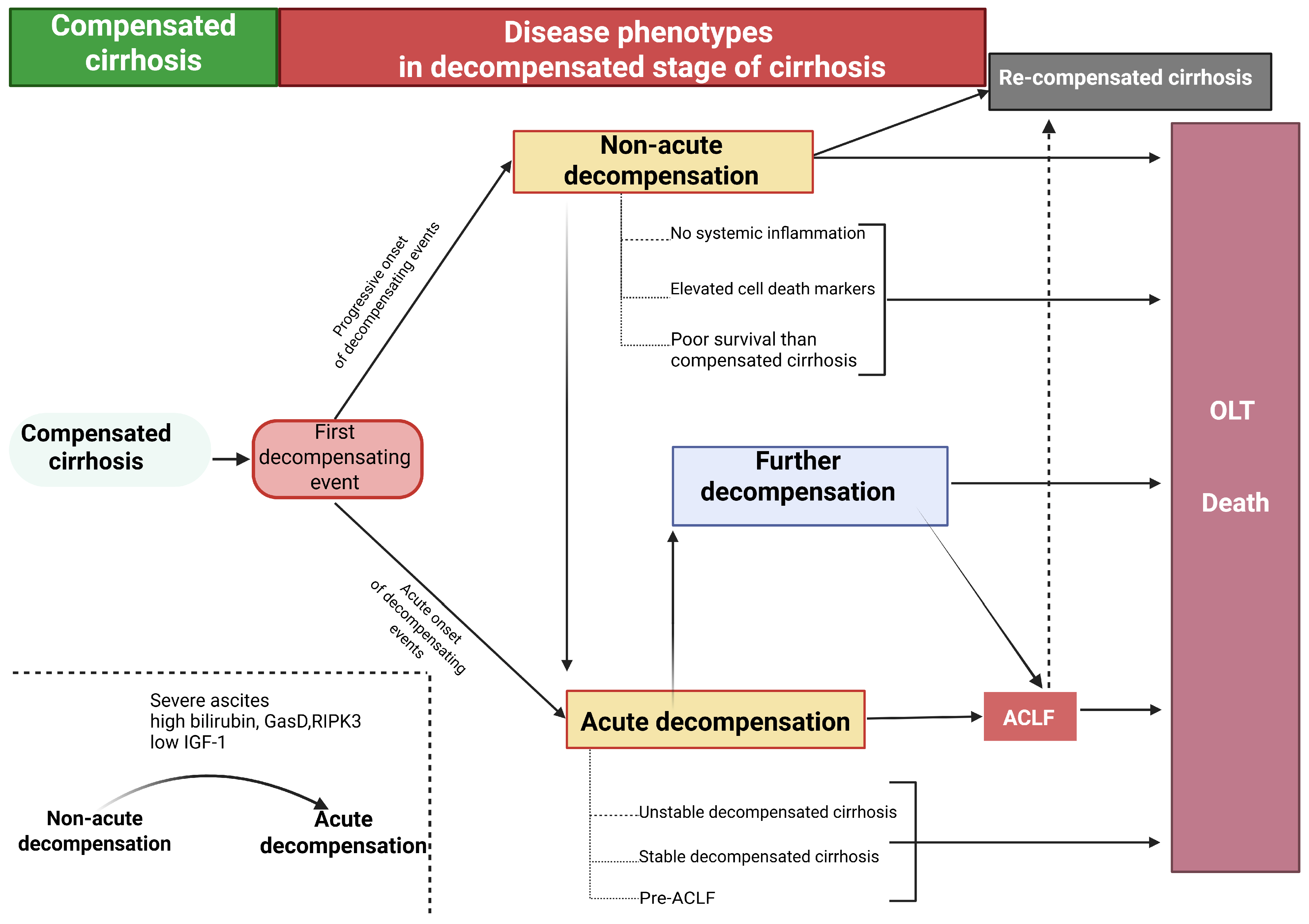
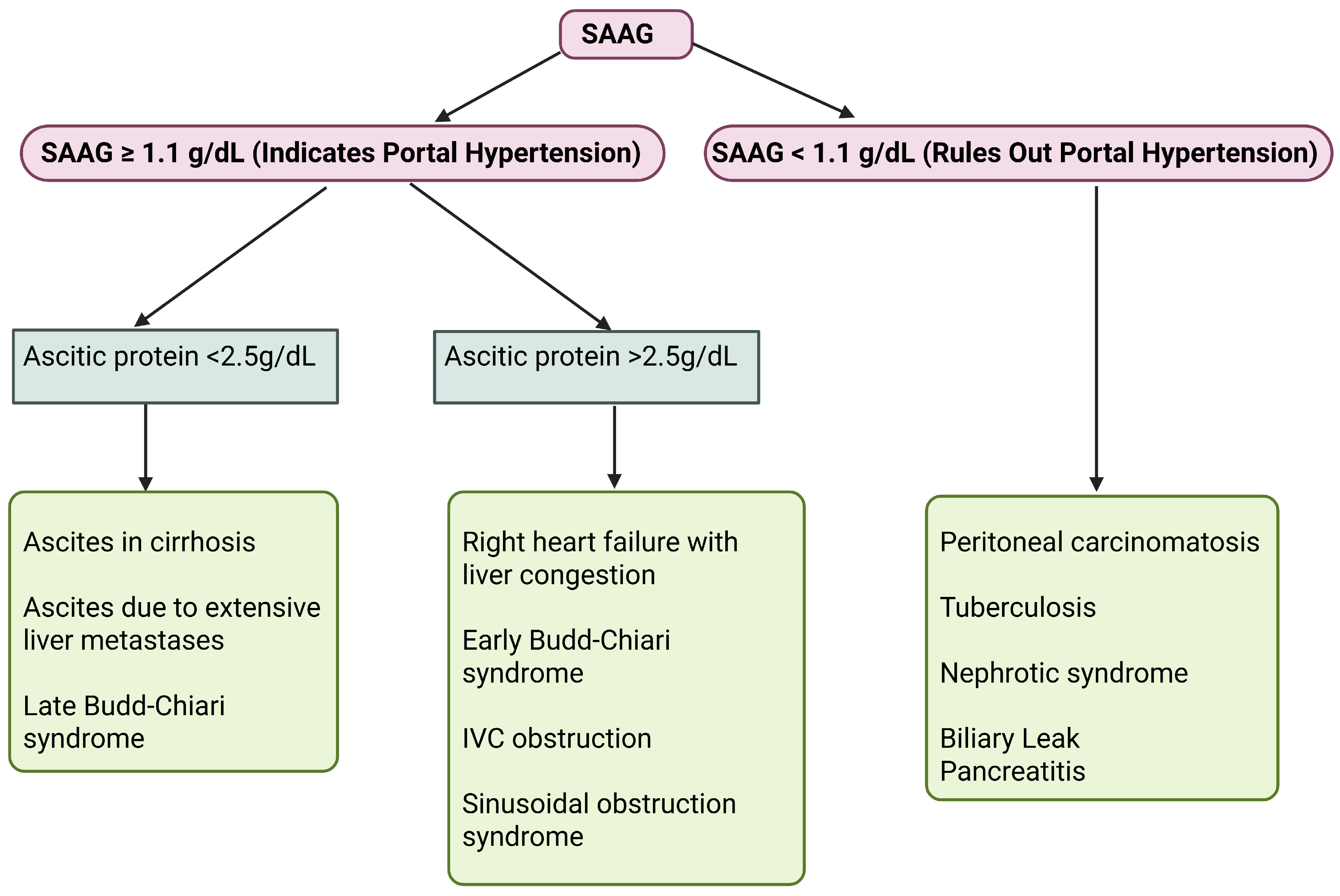
1. Alcohol-related liver disease

2. Metabolic and genetic
MASLD
Haemochromatosis
Wilson’s disease
al-antitryp sin deficiency
Cystic fibrosis
Progressive familial intrahepatic cholestasis
Lysosomal acid lipase deficiency
Tyrosinemia type 1
Type IV glycogen storage disease
3. Auto-immune
Autoimmune hepatitis
Primary biliary cholangitis

4. Biliary
Biliary atresia (paediatrics patients)
Biliary strictures
Primary sclerosing cholangitis

5. Vascular
Budd-Chiari syndrome
Veno-occlusive disease
Fontan-associated liver disease
Cardiac cirrhosis/Right sided heart failure

6. Drugs and toxins (long tenn use)
Methotrexate
Isoniazid
Amiodarone
Methyldopa
Vitamin A
Allopurinol
Valproic Acid
Vinyl chloride
Aflatoxin
Herbs like kava and comfrey

7. Cryptogenic cirrhosis

8. Infections
Viral hepatitis B
Viral hepatitis C
Viral hepatitis D (typically superimposed on hepatitis B infection)
Schistosomiasis
Table 4. The Child–Pugh score.
Points123
EncephalopathyNoneMinimalAdvanced (coma)
AscitesAbsentControlledRefractory
Bilirubin (μmol/L)<3434–51>51
Albumin (g/L)>3528–35<28
Prothrombin (s) a<44–6>6
a Prothrombin time values of 4 and 6 s correspond approximately to 50 and 40% of normal, respectively.
Table 5. Potential therapeutic targets in portal hypertension.
TargetTreatments
Etiological therapyAntiviral therapy (HCV, HBV), immunosuppression (AIH), sustained alcohol abstinence, and therapeutic relapse management
Adoption of healthy lifestyleReduction and cessation of alcohol consumption, regular moderate aerobic exercise, and dietary counseling aimed at maintaining a BMI between 18 and 29 kg/m2. Ensuring adequate protein intake (exceeding 1.2–1.5 g/kg/day), avoiding processed foods, high-sugar and high-fructose corn syrup-sweetened products, excessive salt, and tobacco use, as well as incorporating a nocturnal high-protein snack
Elevated hepatic vascular resistanceCarvedilol, TIPS
Activated hepatic stellate cells (HSCs)Antifibrotic agents (experimental), anticoagulants
Liver sinusoidal endothelial cell (LSEC) dedifferentiationStatins
Hepatocellular damageAntioxidant Therapy
Splanchnic vasodilationNonselective beta-blockers, Carvedilol, Somatostatin, Terlipressin, and analogs
Gut-liver axisNonselective beta-blockers, Carvedilol, probiotics, prebiotics, fecal microbiota transplantation, antibiotics
Collaterals and varicesNonselective beta-blockers, Carvedilol, antiangiogenics (experimental), endoscopic therapy, collateral embolisation, BRTO, PARTO, esophageal stents, balloon tamponade
Endothelial dysfunction and NO imbalanceNO donors (experimental), phosphodiesterase inhibitors (sildenafil, tadalafil)
Microbiome modulationPrebiotics, postbiotics (experimental), dietary fiber interventions
Renal dysfunction and hepatorenal syndromeTerlipressin, albumin, norepinephrine
Liver transplantationLiver transplantation (advanced decompensated cirrhosis)
Abbreviations: AIH, autoimmune hepatitis; BRTO, balloon-occluded retrograde transvenous obliteration; PARTO, plug-occluded retrograde transvenous obliteration; NO, nitric oxide.
Table 6. Common non-selective beta-blockers utilized for portal hypertension management.
TherapyMechanism of ActionInitial DoseTitration StrategyMaximum DoseTherapeutic GoalCommon Side EffectsDuration of Therapy
PropranololReduces cardiac output through beta-1 receptor blockade, which lowers heart rate and contractility; it also decreases portal pressure.20–40 mg twice dailyTitrate gradually every 2–3 days until therapeutic goal is achieved. No ascites: 320 mg/day; with ascites: 160 mg/dayTarget heart rate of 55–60 bpm; maintain SBP ≥ 90 mm HgFatigue, bradycardia, difficulty breathing, low blood pressure, constipationTherapy should be maintained long-term—or until placement of a TIPS or liver transplantation—no routine surveillance via upper endoscopy recommended
NadololInduces constriction of splanchnic arteries by blocking beta-2 receptors, leading to unopposed alpha-adrenergic vasoconstrictive activity.20–40 mg at bedtimeAdjust dose as requiredNo ascites: 160 mg/day; with ascites: 80 mg/dayTarget heart rate of 55–60 bpm; maintain SBP ≥ 90 mm HgTiredness, slow heart rate, low blood pressureTherapy should be maintained long-term—or until placement of a TIPS or liver transplantation—no routine surveillance via upper endoscopy recommended
CarvedilolIn addition to beta-1 and beta-2 receptor blockade, reduces intrahepatic vascular resistance via alpha-adrenergic activity.6.25 mg one time dailyRise to 6.25 mg two times daily after 3 days12.5 mg/day (higher doses possible for non-liver conditions)No specific heart rate target; maintain SBP ≥ 90 mm HgLow blood pressure, dizziness, fatigueTherapy should be maintained long-term—or until placement of a TIPS or liver transplantation—no routine surveillance via upper endoscopy recommended
Abbreviations: SBP, systolic blood pressure; TIPS, transjugular intrahepatic portosystemic shunt.
Disclaimer/Publisher’s Note: The statements, opinions and data contained in all publications are solely those of the individual author(s) and contributor(s) and not of MDPI and/or the editor(s). MDPI and/or the editor(s) disclaim responsibility for any injury to people or property resulting from any ideas, methods, instructions or products referred to in the content.

