Review

Liver and Vascular Involvement in Philadelphia-Negative Chronic Myeloproliferative Neoplasms—A Narrative Review

by
Romeo G. Mihăilă
1,
Samuel B. Todor
1,* and
Marius D. Mihăilă
2
1
Faculty of Medicine, “Lucian Blaga” University of Sibiu, 550024 Sibiu, Romania
2
Faculty of Medicine, “Iuliu Hațieganu” University of Medicine and Pharmacy Cluj-Napoca, 400012 Cluj-Napoca, Romania
*
Author to whom correspondence should be addressed.
Livers 2025, 5(3), 29; https://doi.org/10.3390/livers5030029
Submission received: 19 May 2025 / Revised: 15 June 2025 / Accepted: 25 June 2025 / Published: 30 June 2025

Abstract

Hepatosplenomegaly can occur in extrahepatic diseases such as Philadelphia-negative chronic myeloproliferative neoplasms (MPNs), which may involve the liver and vasculature. In myelofibrosis, extramedullary hematopoiesis can be present in the liver, even within hepatic sinusoids. Liver biopsies in MPN patients have shown platelet aggregates obstructing these sinusoids. Both liver and spleen stiffness are significantly higher in myelofibrosis, correlating with the severity of bone marrow fibrosis. Spleen stiffness is also elevated in myelofibrosis and polycythemia Vera compared to essential thrombocythemia. MPNs are a leading cause of splanchnic vein thrombosis in the absence of cirrhosis or local malignancy, especially in the presence of the JAK2V617F mutation. This mutation promotes thrombosis through endothelial dysfunction and inflammation. It is found in endothelial cells, where it enhances leukocyte adhesion and upregulates thrombogenic and inflammatory genes. Hepatic sinusoidal microthromboses in MPNs may contribute to portal hypertension and liver dysfunction. MPN therapies can also affect liver function. While hepatocytolysis has been reported, agents such as Hydroxycarbamide and Ruxolitinib exhibit antifibrotic hepatic effects in experimental models. Overall, MPNs are linked to chronic inflammation, increased thrombotic risk—particularly splanchnic thrombosis—and atherogenesis.

Graphical Abstract

1. Introduction

The most common types of Philadelphia-negative chronic myeloproliferative neoplasms (MPNs) are polycythemia vera (PV), essential thrombocythemia (ET), and primary myelofibrosis (PMF). They are the consequence of a clonal proliferation of mature blood cells [1,2], a result of the overactivation of the Janus kinase (JAK)/signal transducer and activator of transcription (STAT)-signaling pathway [1]. JAK2V617F is the most frequent mutation present in PV, ET, and PMF, while mutations in calreticulin (CALR) and myeloproliferative leukemia (MPL) genes [1,2,3] also play significant roles [2]. Ninety percent of patients with ET have either JAK2, calreticulin, or MPL mutation [4]. Today, it is considered that JAK2V617F positive ET and PV represent a biological continuum, and ET is an early stage of PV progression [5]. The simultaneous presence of two of the three mutations is generally not considered for characterizing MPNs, leading to misdiagnosis [3]. Patients with concurrent mutational MPN are relatively older, have significantly higher platelet counts, palpable splenomegaly, and significantly higher leukocyte count than their counterparts with single gene mutation [3]. The intriguing lack of disease specificity of these mutations, and of the chronic inflammation associated with MPNs [6], is remarkable. In addition to driver mutations implicated in the pathogenesis of MPNs, concomitant mutations may exist; all have been carefully studied, including their involvement in inflammation, and several pathogenic models have been proposed [6].
Myelofibrosis (MF) is characterized by stem cell-derived clonal proliferation potentially resulting in bone marrow fibrosis. As the disease progresses, extramedullary hematopoiesis is frequently detected in the spleen and the liver and rarely in other organs [7], a common complication of PMF [1]. The dysregulation of the bone marrow microenvironment is the cause of extramedullary hematopoiesis [1]. Such a patient with a history of stable myelofibrosis evolved with progressive hepatosplenomegaly. Magnetic resonance imaging (MRI) and computed tomography (CT) scans showed periportal masses whose biopsy confirmed the suspicion of sclerosing extramedullary hematopoietic tissue [8]. Extramedullary hematopoiesis can sometimes be found even within liver sinusoids in patients with PMF [9].
Bone marrow (BM) biopsy is useful for the diagnosis of most patients with chronic myeloproliferative neoplasms. Its main limitation is the risk of sampling error. Although not currently used in clinical practice, imaging techniques might offer additional information for diagnosis, prognostication, and response monitoring of these patients [10]. Splenic transient elastography is useful to predict BM fibrosis grade in patients with MF. Dynamic contrast-enhanced MRI can be used to differentiate MF patients from ET patients and healthy controls. MRI seems to be promising for the evaluation of BM amount of fat tissue and, indirectly, cellularity/fibrosis in MF, and possibly for evaluating BM cellularity in ET/PV. Also, 18-fluorodeoxyglucose and 18-fluorothymidine positron emission tomography (PET)/CT might be useful for estimating BM fibrosis and has good accuracy for the diagnosis of residual disease [10]. While splenic elastography is rapid, non-invasive, and cost-effective, it may be limited by technical factors such as body habitus or ascites. MRI offers detailed anatomical and fat quantification information, but it is more expensive and less widely available. PET/CT provides functional imaging that can detect metabolic and proliferative activity, but it involves radiation exposure and may lack specificity in distinguishing reactive from clonal processes [10].
MPNs are significantly associated with morbidity and mortality related to an increased risk of thrombo–hemorrhagic events [2,11,12]. Patients with MPNs have chronic inflammation, driven by cytokine release from aberrant leukocytes and platelets, which amplifies cardiovascular risk through various mechanisms, including atherosclerosis and vascular remodeling [2], similarly to patients with metabolic-associated steatotic liver disease (MASLD) who are known to be at risk of developing atherosclerotic vascular illness [13]. Systematic measurement of arterial stiffness can detect early systemic atherosclerosis in both patient groups and may contribute to the early indication of effective treatment. JAK2V617F mutation is particularly connected to the risk of developing cardiovascular disease [14].
In this narrative review we aimed to analyze the connections between MPNs and the liver.

2. Liver and Myelofibrosis

The JAK2V617F mutation induces the generation of reactive oxygen species through deregulation of several oxidative stress and anti-oxidative defense genes. Their role is to maintain genomic stability to regulate the oxidative stress response and modulate the migration or retention of hematopoietic stem cells. Their deregulation increases genomic instability, increased chronic inflammation, and disease progression, characterized by the migration of hematopoietic stem cells from the bone marrow to extramedullary sites such as the liver and spleen [15]. Recombinant Interferon-alpha2 downregulates several upregulated oxidative stress genes and upregulates downregulated antioxidative defense genes and decreases the risk of additional mutations, clonal evolution, and disease progression [15], including progression to myelofibrosis, which frequently evolves with liver myeloid metaplasia.
Various cytokines, hormones, and enzymes are involved in fibrogenesis. Among them, glutathione peroxidase 4 (GPX4), a selenoprotein antioxidant enzyme, is widely found in different tissues, including the liver. GPX4 suppresses phospholipid hydroperoxide at the expense of decreased glutathione (GSH), including autophagy, cell repair, inflammation, ferroptosis, apoptosis, and oxidative stress. These processes are involved in the occurrence of fibrotic disease. It was observed that GPX4 exhibits a decline in fibrotic disease and inhibits fibrosis. This suggests that alterations of GPX4 can change the course or evolution of fibrotic disease, such as liver fibrosis and myelofibrosis [16].
Single-cell RNA sequencing analysis of MF and liver fibrosis showed enrichment in myeloid cell types, including DC3, which express similar inflammatory genes to monocytes. Among the pro-inflammatory genes implicated, recent studies have highlighted the role of cytokines such as IL1A, IL1B, IL12A, IL23A, and IL27, which are upregulated upon DC3 activation via TLR-signaling pathways. These cytokines are also involved in chronic inflammatory conditions, suggesting a potential mechanistic overlap with the inflammatory milieu observed in MF. Understanding their signaling mechanisms across liver and myeloid tissues will be important for future investigation [17] and to find possible connections between liver fibrosis and myelofibrosis.
Fibrotic disorders are defined by the pathological accumulation of extracellular matrix (ECM) components—particularly collagens—in various organs such as the liver and bone marrow, ultimately resulting in tissue scarring and functional impairment. MicroRNAs (miRNAs), a class of small non-coding RNAs approximately 22 nucleotides in length, regulate gene expression by promoting mRNA degradation or inhibiting translation. Several miRNAs have been implicated in the pathogenesis of fibrosis, including miR-101a/b, miR-494, miR-488, miR-382, miR-146, miR-31, and miR-543 [18]. These molecules contribute to the development of a fibrotic phenotype by modulating fibroblast activation and ECM remodeling. Due to their central role, miRNAs have emerged as promising therapeutic targets in fibrotic diseases. A range of strategies has been explored to modulate miRNA activity, including the use of antisense oligonucleotides, small molecule inhibitors, and bioactive natural compounds [18].

3. JAK/STAT Molecular Pathway

There are four JAK proteins (JAK1, JAK2, JAK3, and TYK2) [19] and seven proteins of the STAT family (STAT1, STAT2, STAT3, STAT4, STAT5A, STAT5B, and STAT6) [19] in mammals [20]. The JAK/STAT molecular pathway could be activated by a lot of hormones, cytokines, growth factors, and more and is involved in various processes such as cell proliferation and division, apoptosis, migration, immune regulation, and tumor formation. Activation of this signaling mechanism is present in growth and development, homeostasis, various solid tumors, chronic MPNs, inflammatory illness, and autoimmune diseases [20]. JAK2 molecules are activated by phosphorylation after the binding of the ligand to the tyrosine kinase receptor and form dimers. These activate STAT molecules by phosphorylation. STAT dimers cross the nuclear membrane and activate gene expression. The result is increased erythropoietin synthesis. A deepening understanding of the JAK/STAT pathway has occurred in recent years. In addition, a relationship between JAK/STAT and the pathophysiology of fibrotic diseases was noticed, including the liver and bone marrow [20]. JAK inhibitors have been approved for MF and PV, and, subsequently, the JAK/STAT pathway may serve as a promising target for fibrosis in other organs [20], but this requires future studies. The JAK2—STAT-signaling pathway is represented in Figure 1.
A point mutation (V617F) in the Janus kinase 2 (JAK2) gene leads to the appearance of a disorderly activated tyrosine kinase, involved in the MPNs pathway [21]. JAK2V617F mutation is observed in 60% of ET cases [22], in over 90% of patients with PV, and in about 50% of those with PMF [22,23]. The JAK2V617F mutation is not enough to initiate MPNs. The presence of other factors involved in their pathogenesis could be necessary: different signaling pathways, JAK2V617F mutant allele burden, epigenetic modification, the immune system dysregulation, and lifestyle [5].
Inflammation contributes to the increase in reactive oxygen species (ROS) signaling, which potentially influences the occurrence of MPNs through genomic instability, point mutations, and chromosomal aberrations in a mouse model. ROS was also increased in human hematopoietic cells of human MPN patients compared with healthy subjects [5]. The oxidative stress is more pronounced in ET patients with the JAK2V617F mutation who have experienced a thrombotic episode compared to those who have not [2,24]. Smoking can potentiate the impact of chronic inflammation and oxidative stress in MPNs, favoring genomic instability, subclone formation with disease progression, and higher risk of second malignancies. The patients with JAK2V617F heterozygous mutation expressed p-selectin significantly higher and signaling by tumor necrosis factor-a (TNFa), interleukin 6 (IL6), and interferon gamma (IFNg), which were enriched compared to the cells from healthy subjects [5].
Patients with JAK2V617F-mutated MPN exhibit increased risk of thrombosis [22,23], most frequently in splanchnic veins. It was observed that plasma levels of angiopoietin-2, thrombomodulin, TNF-R1, VWF, and VCAM-1 were higher in JAK2V617F-mutated MPN patients with thrombosis [22]. Thrombosis is mostly visceral [22,23], portal or extra-portal [23]. JAK2V617F mutation was found in 14.4% of 155 patients with splanchnic vein thrombosis (SVT), in a retrospective cohort study. Abdominal pain was their most frequent clinical manifestation [25]. JAK2V617F mutation can also be rarely present in patients with liver metastases of pancreatic neuroendocrine tumor and SVT [26].
The possibility of providing personalized treatment for patients with MPNs requires finding the most useful thrombotic risk biomarker in these patients. Thrombin generation reflects more accurately the balance between pro- and anti-coagulant factors, compared to classic tests that explore the intrinsic, extrinsic, and global coagulation pathways. Endogenous thrombin potential is higher in Philadelphia-negative MPN patients, especially in the presence of JAK2V617F mutation than in healthy controls. The JAK2V617F allele burden correlates more with a higher thrombin generation potential in patients who are not under hydroxycarbamidum treatment. These patients are highly sensitive to hydroxycarbamidum, a fact reflected in lower values of platelet thrombin generation potential [27].
STAT5 phosphorylation in liver and spleen can be effectively achieved with WWQ-131, which is a highly selective JAK2 inhibitor (182-fold and 171-fold more selective to JAK1 and JAK3, respectively) [28]. WWQ-131 has been shown to have stronger therapeutic effects than fedratinib in MPN models in mice [28]. Circulating factors in MPNs with SVT onset are involved in the appearance of endothelial proinflammatory and prothrombotic phenotypes, which can be modulated in vitro with MPN treatment [22]. Mutant JAK2V617F is a positive regulator of DDX5 mRNA expression and DDX5 protein stability by activating STAT5. It was observed that the knockdown of DDX5 significantly suppressed liver hypertrophy and splenomegaly caused by an inoculation of Ba/F3 cells expressing a JAK2V617F mutant and erythropoietin receptor in nude mice. The JAK2V617F/STAT5/DDX5 axis could be a therapeutic target in the treatment of MPN [21].

4. Liver and Spleen Stiffness

Liver stiffness (LS) and spleen stiffness (SS) can be assessed using shear wave point and bidimensional elastography. It was observed that SS values were higher in MF and PV compared to ET. A recent study included 143 patients with chronic MPNs and 75 healthy volunteers who were evaluated by shear wave point elastography. SS and LS were significantly higher in MF patients (40.9 vs. 26.3 kPa, respectively 7.72 vs. 5.52 kPa) [29]. The median LS and SS is higher in patients with higher versus lower grades of bone marrow fibrosis (5.2 vs. 6.65 kPa, respectively 27.2 vs. 37.9 kPa) [29]. This finding suggests that elastography could be a way to estimate the degree of medullary fibrosis without resorting to bone marrow biopsy, which is an invasive procedure, similarly to the assessment of liver fibrosis by elastography, without the need for liver biopsy in most cases. These results require large studies to confirm them.
A recent study included 70 consecutive patients with Philadelphia-negative MPNs, who were examined using ultrasonography and elastography. The median SS was not different between patients with MF and PV, but both MF and PV groups had significantly higher SS compared to the ET group and healthy controls. SS values above 40 kPa were significantly associated with worse progression-free survival (PFS) in patients with MF. SS also correlated with the extension of bone marrow fibrosis and PFS was significantly different in SS with fibrotic stages MF-2 and MF-3 than in those with fibrotic stages MF-0 and MF-1 [12]. LS was significantly higher in the patient cohort with MF compared to healthy controls, but not in the patient cohorts with ET and PV compared to healthy controls [30]. The presented data support the usefulness of visceral stiffness for the clinical work-up of MPNs and can help to define patients at a higher risk of progression [30].

5. Extramedullary Hematopoiesis

MF is known to develop with extramedullary hematopoiesis, including in the liver. Liver biopsy performed for jaundice, unexplained hepatomegaly, or portal hypertension in a series of five patients with myeloid neoplasms revealed platelet aggregates obstructing the hepatic sinusoids and extramedullary hematopoiesis. Two of the five patients had myeloproliferative neoplasm (triple-negative PMF and JAK2-mutated ET). The authors emphasized that hepatic sinusoidal microthromboses present in patients with myeloid neoplasms might cause portal hypertension, independently of the presence of the JAK2V617F mutation and grade of extramedullary hematopoiesis [31].

6. Portal Hypertension

Portal hypertension (an increased pressure within the portal venous system) implies an increased portal pressure gradient (the difference in pressures between the portal venous pressure and the pressure within the inferior vena cava or the hepatic vein) [32]. Portal hypertension leads to pronounced venous collateralization and the appearance of varicose veins [33]. A pressure gradient of 6 mmHg or more suggests the presence of portal hypertension in most cases. When it is greater than 10 mmHg, portal hypertension becomes clinically significant [32,34]. Liver cirrhosis is the most common cause of portal hypertension [32,34], but not all portal hypertension is a consequence of cirrhosis [35]. Besides manifest liver cirrhosis, primarily left-sided portal hypertension is causal for the development of gastric varices [33]. This resistance to portal blood flow can also be outside of the liver, such as prehepatic in portal vein thrombosis or posthepatic in the case of Budd–Chiari syndrome (BCS) or constrictive pericarditis. Identification of the level of resistance to portal blood flow allows the determination of the etiology of portal hypertension [32].
One such patient with recurrent esophageal variceal hemorrhage was initially misdiagnosed with liver cirrhosis. Liver biopsy performed before liver transplantation showed no significant cirrhosis but found the presence of extramedullary hematopoiesis. Necessary investigations followed, establishing the diagnosis of MF, and standard treatment resulted in an absence of subsequent gastrointestinal hemorrhage due to rupture of esophageal varices for nearly 3 years [35].

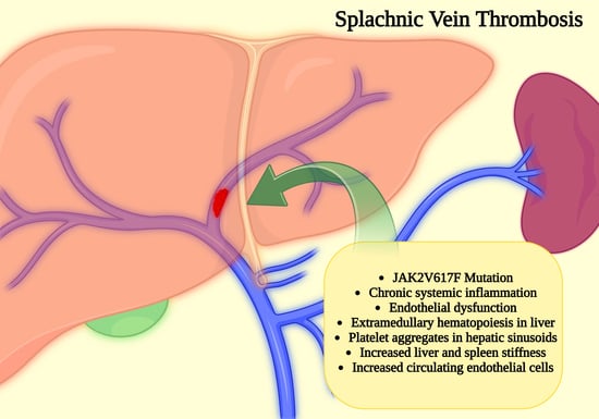
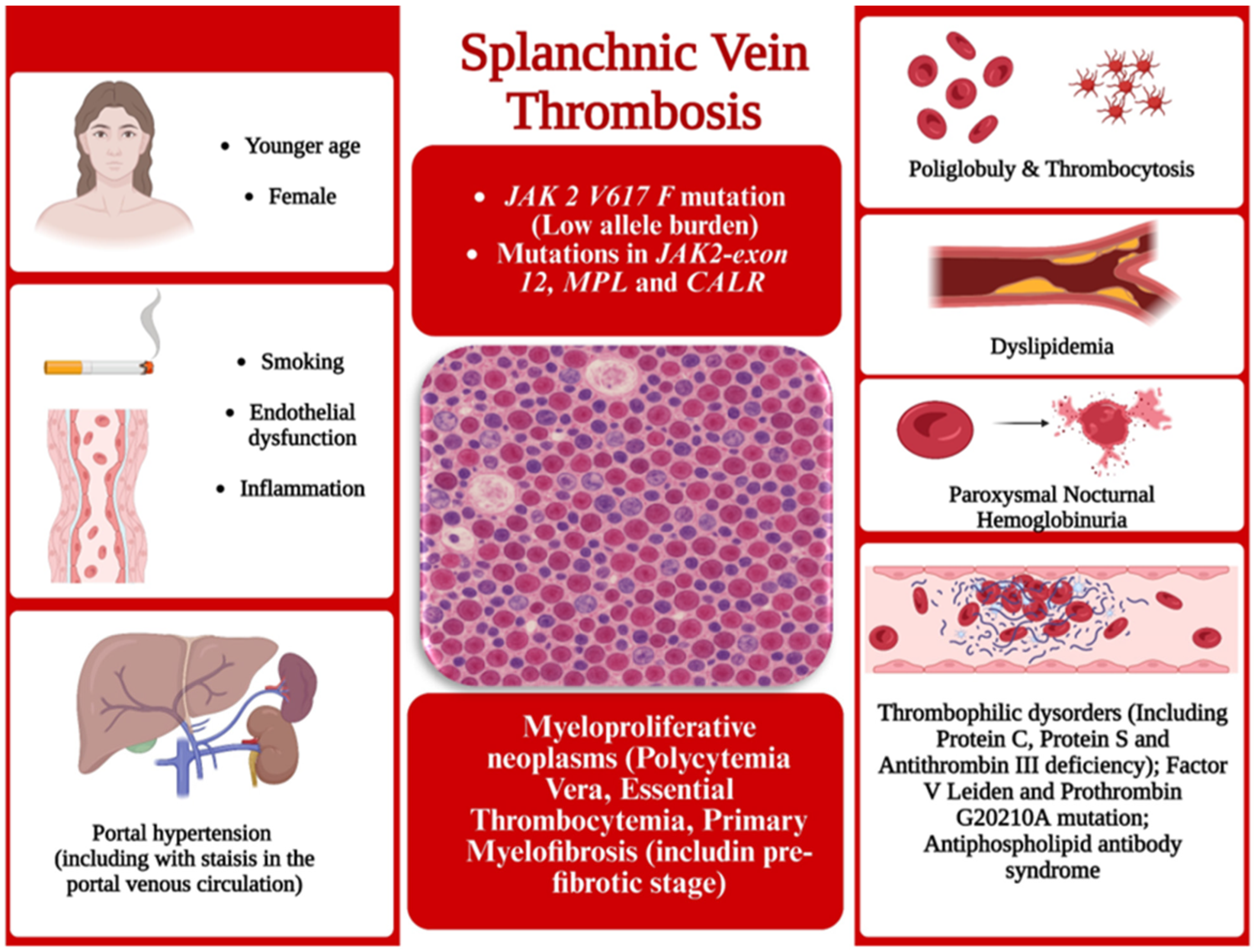
7. Splanchnic Vein Thrombosis

Splanchnic vein thrombosis (SVT) includes portal, mesenteric, or splenic vein thrombosis, or BCS [11,36]. SVT can occur in all MPNs and often affects young patients [37]. It is considered that thrombosis of the portal system in patients who do not have liver cirrhosis is the second-most-common cause of portal hypertension in the Western world [34]. Chronic MPNs are the main cause of splanchnic vein thrombosis in the absence of cirrhosis or nearby malignancy [38,39], including the portal venous thrombosis [34,40,41], and they are diagnosed in 15–30% of patients with portal vein thrombosis [41], 40% of BCS patients, and one-third of those with extrahepatic portal vein obstruction [36]. Risk factors for SVT are presented in Figure 2.
MPNs associated with SVT show a predisposition for younger women, high association with JAK2V617F mutation, low JAK2V617F allele burden (generally <10%), and low rates of CALR, MPL, or JAK2 exon 12 mutations [11]. In SVT patients the JAK2 V617F mutation is detectable in up to 87% of those with overt MPN and up to 26% of those without [36]. In the latter, other molecular markers, such as mutations in JAK2 exon 12, CALR, and MPL genes, can be found, but they are extremely rare [36]. Portal vein thrombosis can involve one or more abdominal vessels [42,43] and may be the first clinical manifestation of occult MPNs [42]. The thrombotic risk is higher in patients carrying the JAK2V617F mutation, which can be found in approximately 90% of PV, 50% of ET, and 50% of PMF patients [34]. About 6.3% of MPNs are associated with hepatic vein-type BCS, and 28.3% of them are associated with extensive non-cirrhotic nonmalignant portal vein thrombosis [44]. CALR mutations were found in 2.7% of SVT patients [44]. A systematic review and meta-analysis studied the prevalence of CALR mutations in patients with SVT. The pooled proportion of CALR mutations was 15.16%, 17.22%, and 31.44% in SVT, BCS, and portal vein thrombosis patients with MPNs but without a JAK2V617F mutation, respectively. The screening for CALR mutations may be useful in this group of patients [45]. Due to the rarity of CALR gene mutations, a prospective cohort study showed that testing for these mutations can be restricted to patients with SVT who have spleen height ≥ 16 cm, a platelet count > 200 × 109/L, and no JAK2V617F mutation. This strategy avoids 96% of unnecessary CALR mutations testing [46]. Table 1 provides a centralized overview of the key clinical, molecular, and imaging features relevant to the diagnosis and management of Philadelphia-negative myeloproliferative neoplasms.
A mutation in exon 12 of JAK2 was identified in two out of five triple-negative patients (those without JAK2V617F, MPL, or CALR mutations) with MPN and SVT. The JAK2-exon 12 mutation was also found in 1/75 patients with idiopathic noncirrhotic SVT [39]. However, peripheral blood counts are within normal ranges in patients with MPN and portal vein thrombosis due to portal hypertension sequelae. Despite suggestive features of MPN in bone marrow, these patients lack adequate diagnostic criteria and are classified as occult MPN. The discovery of recurrent molecular abnormalities, such as the CALR gene exon 9 mutation, contributes to the diagnosis of occult MPNs [41].
The prevalence of antiphospholipid antibody syndrome and protein C, protein S, or antithrombin deficiency in BCS patients was 7.3% and 22.5%, respectively, similarly to that of patients with portal vein thrombosis [44]. Factor V Leiden, prothrombin G20210A mutation, and paroxysmal nocturnal hemoglobinuria (PNH) were found in <1% of both BCS and portal vein thrombosis patients, so that some authors consider that their testing may be unnecessary [44]. But BCS occurring in a patient with PNH can be the cause of death [47].
A thrombophilic disorder was found in 7.8% of 77 patients with non-malignant portal vein thrombosis. One was a carrier of factor V Leiden mutation and had also been diagnosed with PV years before portal vein thrombosis development [48].
Characteristics of patients with MPN and SVT include younger age, female predominance, and low JAK2V617F mutation allele burden. The evolution to MF [12,49], acute leukemia [12,49], or death was observed in 13% of these patients and was associated with a JAK2V617F allele burden ≥ 50%. Apart from the increased level of JAK2V617F allele burden, the presence of the chromatin/spliceosome/TP53 mutation is another high-risk factor [49]. Clinical and molecular characteristics of patients with MPN are different, depending on whether SVT occurs at diagnosis or at follow-up. PV/ET is frequently accompanied by heterozygous JAK2 mutation, low JAK2 allele burden, and no high-risk mutations if the patient has SVT at diagnosis. However, they have a higher risk of death and lower event-free survival compared to age- and sex-matched PV/ET controls. The patient is at an increased risk of venous re-thrombosis if he is classified in the molecular high-risk group. Patients developing SVT during follow-up are more frequently included in the high-risk molecular group than those with this complication present at diagnosis. DNMT3A/TET2/ASXL1 mutations were associated with a higher risk of arterial thrombosis [50].
It was found that all patients had splenomegaly, and three of them had portal vein thrombosis and cavernous degeneration in a case series of non-cirrhotic portal hypertension occurring in five patients with PV. In addition, four patients had moderate or severe esophageal varices. The authors found that patients with non-cirrhotic portal hypertension caused by PV had milder liver function damage compared to those with cirrhosis-induced portal hypertension; splenomegaly, ascites, and esophageal varicose veins were their main manifestations. They are at risk of thrombosis and bleeding, but it remains to be determined whether early antithrombotic therapy can reduce the thrombotic risk without increasing the bleeding risk [51].
An old man presented epigastric pain associated with mild thrombocytosis and elevated levels of aminotransferases, lactate dehydrogenase, and C-reactive protein. Contrast-enhanced abdominal computed tomography found SVT that involved portal, splenic, and superior mesenteric veins, without signs of chronic liver disease. Later, it was found that he was a carrier of the JAK2V617T mutation and had hypercellular bone marrow, with increased myeloid cells and atypical megakaryocytes, which supported the diagnosis of PMF in a prefibrotic stage [38].
ET is a myeloproliferative disorder in which the bone marrow produces excessive number of platelets and can be accompanied by various thrombosis, including extensive portal vein thrombosis, especially in patients carrying the JAK2V617F mutation [52,53]. It can contribute to the appearance of the so-called porto-sinusoidal vascular syndrome. Specific histological changes without cirrhosis are present in porto-sinusoidal vascular syndrome, with or without portal hypertension. The disease is discovered at the time of the appearance of portal hypertension complications. Patients are frequently asymptomatic until then. The pathogenesis of this syndrome is not well understood, but vascular changes present in the liver have been associated with several risk factors, including hypercoagulable states [53]. In addition to hypercoagulability, other risk factors for the development of porto-sinusoidal vascular syndrome in patients with MPNs are: inflammation, endothelial dysfunction, and, in some cases, portal hypertension [53]. Endothelial dysfunction and inflammation increase thrombotic risk. Circulating endothelial cells (CECs) are viable, CD146 positive cells that reflect endothelial injury and are associated with pro-thrombotic conditions [54]. There is recent evidence for EC involvement in the pathobiology of SVT in MPN, although it is not yet fully understood [50]. Blood levels of ECs showed a significant correlation with the extent of venous thrombosis and endothelial cell damage in chronic MPN patients with SVT [54]. Preliminary results support that monitoring CEC levels during cytoreductive and anticoagulant treatments may be useful to improve the outcome of patients with SVT [54]. Stasis in the portal venous circulation favors thrombosis, independently of other risk factors. The hyper-thrombotic state of patients with MPNs can also result in SVT [55]. A case of extensive liver infarction due to PV was recently published [43].
A patient with unusual site of thrombosis, especially visceral, should be investigated, including with JAK2V617F analysis to diagnose a possible MPN, even if the blood counts are normal or he has anemia, because some patients may have PV and blood hemodilution or coincidental blood loss anemia [56].
There are patients with more than one potentially thrombophilic condition, discovered especially in the context of severe thromboses. Such a female patient with increased levels of hematocrit had multiple liver, spleen, and left kidney infarctions and ascites was diagnosed with JAK2V617F-positive PV, but also with paroxysmal nocturnal hemoglobinuria (more than 90% of granulocytes and red blood cells were CD55- CD59-). In particular, she had no clinical manifestations of hemolysis [57].

8. Upper Gastrointestinal Bleeding

Portal hypertension can be complicated by acute upper gastrointestinal bleeding, which is a common medical emergency that has a 10% hospital mortality rate [34]. Acute upper gastrointestinal bleeding can occur from varicose veins and nonvaricose veins. Bleeding from esophageal varices is a life-threatening complication of portal hypertension. Gastrointestinal bleeding was the primary manifestation of the disease in a patient with PMF who also had portal–superior–splenic–mesenteric vein thrombosis [34]. A 36-year-old female patient with splenomegaly, underlying PMF, and a carrier of the somatic JAK2V617F mutation, presented repeated upper gastrointestinal bleeding from isolated gastric varices as a result of underlying left-sided portal hypertension [33].
Portal vein thrombosis with cavernous transformation and a non-cirrhotic liver were found in a patient by abdominal magnetic resonance imaging. He presented to the hospital with ruptured esophageal varices. Then, JAK2V617F mutation was found, and the bone marrow biopsy was consistent with ET. It should be noted that upper digestive hemorrhage and imaging findings were present without peripheral blood alterations [40].
Chronic MPNs should be suspected in patients with upper gastrointestinal hemorrhage due to ruptured esophageal varices who denied a history of liver disease. Abdominal computed tomography revealed cirrhosis, marked splenomegaly, portal vein thrombosis, and portal hypertension in other published cases. Subsequently, bone marrow biopsy and the presence of the JAK2V617F mutation substantiated the diagnosis of ET [23,58].
TIPS (transjugular intrahepatic portosystemic shunt) has proven to be effective in up to 80% of patients presenting with variceal bleeding or refractory ascites. Recurrent variceal bleeding after TIPS placement occurred in 22% of patients and was more frequently observed in patients with lower baseline hemoglobin levels. Patients who developed hepatic encephalopathy after TIPS placement were older and had higher blood levels of creatinine [59].

9. Budd-Chiari Syndrome

BCS is a rare disorder defined by clinical and laboratory signs due to partial or complete hepatic venous outflow obstruction [60,61,62] in the absence of right heart failure or constrictive pericarditis [63,64]. The ultrasonographic and MRI diagnosis of BCS includes direct signs, in particular, the occlusion or compression of the hepatic veins and/or the inferior vena cava and venous collaterals and indirect signs, especially morphological changes in the liver with hypertrophy of the caudate lobe [63], inhomogeneous enhancement, and hypervascular nodules [64].
The number of hospitalizations of BCS patients in the USA over 19 years (from 1998 to 2017) was 8435. The most common risk factor was myeloproliferative disorder (7.75%), followed by a hypercoagulable state (thrombophilia and antiphospholipid antibody syndrome) (7.32%) and paroxysmal nocturnal hemoglobinuria (1.63%). A percentage of 18.7% of them had cirrhosis, 7.9% had portal vein thrombosis, and 6.4% had inferior vena cava thrombosis. Patients may have abdominal pain [62] and ascites [60] (29.9% in the cited study), and, more rarely, liver encephalopathy, acute liver failure, or acute kidney injury [60]. Hypereosinophilic syndrome is rarely associated with BCS, and the JAK2V617F mutation can often be found in these patients [61].
Acute onset, younger age, severe liver injury, high frequency of hepatic vein thrombosis, and poor prognosis characterize the patients with BCS with JAK2V617F gene mutation compared to those without this mutation [65]. Abdominal distension was the clinical presentation of a patient with acute liver failure who was found to have BCS. The patient was found to have JAK2V617F-positive MPN. Angiography revealed extensive thrombotic occlusion of the hepatic veins and liver biopsy revealed submassive hepatic necrosis, which required liver transplantation [66]. Although rare, the MPL mutation was identified in a patient with BCS secondary to an inferior vena cava thrombosis extending into the hepatic veins. He later developed thrombocytosis and was diagnosed with ET [4].
Anticoagulation is indicated immediately in SVT patients with recently occurring thromboses. Upon clinical deterioration, catheter-directed thrombolysis or a transjugular intrahepatic portosystemic shunt is useful in conjunction with anticoagulation [36]. Endovascular management to restore venous patency in patients with BCS, including inferior vena cava angioplasty with stenting, and TIPS placement to reduce liver congestion, is today the standard of care [67]. Orthotopic liver transplantation is the only reliable option in BCS patients with a lack of response to other treatments. The simultaneous presence of MPN is not a contraindication for this operation [36]. One such patient required liver transplantation. Because the stent extended into the thoracic inferior vena cava, it was unable to be removed intraoperatively. The thoracic inferior vena cava was clamped through the diaphragm at the level of the right atrium, and the stent was incorporated within the suprahepatic anastomosis with a good vascular outcome afterwards [67]. Long-term oral anticoagulation with vitamin K antagonists is indicated in all SVT patients with the MPN-related permanent prothrombotic state [36].

10. Liver Diseases and Myeloproliferative Neoplasms

A newly onset elevated liver enzymes appeared during the monitoring of a patient with ET can be a sign of progression to secondary myelofibrosis, and it is necessary to perform bone marrow aspiration and biopsy to reassess hematopoiesis and to look for bone marrow fibrosis, as well as evidence of progression [1]. In a retrospective study, the baseline presence of steatotic liver disease (estimated by ultrasonography) in patients with MPNs did not have any impact on future thrombotic, bleeding, and disease transformation risk, nor patient survival. None of the patients had signs of liver failure during the follow-up [68]. Liver disease was the second-most-significant risk factor for mortality in patients with MPNs in a study that included 82,087 patients [69].

11. Therapeutic Interventions and Liver Consequences

Different types of medicines have been tested in MPNs (Hydroxycarbamidum, Anagrelide, interferons, JAK inhibitors, and Azacytidine, or combinations of those), some of them acting on both the JAK2 pathway and inflammation [6]. Hydroxycarbamidum is the cytoreductive drug of choice in PV and ET, while JAK inhibitors (as Ruxolitinib and Fedratinib) are indicated in intermediaries and high-risk patients with myelofibrosis and in PV patients resistant or intolerant to Hydroxycarbamidum [37].

11.1. Hydroxycarbamidum

Hydroxycarbamidum is an antimetabolite [70] and anti-proliferative medicine [71] frequently used as a cytoreductive drug, especially in patients with hypercellular forms of MPNs. Some of these patients also have hepatic fibrosis of various etiologies. It is interesting to note that Hydroxycarbamidum provided in a CCl4 mouse model of liver fibrosis was able to dose-dependently suppress lipid droplet-loss and mRNA levels of Collagen 1α1 and alpha-smooth muscle actin in transdifferentiating hepatic stellate cells (HSCs)—the main cells involved in liver fibrogenesis, by excessive proliferation and collagen production. Hydroxycarbamidum inhibited HSC proliferation and suppressed accumulation of desmin-positive HSCs and liver collagen deposition after CCl4 treatment. It is able to attenuate early development of hepatic fibrosis in vivo while preserving hepatocyte regeneration [72]. This therapeutic value of Hydroxycarbamidum against liver fibrosis needs to be investigated in large clinical trials. The liver beneficial effects of drugs used for MPNs therapy can be found in Table 2.
But Hydroxycarbamide tolerance should be investigated in all patients because it can rarely produce transient serum enzymes and bilirubin elevations during therapy and has been involved in rare cases of clinically apparent acute hepatic injury with jaundice [70]. Liver side effects of drugs used for MPN therapy can be found in Table 3.
The use of 111 in bone marrow scintigraphy allowed the discovery of extramedullary hematopoiesis in the liver, which coexisted with bilateral paravertebral soft tissue locations and transudative right pleural effusion, which appeared during an exacerbation of thrombocytosis in a patient with MPN. Hydroxycarbamidum simultaneously decreased peripheral blood platelet count and pleural effusion within 2 weeks [71].
The royal jelly has antioxidant, anti-inflammatory, and anti-apoptotic properties against Hydroxycarbamidum-induced hepatic injury in rats and could, therefore, be used as adjuvant therapy in patients with long-term Hydroxycarbamidum medication [93] if future clinical trials confirm these benefits.

11.2. Anagrelide

Anagrelide is a drug used for treatment of MPNs, including ET, with potential liver toxicity. It can produce transaminitis. Liver enzymes should be monitored closely during Anagrelide, and the drug must be discontinued immediately if the above-mentioned adverse events are noted [84].

11.3. Interferon

Interferon (IFN) is used for MPN treatment in some patients, but sometimes it can produce adverse liver effects. IFN-signaling pathways are major immunological checkpoints involved in the pathogenesis of autoimmunity. Ruxolitinib was able to ameliorate murine autoimmune cholangitis induced by IFN overexpression by inhibiting the secretion of IL-6, TNF, and monocyte chemoattractant protein-1, as well as the expression of STAT1 [76]. IFN can induce liver function abnormalities [85].
Ropeginterferon α-2b is a long-acting mono-PEGylated recombinant proline IFN conjugated to a 40 kDa branched polyethylene glycol chain at its N-terminus [73] that has only one major form, as opposed to 8–14 isomers of other on-market pegylated interferon [74,75]. It can be injected every 2-plus weeks with higher tolerability [94].
It is useful for patients with PV but may also be useful for those who have chronic hepatitis C, in addition to MPN. It showed longer effective half life and superior safety profile than PEG-IFN-α2a in a randomized phase 2 clinical trial that included patients with genotype 1 chronic hepatitis C [73]. In addition, Ropeginterferon α-2b, although at only half the number of injections, is as safe and effective as pegylated interferon alfa-2a also for genotype 2 chronic hepatitis C [74] and chronic hepatitis B [75]. Favorable results were also obtained in a phase 3 clinical trial of Ropeginterferon α-2b biweekly compared with the conventional Pegylated IFN-α2b weekly for 24 weeks, combined with Ribavirin in patients with chronic hepatitis C genotype 2. Ropeginterferon α-2b showed a better sustained virologic response rate (79.8%) than pegylated IFN-α2b (71.9%) and had a favorable safety profile [75]. Rarely, Pegylated IFN-α2a can cause: liver failure, cholangitis, and hepatic steatosis [95]. Ropeginterferon α-2b can also produce liver disorder: very frequently: increased plasma levels of gamma-glutamyltransferase; frequently: increased serum alanine aminotransferase, serum aspartate aminotransferase, and blood alkaline phosphatase; infrequently: hepatotoxicity, toxic hepatitis, and hepatomegaly; rarely: hepatic failure [86].

11.4. Ruxolitinib

Ruxolitinib is a tyrosine kinase inhibitor targeting the JAK and STAT pathways [87]. It has selectivity for JAK1 and JAK2 subtypes [96,97]. Ruxolitinib is useful for the treatment of intermediate or high-risk MF [98], resistant forms of PV [87,88,98], and steroid-refractory graft-versus-host disease (GvHD) [99] in the setting of allogeneic stem-cell transplantation [87], but it is potentially hepatototxic [88]. Ruxolitinib can very frequently induce: elevated plasma concentrations of alanine aminotransferase and aspartate aminotransferase [77]. The histopathological examination of the liver biopsies performed in patients with MPNs during Ruxolitinib treatment found a variety of liver pathology, including extramedullary hematopoiesis, obliterative portal venopathy, and drug-induced liver injury. The liver biopsy made in patients with biochemical evidence of liver injury had significant treatment implications [88]. PMF frequently develops hepatosplenomegaly and, sometimes, mild nonspecific liver enzyme abnormalities. Mild increases in bilirubin can occasionally be a consequence of both intravascular hemolysis and extramedullary hematopoiesis. One such patient with marked hyperbilirubinemia had a notable response to Ruxolitinib [89].
Treatment of MF with Ruxolitinib (a JAK1/2 inhibitor) or Fedratinb (a selective JAK2 inhibitor) and PV with Ruxolitinib can reduce spleen size, improve patients’ symptoms, and increase their survival. Activation of JAK-STAT signaling in MPN results in dysregulation of key downstream pathways, including a notably increased expression of cell-cycle mediators such as CDC25A and the PIM kinases. Exposure of murine models of MPN to the combination of Ruxolitinib with a CDK4/6 inhibitor (LEE011) and a PIM kinase inhibitor (PIM447) led to reductions in spleen and liver size, reduction of bone marrow reticulin fibrosis, and improved overall survival [78].
Ruxolitinib metabolism is mainly hepatic through CYP3A4 and can be altered by CYP3A4 inducers and inhibitors. Liver dysfunction can affect some of the pharmacokinetic variables of ruxolitinib and require dose reductions, although the main route of elimination of ruxolitinib metabolites is renal [87]. In these situations, transient and usually mild elevations of liver enzyme levels and rare instances of self-limited acute liver injury can be observed [87,98].
Concanavalin A (Con A) can induce immune hepatitis and systemic hyperinflammation in mice to mimic the context occurring in COVID-19 patients. Ruxolitinib showed significant liver protection against Concanavalin A toxicity via curbing the inflammatory cytokine storm triggered by TNF-α, IFN-γ, and IL-17A [79].
Patients with MPNs can have various infections. Those with MF are even prone to repeated infections. Interleukin-22 (IL-22) is a cytokine up-regulated in inflammatory situations and known to exert various hepatic effects. It was shown that the exposure of differentiated hepatoma HepaRG cells or primary human hepatocytes to IL-22 activates the JAK/STAT3 pathway. This is the pathophysiological mechanism by which IL-22 produces the repression of mRNA expression of cytochrome P-450 (CYP) 1A2, CYP3A4, CYP2B6, and CYP2C9, and of the sinusoidal sodium-taurocholate co-transporting polypeptide, in a dose-dependent manner. This down-regulation of liver drug detoxifying proteins, notably of CYPs, by IL-22 may contribute to the alteration of pharmacokinetics in patients suffering from acute and chronic inflammatory diseases. Ruxolitinib provided in this experimental model was able to fully prevent the IL-22-mediated CYP3A4, CYP2B6, and NTCP repression in HepaRG cells [80]. MPN patients receiving Ruxolitinib treatment could benefit from this liver protection during various infections, but clinical studies are needed to confirm this hypothesis.
JAK1 and JAK2 are involved in liver fibrogenesis and Ruxolitinib, a JAK1/2 selective inhibitor useful for MPNs therapy, significantly attenuated fibrosis progression, improved cell damage, and accelerated fibrosis reversal in the liver of mice treated with CCl4 or Thioacetamide [81].
Ruxolitinib is associated with transient and frequently mild elevations in serum aminotransferase during therapy and to rare instances of self-limited, clinically apparent idiosyncratic acute liver injury. It may favor opportunistic infections, including reactivation of hepatitis B virus [98]. One such case developed with the elevation of aspartate aminotransferase and alanine aminotransferase. Treatment with entecavir started immediately, resulting in a decrease in the HBV viral load with an improvement in liver function [82]. The FDA Adverse Event Reporting System (FAERS) pharmacovigilance database from Q4 2011 to Q1 2022 contained a number of 2097 reports of HBV reactivation, of which only 41 (1.96%) were associated with JAK inhibitors treatment. Baricitinib appeared to have the strongest signal among four JAK inhibitors, followed by Ruxolitinib. The conclusion of this pharmacovigilance study is that the association between JAK inhibitors and hepatitis B virus (HBV) reactivation is a numerically uncommon occurrence [100].
Hepatitis E virus infection can become chronic in immunosuppressed patients. Viral replication could not be controlled until Ruxolitinib suspension in an MF patient. Ruxolitinib was reintroduced after normalization of liver enzymes and clearance of Hepatitis E virus with no disease relapse, suggesting spontaneous eradication of the virus [90].
Although Ruxolitinib has immunomodulatory properties and can lead to severe infections, it seems a feasible treatment option for patients with MF and PV after liver transplantation [91]. Peritransplant administration of Ruxolitinib in patients with MF is safe at 10 mg twice daily, but 4 of 18 patients died, and the cause in one of them was GvHD of the liver [101]. Ruxolitinib can improve outcomes in steroid refractory cases of acute GvHD after allogenic hematopoietic stem cell transplantation [102]. According to a meta-analysis, the response rates to Ruxolitinib provided to patients with acute liver GvHD were between 41.8 and 71.8% [99].
Liver insufficiency may be the cause of impaired vitamin D activation, one of the risk factors for severe hypocalcemia observed in patients undergoing treatment with Ruxolitinib for MF [83].

11.5. Fedratinib

Fedratinib is an oral selective JAK-2 inhibitor used in the therapy of intermediate or high-risk, primary, or secondary MF. It frequently produces serum liver enzyme elevations but has been associated with only rare cases of clinically apparent acute liver injury [103]. The elevation in liver transaminases is one of the most common adverse effects associated with Fedratinib provided to patients with MF [92]. In a hepatic study, the Fedratinib area under the plasma concentration-time curve from time 0 to infinity did not significantly differ between patients with mild liver involvement and matched healthy subjects. Therefore, mild hepatic involvement does not necessitate Fedratinib dosage adjustments [104].

11.6. Anticoagulant Treatment

Outcomes of JAK2V617F-positive patients with non-cirrhotic portal vein thrombosis under anticoagulation treatment alone were compared with that of patients with other etiologies. Recanalization of occlusive portal vein thrombosis was observed only in 16% of JAK2V617F-positive patients, compared to 33% in those with no obvious etiology and 49% in those with other obvious etiology. Significant portal hypertension rates were 49%, 32%, and 17% for the JAK2V617F-positive patients, those with no obvious etiology, and other obvious etiology patient groups, respectively. These results support the poor outcomes of patients harboring JAK2V617F mutation, and that anticoagulation alone does not appear to be adequate therapy for this cohort [105].
The therapeutic goals in patients with SVT are: preventing thrombosis recurrence, managing the underlying MPN, and treating liver dysfunction. The management of SVT in MPNs requires a multidisciplinary team that may include a hematologist, a gastroenterologist, an interventional radiologist, and a surgeon [37]. Patients with MPNs who have a thrombotic episode normally require lifelong anticoagulation [4].
Long-term treatment with vitamin K antagonists is essential for patients with MPNs who have developed SVT. However, recurrences of SVT or other thrombosis may occur in about 15–20% of patients. Direct oral anticoagulants can be an alternative and preliminary date enclosure comparative studies [37]. The outcomes in Dabigatran-treated patients with non-tumoral acute and acute-on-chronic portal vein thrombosis (of which only 10.1% were JAK2V617F-positive) showed a recanalization rate of 47.1% (of which 21% had complete recanalization), compared with a spontaneous rate of 21.4% in not anticoagulated patients, over 32 months of follow-up. High Factor VIII:Ag levels were a predictor of non-recanalization. Dabigatran was safe in cirrhosis (Child-Pugh class A and B) [106], but the results are less encouraging in studies that only included JAK2V617F-positive anticoagulated patients.

12. Discussion

MPNs associated with SVT have a predilection for younger women, high association with JAK2V617F mutation, low JAK2V617F variant allele frequency (usually <10%), and low rates of CALR, MPL, or JAK2 exon 12 mutations [32]. Next-Generation Sequencing (NGS) techniques have contributed to deepening our knowledge of the molecular mutations in MPNs, with potential diagnostic and prognostic implications. NGS allows the simultaneous identification of multiple genes carrying mutations even at very low levels. Thus, JAK2-exon12, exon13, or exon14 mutations not previously detected by conventional techniques can be detected by NGS [39]. In addition to driver mutations, there are other risk factors for SVT: thrombophilia, hyperviscosity (present in PV with insufficient therapeutic control), hyperhomocysteinemia, antiphospholipid syndrome, paroxysmal nocturnal hemoglobinuria, chronic liver disease, portal hypertension, low portal venous flow velocity (risk factor independent of other factors), inflammation, and endothelial dysfunction. Thrombocytosis in ET favors thrombosis at a platelet count above normal, but at values above 1,500,000/mm3, patients have bleeding risk due to possible acquired von Willebrand disease. It is worth emphasizing that thrombocytosis present in some patients with PV does not increase the thrombotic risk, but polycythemia increases it.
The overproduction of cytokines found in MPNs leads to the activation of thrombocytes, leukocytes, and endothelial cells. This activation leads to mixed leukocyte–platelet aggregates, which disrupt normal coagulation, followed by thrombosis and tissue ischemia [2]. JAK2V617F mutation has been identified even in ECs, where it has been shown that leukocytes attach more firmly to JAK2V617F-positive ECs than to regular ECs. Many genes associated with inflammation and thrombogenic cascades are upregulated in JAK2V617F-positive ECs [107,108]. Aberrant secretion of inflammatory cytokines is involved in the formation of thrombosis through the amplification of vascular cell adhesion molecule 1 and intercellular adhesion molecule 1 receptors on ECs, activation of integrins and neutrophil enrollment, generation of neutrophil extracellular traps, thrombocytes activation and aggregation, and tissue factor release [109]. NF-κB signaling is induced by free radicals, which enhances immune response by promoting the production of proinflammatory cytokines and chemokines [110].
It is useful to know the coagulation status of patients with chronic liver diseases, as some may also have Philadelphia-negative MPN. Patients with chronic liver disease present complex hemostatic changes leading to a state of “rebalanced hemostasis” that may shift towards bleeding or thrombosis due to their possible complications. Although in the past, cirrhosis was considered a bleeding disorder, recent evidence highlights a higher venous thromboembolism incidence in hospitalized cirrhotic patients. Low molecular weight heparin is today considered safe and effective in cirrhotic patients, supporting more evidence-based thromboprophylaxis. Therapeutic anticoagulation is recommended nowadays for thrombotic events and may offer additional benefits, such as reducing liver fibrosis and portal hypertension [111]. However, anticoagulation is not established as a core therapy for cirrhotic patients due to safety concerns in advanced disease. The hemorrhagic risk remains a significant challenge in cirrhosis [111], favored especially by local causes (e.g., esophageal varices) or surgical interventions.
The relationship between hypercoagulability and liver fibrogenesis is known. Microthrombi formed in the intrahepatic vascular tree lead to ischemic lesions, followed by parenchymal extinction or hepatocytes collapse and hepatic stellate cells activation through a direct thrombin-mediated pathway or preceded by an inflammatory response. A fibrous tissue remodeling occurs later. The hepatitis C virus can produce direct endothelial lesions involved in tissue factor activation, alteration of fibrinolysis, and increased platelet activity and aggregability. The patients with chronic hepatitis C and detectable tissue factor microparticles activity had a higher mean liver stiffness. Liver fibrogenesis is faster in patients with chronic liver diseases who are carriers of some thrombophilic markers [112]. Liver fibrosis can be involved in the emergence of portal hypertension and the decrease in portal venous flow velocity, which increases the risk of SVT.
Smoking and dyslipidemia are other thrombotic risk factors. Prophylaxis of venous thrombosis in chronic MPNs is done with long-term platelet antiaggregants, and that of arterial thrombosis with limited-term anticoagulants. The association of prothrombotic factors is possible in the same patient: thrombophilic markers coexisting with mutations present in MPNs [48].
Chronic inflammation is considered a major risk factor for clonal expansion and evolution in the Philadelphia-negative MPNs. One of the key mutation drivers is the JAK2V617F mutation, which can induce the generation of ROS. Studies based on whole blood gene expression profiling have identified deregulation of several oxidative stress and anti-oxidative defense genes in MPNs, including significant downregulation of TP53, the NFE2L2, or NRF2 genes. These genes are involved in maintaining genomic stability, regulation of the oxidative stress response, and in modulating migration or retention of hematopoietic stem cells. Their deregulation produces increasing genomic instability, increased chronic inflammation, and disease progression with egress of hematopoietic stem cells from the bone marrow to seed in the liver, spleen, and elsewhere [15]. Interferon-alpha2 (rIFNα) is increasingly being recognized as the drug of choice for the treatment of patients with MPNs. It downregulates several up-regulated oxidative stress genes and up-regulates downregulated antioxidative defense genes. More precisely, rIFNα produced up-regulation of 19 genes in ET and 29 genes in PV, including CXCR4 and TP53. rIFNα can diminish genotoxic damage in hematopoietic cells and may ultimately diminish the risk of additional mutations and, subsequently, clonal evolution and disease progression towards myelofibrotic and acute leukemic transformation [15]. The divergent development is influenced by the acquisition of additional mutations [14]. It is considered that about 50% of ET patients harbor other mutations, the most frequent being ASXL1 (7–20%), TET2 (9–11%), DNMT3A (7%), and SF3B1 (5%). Abnormal karyotype can be found in <10% of patients and includes +9/20q-/13q- [12]. These are risk factors for disease progression. MPL and CALR-1 mutations increase the risk of MF transformation of ET, TP53 predisposes to leukemic transformation, and JAK2V617F mutation increases thrombotic risk. Leukemic transformation rate at 10 years is <1% but can be higher in JAK2V617F-positive ET patients with high thrombocytosis and those with abnormal karyotype [12].
Circulating CECs are viable, apoptotic, or necrotic CD146-positive cells, and they can be used as a biomarker of thrombotic risk due to their active involvement in inflammatory, procoagulant, and immune processes of the vascular compartment. It was shown that CEC levels were significantly correlated with the extent of venous thrombosis and endothelial cell damage in MPN patients with SVT [54].
Further research is needed to explain the interindividual variability of the Ruxolitinib pharmacokinetic variables, including depending on the genetic particularities of each subject and the associated liver diseases, and to optimize individual treatment [87].

13. Conclusions

MF is known to develop with extramedullary hematopoiesis, including in the liver. Extramedullary hematopoiesis can sometimes be found even within liver sinusoids in patients with PMF. Liver biopsies performed in patients with MPNs have revealed both platelet aggregates obstructing the hepatic sinusoids and extramedullary hematopoiesis. Hepatic sinusoidal microthromboses, observed in MPN patients, may contribute to portal hypertension or liver dysfunction independently of the presence of the JAK2V617F mutation or the grade of extramedullary hematopoiesis.
LS and SS are significantly higher in MF patients. Median LS and SS values are elevated in patients with higher versus lower grades of bone marrow fibrosis. SS values are also higher in MF and PV compared to ET. These findings suggest that elastography may offer a non-invasive method to estimate the degree of medullary fibrosis without the need for bone marrow biopsy.
MPNs are characterized by a chronic proinflammatory state, which is closely linked to increased thrombotic risk and atherogenesis. Chronic MPNs are the leading cause of splanchnic vein thrombosis in the absence of cirrhosis or adjacent malignancy. Thrombotic risk is notably higher in patients carrying the JAK2V617F mutation. Endothelial dysfunction and inflammation further enhance this risk. CECs—viable, apoptotic, or necrotic CD146-positive cells—are considered biomarkers of thrombotic risk.
Thrombin generation testing more accurately reflects the balance between procoagulant and anticoagulant factors than traditional coagulation assays. Endogenous thrombin potential is elevated in Philadelphia-negative MPN patients, particularly in those with the JAK2V617F mutation, compared to healthy controls.
Chronic inflammation in MPNs is driven by cytokine release from aberrant leukocytes and platelets, amplifying cardiovascular risk via mechanisms such as atherosclerosis and vascular remodeling.
Hydroxycarbamide inhibits HSC proliferation, reduces desmin-positive HSC accumulation, and suppresses liver collagen deposition in experimental models. It may attenuate early hepatic fibrosis while preserving hepatocyte regeneration, though it can cause liver injury in animal models.
Ropeginterferon alfa-2b is effective in PV and in patients with concurrent chronic hepatitis C. It offers a longer half life and a better safety profile than PEG-IFN-α2a in genotype 1 chronic hepatitis C and demonstrates comparable efficacy with fewer injections in genotypes 2 and B. It has shown superior sustained virologic response rates and favorable safety in these populations.
Anagrelide, used in MPNs, including ET, has potential hepatotoxicity and may cause transaminitis.
Ruxolitinib, beneficial for MPN treatment, has demonstrated significant antifibrotic effects in murine models of liver fibrosis (CCl4 or thioacetamide-induced), improving cellular damage and promoting fibrosis reversal. However, liver dysfunction can alter the pharmacokinetics of ruxolitinib and necessitate dose adjustments. In such cases, transient and usually mild elevations in liver enzymes, as well as rare, self-limited acute liver injury, may occur. Fedratinib is associated with more frequent and severe hepatic adverse events compared to ruxolitinib.

14. Future Directions

It is considered that MPNs are under-diagnosed, and due to their connection with CVD, early detection of phenotypic driver mutations (JAK2V617F, CALR, MPL) and therapeutic clinical intervention is indicated [14]. Detection of driver mutations is a starting point for investigating possible liver damage through infiltration with hematopoietic tissue, predisposition to SVT, or adverse effects of drugs used for MPN therapy.
Further studies are needed to improve our understanding of the SVT pathway in patients with MPNs and their outcomes with this debilitating complication [50]. Reducing thrombotic risk through appropriate treatment could contribute to reducing hepatic fibrogenesis and, perhaps, reducing portal hypertension.
The therapeutic potential of Hydroxycarbamide in attenuating liver fibrosis warrants investigation in large-scale clinical trials. Due to its known hepatotoxic risks, such trials should also include safety monitoring protocols and explore adjunctive strategies to mitigate adverse hepatic effects [93].
WWQ-131 is a highly selective JAK2 inhibitor, with it being 182-fold and 171-fold more selective to JAK1 and JAK3, respectively. In a dose of 75 mg/kg, it showed stronger therapeutic effects than Fedratinib (120 mg/kg) in experimental models. It suppressed STAT5 phosphorylation in the liver and spleen and inhibited Ba/F3_JAK2V617F cells spreading and proliferation in vivo. In addition, it suppressed rh-erythropoietin-induced extramedullary erythropoiesis and polycythemia in mice. Therefore, WWQ-131 could be a promising candidate for the treatment of MPNs [28].
Monitoring CEC levels during cytoreductive and anticoagulant treatments may contribute to improving SVT outcomes in MPN patients [54].
It would be useful to analyze whether there is any correlation between LS and SS and arterial stiffness—a marker of incipient systemic atherosclerosis. It would also be useful to compare arterial stiffness with the degree of medullary fibrosis.
The association of statin therapy, which has vascular pleiotropic effects, should be investigated in association with cytoreductive therapy in MPN patients to reduce SVT risk and the progression of atherogenesis.

Author Contributions

Conceptualization R.G.M. and M.D.M.; methodology, R.G.M.; software, S.B.T.; validation, R.G.M., M.D.M. and S.B.T.; formal analysis, R.G.M.; investigation, R.G.M.; resources, S.B.T.; data curation, S.B.T.; writing—original draft preparation, R.G.M.; writing—review and editing, M.D.M.; visualization, S.B.T. All authors have read and agreed to the published version of the manuscript.

Funding

This research received no external funding.

Institutional Review Board Statement

Not applicable.

Informed Consent Statement

Not applicable.

Data Availability Statement

No new data were created or analyzed in this study. Data sharing is not applicable to this article.

Acknowledgments

The authors thank the editors of Livers magazine for the invitation to publish this article and for their ongoing support.

Conflicts of Interest

The authors declare no conflicts of interest.

References

  1. McArthur, K.I.; Papayanis, P.N.; Nguyen, M.H.K.; Daneshbod, Y.; Akhtari, M. A Case Report on Hepatic Extramedullary Hematopoiesis as the Manifestation of Progression to Secondary Myelofibrosis in a Patient with Essential Thrombocytopenia. Hematol. Rep. 2022, 14, 286–289. [Google Scholar] [CrossRef] [PubMed]
  2. Todor, S.B.; Ichim, C.; Boicean, A.; Mihaila, R.G. Cardiovascular Risk in Philadelphia-Negative Myeloproliferative Neoplasms: Mechanisms and Implications—A Narrative Review. Curr. Issues Mol. Biol. 2024, 46, 8407–8423. [Google Scholar] [CrossRef] [PubMed]
  3. Wang, Y.; Ran, F.; Lin, J.; Zhang, J.; Ma, D. Genetic and Clinical Characteristics of Patients with Philadelphia-Negative Myeloproliferative Neoplasm Carrying Concurrent Mutations in JAK2V617F, CALR, and MPL. Technol. Cancer Res. Treat. 2023, 22, 15330338231154092. [Google Scholar] [CrossRef] [PubMed]
  4. Ammad Ud, D.M.; Liaqat, H.; Osama, M. MPL-Positive Essential Thrombocytosis Presenting as Budd-Chiari Syndrome in a Middle-Aged Woman with an Initially Normal Platelet Count. Eur. J. Case Rep. Intern. Med. 2021, 8, 003081. [Google Scholar] [CrossRef]
  5. Gou, P.; Zhang, W.; Giraudier, S. Insights into the Potential Mechanisms of JAK2V617F Somatic Mutation Contributing Distinct Phenotypes in Myeloproliferative Neoplasms. Int. J. Mol. Sci. 2022, 23, 1013. [Google Scholar] [CrossRef]
  6. Hermouet, S. Mutations, inflammation and phenotype of myeloproliferative neoplasms. Front. Oncol. 2023, 13, 1196817. [Google Scholar] [CrossRef]
  7. Watanabe, H.; Fujishima, F.; Fukuhara, N.; Taniuchi, S.; Joh, K.; Sasano, H. Advanced extramedullary hematopoiesis with a marked increase in reticulin fibers and hemorrhage on various organs: The first autopsy case report. Med. Mol. Morphol. 2022, 55, 68–75. [Google Scholar] [CrossRef]
  8. Crawford, C.K.; Arshad, H.; Kawamoto, S.; Fishman, E.K. Periportal extramedullary hematopoiesis in a patient with primary myelofibrosis: A great mimicker of an infiltrating tumor in the liver. Radiol. Case Rep. 2025, 20, 2482–2486. [Google Scholar] [CrossRef]
  9. Yamazaki, K.; Suzuki, S.; Kawaguchi, T. Extramedullary haematopoiesis within hepatic sinusoids of a patient with primary myelofibrosis. Br. J. Haematol. 2022, 196, 1130. [Google Scholar] [CrossRef]
  10. Slot, S.; van de Donk, N.W.C.J.; Otten, R.H.J.; Boden, B.J.H.; Zijlstra, J.; Raijmakers, P.G.H.M.; Zweegman, S. The value of bone marrow, liver, and spleen imaging in diagnosis, prognostication, and follow-up monitoring of myeloproliferative neoplasms: A systematic review. Cancer Imaging 2021, 21, 36. [Google Scholar] [CrossRef]
  11. Pescia, C.; Lopez, G.; Cattaneo, D.; Bucelli, C.; Gianelli, U.; Iurlo, A. The molecular landscape of myeloproliferative neoplasms associated with splanchnic vein thrombosis: Current perspective. Leuk. Res. 2024, 136, 107420. [Google Scholar] [CrossRef] [PubMed]
  12. Tefferi, A.; Vannucchi, A.M.; Barbui, T. Essential thrombocythemia: 2024 update on diagnosis, risk stratification, and management. Am. J. Hematol. 2024, 99, 697–718. [Google Scholar] [CrossRef]
  13. Solomon, A.; Negrea, M.O.; Cipăian, C.R.; Boicean, A.; Mihaila, R.; Rezi, C.; Cristinescu, B.A.; Berghea-Neamtu, C.S.; Popa, M.L.; Teodoru, M.; et al. Interactions between Metabolic Syndrome, MASLD, and Arterial Stiffening: A Single-Center Cross-Sectional Study. Healthcare 2023, 11, 2696. [Google Scholar] [CrossRef]
  14. Kjær, L. Clonal Hematopoiesis and Mutations of Myeloproliferative Neoplasms. Cancers 2020, 12, 2100. [Google Scholar] [CrossRef]
  15. Skov, V.; Thomassen, M.; Kjær, L.; Ellervik, C.; Larsen, M.K.; Knudsen, T.A.; Kruse, T.A.; Hasselbalch, H.C. Interferon-alpha2 treatment of patients with polycythemia vera and related neoplasms favorably impacts deregulation of oxidative stress genes and antioxidative defense mechanisms. PLoS ONE 2022, 17, e0270669. [Google Scholar] [CrossRef] [PubMed]
  16. Li, Z.; Zhu, Z.; Liu, Y.; Liu, Y.; Zhao, H. Function and regulation of GPX4 in the development and progression of fibrotic disease. J. Cell. Physiol. 2022, 237, 2808–2824. [Google Scholar] [CrossRef] [PubMed]
  17. Odell, I.D. Cross-tissue organization of myeloid cells in scleroderma and related fibrotic diseases. Curr. Opin. Rheumatol. 2024, 36, 379–386. [Google Scholar] [CrossRef]
  18. Mehjabin, A.; Kabir, M.; Micolucci, L.; Akhtar, M.M.; Mollah, A.K.M.M.; Islam, M.S. MicroRNA in Fibrotic Disorders: A Potential Target for Future Therapeutics. Front. Biosci. 2023, 28, 317. [Google Scholar] [CrossRef]
  19. Aaronson, D.S.; Horvath, C.M. A road map for those who don’t know JAK-STAT. Science 2002, 296, 1653–1655. [Google Scholar] [CrossRef]
  20. Liu, J.; Wang, F.; Luo, F. The Role of JAK/STAT Pathway in Fibrotic Diseases: Molecular and Cellular Mechanisms. Biomolecules 2023, 13, 119. [Google Scholar] [CrossRef]
  21. Takeda, K.; Tago, K.; Funakoshi-Tago, M. The indispensable role of the RNA helicase DDX5 in tumorigenesis induced by the myeloproliferative neoplasm-associated JAK2V617F mutant. Cell. Signal. 2023, 102, 110537. [Google Scholar] [CrossRef] [PubMed]
  22. De Moner, B.; Martinez-Sanchez, J.; Garrote, M.; Ramos, A.; Ventosa-Capell, H.; Moreno-Castaño, A.; Nomdedeu, M.; Ojeda, A.; Escolar, G.; Garcia-Pagan, J.C.; et al. Endothelial Damage in JAK2V617F Myeloproliferative Neoplasms with Splanchnic Vein Thrombosis. Thromb. Haemost. 2025. Available online: https://www.thieme-connect.com/products/ejournals/abstract/10.1055/a-2498-4849 (accessed on 19 May 2025). [CrossRef] [PubMed]
  23. Yakami, Y.; Yagyu, T.; Bando, T.; Hanada, M. Asymptomatic Essential Thrombocytosis Presenting with Extrahepatic Portal Vein Thrombosis: A Case Report. Am. J. Case Rep. 2023, 24, e938547. [Google Scholar] [CrossRef]
  24. Moisa, C.; Gaman, M.A.; Diaconu, C.C.; Gaman, A.M. Oxidative Stress Levels, JAK2V617F Mutational Status and Thrombotic Complications in Patients with Essential Thrombocythemia. Rev. Chim. 2019, 70, 2822–2825. [Google Scholar] [CrossRef]
  25. Kawata, E.; Siew, D.A.; Payne, J.G.; Louzada, M.; Kovacs, M.J.; Lazo-Langner, A. Splanchnic vein thrombosis: Clinical manifestations, risk factors, management, and outcomes. Thromb. Res. 2021, 202, 90–95. [Google Scholar] [CrossRef] [PubMed]
  26. Sira, L.; Zsíros, N.; Bidiga, L.; Barna, S.; Kanyári, Z.; Nagy, E.B.; Guillaume, N.; Wild, D.; Rázsó, K.; Andó, S.; et al. Case report: Metastatic pancreatic neuroendocrine tumour associated with portal vein thrombosis; successful management with subsequent pregnancies. Front. Endocrinol. 2023, 14, 1095815. [Google Scholar] [CrossRef]
  27. Mihaila, R.G. Thrombin generation—A potentially useful biomarker of thrombotic risk in Philadelphia-negative myeloproliferative neoplasms. Biomed. Pap. 2017, 161, 50–53. [Google Scholar] [CrossRef]
  28. Ge, H.; Wang, C.; Tian, C.; Diao, Y.; Wang, W.; Ma, X.; Zhang, J.; Li, H.; Zhao, Z.; Zhu, L. Efficacy of WWQ-131, a highly selective JAK2 inhibitor, in mouse models of myeloproliferative neoplasms. Biomed. Pharmacother. 2022, 156, 113884. [Google Scholar] [CrossRef]
  29. Sansone, V.; Auteri, G.; Tovoli, F.; Mazzoni, C.; Paglia, S.; Di Pietro, C.; Vianelli, N.; Cavo, M.; Palandri, F.; Piscaglia, F. Liver and spleen shear-wave elastography in the diagnosis and severity staging of myeloproliferative diseases and myelofibrosis. J. Ultrasound. 2024, 27, 715–722. [Google Scholar] [CrossRef]
  30. Benedetti, E.; Tavarozzi, R.; Morganti, R.; Bruno, B.; Bramanti, E.; Baratè, C.; Balducci, S.; Iovino, L.; Ricci, F.; Ricchiuto, V.; et al. Organ Stiffness in the Work-Up of Myelofibrosis and Philadelphia-Negative Chronic Myeloproliferative Neoplasms. J. Clin. Med. 2020, 9, 2149. [Google Scholar] [CrossRef]
  31. Normand, A.; Le Bris, Y.; Loussouarn, D.; Gournay, J.; Mosnier, J.F. Obliteration of liver sinusoids through platelet aggregates associated to extramedullary haematopoiesis in myeloid neoplasms. Virchows Arch. 2025, 486, 491–497. [Google Scholar] [CrossRef] [PubMed]
  32. Oliver, T.I.; Sharma, B.; John, S. Portal Hypertension. In StatPearls [Internet]; StatPearls Publishing: Treasure Island, FL, USA, 2025. Available online: https://www.ncbi.nlm.nih.gov/books/NBK507718/ (accessed on 19 May 2025).
  33. Giszas, B.; Weber, M.; Heidel, F.H.; Reuken, P.A. Recurrent Upper Gastrointestinal Bleeding from Isolated Gastric Varices as Primary Symptom of Myelofibrosis: A Case Report on Combining Interventional and Pharmacologic Treatment Options. Dig. Dis. 2022, 40, 530–534. [Google Scholar] [CrossRef]
  34. Chen, Y.; Kong, B.B.; Yin, H.; Liu, H.; Wu, S.; Xu, T. Acute upper gastrointestinal bleeding due to portal hypertension in a patient with primary myelofibrosis: A case report. World J. Clin. Cases 2024, 12, 2621–2626. [Google Scholar] [CrossRef]
  35. Gan, X.; Yu, S.; Zhu, M.; Ning, B.; He, S.; Xie, X.; Tu, L.; Yu, H. Case report: Primary myelofibrosis presenting with portal hypertension mimicking cirrhosis. Front. Med. 2024, 11, 1375571. [Google Scholar] [CrossRef]
  36. De Stefano, V.; Qi, X.; Betti, S.; Rossi, E. Splanchnic vein thrombosis and myeloproliferative neoplasms: Molecular-driven diagnosis and long-term treatment. Thromb. Haemost. 2016, 115, 240–249. [Google Scholar]
  37. Finazzi, G.; De Stefano, V.; Barbui, T. Splanchnic vein thrombosis in myeloproliferative neoplasms: Treatment algorithm 2018. Blood Cancer J. 2018, 8, 64. [Google Scholar] [CrossRef]
  38. Dias, E.; Liberal, R.; Costa-Moreira, P.; Príncipe, F.; Fonseca, E.; Macedo, G. Primary Myelofibrosis in the Prefibrotic Stage Presenting as Portal, Splenic, and Superior Mesenteric Vein Thrombosis: A Case Report and Review of the Literature. GE Port. J. Gastroenterol. 2021, 29, 125–131. [Google Scholar] [CrossRef] [PubMed]
  39. Magaz, M.; Alvarez-Larrán, A.; Colomer, D.; López-Guerra, M.; García-Criado, M.Á.; Mezzano, G.; Belmonte, E.; Olivas, P.; Soy, G.; Cervantes, F.; et al. Next-generation sequencing in the diagnosis of non-cirrhotic splanchnic vein thrombosis. J. Hepatol. 2021, 74, 89–95. [Google Scholar] [CrossRef] [PubMed]
  40. Brito, M.; Nunes, G.; Laranjo, A.; Sabino, J.; Oliveira, C.; Valle, S.; Gonçalves, D.; Fonseca, J. A unique case of bleeding from esophageal varices as the first sign of essential thrombocythemia. Clin. J. Gastroenterol. 2021, 14, 1612–1616. [Google Scholar] [CrossRef]
  41. Karam, D.; Iyer, V.; Agrawal, B. Occult myeloproliferative neoplasms: Not so occult any more. BMJ Case Rep. 2017, 2017, bcr2017219388. [Google Scholar] [CrossRef]
  42. Jeon, W.J.; Mehta, A.; Hudson, J.; Castillo, D.R.; Wang, J.; Nguyen, A.; Akhtari, M. Portal vein thrombosis as the presenting manifestation of JAK2 positive myeloproliferative neoplasm. Am. J. Med. Sci. 2023, 365, 457–461. [Google Scholar] [CrossRef] [PubMed]
  43. Furukawa, K.; Urano, F.; Okamura, S.; Kawashima, H. Extensive Hepatic Infarction due to Polycythemia Vera. J. Gastrointest. Liver Dis. 2024, 33, 18. [Google Scholar] [CrossRef] [PubMed]
  44. Fan, J.; Wang, Q.; Luo, B.; Chen, H.; Wang, Z.; Niu, J.; Yuan, J.; Yuan, X.; Bai, W.; He, C.; et al. Prevalence of prothrombotic factors in patients with Budd-Chiari syndrome or non-cirrhotic nonmalignant portal vein thrombosis: A hospital-based observational study. J. Gastroenterol. Hepatol. 2020, 35, 1215–1222. [Google Scholar] [CrossRef]
  45. Li, M.; De Stefano, V.; Song, T.; Zhou, X.; Guo, Z.; Zhu, J.; Qi, X. Prevalence of CALR mutations in splanchnic vein thrombosis: A systematic review and meta-analysis. Thromb. Res. 2018, 167, 96–103. [Google Scholar] [CrossRef]
  46. Poisson, J.; Plessier, A.; Kiladjian, J.J.; Turon, F.; Cassinat, B.; Andreoli, A.; De Raucourt, E.; Goria, O.; Zekrini, K.; Bureau, C.; et al. French national network for vascular liver diseases. Selective testing for calreticulin gene mutations in patients with splanchnic vein thrombosis: A prospective cohort study. J. Hepatol. 2017, 67, 501–507. [Google Scholar] [CrossRef] [PubMed]
  47. Füreder, W.; Sperr, W.R.; Heibl, S.; Zebisch, A.; Pfeilstöcker, M.; Stefanzl, G.; Jäger, E.; Greiner, G.; Schwarzinger, I.; Kundi, M.; et al. Prognostic factors and follow-up parameters in patients with paroxysmal nocturnal hemoglobinuria (PNH): Experience of the Austrian PNH network. Ann. Hematol. 2020, 99, 2303–2313. [Google Scholar] [CrossRef]
  48. Fortea, J.I.; Carrera, I.G.; Puente, Á.; Cuadrado, A.; Huelin, P.; Tato, C.Á.; Fernández, P.Á.; Montes, M.D.R.P.; Céspedes, J.N.; López, A.B.; et al. Portal Thrombosis in Cirrhosis: Role of Thrombophilic Disorders. J. Clin. Med. 2020, 9, 2822. [Google Scholar] [CrossRef]
  49. Debureaux, P.E.; Cassinat, B.; Soret-Dulphy, J.; Mora, B.; Verger, E.; Maslah, N.; Plessier, A.; Rautou, P.E.; Ollivier-Hourman, I.; De Ledinghen, V.; et al. Molecular profiling and risk classification of patients with myeloproliferative neoplasms and splanchnic vein thromboses. Blood Adv. 2020, 4, 3708–3715. [Google Scholar] [CrossRef]
  50. Tremblay, D.; Winters, A.; Beckman, J.D.; Naymagon, L.; Patel, R.; Mascarenhas, J.; Schiano, T.D. Splanchnic vein thrombosis associated with myeloproliferative neoplasms. Thromb. Res. 2022, 218, 8–16. [Google Scholar] [CrossRef]
  51. Zhang, L.; Wang, Y.; Lv, W.; Hu, J.; Gou, C. Association of polycythemia vera with non-cirrhotic portal hypertension in five patients: A case series. Exp. Ther. Med. 2023, 25, 149. [Google Scholar] [CrossRef]
  52. Iwaki, K.; Ueda, Y.; Mishima, M.; Narukami, S.; Shiokawa, M.; Sawada, K.; Kanda, J.; Takahashi, K.; Seno, H. Portal vein thrombosis due to essential thrombocythemia with limited cutaneous systemic sclerosis. Clin. J. Gastroenterol. 2021, 14, 293–296. [Google Scholar] [CrossRef]
  53. Silva, L.; Moura, R.; Rocha, L.; Costa, T.; Breda, F.; Cochicho, J. A Case of Porto-Sinusoidal Vascular Disease. Eur. J. Case Rep. Intern. Med. 2023, 10, 004138. [Google Scholar] [CrossRef] [PubMed]
  54. Giordano, G.; Napolitano, M.; Cellurale, M.; Di Carlo, P.; Musuraca, G.; Micucci, G.; Lucchesi, A. Circulating Endothelial Cell Levels Correlate with Treatment Outcomes of Splanchnic Vein Thrombosis in Patients with Chronic Myeloproliferative Neoplasms. J. Pers. Med. 2022, 12, 364. [Google Scholar] [CrossRef]
  55. Zafar, M.; Heslop-Harrison, W.; Loterh, L.; Ofuafor, K. Splenic Vein Thrombosis: A Case Series of Consequential Chronic Pancreatitis and Sequential Myeloproliferative Disorder. Cureus 2022, 14, e25924. [Google Scholar] [CrossRef]
  56. Erdinc, B.; Ramachandran, P.; Boris, A. Polycythemia Vera Presenting with Normal Hemoglobin and Hematocrit: A Rare Variant. Cureus 2020, 12, e8404. [Google Scholar] [CrossRef] [PubMed]
  57. Chatzidavid, S.; Giannakopoulou, N.; Diamantopoulos, P.T.; Gavriilaki, E.; Katsiampoura, P.; Lakiotaki, E.; Sakellariou, S.; Viniou, N.A.; Dryllis, G. JAK2V617F positive polycythemia vera with paroxysmal nocturnal hemoglobinuria and visceral thromboses: A case report and review of the literature. Thromb. J. 2021, 19, 16. [Google Scholar] [CrossRef] [PubMed]
  58. Wang, J.B.; Gao, Y.; Liu, J.W.; Dai, M.G.; Yang, S.W.; Ye, B. Gastroesophageal varices in a patient presenting with essential thrombocythemia: A case report. World J. Clin. Cases 2021, 9, 1871–1876. [Google Scholar] [CrossRef]
  59. Santos, S.; Dantas, E.; Veloso Gomes, F.; Luz, J.H.; Vasco Costa, N.; Bilhim, T.; Calinas, F.; Martins, A.; Coimbra, É. Retrospective Study of Transjugular Intrahepatic Portosystemic Shunt Placement for Cirrhotic Portal Hypertension. GE Port. J. Gastroenterol. 2020, 28, 5–126. [Google Scholar] [CrossRef]
  60. Alukal, J.J.; Zhang, T.; Thuluvath, P.J. A Nationwide Analysis of Budd-Chiari Syndrome in the United States. J. Clin. Exp. Hepatol. 2021, 11, 181–187. [Google Scholar] [CrossRef]
  61. Li, Z.; Li, N.; Ji, Z.; Shi, J.; Xin, G. Buddi-Chiari syndrome associated with hypereosinophilic syndrome: A case report. Medicine 2023, 102, e34291. [Google Scholar] [CrossRef]
  62. Camões Neves, J.; Rodrigues, F.; Apolinário, I.; Alves, M.; Sousa Caetano, O. Budd-Chiari Syndrome Caused by Polycythemia Vera: A Case Report. Cureus 2023, 15, e45527. [Google Scholar] [CrossRef] [PubMed]
  63. Van Wettere, M.; Bruno, O.; Rautou, P.E.; Vilgrain, V.; Ronot, M. Diagnosis of Budd-Chiari syndrome. Abdom. Radiol. 2018, 43, 1896–1907. [Google Scholar] [CrossRef] [PubMed]
  64. Porrello, G.; Mamone, G.; Miraglia, R. Budd-Chiari Syndrome Imaging Diagnosis: State of the Art and Future Perspectives. Diagnostics 2023, 13, 2256. [Google Scholar] [CrossRef]
  65. Chen, H.L.; Zhang, Q.Q.; Xu, H.; Xiao, J.C.; Wei, N.; Cui, Y.F.; Liu, H.T.; Wang, W.L.; Zu, M.H. Comparison of clinical features of JAK2V617F gene mutation and non-mutation in patients with Budd-Chiari syndrome. Zhonghua Gan Zang Bing Za Zhi 2022, 30, 1365–1369. [Google Scholar]
  66. Kimura, K.; Osaki, A.; Hirata, Y.; Egawa, H.; Kogiso, T.; Nakamura, G.; Hashidate, H.; Wakabayashi, T.; Sato, M.; Waguri, N. A case of acute liver failure caused by Budd-Chiari syndrome salvaged by brain-dead donor liver transplantation. Clin. J. Gastroenterol. 2024, 17, 84–92. [Google Scholar] [CrossRef] [PubMed]
  67. Coe, T.M.; Tanaka, M.; Bethea, E.D.; D’Alessandro, D.A.; Kimura, S.; Yeh, H.; Markmann, J.F. Liver transplantation with suprahepatic caval anastomosis including inferior vena cava stent. Transpl. Rep. 2020, 5, 100062. [Google Scholar] [CrossRef]
  68. Krečak, I.; Bačić, J.A.; Šimunić, N.; Bušac, V.; Pivac, L.; Čubrić, E.; Skelin, M.; Lucijanić, M. No impact of steatotic liver disease on clinical outcomes in patients with essential thrombocythemia and polycythemia vera: A pilot study. Heliyon 2024, 10, e32827. [Google Scholar] [CrossRef]
  69. Gonzalez-Mosquera, L.F.; Moscoso, B.; Tobar, P.; Cardenas-Maldonado, D.; Podrumar, A.I.; Mesa, R.; Cuenca, J.A. Sepsis-Related Outcomes of Patients with Philadelphia-Negative Myeloproliferative Neoplasms. Cancer Investig. 2023, 41, 423–431. [Google Scholar] [CrossRef]
  70. Hydroxyurea. In LiverTox: Clinical and Research Information on Drug-Induced Liver Injury [Internet]; National Institute of Diabetes and Digestive and Kidney Diseases: Bethesda, MD, USA, 2012. Available online: https://www.ncbi.nlm.nih.gov/books/NBK547852/ (accessed on 19 May 2025).
  71. Enokida, K.; Tomomatsu, K.; Okada, N.; Takeuchi, T.; Takihara, T.; Amaki, J.; Hayama, N.; Oguma, T.; Aoki, T.; Asano, K. Transudative Pleural Effusion Associated with Extramedullary Hematopoiesis. Intern. Med. 2021, 60, 449–452. [Google Scholar] [CrossRef]
  72. Haijer, F.; Koets-Shajari, S.; Heegsma, J.; Serna-Salas, S.; Blokzijl, T.; Buist-Homan, M.; Moshage, H.; Faber, K.N. Hydroxyurea attenuates hepatic stellate cell proliferation in vitro and liver fibrogenesis in vivo. FASEB J. 2023, 37, e23124. [Google Scholar] [CrossRef]
  73. Lin, H.H.; Hsu, S.J.; Lu, S.N.; Chuang, W.L.; Hsu, C.W.; Chien, R.N.; Yang, S.S.; Su, W.W.; Wu, J.C.; Lee, T.H.; et al. Ropeginterferon alfa-2b in patients with genotype 1 chronic hepatitis C: Pharmacokinetics, safety, and preliminary efficacy. JGH Open 2021, 5, 929–940. [Google Scholar] [CrossRef] [PubMed]
  74. Hsu, S.J.; Yu, M.L.; Su, C.W.; Peng, C.Y.; Chien, R.N.; Lin, H.H.; Lo, G.H.; Su, W.W.; Kuo, H.T.; Hsu, C.W.; et al. Ropeginterferon Alfa-2b administered every two weeks for patients with genotype 2 chronic hepatitis C. J. Formos. Med. Assoc. 2021, 120, 956–964. [Google Scholar] [CrossRef]
  75. Chen, C.Y.; Chuang, W.L.; Qin, A.; Zhang, W.H.; Zhu, L.Y.; Zhang, G.Q.; Chen, J.J.; Lo, C.C.; Zhou, X.; Mao, X.; et al. A Phase 3 clinical trial validating the potency and safety of an innovative, extra-long-acting interferon in chronic hepatitis C. JGH Open 2022, 6, 782–791. [Google Scholar] [CrossRef]
  76. Shao, T.; Leung, P.S.C.; Zhang, W.; Tsuneyama, K.; Ridgway, W.M.; Young, H.A.; Shuai, Z.; Ansari, A.A.; Gershwin, M.E. Treatment with a JAK1/2 inhibitor ameliorates murine autoimmune cholangitis induced by IFN overexpression. Cell. Mol. Immunol. 2022, 19, 1130–1140. [Google Scholar] [CrossRef] [PubMed]
  77. Ruxolitinib. Summary of Product Characteristics. Available online: https://www.ema.europa.eu/en/homepage (accessed on 12 May 2025).
  78. Rampal, R.K.; Pinzon-Ortiz, M.; Somasundara, A.V.H.; Durham, B.; Koche, R.; Spitzer, B.; Mowla, S.; Krishnan, A.; Li, B.; An, W.; et al. Therapeutic Efficacy of Combined JAK1/2, Pan-PIM, and CDK4/6 Inhibition in Myeloproliferative Neoplasms. Clin. Cancer Res. 2021, 27, 3456–3468. [Google Scholar] [CrossRef]
  79. Shaker, M.E.; Hendawy, O.M.; El-Mesery, M.; Hazem, S.H. The JAK inhibitor ruxolitinib abrogates immune hepatitis instigated by concanavalin A in mice. Int. Immunopharmacol. 2022, 103, 108463. [Google Scholar] [CrossRef]
  80. Le Vée, M.; Bruyère, A.; Jouan, E.; Fardel, O. Janus kinase-dependent regulation of drug detoxifying protein expression by interleukin-22 in human hepatic cells. Int. Immunopharmacol. 2020, 83, 106439. [Google Scholar] [CrossRef] [PubMed]
  81. Song, Z.; Liu, X.; Zhang, W.; Luo, Y.; Xiao, H.; Liu, Y.; Dai, G.; Hong, J.; Li, A. Ruxolitinib suppresses liver fibrosis progression and accelerates fibrosis reversal via selectively targeting Janus kinase 1/2. J. Transl. Med. 2022, 20, 157. [Google Scholar] [CrossRef]
  82. Kirito, K.; Sakamoto, M.; Enomoto, N. Elevation of the Hepatitis B Virus DNA during the Treatment of Polycythemia Vera with the JAK Kinase Inhibitor Ruxolitinib. Intern. Med. 2016, 55, 1341–1344. [Google Scholar] [CrossRef]
  83. Kurumazaki, M.; Ogawa, N.; Kobayashi, M.; Ikejiri, F.; Kanasaki, K. A Case of Severe Hypocalcemia During JAK1/2 Inhibitor Therapy for Myelofibrosis in a Patient with Liver Cirrhosis. Intern. Med. 2025. Available online: https://www.jstage.jst.go.jp/article/internalmedicine/advpub/0/advpub_4723-24/_article (accessed on 19 May 2025). [CrossRef]
  84. Siddiqui, M.U.; Siddiqui, M.D.; Hameed, S.; Naeem, Z.; Siddiqui, M. Platelet Anti-Aggregator Drowns the Heart. S D Med. 2021, 74, 502–505. [Google Scholar] [PubMed]
  85. Interferon. Summary of Product Characteristics. Available online: https://www.ema.europa.eu/en/homepage (accessed on 12 May 2025).
  86. Ropeginterferon Alpha2b. Summary of Product Characteristics. Available online: https://www.ema.europa.eu/en/homepage (accessed on 12 May 2025).
  87. Appeldoorn, T.Y.J.; Munnink, T.H.O.; Morsink, L.M.; Hooge, M.N.L.; Touw, D.J. Pharmacokinetics and Pharmacodynamics of Ruxolitinib: A Review. Clin. Pharmacokinet. 2023, 62, 559–571. [Google Scholar] [CrossRef]
  88. Tremblay, D.; Putra, J.; Vogel, A.; Winters, A.; Hoffman, R.; Schiano, T.D.; Fiel, M.I.; Mascarenhas, J.O. The Implications of Liver Biopsy Results in Patients with Myeloproliferative Neoplasms Being Treated with Ruxolitinib. Case Rep. Hematol. 2019, 6, 3294046. [Google Scholar] [CrossRef]
  89. Sandhu, S.; Chaudhry, H.; Zhang, S.; Prajapati, D. Marked Hyperbilirubinemia Associated with Primary Myelofibrosis Responsive to Ruxolitinib. Case Rep. Hepatol. 2022, 2022, 9630996. [Google Scholar] [CrossRef] [PubMed]
  90. Ribeiro da Cunha, M.; Marques, T. A Case of Hepatitis E Persistence in a Patient with Myelofibrosis Under Ruxolitinib. ACG Case Rep. J. 2021, 8, e00674. [Google Scholar] [CrossRef]
  91. Dold, L.; Lutz, P.; Heine, A.; Weismüller, T.J.; Strassburg, C.P.; Spengler, U. Ruxolitinib for treatment of polycythemia vera and myelofibrosis in patients after liver transplantation. Clin. Case Rep. 2021, 9, e04782. [Google Scholar] [CrossRef]
  92. Schiffer, M.; Kowalski, A.; Zhao, J.; Bewersdorf, J.P.; Lewis, R.S., Jr.; Zeidan, A.M. Fedratinib hydrochloride to treat intermediate-2 or high-risk primary or secondary myelofibrosis. Drugs Today 2020, 56, 755–768. [Google Scholar] [CrossRef] [PubMed]
  93. Tohamy, H.G.; El-Neweshy, M.S.; Soliman, M.M.; Sayed, S.; Shukry, M.; Ghamry, H.I.; Abd-Ellatieff, H. Protective potential of royal jelly against hydroxyurea -induced hepatic injury in rats via antioxidant, anti-inflammatory, and anti-apoptosis properties. PLoS ONE 2022, 17, e0265261. [Google Scholar] [CrossRef]
  94. Huang, Y.W.; Hsu, C.W.; Lu, S.N.; Yu, M.L.; Su, C.W.; Su, W.W.; Chien, R.N.; Hsu, C.S.; Hsu, S.J.; Lai, H.C.; et al. Ropeginterferon alfa-2b every 2 weeks as a novel pegylated interferon for patients with chronic hepatitis B. Hepatol. Int. 2020, 14, 997–1008. [Google Scholar] [CrossRef]
  95. Peginterferon Alfa-2a. Summary of Product Characteristics. Available online: https://www.ema.europa.eu/en/homepage (accessed on 12 May 2025).
  96. Mesa, R.A. Ruxolitinib, a selective JAK1 and JAK2 inhibitor for the treatment of myeloproliferative neoplasms and psoriasis. IDrugs 2010, 13, 394–403. [Google Scholar]
  97. Pardanani, A.; Tefferi, A. Targeting myeloproliferative neoplasms with JAK inhibitors. Curr. Opin. Hematol. 2011, 18, 105–110. [Google Scholar] [CrossRef] [PubMed]
  98. Ruxolitinib. In LiverTox: Clinical and Research Information on Drug-Induced Liver Injury [Internet]; National Institute of Diabetes and Digestive and Kidney Diseases: Bethesda, MD, USA, 2012. Available online: https://www.ncbi.nlm.nih.gov/books/NBK547852/ (accessed on 12 May 2025).
  99. Zhang, M.Y.; Zhao, P.; Zhang, Y.; Wang, J.S. Efficacy and safety of ruxolitinib for steroid-refractory graft-versus-host disease: Systematic review and meta-analysis of randomised and non-randomised studies. PLoS ONE 2022, 17, e0271979. [Google Scholar] [CrossRef]
  100. Pan, C.; Cao, M.; Yan, C.; Ou, X.; Zhang, X.; Xu, W.; Xu, Y.; Cui, X. Hepatitis B virus reactivation associated with Janus kinase (JAK) inhibitors: A retrospective study of pharmacovigilance databases and review of the literature. Expert. Opin. Drug Saf. 2023, 22, 469–476. [Google Scholar] [CrossRef]
  101. Ali, H.; Tsai, N.C.; Synold, T.; Mokhtari, S.; Tsia, W.; Palmer, J.; Stiller, T.; Al Malki, M.; Aldoss, I.; Salhotra, A.; et al. Peritransplantation ruxolitinib administration is safe and effective in patients with myelofibrosis: A pilot open-label study. Blood Adv. 2022, 6, 1444–1453. [Google Scholar] [CrossRef]
  102. Kim, E.; Adeel, A.; Bozorgzadeh, A.; Amano, S.; Barry, C.T.; Daly, J.S.; Devuni, D.; Elaba, Z.; Houk, L.; Martins, P.N.; et al. Treatment of Acute Graft-versus-Host Disease in Liver Transplant Recipients. Case Rep. Transplant. 2021, 2021, 8981429. [Google Scholar] [CrossRef] [PubMed]
  103. Fedratinib. In LiverTox: Clinical and Research Information on Drug-Induced Liver Injury [Internet]; National Institute of Diabetes and Digestive and Kidney Diseases: Bethesda, MD, USA, 2012. Available online: https://www.ncbi.nlm.nih.gov/books/NBK547852/ (accessed on 12 May 2025).
  104. Ogasawara, K.; Smith, W.B.; Xu, C.; Yin, J.; Palmisano, M.; Krishna, G. Pharmacokinetics and tolerability of fedratinib, an oral, selective Janus kinase 2 inhibitor, in subjects with renal or hepatic impairment. Cancer Chemother. Pharmacol. 2020, 85, 1109–1117. [Google Scholar] [CrossRef] [PubMed]
  105. Naymagon, L.; Tremblay, D.; Zubizarreta, N.; Moshier, E.; Schiano, T.; Mascarenhas, J. Portal vein thrombosis patients harboring JAK2V617F have poor long-term outcomes despite anticoagulation. J. Thromb. Thrombolysis 2020, 50, 652–660. [Google Scholar] [CrossRef]
  106. Premkumar, M.; Bhujade, H.; Sharma, P.; Nain, J.; Ahluwalia, J.; Sandhu, A.; Kumar, Y.; Rathi, S.; Taneja, S.; Duseja, A.K.; et al. Experience With Dabigatran on Rate of Portal Vein Thrombosis Recanalization, Disease Progression and Survival. Aliment. Pharmacol. Ther. 2025, 6, 971–987. [Google Scholar] [CrossRef]
  107. Guy, A.; Gourdou-Latyszenok, V.; Le Lay, N.; Peghaire, C.; Kilani, B.; Dias, J.V.; Duplaa, C.; Renault, M.-A.; Denis, C.; Villeval, J.L.; et al. Vascular endothelial cell expression of JAK2 V617F is sufficient to promote a pro-thrombotic state due to increased P-selectin expression. Haematologica 2019, 104, 70–81. [Google Scholar] [CrossRef]
  108. Guadall, A.; Lesteven, E.; Letort, G.; Awan Toor, S.; Delord, M.; Pognant, D.; Brusson, M.; Verger, E.; Maslah, N.; Giraudier, S.; et al. Endothelial Cells Harbouring the JAK2V617F Mutation Display Pro-Adherent and Pro-Thrombotic Features. Thromb. Haemost. 2018, 118, 1586–1599. [Google Scholar] [CrossRef]
  109. Najem, M.Y.; Couturaud, F.; Lemarié, C.A. Cytokine and chemokine regulation of venous thromboembolism. J. Thromb. Haemost. 2020, 18, 1009–1019. [Google Scholar] [CrossRef] [PubMed]
  110. Bjørn, M.E.; Hasselbalch, H.C. The Role of Reactive Oxygen Species in Myelofibrosis and Related Neoplasms. Mediat. Inflamm. 2015, 2015, 648090. [Google Scholar] [CrossRef] [PubMed]
  111. Ferdinande, K.; Raevens, S.; Decaestecker, J.; De Vloo, C.; Seynhaeve, L.; Hoof, L.; Verhelst, X.; Geerts, A.; Devreese, K.M.J.; Degroote, H.; et al. Unravelling the coagulation paradox in liver cirrhosis: Challenges and insights. Acta Clin. Belg. 2024, 79, 451–461. [Google Scholar] [CrossRef]
  112. Mihaila, R.; Boicean, A.; Birlutiu, V. The Connection between Hypercoagulability and Fibrogenesis in Chronic Liver Diseases. Rom. Biotechnol. Lett. 2020, 25, 1851–1860. [Google Scholar] [CrossRef]
Figure 1. JAK2—STAT-signaling pathway; JAK = Janus kinase; STAT = signal transducer and activator of transcription proteins.
Livers 05 00029 g001
Figure 2. Risk factors for SVT; CALR = calreticulin; JAK 2 = Janus kinase 2; MPL = myeloproliferatie leukemia.
Livers 05 00029 g002
Table 1. Diagnostic and Prognostic Implications of Molecular and Imaging Biomarkers in Philadelphia-Negative Myeloproliferative Neoplasms.
ParameterClinical RoleCommentsReference(s)
JAK2V617F mutationMost frequent mutation in PV, ET, PMF; driver mutation of JAK/STAT pathway overactivationPresent in ~90% PV, ~50% ET, ~50% PMF; associated with higher cardiovascular risk[1,2,3,4,14]
CALR mutationRare in SVT patients; testing recommended selectively in SVT with certain criteriaImportant mutation in ET and PMF; useful for diagnosis when JAK2 negative[2,3,11,44,45,46]
MPL mutationUsually exclusive with other mutationsLess frequent driver mutation in MPNs[1,2,3]
JAK2 exon 12 mutationLess commonRare; found in some SVT and JAK2-negative MPN cases[36]
Concurrent mutationsGenerally, not used diagnostically due to rarityMay be present; associated with older age, higher platelet count, palpable splenomegaly, leukocytosis[3]
Bone marrow biopsyGold standard for diagnosis; detects marrow fibrosis and cellularityRisk of sampling error; invasive[1,7,10]
Splenic transient elastographyLimited by body habitus, ascitesNon-invasive prediction of bone marrow fibrosis grade[10,12,29,30]
Dynamic contrast-enhanced MRIExpensive, less availableDifferentiates MF from ET and healthy controls; estimates BM fat content (cellularity/fibrosis)[10]
18-FDG and 18-FLT PET/CTRadiation exposure, limited specificityEstimates bone marrow fibrosis; assesses residual disease[10]
LS and SS via elastographyNeeds further validation in large cohortsLS and SS correlate with bone marrow fibrosis grade; SS higher in MF and PV vs ET[12,29,30]
EMH in liverMay cause microthromboses and portal hypertension independent of mutationsSeen in PMF; causes hepatosplenomegaly and portal hypertension[7,8,9,31,35]
Portal hypertensionGradient > 6 mmHg suggests portal hypertension; >10 mmHg clinically significantOccurs due to portal vein thrombosis, extramedullary hematopoiesis, or cirrhosis[32,33,34,35]
Inflammatory cytokines and chronic inflammationRelated to aberrant leukocytes and plateletsContribute to thrombo-hemorrhagic complications and cardiovascular risk[2,6,11,12,13]
BM (bone marrow), CALR (calreticulin), EMH (extramedullary hematopoiesis), ET (essential thrombocythemia), FDG (fluorodeoxyglucose), FLT (fluorothymidine), JAK (Janus kinase), LS (liver stiffness), MF (myelofibrosis), MPN (myeloproliferative neoplasm), MPL (myeloproliferative leukemia virus oncogene), MRI (magnetic resonance imaging), PET/CT (positron emission tomography/computed tomography), PMF (primary myelofibrosis), PV (polycythemia vera), SS (spleen stiffness), and SVT (splanchnic vein thrombosis).
Table 2. Liver beneficial effects of drugs used for MPN therapy.
DrugLiver Beneficial EffectsReferences
HydroxycarbamidumInhibits HSC proliferation and suppresses accumulation of desmin-positive HSCs and liver collagen deposition after CCl4 treatment; attenuates early development of hepatic fibrosis in vivo while preserving hepatocyte regeneration[72]
PEG-IFN-α2aUseful for the treatment of genotype 1 and 2 of chronic HCV[73,74]
Ropeginterferon α-2bUseful for the treatment of genotype 2 of chronic HCV and chronic HBV[74,75]
RuxolitinibAmeliorates murine autoimmune cholangitis induced by IFN overexpression by inhibiting the secretion of IL-6, TNF, and monocyte chemoattractant protein-1, as well as the expression of STAT1; reduces marked hyperbilirubinemia; reduces liver size in murine models of MPN in combination with a CDK4/6 inhibitor (LEE011) and a PIM kinase inhibitor (PIM447); produces significant liver protection against Concanavalin A toxicity, via curbing the inflammatory cytokine storm; fully prevents the IL-22-mediated CYP3A4, CYP2B6, and NTCP repression in HepaRG cells (96); could offer liver protection of MPN patients during various infections, but clinical studies are needed to confirm this hypothesis; significantly attenuates fibrosis progression, improves cell damage, and accelerates fibrosis reversal in the liver of mice treated with CCl4 or Thioacetamide; improves outcomes in steroid refractory cases of acute GvHD after allogenic hematopoietic stem cell transplantation[76,77,78,79,80,81,82,83]
Abbreviations: CCl4: Carbon Tetrachloride. CDK4/6: Cyclin-Dependent Kinases 4 and 6. CYP2B6: Cytochrome P450 2B6. CYP3A4: Cytochrome P450 3A4. GvHD: Graft-versus-Host Disease. HBV: Hepatitis B Virus. HCV: Hepatitis C Virus. HSCs: Hematopoietic Stem Cells. IFN: Interferon. IL-6: Interleukin-6. IL-22: Interleukin-22. MPN: Myeloproliferative Neoplasm. NTCP: Sodium Taurocholate Co-Transporting Polypeptide. PIM: Proviral Integration site for Moloney murine leukemia virus (a kinase family). STAT1: Signal Transducer and Activator of Transcription 1. TNF: Tumor Necrosis Factor.
Table 3. Liver side effects of drugs used for MPN therapy.
DrugLiver Side EffectsReferences
HydroxycarbamidumTransient serum enzymes and bilirubin elevations, rare cases of clinically apparent acute hepatic injury with jaundice[72]
AnagrelideIncreased serum transaminase levels[84]
InterferonLiver function abnormalities[85]
Ropeginterferon α-2bVery frequently: increased plasma levels of gamma-glutamyltransferase; frequently: increased serum ALT, serum AST, blood alkaline phosphatase; infrequently: hepatotoxicity, toxic hepatitis, hepatomegaly; rarely: hepatic failure[86]
RuxolitinibFrequently: elevated plasma concentrations of alanine aminotransferase and aspartate aminotransferase; rare instances of self-limited acute liver injury; it may favor opportunistic infections, including reactivation of the HBV and persistent HEV replication in immunosuppressed patients[87,88,89,90,91]
FedratinibFrequently: serum liver enzyme elevations; rarely: clinically apparent acute liver injury[92]
Abbreviations: ALT: Alanine Aminotransferase. AST: Aspartate Aminotransferase. HBV: Hepatitis B Virus. HEV: Hepatitis E Virus.
Disclaimer/Publisher’s Note: The statements, opinions and data contained in all publications are solely those of the individual author(s) and contributor(s) and not of MDPI and/or the editor(s). MDPI and/or the editor(s) disclaim responsibility for any injury to people or property resulting from any ideas, methods, instructions or products referred to in the content.

