Article

Predominance of Genotype 5 Hepatitis Delta Virus Infection in a Portuguese Hepatology Unit

by
Mariana Ferreira Cardoso
1,*,
Henrique Coelho
1,
Joana Carvalho e Branco
1,
Sofia Bragança
1,
Gonçalo Alexandrino
1,
Mariana Nuno Costa
1,
Rita Carvalho
1,
Elizabeth Pádua
2,† and
Alexandra Martins
1,†
1
Gastroenterology Department, Hospital Prof. Doutor Fernando Fonseca, 2720-276 Amadora, Portugal
2
Infectious Disease Department, National Institute of Health, 1649-016 Lisbon, Portugal
*
Author to whom correspondence should be addressed.
These authors contributed equally to this work.
Livers 2024, 4(3), 388-397; https://doi.org/10.3390/livers4030028
Submission received: 29 June 2024 / Revised: 8 August 2024 / Accepted: 19 August 2024 / Published: 21 August 2024
(This article belongs to the Special Issue Viral Hepatitis: Prevention, Infection, and Treatment)

Abstract

:
Hepatitis delta virus (HDV) infection is the most severe form of viral hepatitis. Genotype 1 (HDV-1) is by far the most prevalent in Europe and globally, while HDV-5 predominates in Western Africa. Data about HDV seroprevalence in Portugal are scarce and genotyping studies have not been performed yet. We aimed to assess the seroprevalence and genotypes of HDV in a large cohort of HBsAg-positive patients followed in our Hepatology Unit between 2012 and 2022. The anti-HDV-positive patients were subjected to a cross-sectional analysis, including blood sample collection for HDV RNA testing and genotype determination. In the cohort of HBsAg-positive patients, 57.5% (480/835) were born in African countries and 665/835 (79.6%) had been screened for anti-HDV antibodies. The HDV seroprevalence obtained was 6.5% (43/665). Twenty-one patients (age 41.2 ± 9.9 years; 57.1% male) were included in further molecular analyses. HDV RNA was positive in 8/21 (38.0%) and classified as HDV-5 in 7 patients (6 from Guinea-Bissau and 1 from Cape Verde) and HDV-1 in 1 patient (from Ukraine). In the largest and most comprehensive study performed in Portugal regarding HDV epidemiology to date, seroprevalence and genotype distribution of HDV (with predominance of HDV-5) were strongly influenced by immigration, notably from African countries.

1. Introduction

Hepatitis delta virus (HDV) is a defective RNA virus that requires the presence of hepatitis B virus (HBV) surface antigens (HBsAg) for assembly and release [1]. Chronic HDV infection is considered the most severe form of viral hepatitis. When compared with HBV monoinfection, HBV/HDV coinfection is associated with higher rates of liver cirrhosis, hepatocellular carcinoma, liver transplantation, and liver-related death [2,3,4,5,6].
The global seroprevalence of anti-HDV antibodies remains uncertain, with three meta-analyses pointing to 4.5–13% of HBsAg-positive patients, corresponding to 12–60 million people worldwide [7,8,9], of whom 58.8% are thought to be RNA positive [7]. For comparison purposes, the World Health Organization estimates that, in 2022, 50 million people were living with hepatitis C virus (HCV) infection and 39.0 million with human immunodeficiency virus (HIV) infection [10,11].
To date, eight HDV genotypes (HDV-1 to HDV-8) have been described, based on the nucleotide sequence diversity characterized by means of phylogenetic analyses of full-length genome sequences [12]. While HDV-1 is the most common, accounting for around 90% of infections worldwide, others were found to be predominant in specific areas, such as HDV-2 in Asia, HDV-3 in Latin America, HDV-4 in Japan and Taiwan, HDV-5 in Western Africa, and the remaining genotypes 6–8 in Central Africa [7]. Both the HDV genotype and the place of birth of infected patients seem to influence the severity of liver disease and response to treatment [13,14,15]. In a French nationwide study, European patients, almost exclusively infected with HDV-1, were at higher risk of cirrhosis than Sub-Saharan African patients. However, in the latter group, HDV-5 appeared more fibrogenic than HDV-1 [14]. In addition, African patients have been shown to display a higher rate of past HDV infection and a better response to antiviral treatment with interferon than non-African patients [14,15].
In the last decades, the epidemiology of HDV has changed. In fact, since the introduction of vaccination programs for HBV in high-income countries in the 90s, domestic infections have decreased and are now mostly related to intravenous drug use, whereas cases introduced by immigration from endemic areas increased [16]. In a large international registry, patients infected with HDV were young (mean age 36.7 years), more often male (62%), and most (77%) were HBeAg negative [13].
Several markers can be used to establish and characterize HDV infection. Anti-HDV total antibodies are generally used to screen patients infected with HBV. According to current European guidelines, this strategy should be applied to all patients with chronic HBV, regardless of risk factors for HDV [1]. Anti-HDV-positive individuals should be tested for HDV RNA, using a standardized and sensitive RT-PCR assay. Detectable HDV RNA indicates active replication, whereas negative RNA is consistent with past exposure. However, due to the fluctuating nature of HDV viral load, HDV RNA should be determined at least twice in order to correctly ascertain replicative status [1]. If RNA testing is unavailable, anti-HDV IgM can be used as a surrogate marker, as it correlates with disease activity [17]. Furthermore, quantitative HBsAg (HBV surface antigen) has been shown to correlate with HDV viraemia and with histological activity in chronic HDV infection [18,19].
There are, however, several shortcomings in the diagnosis. Firstly, adherence to European guidelines regarding HDV screening have been systematically reported to be suboptimal. To overcome underdiagnosis, reflex testing of all HBsAg-positive patients for anti-HDV antibodies has recently been implemented in some centers, with excellent results [20,21]. On the other hand, HDV RNA tests are not widely available and often have low sensitivity, especially when considering non-HDV-1 genotypes [22].
The association of HDV infection with positive autoantibodies and autoimmune features has been known for decades [23,24]. In fact, positive titers of autoantibodies, such as antinuclear (ANA) and anti-smooth muscle (SMA) antibodies, and elevated IgG (immunoglobulin G), which are hallmark features of classical autoimmune hepatitis, are common in HDV infection [25,26]. The clinical significance of these findings remains, however, elusive. It is still unclear whether these autoantibodies correlate with disease severity, as has been described for hepatitis C virus (HCV) [27]. Both HBV and HCV have been linked to overt autoimmune diseases [27]. Whether HDV can trigger autoimmune hepatitis in genetically predisposed individuals is currently unknown.
Until recently, the only available treatment for HDV was peginterferon, with limited efficacy and extensive side effects [28]. The field of HDV has gained renewed attention since 2020 with the approval by the European Medicines Agency (EMA) of the first drug for the treatment of chronic HDV infection: the entry inhibitor bulevirtide. Other drugs in the pipeline include lonafarnib, nucleic acid polymers, small-interfering RNAs and interferon lambda [1].
In Portugal, data about seroprevalence and clinical features of HDV are scarce. In early publications from the ‘80s to ‘90s, HDV seroprevalence in chronic HBsAg carriers ranged from 2.2% to 12.6% [29,30,31]. In one of those cohorts, HDV was predominantly found in intravenous drug users [31]. A multicentric study presented in 2008, involving 735 patients from 15 hospitals, showed a HDV seroprevalence of 3.5% [32]. More recently, data from single-center studies developed in Lisbon and Porto have reported seroprevalences of 14.3% (28/196) and 3.4% (20/580), respectively [33,34].
HDV RNA testing and genotype determination are not easily available in most centers in Portugal. In the current study, we aimed to assess the seroprevalence and genotypes of HDV in a large cohort of HBsAg-positive patients and to describe their epidemiologic and clinical features.

2. Materials and Methods

2.1. Target Population

We performed a single-center retrospective study including all adult patients with chronic HBV infection followed in our Hepatology Unit between 2012 and 2022. These patients were characterized by age, gender, country of origin, and anti-HDV antibody status.
From the total cohort of patients, those who were anti-HDV-positive and actively followed were subjected to a cross-sectional analysis, including blood sample collection for HDV RNA detection and genotype determination, after providing written informed consent. Genotyping determination was performed regardless of previous HDV RNA status. Patients who had missed clinical follow-up were booked for a new appointment in our clinic. All anti-HDV-positive patients underwent a complete clinical characterization.
This study was conducted in accordance with the ethical principles outlined in the Declaration of Helsinki and approved by our local Ethics Committee.

2.2. Sample Collection, HDV RNA Detection and Genotype Assessment

A blood sample of 4 mL was collected from anti-HDV-positive patients and sent to reference laboratory at National Institute of Health (INSA) for analysis. HDV RNA extraction was performed in plasma using the QIAamp Viral RNA mini kit (QIAGEN, Hilden, Germany) and complementary DNA (cDNA) was synthetized by using the RT-kit plus (Nanogen Advanced Diagnostics, Buttigliera Alta, Italy) according to manufacturer’s instructions. HDV RNA detection and genotype determination was based on PCR amplification of the 3′-terminal part of the HD gene of the HDV genome using the primer forward 900 s, 5′-CATGCCGACCCGAAGAGGAAAG-3′ (885–908), and the primer reverse 1280 as, 5′-GAAGGAAGGCCCTCGAGAACAAGA-3′ (1285–1261). Both were described by Ivaniushina et al. (2001) [35] and are frequently used in HDV genotyping studies. For PCR amplification, illustra puReTaq Ready-To-Go™ PCR Beads (GE Healthcare, Amersham, Buckinghamshire, UK) were used, with the set of primers’ concentration at 0.4 µM. The synthetized cDNA was added to reaction mixture, and the PCR was carried out under the following conditions: an initial denaturation at 95 °C for 5 min, followed by 10 cycles at 94 °C for 30 s, 51 °C+ 0.4 °C/cycle for 30 s and 72 °C for 1 min, followed by additional 30 cycles at 94 °C for 30 s, 58 °C for 30 s, 72 °C for 1 min and a final extension at 72 °C for 5 min. PCR products obtained were confirmed by electrophoresis through a 1.8% Agarose gel (SeaKem LE agarose–Lonza, Basel, Switzerland), purified by ExoSAP-IT (USB, Cleveland, OH, USA) and sequenced with the BigDye Terminator Cycle Sequencing Ready Reaction kit (Applied Biosystems, Foster City, CA, USA) according to manufacturer’s instructions, using ABI Prism 3130xl Genetic Analyzer equipment (Applied Biosystems, Foster City, CA, USA).
Sequences obtained (direct and complementary reverse) were analyzed using Chromas v.2.6.6, to correct nucleotide misreads, and then imported to BioEdit Sequence Alignment Editor v.7.2.5 12, for alignment and construction of the partial HD gene consensus sequence, representative of each sample/individual. The sequences were genotyped with BLAST algorithms to identify similar nucleotide sequences in the Hepatitis Delta Virus Database (HDVdb) (https://hdvdb.bio.wzw.tum.de/hdvdb/) [36,37] and in an international public database (https://blast.ncbi.nlm.nih.gov/Blast.cgi), both accessed between April and December 2022.

2.3. Epidemiological and Clinical Characterization of Participants

The cohort of patients that participated in the molecular analysis was characterized by age, gender, country of origin, date of HBV and HDV diagnosis and route of infection. Comorbidities including HCV or HIV coinfection, excessive alcohol consumption (defined as 20 g of alcohol for females and 30 g for males), arterial hypertension, diabetes mellitus, dyslipidemia and other chronic liver diseases were recorded.
The most recent laboratory data were collected, including hepatitis B surface antigen (HBsAg), surface antibody (HBsAb), core antibody (HBcAb), e antigen (HBeAg), e antibody (HBeAb), HBV DNA (as well as HBV DNA at diagnosis), HBV genotype, total and IgM anti-HDV antibodies, HDV RNA, aspartate aminotransferase (AST), alanine aminotransferase (AST), gamma alkaline phosphatase (ALP), gamma-glutamyl transpeptidase (GGT), total and direct bilirubin, albumin, international normalized ratio (INR), platelets, creatinine and sodium. The presence of antinuclear antibodies (ANAs) and hypergammaglobulinemia were recorded. Fibrosis was assessed by transient liver elastography using FibroScan® or by percutaneous liver biopsy, upon decision by the attending physician. The results of upper endoscopy for portal hypertension assessment were also recorded. Patients who had missed follow-up and attended their appointment after being recalled were thoroughly restaged.
Patients were considered to have cirrhosis by taking into account the combination of liver stiffness values, liver histology, laboratory and imaging data. These patients were further characterized using the Child–Pugh–Turcotte classification.

2.4. Statistical Analysis

Fischer exact test was used to compare categorical variables and t-Student or Mann–Whitney tests were used to compare continuous variables. A p-value < 0.05 was considered significant. IBM® SPSS® Statistics 28.0 was used for statistical analysis.

3. Results

A total of 835 HBsAg-positive patients (age 48.3 ± 15.2, (19–88) years; 56.6% male) were included in the HBV cohort followed in our Hepatology Unit. Portuguese patients represented 35.9% (n = 300/835) of the total, while the majority of patients were born in African countries (57.5%, n = 480/835), namely Guinea-Bissau (20.2%, n = 169), Cape Verde (16.0%, n = 134), Angola (12.6%, n = 106) and São Tomé and Príncipe (7.1%, n = 59). There were 31 patients (3.7%) originating from other European countries, notably Romania and Ukraine, 14 (1.7%) from Asia and 10 (1.2%) from South America, mostly from Brazil. Portuguese HBsAg-positive patients were older than those originating from African countries (57.2 vs. 42.9 years, p < 0.001).
In the total HBV cohort, 665/835 (79.6%) patients were screened for anti-HDV antibodies (Figure 1). The overall HDV seroprevalence obtained was 43/665 (6.5%).
Seroprevalence for HDV was 3.0% (7/232) in patients born in Portugal, 12.0% (3/25) in those originating from Central and Eastern Europe, and 8.3% (32/387) in patients born in Africa, being as high as 20.3% (29/143) in Guinea-Bissau. The seroprevalence per area and country of the patient’s geographic origin is shown in Table 1.
When comparing patients according to seropositivity for HDV, anti-HDV-positive patients were younger than anti-HDV-negative patients (41.3 vs. 48.9 years, respectively; p < 0.001), but no statistical difference was found in gender (60.5% female vs. 58.7% male; p = 0.874).
From the group of 43 anti-HDV-positive patients, only 21 patients were included in the cross-sectional analysis, which comprised 19 patients currently followed in the Hepatology Unit and 2 who were successfully recalled (Figure 1). The remaining 22 patients were either lost to clinical follow-up (n = 17) or deceased with decompensated cirrhosis or hepatocellular carcinoma (n = 3), while 1 patient received a liver transplant and was no longer followed at our unit and 1 was unable to come to the hospital for blood collection.
The small subgroup of anti-HDV-positive patients born in Portugal (n = 7) included five men aged 35–54 years who were no longer followed-up (death: two; loss of follow-up: two; liver transplantation: one), three of which had a previous history of intravenous drug use, and two women who were actively followed (ages 53 and 68 years), only one of which showed up for blood sampling.
The cohort of 21 patients that underwent subsequent cross-sectional evaluation included 12 (57.1%) men and 9 (42.9%) women (Table 2). The mean age was 41.2 ± 9.9 years (ranging from 20 to 68 years). The majority of patients originated from Guinea-Bissau (16/21; 76.2%), while one patient originated from each of the following countries: Portugal, Cape Verde, Equatorial Guinea, Ukraine and Romania. Only one patient had a positive IgM anti-HDV antibody test. Transmission was presumed to be vertical in the majority of patients (17/21; 81.0%), sexual in two patients, iatrogenic in one and unknown in one, while no patient reported current or past use of intravenous drugs. Regarding comorbidities, one patient was followed and treated for HIV (this patient had been referred to Hepatology for abnormal liver function tests) and no patients were infected with HCV. Two patients mentioned significant alcohol consumption. Three patients had arterial hypertension, and no patients were obese or diabetic.
Most patients were HBeAg-negative (85.7% at baseline, and currently 95.2%). The median liver stiffness was 7.8 kPa. One third of patients (7/21) were considered to have Child–Pugh A cirrhosis, based on liver stiffness values (17.3–75 kPa), histology (one patient with an Ishak score of 5/6) or, in one case, imaging findings of cirrhosis and biopsy-proven cholangiocarcinoma. Most patients (71.4%) were treated with nucleos(t)ide analogs for HBV. One third of patients (7/21) had been treated with peginterferon, resulting in sustained HDV clearance in two and non-response in four, while one was intolerant. Five patients started therapy with bulevirtide through the national early access program, including one patient from Ukraine, one from Cape Verde and three from Guinea-Bissau.
In the current cross-sectional study, HDV RNA was positive in 8/21 (38.0%) patients. HDV was classified as genotype 5 in seven patients (six from Guinea-Bissau and one from Cape Verde) and genotype 1 in one patient (originating from Ukraine).
Patients with active replicating HDV were younger than with those with negative HDV RNA, who were considered to have resolved infections (35.9 vs. 44.5 years, p = 0.053; borderline significance). The proportion of cirrhotic patients was not significantly different between groups (37.5% vs. 30.8%, p = 1.000).
Regarding autoimmune features, antinuclear antibodies were present in 14/18 patients (77.8%, including 5 with a titer ≥ 1:320) and IgG was elevated in 10/16 patients (62.5%). Three patients (14.3%) underwent liver biopsy and all had autoimmune features on liver histology. One of these patients had been diagnosed as autoimmune hepatitis and treated with immunosuppression prior to inclusion in the current study (this case has been detailed elsewhere) [38]. A second case concerns a 20-year-old male who presented with acute hepatitis and jaundice without encephalopathy (maximum bilirubin 18 mg/dL, AST 2604, ALT 3283 IU/L, INR 1.4; ANA 1/160 and normal IgG), prompting a new diagnosis of HBe-negative HBV infection (HBV DNA 78 IU/mL) with a positive anti-HDV antibody and negative HDV RNA (RealStar® HDV RT-PCR, Altona Diagnostics). He started treatment with tenofovir and, one month later, due to persistent jaundice and considering the autoimmune features in his liver biopsy (with an activity index of 9/18), initiated prednisolone at 1 mg/kg/day, with a dramatic improvement in 2 weeks, after which he abandoned follow-up. HDV RNA in the present study, before the initiation of steroids, was positive. The third case refers to a young patient with HIV-HBV-HDV coinfection with cirrhosis, with abnormal liver function tests, positive ANA and markedly elevated IgG (8031 mg/dL). All three patients were from Guinea-Bissau and were infected with HDV genotype 5.

4. Discussion

This is the largest and most comprehensive study performed in Portugal regarding HDV epidemiology to date. In fact, we describe a large cohort of HBV patients (n = 835) and present HDV RNA data, which were lacking in previous studies [29,30,31,32,33,34]. In addition, we describe HDV genotypes for the first time, as this technique was not previously available in our country.
We consider that our HDV screening rate (79.6%) was very acceptable. In fact, it has long been our routine practice to screen HBV patients for HDV regardless of risk factors, as recommended by the European guidelines. Nevertheless, this rate can be optimized in the future through the implementation of reflex testing, as has been demonstrated in other centers [20]. This could not only increase the diagnosis of HDV but also provide a more accurate estimate of HDV seroprevalence [21,39], as well as aid in identifying patients that could benefit from treatment.
The value for HDV seroprevalence that we found in our unit (6.5%) is in agreement with published data (4.5–13%) [7,8,9]. However, we do not believe this number to be representative of the whole country. In fact, our hospital covers two large suburban municipalities in the Lisbon metropolitan area, where immigration from Portuguese-speaking African countries is very high. A large proportion of our patients originated from Guinea-Bissau, a Western African country that is endemic for HDV. Therefore, we believe that HDV prevalence is probably disproportionately high in our area. Indeed, in a study from a large central hospital in Porto, in the north of Portugal, HDV seroprevalence was lower (3.4%) and, conversely from our unit, mostly related to intravenous drug use [34].
The cohort of anti-HDV-positive patients (n = 21) studied has similar epidemiologic characteristics to those described in other centers, namely male predominance, young age (although slightly higher than reported in other large cohorts) [13,14] and high predominance of HBe-negative status [13,16]. Because a large proportion of patients were on nucleos(t)ide analog treatment at the time of blood collection for HDV RNA detection, it is not possible to ascertain a possible suppressive effect of HDV on HBV viral load. The proportion of patients with cirrhosis (33.3%) is also similar to other HDV cohorts studied [14,15].
The HDV RNA positivity rate in our cohort (8/21, 38.0%) lies on the lower side of the range that has been described in the literature (40–85%) [13,14,15,39]. We believe that this could in part be explained by the fact that these results refer to a single cross-sectional analysis. It is known that HDV RNA levels can fluctuate and, therefore, this rate could have been higher had more measurements been performed. On the other hand, resolved or past HDV infection has been shown to be more common in African populations [14,15]. In fact, in a cohort from the United Kingdom with 64% of patients being of African origin (as compared to 18/21, or 85.7%, in our cohort), HDV RNA was positive in 40.2% of anti-HDV-positive patients, similar to this study [15].
Considering HDV genotypes, it is not surprising that, given strong immigration from Western African countries, and especially from Guinea-Bissau, genotype 5 was predominant in our cohort. In fact, seven out of eight RNA-positive patients were classified as genotype 5, all of them originating from Western Africa. The only patient that was classified as genotype 1 originated from Ukraine, where this genotype predominates [7]. Interestingly, in our cohort, no patient with actively replicating HDV was born in Portugal. Unfortunately, little information can be drawn regarding domestic HDV infections, namely replicative status and HDV genotype, as most of the anti-HDV-positive patients were no longer followed at our Unit. It should be stressed, however, that while intravenous drug use was common in the subgroup of Portuguese patients (43%, 3/7), this was no longer observed in the current cross-sectional analysis, which is in keeping with previous reports showing a shift in the epidemiology of HDV from domestic infections related to drug use to immigration from endemic countries [16].
We believe that a thorough knowledge of local HDV epidemiology is of the utmost importance in the approach of this particular population, especially as new treatments are becoming available. Bulevirtide was approved for the treatment of chronic hepatitis delta in Europe in 2020. Since then, increasing data about treatment efficacy and safety have been collected. However, very few patients with non-HDV-1 genotypes have been included both in clinical trials and in real-life cohorts [40,41]. In a European multicentric cohort of 244 patients with cirrhosis and chronic hepatitis delta treated with bulevirtide, only 82 (34%) had information on genotype, most of them being HDV-1 (94%) [32], which limits the extrapolation of the results to other less common genotypes. Indeed, HDV genotype has been shown to influence the response to interferon, for example [14].
Finally, we describe autoimmune features in a large proportion of patients in our cohort. Although autoantibodies and other autoimmune features have been described in association with HDV infection, its clinical significance remains unknown [25,26]. This study was not specifically designed to address this question. However, as we have faced challenges in the management of patients with autoimmune or “autoimmune-like” hepatitis in the context of chronic HDV infection [38,42], we felt that one case was worth a brief mention in the present report, due to the exuberant clinical presentation and response to steroids.
In conclusion, in the largest national cohort to date, seroprevalence and genotype distribution of HDV, with predominance of genotype 5, were strongly influenced by immigration, notably from Western African countries. In the future, we plan to apply the knowledge gathered in the molecular characterization of HDV in a wider scale and to perform a comprehensive characterization of HDV infection in Portugal.

Author Contributions

Conceptualization, M.F.C., J.C.e.B. and E.P.; methodology, M.F.C., J.C.e.B. and E.P.; software, M.F.C.; validation, E.P. and A.M.; formal analysis, M.F.C. and H.C.; investigation, M.F.C. and E.P.; resources, M.F.C., H.C., J.C.e.B., S.B., G.A., M.N.C., R.C., E.P. and A.M.; data curation, M.F.C. and H.C.; writing—original draft preparation, M.F.C.; writing—review and editing, E.P. and A.M.; visualization, M.F.C.; supervision, E.P. and A.M.; project administration, E.P. and A.M. All authors have read and agreed to the published version of the manuscript.

Funding

This research received no external funding.

Institutional Review Board Statement

The study was conducted in accordance with the Declaration of Helsinki and approved by the Institutional Ethics Committee of Hospital Prof. Doutor Fernando Fonseca (38/2022, approved on 4 May 2022).

Informed Consent Statement

The original contributions presented in the study are included in the article. Further inquiries can be directed to the corresponding author.

Data Availability Statement

HDV sequences obtained in the current study were submitted to the GenBank database under the accession numbers PQ149940 to PQ149945 and OR428249 to OR428250. The remaining data presented in this study are available on request from the corresponding author due to privacy.

Acknowledgments

We would like to acknowledge the contribution of the nurses who participated in this study, including José António Silva, Fátima Manica, Sílvia Fernandes and Lídia Jerónimo.

Conflicts of Interest

The authors declare no conflicts of interest.

References

  1. Brunetto, M.R.; Ricco, G.; Negro, F.; Wedemeyer, H.; Yurdaydin, C.; Asselah, T.; Papatheodoridis, G.; Gheorghe, L.; Agarwal, K.; Farci, P.; et al. EASL Clinical Practice Guidelines on hepatitis delta virus. J. Hepatol. 2023, 79, 433–460. [Google Scholar] [CrossRef]
  2. Fattovich, G. Influence of hepatitis delta virus infection on morbidity and mortality in compensated cirrhosis type B. Gut 2000, 46, 420–426. [Google Scholar] [CrossRef]
  3. Alfaiate, D.; Clément, S.; Gomes, D.; Goossens, N.; Negro, F. Chronic hepatitis D and hepatocellular carcinoma: A systematic review and meta-analysis of observational studies. J. Hepatol. 2020, 73, 533–539. [Google Scholar] [CrossRef]
  4. Hughes, S.A.; Wedemeyer, H.; Harrison, P.M. Hepatitis delta virus. Lancet 2011, 378, 73–85. [Google Scholar] [CrossRef] [PubMed]
  5. Da, B.L.; Heller, T.; Koh, C. Hepatitis D infection: From initial discovery to current investigational therapies. Gastroenterol. Rep. 2019, 7, 231–245. [Google Scholar] [CrossRef]
  6. Wranke, A.; Heidrich, B.; Deterding, K.; Hupa-Breier, K.L.; Kirschner, J.; Bremer, B.; Cornberg, M.; Wedemeyer, H. Clinical long-term outcome of hepatitis D compared to hepatitis B monoinfection. Hepatol Int. 2023, 17, 1359–1367. [Google Scholar] [CrossRef]
  7. Wranke, A.; Heidrich, B.; Deterding, K.; Hupa-Breier, K.L.; Kirschner, J.; Bremer, B.; Cornberg, M.; Wedemeyer, H. The global prevalence of hepatitis D virus infection: Systematic review and meta-analysis. J. Hepatol. 2020, 73, 523–532. [Google Scholar] [CrossRef]
  8. Chen, H.-Y.; Shen, D.-T.; Ji, D.-Z.; Han, P.-C.; Zhang, W.-M.; Ma, J.-F.; Chen, W.-S.; Goyal, H.; Pan, S.; Xu, H.-G. Prevalence and burden of hepatitis D virus infection in the global population: A systematic review and meta-analysis. Gut 2019, 68, 512–521. [Google Scholar] [CrossRef]
  9. Miao, Z.; Zhang, S.; Ou, X.; Li, S.; Ma, Z.; Wang, W.; Peppelenbosch, M.P.; Liu, J.; Pan, Q. Estimating the global prevalence, disease progression, and clinical outcome of hepatitis delta virus infection. J. Infect. Dis. 2020, 221, 1677–1687. [Google Scholar] [CrossRef]
  10. Geneva: World Health Organization. HIV and AIDS. Available online: https://www.who.int/news-room/fact-sheets/detail/hiv-aids (accessed on 13 June 2024).
  11. World Health Organization. Global Hepatitis Report 2024: Action for Access in Low- and Middle-Income Countries. 2024. Available online: https://www.who.int/publications/i/item/9789240091672 (accessed on 13 June 2024).
  12. Dény, P. Hepatitis Delta Virus Genetic Variability: From Genotypes I, II, III to Eight Major Clades? Curr. Top. Microbiol. Immunol. 2006, 307, 151–171. [Google Scholar] [CrossRef] [PubMed]
  13. Wranke, A.; Borzacov, L.M.P.; Parana, R.; Lobato, C.; Hamid, S.; Ceausu, E.; Dalekos, G.N.; Rizzetto, M.; Turcanu, A.; Niro, G.A.; et al. Clinical and virological heterogeneity of hepatitis delta in different regions world-wide: The Hepatitis Delta International Network (HDIN). Liver Int. 2018, 38, 842–850. [Google Scholar] [CrossRef]
  14. Roulot, D.; Brichler, S.; Layese, R.; BenAbdesselam, Z.; Zoulim, F.; Thibault, V.; Scholtes, C.; Roche, B.; Castelnau, C.; Poynard, T.; et al. Origin, HDV genotype and persistent viremia determine outcome and treatment response in patients with chronic hepatitis delta. J. Hepatol. 2020, 73, 1046–1062. [Google Scholar] [CrossRef]
  15. Spaan, M.; Carey, I.; Bruce, M.; Shang, D.; Horner, M.; Dusheiko, G.; Agarwal, K. Hepatitis delta genotype 5 is associated with favourable disease outcome and better response to treatment compared to genotype 1. J. Hepatol. 2020, 72, 1097–1104. [Google Scholar] [CrossRef]
  16. Rizzetto, M.; Hamid, S.; Negro, F. The changing context of hepatitis D. J. Hepatol. 2021, 74, 1200–1211. [Google Scholar] [CrossRef]
  17. Wranke, A.; Heidrich, B.; Ernst, S.; Calle Serrano, B.; Caruntu, F.A.; Curescu, M.G.; Yalcin, K.; Gürel, S.; Zeuzem, S.; Erhardt, A.; et al. Anti-HDV IgM as a Marker of Disease Activity in Hepatitis Delta. PLoS ONE. 2014, 9, e101002. [Google Scholar] [CrossRef]
  18. Zachou, K.; Yurdaydin, C.; Drebber, U.; Dalekos, G.N.; Erhardt, A.; Cakaloglu, Y.; Degertekin, H.; Gurel, S.; Zeuzem, S.; Bozkaya, H.; et al. Quantitative HBsAg and HDV-RNA levels in chronic delta hepatitis. Liver Int. 2010, 30, 430–437. [Google Scholar] [CrossRef]
  19. Ebik, B.; Cangul, M.S.; Yalçin, K. What does quantitative HBsAg level mean in chronic hepatitis D infection? Eur. J. Gastroenterol. Hepatol. 2023, 35, 320–326. [Google Scholar] [CrossRef]
  20. Palom, A.; Rando-Segura, A.; Vico, J.; Pacín, B.; Vargas, E.; Barreira-Díaz, A.; Rodríguez-Frías, F.; Riveiro-Barciela, M.; Esteban, R.; Buti, M. Implementation of anti-HDV reflex testing among HBsAg-positive individuals increases testing for hepatitis D. JHEP Rep. 2022, 4, 100547. [Google Scholar] [CrossRef]
  21. Razavi, H.A.; Buti, M.; Terrault, N.A.; Zeuzem, S.; Yurdaydin, C.; Tanaka, J.; Aghemo, A.; Akarca, U.S.; Al Masri, N.M.; Alalwan, A.M.; et al. Hepatitis D double reflex testing of all hepatitis B carriers in low-HBV- and high-HBV/HDV-prevalence countries. J. Hepatol. 2023, 79, 576–580. [Google Scholar] [CrossRef]
  22. Le Gal, F.; Brichler, S.; Sahli, R.; Chevret, S.; Gordien, E. First international external quality assessment for hepatitis delta virus RNA quantification in plasma. Hepatology 2016, 64, 1483–1494. [Google Scholar] [CrossRef]
  23. Zauli, D.; Crespi, C.; Bianchi, F.B.; Craxi, A.; Pisi, E. Autoimmunity in Chronic Liver Disease Caused by Hepatitis Delta Virus. J. Clin. Pathol. 1986, 39, 897–899. [Google Scholar] [CrossRef] [PubMed]
  24. Obermayer-Straub, P.; Manns, M.P. Hepatitis C and D, Retroviruses and Autoimmune Manifestations. J. Autoimmun. 2001, 16, 275–285. [Google Scholar] [CrossRef] [PubMed]
  25. Hermanussen, L.; Lampalzer, S.; Bockmann, J.H.; Ziegler, A.E.; Piecha, F.; Dandri, M.; Pischke, S.; Haag, F.; Lohse, A.W.; Lütgehetmann, M.; et al. Non-organ-specific autoantibodies with unspecific patterns are a frequent para-infectious feature of chronic hepatitis D. Front. Med. 2023, 10, 1169096. [Google Scholar] [CrossRef]
  26. Zachou, K.; Yurdaydin, C.; Drebber, U.; Schlaphoff, V.; Dienes, H.; Manns, M.; Wedemeyer, H.; Dalekos, G. Impact of organ and non-organ-specific autoantibodies on the treatment outcome of patients with hepatitis D virus infection. J. Hepatol. 2015, 62, S531–S532. [Google Scholar] [CrossRef]
  27. Vergani, D.; Mieli-Vergani, G. Autoimmune manifestations in viral hepatitis. Semin. Immunopathol. 2013, 35, 73–85. [Google Scholar] [CrossRef] [PubMed]
  28. Sandmann, L.; Wedemeyer, H. Interferon-based treatment of chronic hepatitis D. Liver Int. 2023, 43, 69–79. [Google Scholar] [CrossRef] [PubMed]
  29. Ramalho, F.; Velosa, J.; Carneiro de Moura, M. Infecção com o agente delta na doença hepática crónica e carcinoma hepatocelular. Um achado pouco frequente em Portugal. J. Médico. 1985, 118, 405–409. [Google Scholar]
  30. Velosa, J.; Ramalho, F.; Serejo, F.; Marinho, R.; Carneiro de Moura, M. Hepatite crónica: O espectro etiológico. Acta Med. Port. 1993, 6, 233–238. [Google Scholar] [PubMed]
  31. Ramalho, F.; Carvalho, G.; Bonino, F.; Baptista, A.; Carneiro de Moura, M. Clinical and epidemiological significance of hepatitis delta virus (HDV) infection in chronic HBV carriers in Portugal. Prog. Clin. Biol. Res. 1987, 234, 409–417. [Google Scholar] [PubMed]
  32. Carneiro de Moura, M.; Gameiro, R.; Matos, L.; Macedo, G.; Areias, J.; Carvalho, A.; Moneiro, E.; Rodrigues, B.; Marcelino, M. Hepatite crónica B em Portugal: Estudo multicêntrico de 735 doentes observados em Hospitais do Serviço Nacional de Saúde. In Proceedings of the “8ª Reunião da Associação Portuguesa Para o Estudo do Fígado”, Cascais, Portugal, 3–6 April 2008. [Google Scholar]
  33. Gamelas, V.; Saraiva, R.; Silva, M.J.; Corte-Real, R.; Calinas, F. Aspectos epidemiológicos da infecção por vírus da hepatite delta em Portugal. In Proceedings of the “Congresso Português de Hepatologia 2021”, Fátima, Portugal, 27–29 May 2021. [Google Scholar]
  34. Garrido, I.; Liberal, R.; Teixeira, S.; Koch, C.; Macedo, G. Hepatite delta em Portugal. In Proceedings of the “Congresso Português de Hepatologia 2022”, Fátima, Portugal, 28–30 April 2022. [Google Scholar]
  35. Ivaniushina, V.; Radjef, N.; Alexeeva, M.; Gault, E.; Semenov, S.; Salhi, M.; Kiselev, O.; Dény, P. Hepatitis delta virus genotypes I and II cocirculate in an endemic area of Yakutia, Russia. J. Gen. Virol. 2001, 82, 2709–2718. [Google Scholar] [CrossRef] [PubMed]
  36. Altschul, S.F.; Gish, W.; Miller, W.; Myers, E.W.; Lipman, D.J. Basic local alignment search tool. J. Mol. Biol. 1990, 215, 403–410. [Google Scholar] [CrossRef] [PubMed]
  37. Usman, Z.; Velkov, S.; Protzer, U.; Roggendorf, M.; Frishman, D.; Karimzadeh, H. HDVdb: A Comprehensive Hepatitis D Virus Database. Viruses 2020, 12, 538. [Google Scholar] [CrossRef] [PubMed]
  38. Cardoso, M.F.; Carvalho, R.; Correia, F.P.; Branco, J.C.; Nuno Costa, M.; Martins, A. Autoimmune Hepatitis Induced by Hepatitis Delta Virus: A Conundrum. GE Port. J. Gastroenterol. 2024, 31, 203–208. [Google Scholar] [CrossRef] [PubMed]
  39. Razavi-Shearer, D.; Child, H.; Razavi-Shearer, K.; Voeller, A.; Razavi, H.; Buti, M.; Tacke, F.; Terrault, N.; Zeuzem, S.; Abbas, Z.; et al. Adjusted estimate of the prevalence of hepatitis delta virus in 25 countries and territories. J. Hepatol. 2024, 80, 232–242. [Google Scholar] [CrossRef]
  40. Degasperi, E.; Anolli, M.P.; Jachs, M.; Reiberger, T.; de Lédinghen, V.; Metivier, S.; D’Offizi, G.; De Maria, F.; Schramm, C.; Schmidt, H.; et al. WED-391 Long-term virological and clinical outcomes of patients with HDV-related compensated cirrhosis treated with bulevirtide monotherapy for up to 120 weeks: A retrospective multicenter european study (SAVE-D). J. Hepatol. 2024, 80, S793–S794. [Google Scholar] [CrossRef]
  41. Lampertico, P.; Roulot, D.; Wedemeyer, H. Bulevirtide with or without pegIFNα for patients with compensated chronic hepatitis delta: From clinical trials to real-world studies. J. Hepatol. 2022, 77, 1422–1430. [Google Scholar] [CrossRef] [PubMed]
  42. Gigi, E.; Lagopoulos, V.; Liakos, A. Management of autoimmune hepatitis induced by hepatitis delta virus. World J. Gastroenterol. 2024, 30, 799–805. [Google Scholar] [CrossRef] [PubMed]
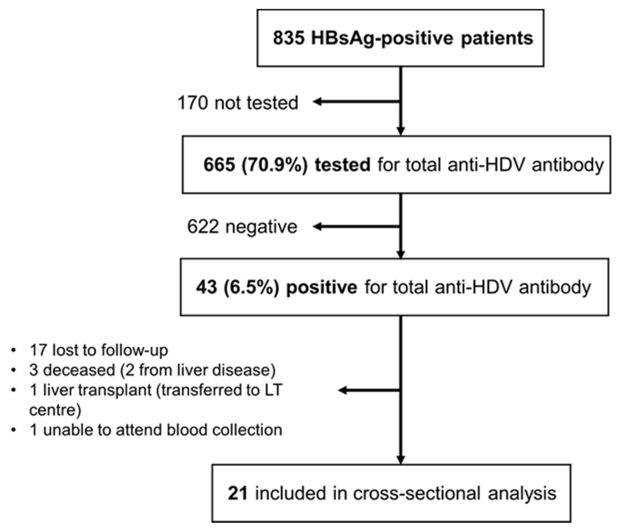
Figure 1. Flowchart showing the total population of HBsAg-positive patients analyzed, the proportion that was screened for anti-HDV antibodies, HDV seroprevalence results and, finally, the cohort that was included in the cross-sectional analysis (HDV RNA determination and genotyping). HDV: hepatitis delta virus; LT: liver transplant.
Livers 04 00028 g001
Table 1. HDV seroprevalence per area and country of origin.
Area and Country of OriginHDV Seroprevalence–Positive/Total (%)
Europe10/258 (3.9%)
   Portugal7/232 (3.0%)
   Central and Eastern Europe3/25 (12.0%)
     Romania1/14 (7.1%)
     Ukraine2/6 (33.3%)
     Bulgaria0/2 (0.0%)
     Croatia0/1 (0.0%)
     Latvia0/1 (0.0%)
     Russia0/1 (0.0%)
   Luxembourg0/1 (0.0%)
Africa32/387 (8.3%)
   Guinea-Bissau29/143 (20.3%)
   Cape Verde1/99 (1.0%)
   Angola0/84 (0.0%)
   São Tomé and Príncipe0/51 (0.0%)
   Mozambique0/5 (0.0%)
   Senegal1/3 (33.3%)
   Equatorial Guinea1/1 (100%)
   Gambia0/1 (0.0%)
Asia0/12 (0.0%)
   China0/5 (0.0%)
   Pakistan0/3 (0.0%)
   India0/2 (0.0%)
   Nepal0/1 (0.0%)
   Bangladesh0/1 (0.0%)
South America1/8 (12.5%)
   Brazil1/7 (14.3%)
   Paraguay0/1 (0.0%)
Total43/665 (6.5%)
Table 2. Clinical characteristics of anti-HDV-positive patients.
Age—M ± SD years41.2 ± 9.9
Male—n (%)12 (57.1%)
HBeAg-negative at diagnosis—n (%)18 (85.7%)
Anti-HDV IgM positive—n (%)1 (4.8%)
Cirrhosis *—n (%)7 (33.3%)
Median liver stiffness (kPa)7.8
Treatment with nucleos(t)ide analogs—n (%)15 (71.4%)
Previous treatment with peginterferon **—n (%)7 (33.3%)
HDV RNA positivity—n (%)8 (38.1%)
HDV genotype 1/genotype 5—n (%)1 (12.5%)/7 (87.5%)
* Liver stiffness ≥ 14 kPa: 5; Ishak score of 5/6: 1; imaging findings of cirrhosis and cholangiocarcinoma: 1 ** HDV clearance: 2; non-response: 4; intolerance: 1. HDV: hepatitis delta virus; M: mean; SD: standard deviation.
Disclaimer/Publisher’s Note: The statements, opinions and data contained in all publications are solely those of the individual author(s) and contributor(s) and not of MDPI and/or the editor(s). MDPI and/or the editor(s) disclaim responsibility for any injury to people or property resulting from any ideas, methods, instructions or products referred to in the content.