1. Introduction
Polysaccharides derived from
Lycium barbarum L. (LL) leaves have recently gained considerable interest because of their exceptionally high levels of bioactive compounds and nutrients. Prior research has demonstrated that polysaccharides derived from the leaves of
Lycium barbarum L. (LBP) have several biological properties, including anti-inflammatory, lipid-lowering, anti-radiation, anti-oxidative, anticoagulant, and blood sugar-regulating effects [
1,
2]. Additionally, the isolated water-soluble polysaccharides from the
Lycium barbarum L. leaves after functional group analysis via FT-IR analysis indicated carboxylate (COO−) and uronic acid moieties in LBP, which facilitate Ag
+ binding and AgNP formation [
3]. Polysaccharides, originating from natural sources, represent a class of hydrophilic polymers that have garnered significant attention due to their remarkable biocompatibility [
3]. These versatile molecules have found diverse applications, particularly in the burgeoning field of nanotechnology. Their intrinsic characteristics, including biodegradability, biocompatibility, and low toxicity, have made them indispensable in various biological systems, contributing to their frequent adoption in nanoparticle (NP) synthesis [
4,
5]. In the context of medical therapy, polysaccharide-based nanoparticles (NPs) hold immense promise [
6]. They possess several advantages, including accommodating high drug loads, exhibiting rapid drug release kinetics, and providing efficient targeting capabilities. Additionally, these NPs demonstrate notable stability and low toxicity when placed within physiological environments [
7]. The advantageous attributes of polysaccharides are not limited to their biocompatibility and biodegradability. These polymers are also celebrated for their abundant availability, ease of processing, and sustainable sourcing. The chemical modification of polysaccharides often involves leveraging the carboxyl and hydroxyl groups present along their backbones [
8]. These reactive groups facilitate the development of customized polysaccharide derivatives with specific properties, such as hydrophobicity and solubility, thereby further enhancing the versatility and utilization of polysaccharides across various application domains [
9].
Precisely, carboxyl and hydroxyl functional groups in polysaccharide-containing groups offer distinct advantages in the synthesis of nanoparticles when compared to their neutral counterparts. The polysaccharide carbonyl groups are versatile, functional entities, readily participating in chemical reactions [
10]. This facilitates precise control over the nanoparticle synthesis process, allowing for the attachment of specific molecules or ligands to the nanoparticles [
11]. Additionally, carboxylated polysaccharides are crucial in stabilizing nanoparticles due to their negatively charged nature. This electrostatic repulsion effectively prevents nanoparticle aggregation, maintaining their stability.
Furthermore, these polysaccharides can control nanoparticle size and shape, enabling fine-tuning of the desired properties [
12,
13]. Polysaccharides with carbonyl groups are well-suited for coating with silver because the carbonyl groups present on these polysaccharides provide binding sites for silver ions (Ag
+), promoting the formation of silver nanoparticles [
14]. These functional groups can chemically interact with silver ions through coordination bonds, facilitating the reduction of silver ions to metallic silver atoms, which subsequently aggregate to form nanoparticles. This controlled reduction process is vital for producing stable, well-dispersed silver nanoparticles [
15]. Using silver nanoparticles, polysaccharides (AgNPs), and naturally occurring polysaccharides together creates a class of biodegradable and environmentally friendly polymers with exceptional and all-around antibacterial properties [
16,
17].
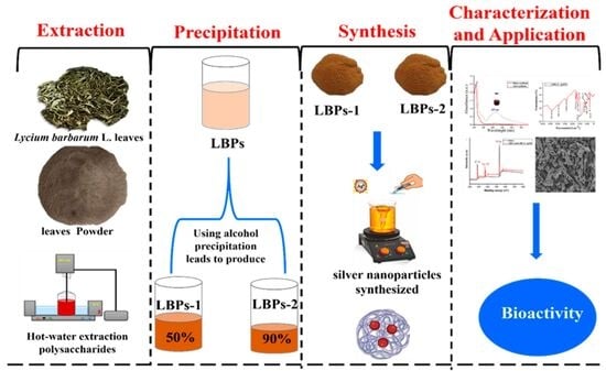
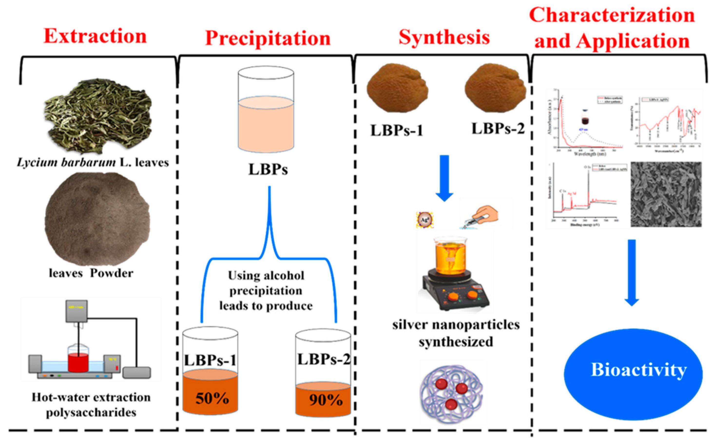
This work exploits the intrinsic chemical characteristics and interactions of water-soluble polysaccharides isolated from the
Lycium barbarum L. leaves (LBP) to engineer drug delivery systems through an environmentally friendly pathway [
18]. To the best of our knowledge, this is the first extensive, systematic characterization of the three-dimensional structure, morphology, and crystallinity of LBP-AgNPs, which not only demonstrates the successful synthesis of well-formed NPs dispersed evenly but also reveals a remarkable difference in their average sizes based on the method of synthesis [
19]. The size-dependent characteristics show an extraordinary potential for the design of customizable functionalities [
20]. Additionally, utilizing these NPs as potent antioxidants expands their application potential to improve the therapeutic activity of existing drugs [
21]. Silver nanoparticles (AgNPs) were specifically selected for this study because they offer unique advantages for antioxidant and drug-delivery applications: (i) intrinsic antimicrobial and redox-modulating properties that complement the bioactivity of polysaccharides, (ii) strong surface plasmon resonance (SPR) that enables easy tracking and characterization, and (iii) controllable surface chemistry when capped with polysaccharides, allowing tunable size, stability, and targeting potential [
22,
23,
24,
25,
26,
27,
28,
29,
30,
31]. The carboxyl (-COO
−) and hydroxyl (-OH) groups in
Lycium barbarum polysaccharides (LBP) act as both reducing agents (converting Ag
+ to Ag
0) and capping agents (stabilizing the nanoparticles), enabling green synthesis of monodisperse, biocompatible AgNPs without toxic chemicals [
32]. This polysaccharide–AgNP combination creates biodegradable nanomaterials with enhanced antibacterial and antioxidant properties suitable for biomedical applications [
33,
34,
35].
Molecular docking was performed to investigate how LBP interacts with bovine serum albumin (BSA), a model carrier protein. This computational approach helps predict binding sites and interaction modes, supporting interpretation of the antioxidant activity and drug-delivery potential observed experimentally [
36,
37].
3. Chemical Characterizations of Synthesized LBP-AgNPs
3.1. UV–Visible Test
The colloidal dispersions of PS-AgNPs were analyzed for their UV-Vis spectra in the 200–900 nm wavelength range using a TU-1901 UV-Vis spectrophotometer from Purkinje General Instrument Co., Ltd., in Beijing, China. For UV-Vis analysis, in addition to the synthesized LBP-AgNPs, several control samples were prepared: (1) a solution of LBP alone, (2) a solution of AgNO3 alone, and (3) a physical mixture of LBP and AgNO3 without allowing the reduction reaction to occur (e.g., by immediate dilution or analysis before significant reaction time). These controls were analyzed under the same conditions as the LBP-AgNPs.
3.2. Fourier Transform Infrared (FTIR) Test
The functional groups present in the samples were identified using Fourier transform infrared spectroscopy (FTIR-8400S, Shimadzu, Kyoto, Japan). The LBP-AgNP sample was combined with KBr and compacted into pellets, and the analysis was conducted within the frequency range of 4000–400 cm−1.
3.3. X-Ray Photoelectron Spectroscopy (XPS) Test
XPS spectroscopy was performed to confirm the existence of the elements and the chemical state on the nanoparticle surface. The narrow scan spectra define the successful introduction of silver nanoparticles on the surface of the LBP-AgNPs by radiation, where the new doublet peak of Ag (0) is at 368 eV (3d5/2) and 374 eV (3d3/2).
3.4. Transmission Electron Microscopy (TEM) Test
The nanoparticle samples were separated into pellets using centrifugation at 8964 RCF for 30 min in a Thermo Scientific Sorvall ST 8 centrifuge (Thermo Fisher Scientific, Waltham, MA, USA). After that, the pellets were mixed with distilled water and sonicated to disperse any aggregates. The microstructures of the biogenic LBP-AgNPs were observed using a Joel 6360LA transmission electron microscope (JEOL Ltd., Akishima, Tokyo, Japan).To make the recovered LBP-AgNPs, 5 L of the colloid solution was applied to a carbon-coated 3 mm copper grid using double-sided sticky tape. The excess sample was removed using filter paper and left to dry at room temperature. The evaluation was performed using ImageJ software (Version 1.54r, National Institutes of Health, Bethesda, MD, USA).
3.5. X-Ray Diffractometer (XRD) Test
The X-ray diffractometer (D8 Advance, Bruker AXS, Karlsruhe, Germany) was used to examine the crystalline structure of the biosynthesized LBP-AgNPs. It was operated at 40 kV using Cu-Kα radiation with λ of 1.54 Å and a scanning rate of 0.1° in the 2θ range from 5° to 80°. The elemental composition of the LBP-Ag-NPs was identified by scanning electron microscopy (JEOL JSM-6510LV JEOL Ltd., Akishima, Tokyo, Japan) with energy-dispersive spectroscopy (EDS) attached between 0 and 20 kV.
3.6. Scanning Electron Microscopy (SEM) Test
The size and shape of the LBP-AgNPs produced were examined using a scanning electron microscope (SEM), specifically the JEOL JSM-6510LV model (JEOL Ltd., Akishima, Tokyo, Japan). The SEM operated at an accelerating voltage of 30 kV, and a very small sample was placed on a copper grid coated with carbon. The sample was allowed to dry at room temperature before SEM analysis.
3.7. Atomic Force Microscopy (AFM) Test
The size, shape, and spatial distribution of LBP-AgNPs have been identified using AFM analysis using the FLEX-AFM device (Nanosurf AG, Liestal, Switzerland) [
18].
3.8. Thermogravimetric Analysis (TGA) Test
The thermal stability of the LBP-AgNPs was evaluated using a DSC-200PC instrument (NETZSCH, Gerätebau GmbH, Selb, Germany) through thermogravimetric analysis (TGA) in the presence of air. The samples were heated from 25 to 900 °C at a rate of 10 °C/min [
19].
5. Results and Discussion
5.1. Characterization of LBP-1 and LBP-2 Polysaccharides
To address the reviewer’s request for characterization data of LBP-1 and LBP-2, we refer to our previous comprehensive study on the crude
Lycium barbarum L. leaf polysaccharide (CLP) [
35]. The LBP preparations (LBP-1 and LBP-2) used in the current study are derived from this same source and are considered to possess largely similar fundamental structural characteristics as the previously characterized CLP.
As detailed in Al-Wraikat et al. (2022) [
35], the CLP exhibited a molecular weight (Mw) of 223.5 ± 1.13 kDa, a uronic acid content of 13.7 ± 1.7%, and a protein content of 2.5 ± 0.1%. The monosaccharide composition of CLP was determined to be mannose, ribose, glucuronic acid, glucose, xylose, and galactose at molar ratios of 4.1, 1.7, 3.1, 1.5, 3.26, and 1.9, respectively. These key characteristics are summarized in
Table 1.
The low protein content (2.5 ± 0.1%) in the LBP preparations indicates a high degree of polysaccharide purity. This minimal protein contamination is crucial, as it strongly suggests that proteins are unlikely to be the primary reducing or stabilizing agents in the silver nanoparticle synthesis process. Instead, the green synthesis of silver nanoparticles using LBP is predominantly driven by the inherent reducing and stabilizing properties of the polysaccharide’s functional groups. Specifically, these are the hydroxyl (-OH) and carboxyl (-COO-) groups, characteristic of the uronic acid residues within the polysaccharide structure, act as effective reducing agents for silver ions (Ag
+) to metallic silver (Ag
0) and subsequently stabilize the formed nanoparticles, preventing aggregation. This mechanism is further supported by the observed shifts in FTIR spectra (
Section 5.3.2) and XPS analysis (
Section 5.3.3), which indicate direct interaction between the polysaccharide functional groups and the silver nanoparticles.
While the AgNPs synthesized from LBP-1 and LBP-2 show some differences in size and antioxidant activity (as discussed in subsequent sections), these are likely attributable to subtle variations in the specific conditions during nanoparticle synthesis or minor batch-to-batch differences in polysaccharide preparations, rather than major structural disparities in the parent polysaccharides themselves.
5.2. Synthesis Observations and Initial Characterization
The successful formation of LBP-AgNPs was visually indicated by a distinct color transformation of the solution from pale yellow to a characteristic yellowish-brown. This color change, observed within 2 h of continuous stirring and progressing over the 6 h reaction period, is a well-known indicator of the reduction of silver nitrate to silver nanoparticles due to the excitation of surface plasmon resonance (SPR).
5.3. Characterization Analysis Result of LBP-AgNPs
5.3.1. UV-Visible Absorption Analysis
As shown in
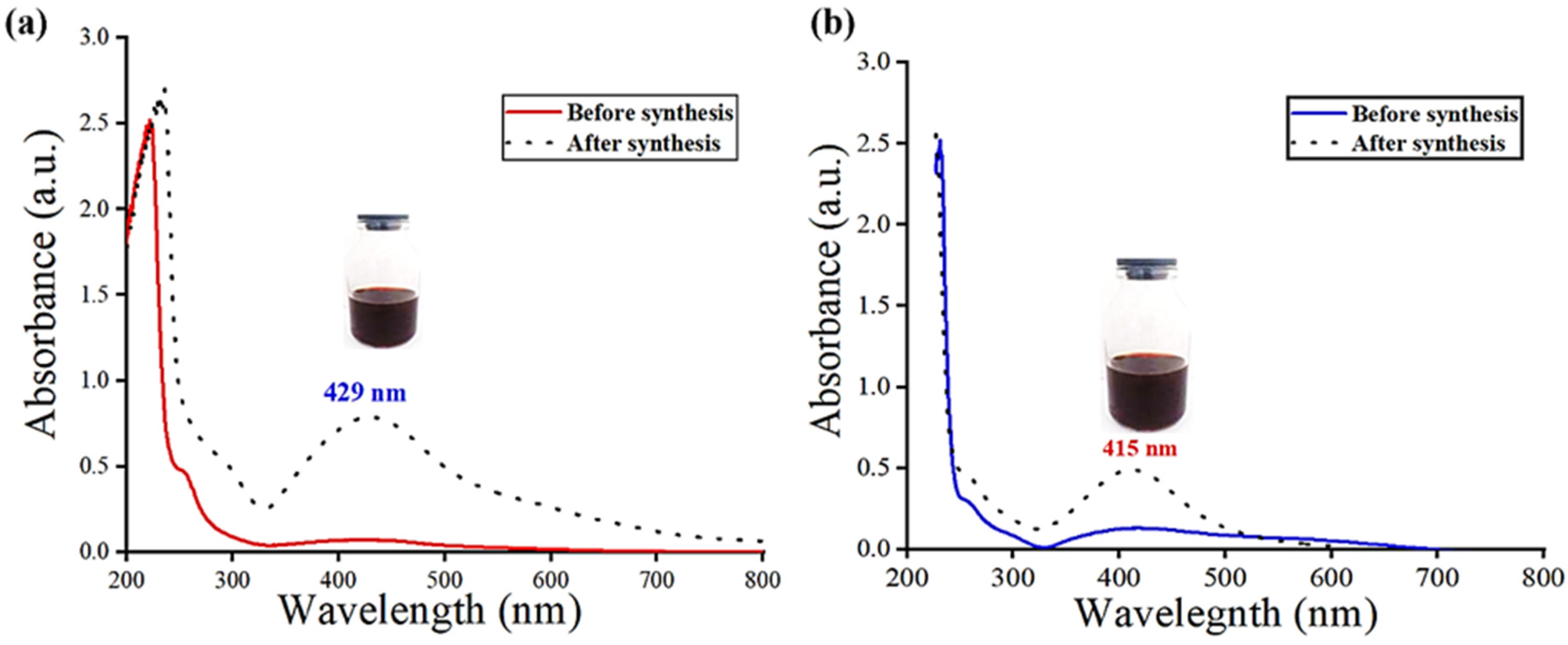
Figure 1, the production of silver nanoparticles in an aqueous solution was first examined using ultraviolet-visible spectroscopy. To create LBP-AgNPs, the polysaccharides and AgNO
3 were then treated to a bio-reduction reaction, which resulted in a color shift to a yellowish brown. At 428 nm, the silver surface plasmon resonance was discovered [
22]. Generally, AgNO
3’s absorbance is 400–450 nm, and the reduction of AgNO
3 to the polysaccharides is due to the hydroxyl group -OH in the carboxylic functional group [
23]. The successful formation of LBP-AgNPs was visually indicated by a distinct color transformation of the solution from pale yellow to a characteristic yellowish-brown. This color change is a well-known indicator of the reduction of silver nitrate to silver nanoparticles due to the excitation of surface plasmon resonance (SPR).
As shown in
Figure 1, UV-visible spectroscopy confirmed the production of silver nanoparticles. The LBP-AgNPs displayed a prominent SPR absorption peak at 428 nm. In contrast, control spectra for the polysaccharide alone and AgNO
3 alone showed no significant absorption in the 400–450 nm range, confirming that the observed peak is specific to the formation of nanoparticles. The intensity of the peak increased over the 6 h reaction period, reflecting the progressive reduction of silver ions by the LBP functional groups.
5.3.2. FTIR Analysis
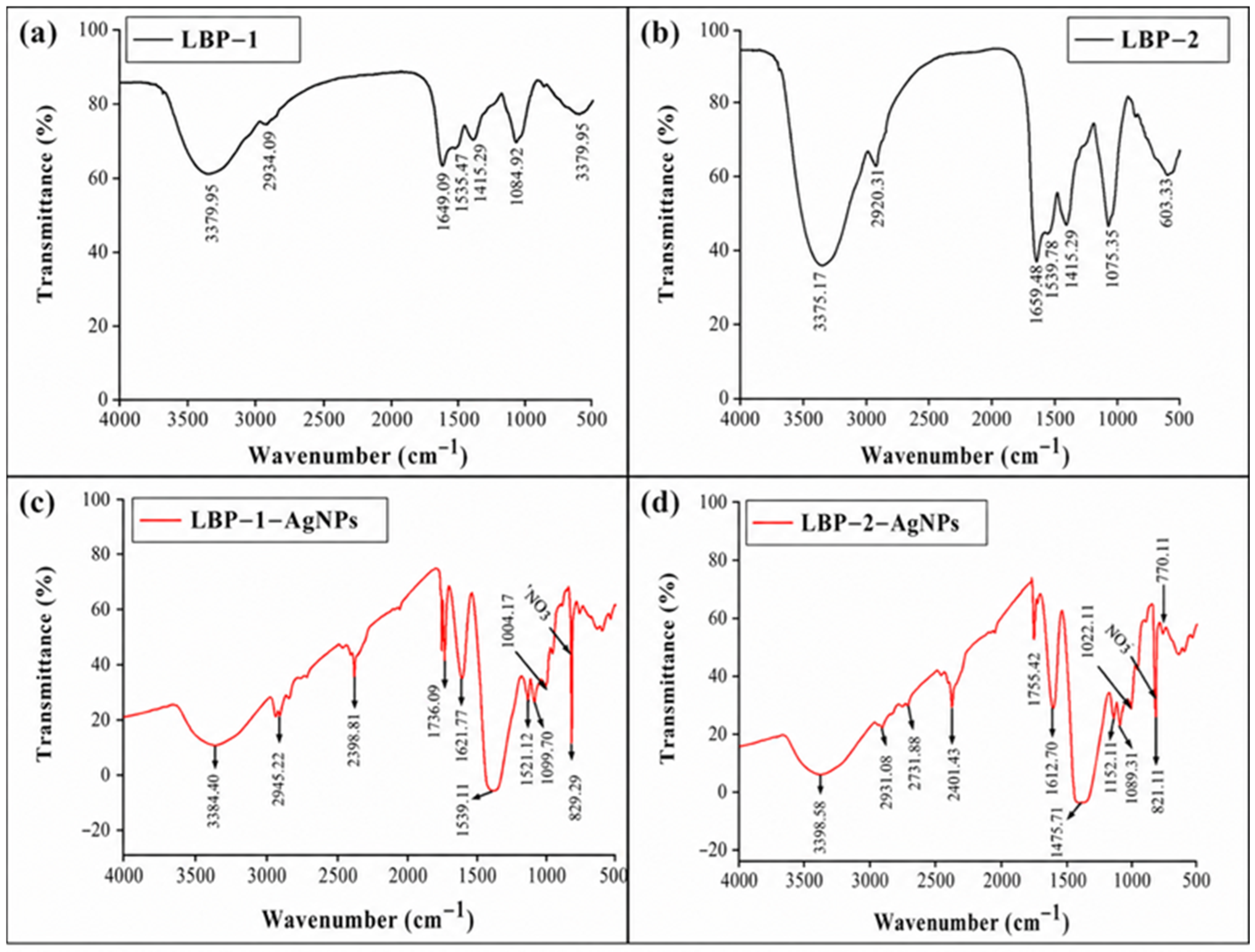
Employing acidic polysaccharides in silver nanoparticle (AgNP) synthesis induces noticeable alterations in Fourier-transform infrared spectroscopy (FTIR) results. The formation of LBP-AgNPs causes significant shifts in FTIR spectra, indicating their interaction, as illustrated in
Figure 2. These shifts suggest potential binding of polysaccharide functional groups to nanoparticle surfaces. Moreover, novel spectral characteristics appear, attributed to vibrational modes linked with AgNPs or interactions between nanoparticles and polysaccharides. Furthermore, changes in peak intensities are observable, indicating structural adjustments or the creation of new chemical bonds between the polysaccharides and AgNPs.
Variations in the broadness or position of hydroxyl -OH bands, characteristic of polysaccharides, indicate changes in hydrogen bonding or interactions between polysaccharide hydroxyl groups and LBP-AgNP. Moreover, LBP-AgNP formation often involves interactions with carboxylate groups -COO- present in polysaccharides with uronic acid, leading to reduced intensity or disappearance of carboxylate-related FTIR bands. The presence of the -COO- functional group, which belongs to the carbonyl groups of uronic acid residues and the oxidizable hydroxyl group of polysaccharides, was detected by FTIR spectroscopy in both fractions of capped silver nanoparticles [
24]. Furthermore, the observed elimination or disappearance of certain peaks suggests significant interactions between AgNO
3 and the polysaccharide fractions.
5.3.3. XPS Analysis
In the synthesis of silver nanoparticles AgNPs with LBP polysaccharides, X-ray photoelectron spectroscopy (XPS) reveals significant changes. XPS analyzes material chemistry and surface properties, particularly within composite systems. In the case of acidic polysaccharide LBP-AgNP composites, XPS uncovers shifts in surface composition, which is evident through new peaks or binding energy alterations. These changes signify the presence of AgNPs on the material’s surface, indicating silver integration. XPS further identifies modifications in chemical states, binding energies, and valence states of elements (e.g., carbon, oxygen, silver) due to interactions between acidic polysaccharides and AgNPs. Depth profiling in XPS delineates element distribution, showcasing variations in silver and polysaccharide distribution with depth. This analysis enhances comprehension of the composite’s chemical attributes, structure, and interactions. C1s, O1s, and Ag3d XPS spectra were measured to gain an understanding of the adsorption of RCH2OCOO from galacturonic acid onto the LBP-AgNP surface derived from LBP-1 and the substances used in this study are LBP-2 AgNPs. In addition to being derived from certain powdered samples, the results are shown in
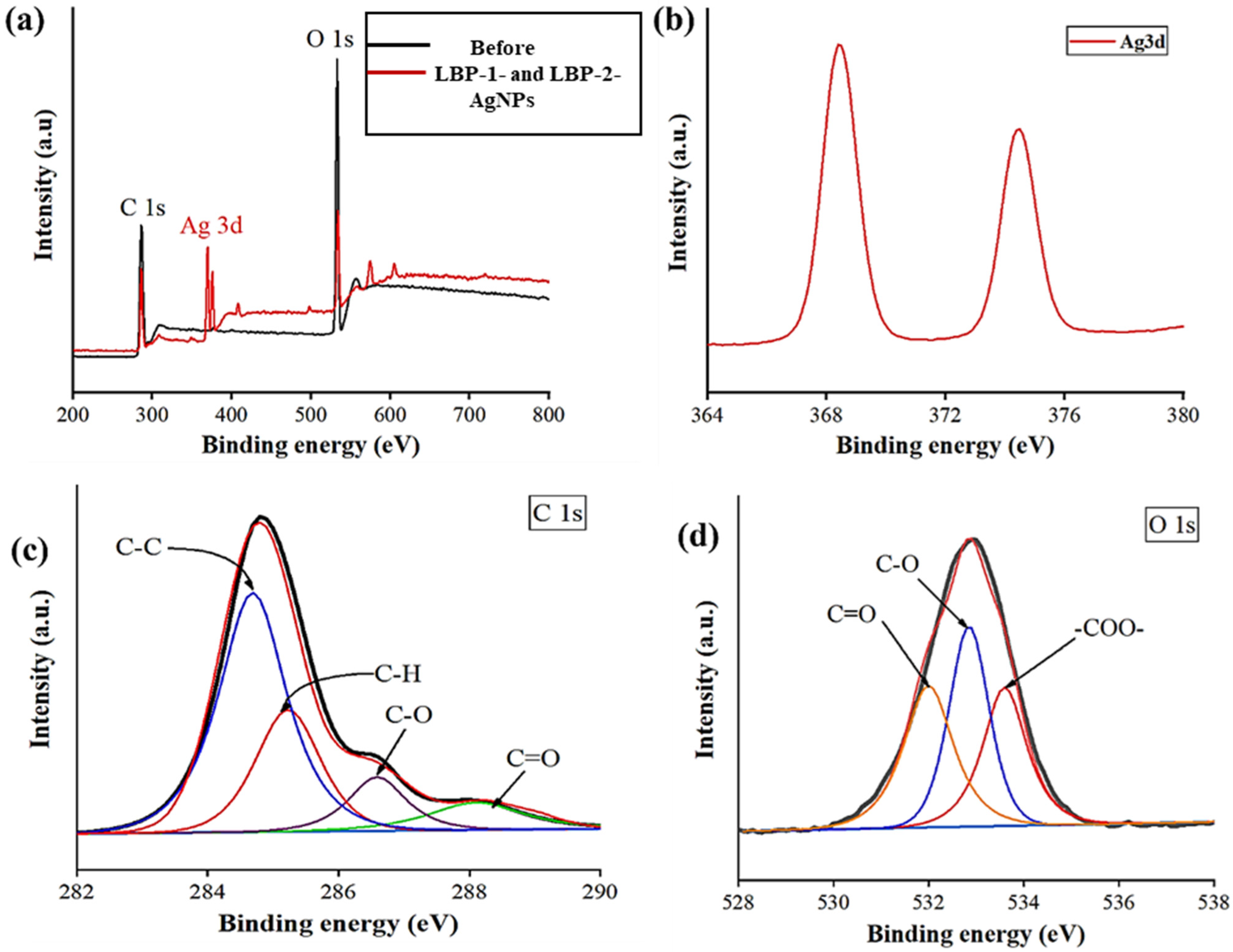
Figure 3. In the two fractions in
Figure 3a, the results of the XPS spectra survey before and after interaction with the silver are shown. The C1s XPS spectra of LBP-1 and LBP-2-AgNPs shown in
Figure 3c show four peaks: 284.6, 285.2, 286.6 and 288.1 eV in LBP-AgNPs, respectively, which are ascribed to C in C-C, C-H, C-O, and C=O, respectively. The O1s spectrum exhibited that there are three peaks at 532.0, 532.8, and 533.6 eV in LBP-1-AgNPs and 531.9, 532.7, and 533.5 eV related to C-O, C=O, and COOH, respectively; the presence of -COO- groups is related to the acidic residues in the polysaccharide’s fractions LBP-1 and LBP-2.
These samples’ Ag3d spectra also show peaks at 368.4 eV, associated with photoelectrons released from the 3d3/2 and 3d5/2 states, respectively. The energy required to bind such emissions differs by (6 eV) in all cases, proving the presence of metallic silver, Ag
0, in the samples [
25,
26]. Overall, XPS is a powerful tool for investigating the surface chemistry of polysaccharide-encapsulated AgNPs and can provide essential insights into their properties and performance. The successful removal of free silver ions and the complete reduction to metallic silver were confirmed through UV-Vis spectroscopy and XPS analysis. In the UV-Vis spectra (
Figure 1), the disappearance of the characteristic AgNO
3 absorption and the appearance of a sharp surface plasmon resonance (SPR) peak at 415–429 nm indicates the formation of nanoparticles. Furthermore, the XPS Ag3d spectra (
Figure 3b) showed peaks at 368.4 eV, which are specifically associated with metallic silver (Ag
0). The absence of significant peaks corresponding to silver oxides or unreduced Ag
+ ions in the purified samples confirms that the antioxidant activity is attributable to the LBP-AgNPs rather than free silver ions.
5.3.4. SEM Analysis
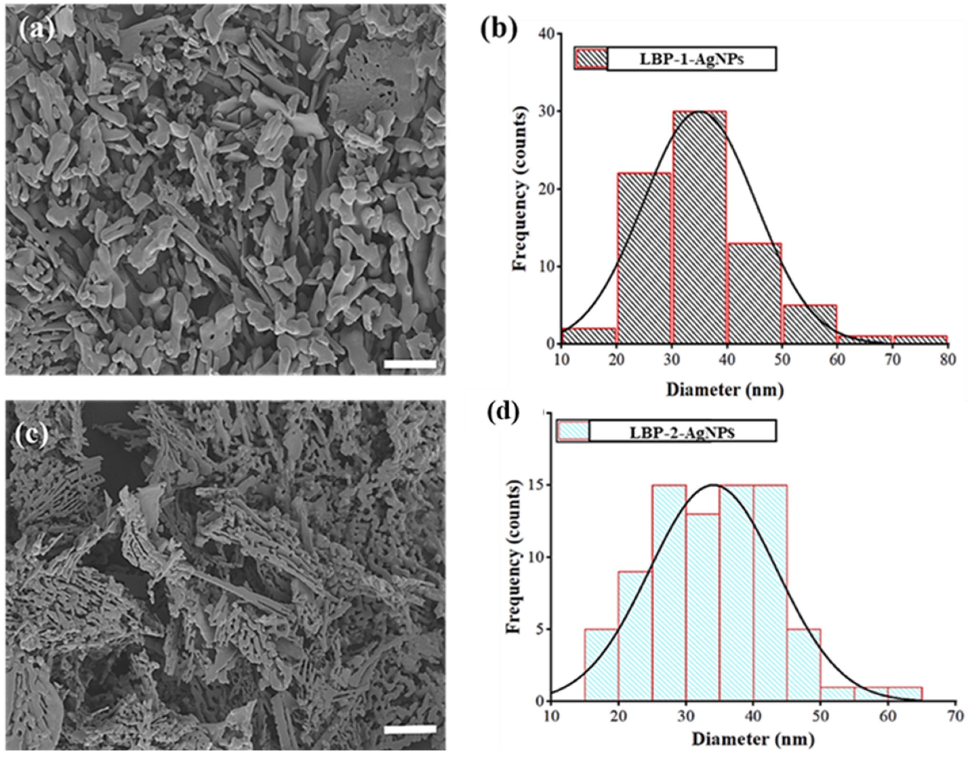
The surface characteristics of LBP-AgNP polysaccharides were analyzed using scanning electron microscopy (SEM). The SEM images show a uniform surface with randomly distributed particles, as illustrated in
Figure 4. Additionally, the SEM analysis was carried out to determine the structure and size of the nanoparticles. According to the results of the current investigation, the mean average size of LBP-AgNPs was determined to be 10–80 nm, as shown in
Figure 4a,c. For several biological applications, including targeted medication delivery and nano-drug formulations, complete reporting of the morphology and size of the nanoparticles is crucial [
27,
28]. The LBP-1-AgNPs showed an average size of 36.2 ± 1.03 nm and the LBP-2-AgNPs were 49.9 ± 1.03 nm. To provide a statistically significant representation of the nanoparticle size, the diameters of at least 100 individual nanoparticles were measured from multiple TEM and SEM images using ImageJ software. The resulting size distribution (as shown in the histograms in
Figure 4b,d) confirms that LBP-1-AgNPs had a mean diameter of 36.2 ± 1.03 nm, while LBP-2-AgNPs were slightly larger with a mean diameter of 49.9 ± 1.03 nm. The narrow size distribution confirms the monodisperse nature of the synthesized nanoparticles.
5.3.5. TEM Analysis
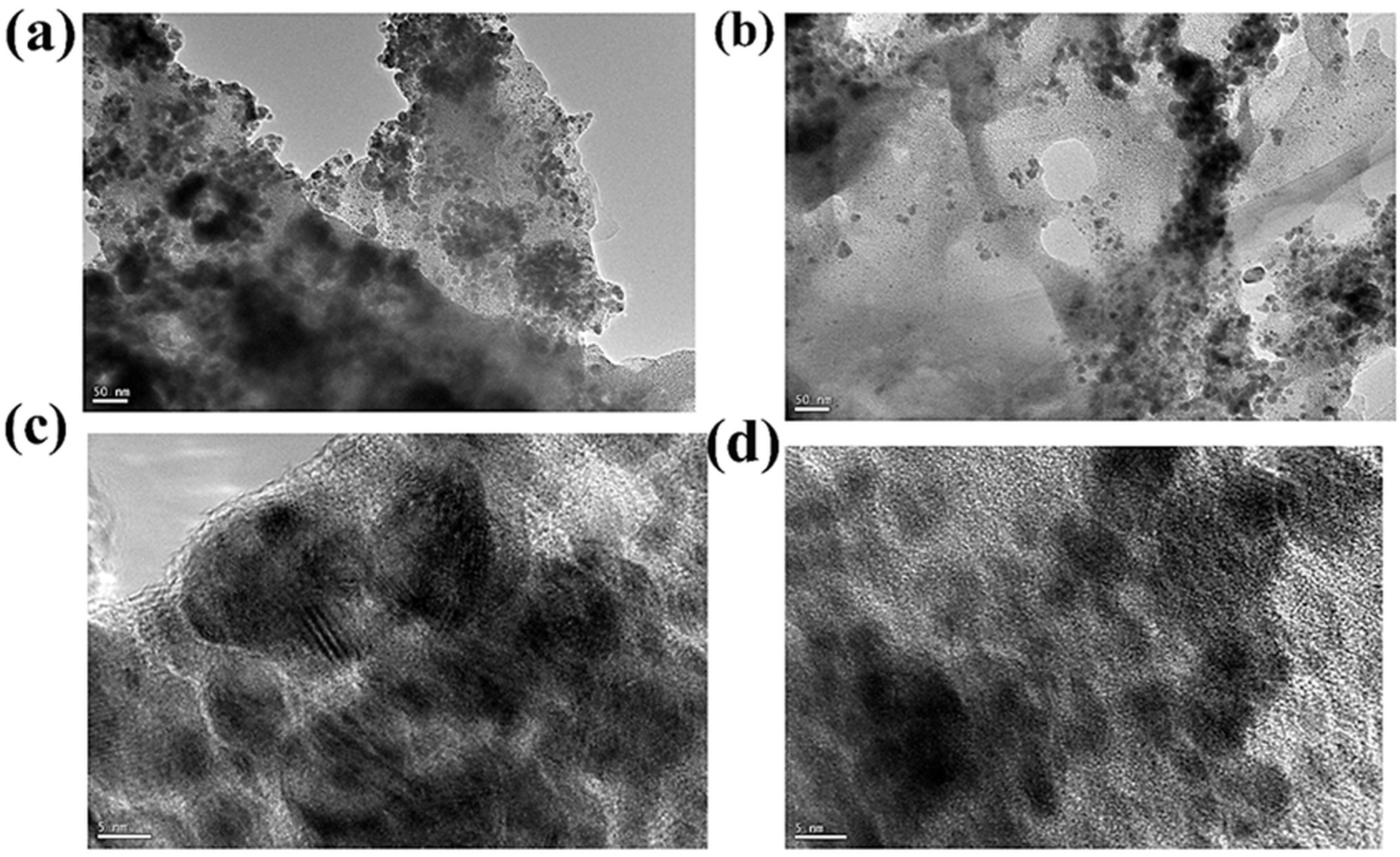
Transmission electron microscopy (TEM) is a qualitative tool for examining the surface morphology of polysaccharides, as shown in
Figure 5 [
29]. The TEM analysis of LBP-1—AgNPs and LBP-2—AgNPs revealed that the particles were predominantly spherical and ranged in size from 10 to 80 nm, confirming their monodisperse nature.
These sub-micron-sized particles are well-suited for use as drug carriers or in the delivery of various biomolecules. At low magnification, clusters of polysaccharides doped with AgNPs could be observed. Moreover, the high-magnification image clearly shows the nanocomposite structure at the nanoscale, where particles as small as 100 nm and LBP-AgNPs were distinctly visible as nanocomposite particles. Additionally, the diffraction pattern highlighted the nanocomposite’s crystalline structure, which could result from adding LBP-AgNPs to a tertiary composite. The production of tritely composite doped LBP-AgNPs was successful, and instrumental analysis showed that the size of the nanocomposite particles was less than 100 nm [
30].
5.3.6. XRD Analysis
The XRD measurement is valuable for analyzing newly produced compounds and their phases [
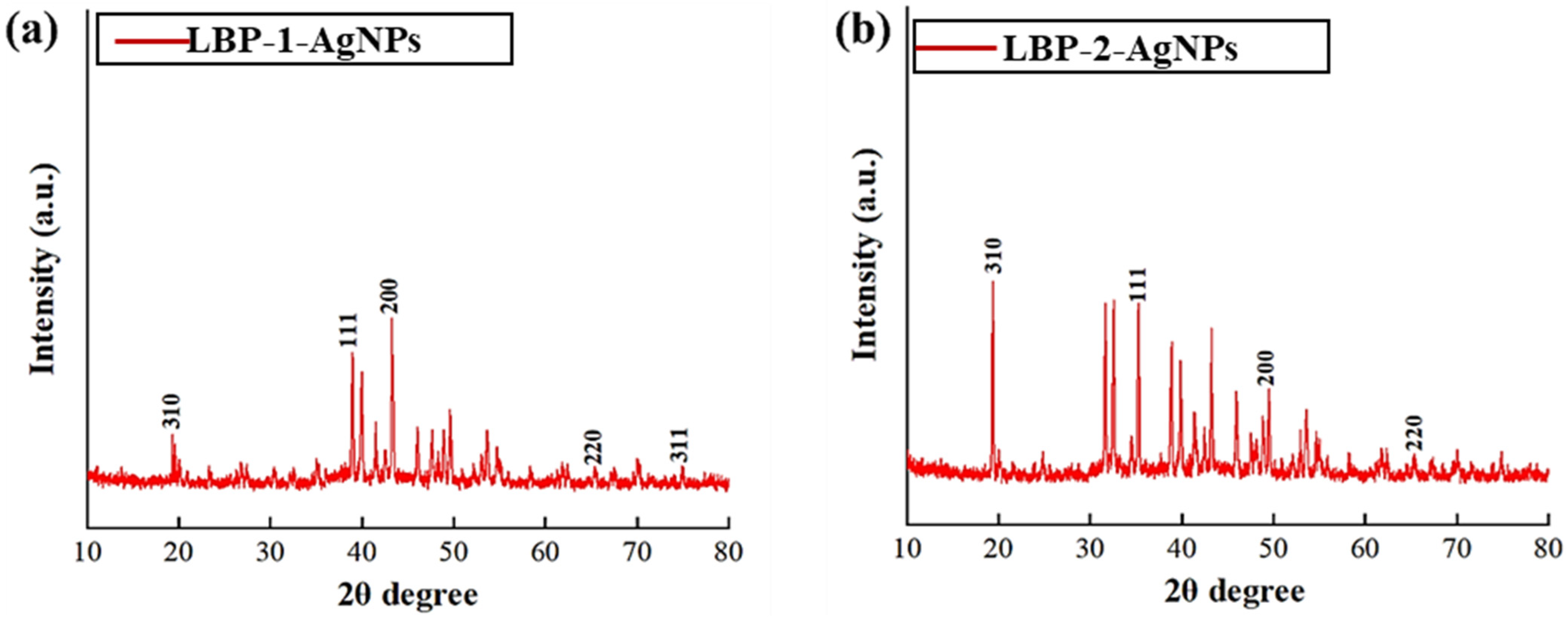
31]. X-ray diffraction (XRD) further confirmed the generation of Ag0. Vacuum-dried silver nanoparticles’ XRD patterns can be examined to find sharp diffraction lines at low angles of 5–80° shown in
Figure 6. The crystallinity of LBP-AgNPs shown in
Figure 6a,b was established by XRD analysis; the spectra of LBP-AgNPs displayed four identical peaks appearing at 2θ = 38°, 44°, 64° and 78° conforming to the (111), (200), (220) and (311) facets of silver, respectively [
32].
5.3.7. AFM Analysis
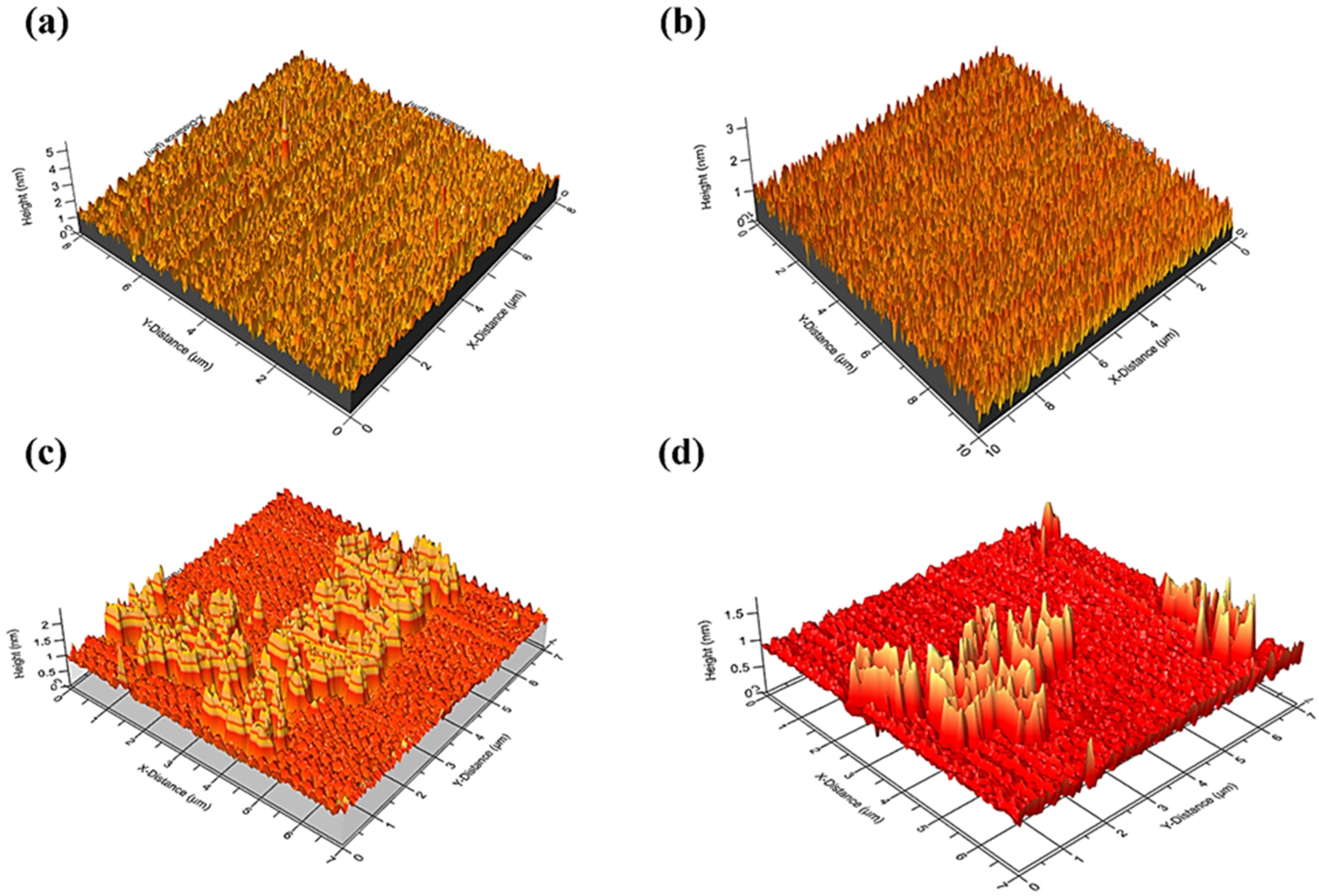
As shown in
Figure 7, recently, AFM probe analysis for measuring nanoparticles has been used [
33]. The AFM probe then scans across the surface of the sample, detecting changes in height and producing a 3D topographical map of the sample surface. AFM, after synthesis, showed an apparent change in the bumps and aggregations of LBP-1 and LBP-2, as shown in
Figure 7a,b, which resulted in less distribution and roughness. These results indicated the successful construction of LBP-AgNP, as shown in
Figure 7c,d. As shown, the surface in AFM of the LBP-AgNPs is consistent with the average of the particles in the SEM result. Overall, AFM analysis can be a powerful tool for characterizing the size, shape, and distribution of silver nanoparticles within a polysaccharide matrix, as well as the mechanical properties of the composite. This information can be helpful for understanding the behavior of the composite in various applications, such as in antimicrobial coatings or wound dressings.
5.3.8. TGA Results Analysis
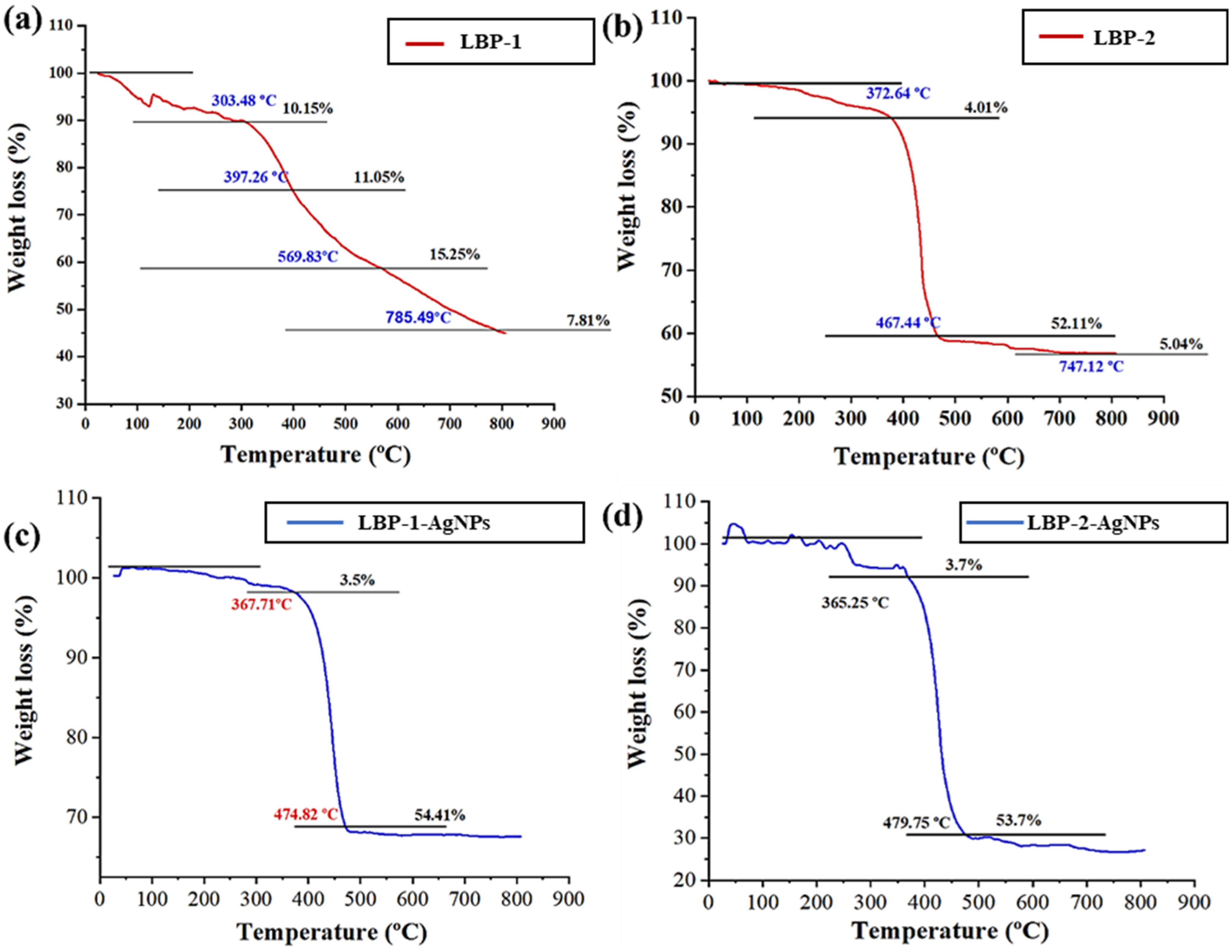
The LBP-AgNPs synthesis alters thermal decomposition behavior per thermogravimetric analysis (TGA), as shown in
Figure 8. TGA assesses material stability, revealing shifts in decomposition profiles, onset temperatures, rates, and patterns. These signify interactions between acidic polysaccharides and AgNPs, influencing each other’s thermal stability. After analyzing known quantities of LBP-AgNP samples using TGA, we observed distinct hydrogen-bonded water degradation stages. LBP exhibited initial stages at 303.48 °C and 372.64 °C, shown in
Figure 8a,b, while LBP-AgNPs displayed them at 367.71 °C and 365.25 °C shown in
Figure 8c,d.
This distinction is directly associated with the findings from the TGA results, showcasing the dynamic changes in the thermal behavior of acidic polysaccharides in response to the synthesis process involving nano-silver polysaccharides.
5.4. Antioxidant Activity Analysis
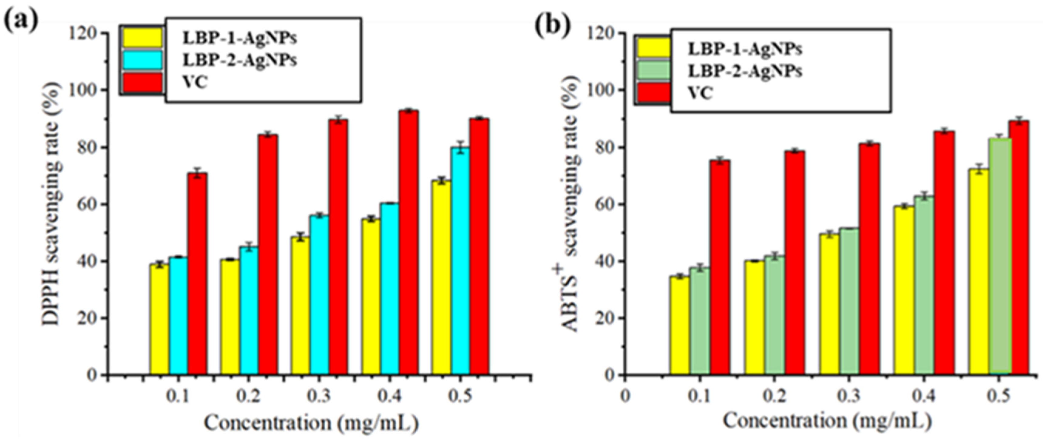
After synthesizing LBP-AgNPs from polysaccharides, a significant change in antioxidant activity was noted, as shown in
Figure 9. The DPPH free radical scavenging ability of LBP-AgNPs showed notable differences at both low and high inhibition points compared to the original acidic polysaccharides
Figure 9a. At a 0.1 mg/mL dose, VC showed an inhibition rate of 71.02 ± 1.56%, while LBP-1-AgNPs were 39.00 ± 1.06%, and LBP-2-AgNPs were 41.60 ± 1.17%. At 0.5 mg/mL, VC showed an inhibition rate of 90.23 ± 0.47%, while LBP-1-AgNPs and LBP-2-AgNPs exhibited rates of 68.44 ± 1.12% and 80.01 ± 2.13%, respectively. In
Figure 9b, with a 0.1 mg/mL dose of ABTS
+, VC exhibited an inhibition rate of 75.43 ± 1.19%, while LBP-1-AgNPs were 34.79 ± 0.89%, and LBP-2-AgNPs were 37.78 ± 1.23%. At 0.5 mg/mL, VC showed an inhibition rate of 89.34 ± 1.23%, although LBP-1-AgNPs and LBP-2-AgNPs had rates of 72.45 ± 1.56% and 83.11 ± 1.43%, respectively. To ensure that the observed radical scavenging activity was due to the synthesized nanoparticles and non-residual reactants, only purified and washed LBP-AgNPs were used in the DPPH and ABTS
+ assays. The significant inhibition observed (up to 80–83% for LBP-2-AgNPs) demonstrates the potent antioxidant efficacy of the polysaccharide-stabilized silver nanoparticles.
While DPPH and ABTS+ are the primary assays used in this study, they were selected because they cover both organic (DPPH) and inorganic (ABTS+) radical species, providing a comprehensive overview of the total antioxidant capacity (TAC) of the LBP-AgNPs. These assays are widely recognized as standard, reproducible methods for evaluating the electron-transfer and hydrogen-atom-transfer capabilities of green-synthesized nanomaterials. The high scavenging rates observed in both assays provide sufficient evidence of the antioxidant potential of LBP-AgNPs for biomedical applications.
5.5. Molecular Docking Analysis
Docking was performed as described in
Section 2.4 to investigate how LBP interacts with bovine serum albumin (BSA), a model carrier protein for predicting circulation behavior and cellular uptake. This computational approach helps predict binding sites and interaction modes, supporting interpretation of the antioxidant activity and drug-delivery potential observed experimentally.
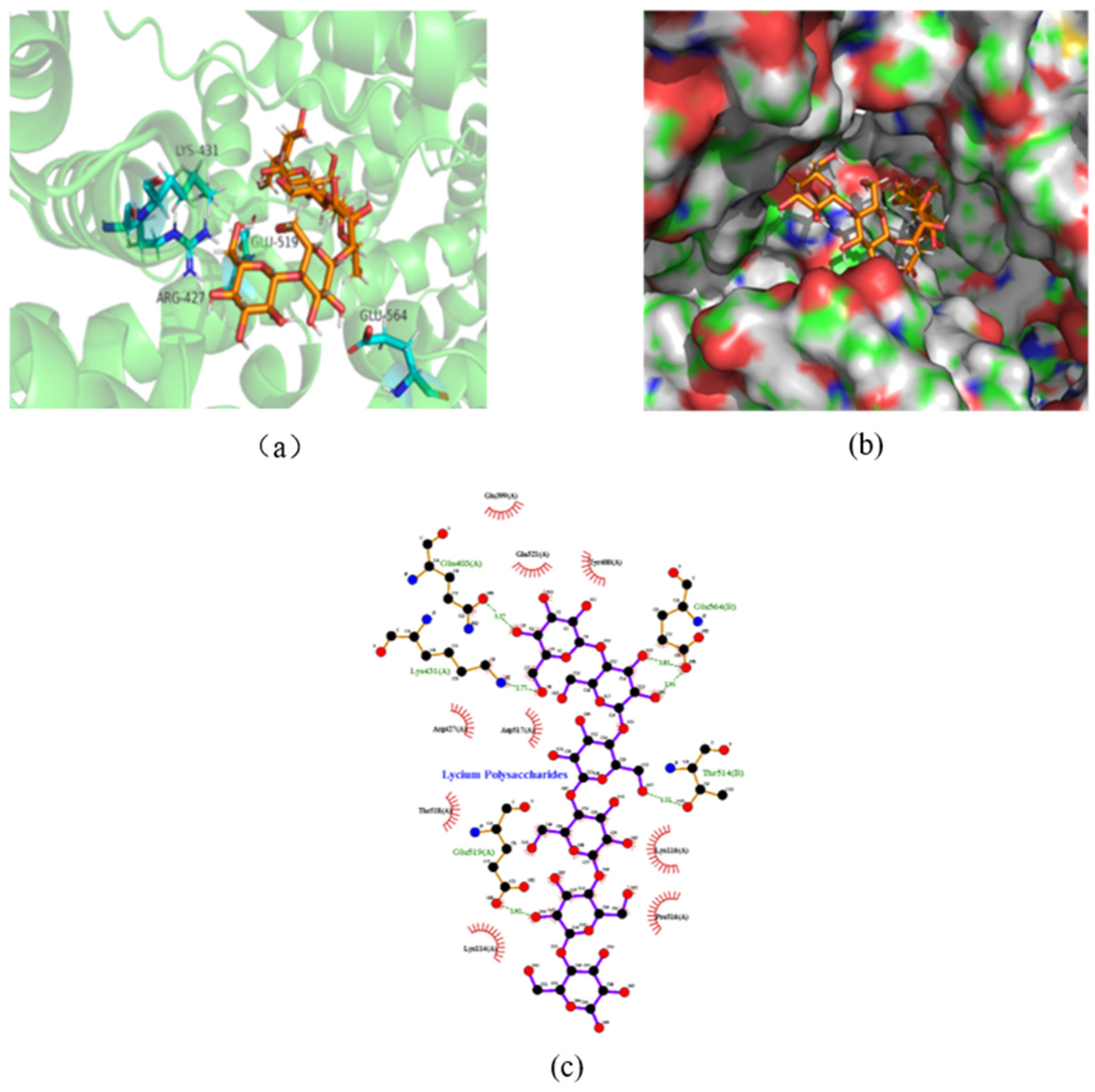
Molecular docking analysis revealed critical amino acid residues at the LBP–BSA binding interface. Hydrogen bond interactions were observed between LBP and Glu564, Thr514, Glu519, Lys431, and Gln403 (
Figure 10a–c). Hydrophobic interactions further stabilized the complex through contacts with Lys116, Pro516, Lys114, Thr518, Arg427, Asp427, Glu399, Gln521, and Tyr400.
Notably, hydrogen bond lengths between the phenolic hydroxyl groups on the LBP structure and Thr514, Glu519, Lys431, and Gln403 were 3.11 Å, 2.92 Å, 2.77 Å, and 3.17 Å, respectively. Glu564 interacted with the sugar ring’s hydroxyl groups with hydrogen bond lengths of 3.01 Å and 2.91 Å.
These findings elucidate the non-covalent interactions between LBP and BSA, providing valuable insights into the molecular-level binding mechanism. The observed hydrogen-bonding network suggests that LBP can bind BSA at surface-exposed sites, which may influence nanoparticle–protein corona formation, modulate systemic circulation half-life, and affect cellular uptake and antioxidant delivery. This supports the hypothesis that polysaccharide capping enhances colloidal stability and offers surface ligands that mediate protein interactions relevant to in vivo behavior.