Article

Chitosan Combined with Methanolic Plants Extracts: Antifungal Activity, Phytotoxicity and Acute Toxicity

by
Sofía de Gante-de la Maza
1,
Maribel Plascencia-Jatomea
1,*,
Mario Onofre Cortez-Rocha
1,
Reyna Isabel Sánchez-Mariñez
2,
Salvador Enrique Meneses-Sagrero
3,
Alma Carolina Gálvez-Iriqui
1 and
Ana Karenth López-Meneses
2,*
1
Departamento de Investigación y Posgrado en Alimentos, Universidad de Sonora, Hermosillo 83000, Mexico
2
Departamento de Ciencias Químico-Biológicas, Universidad de Sonora, Hermosillo 83000, Mexico
3
Departamento de Agricultura y Ganadería, Universidad de Sonora, Hermosillo 83323, Mexico
*
Authors to whom correspondence should be addressed.
Polysaccharides 2025, 6(2), 52; https://doi.org/10.3390/polysaccharides6020052
Submission received: 15 March 2025 / Revised: 16 April 2025 / Accepted: 16 June 2025 / Published: 18 June 2025

Abstract

:
Anthracnose is a disease caused by phytopathogenic fungi such as Colletotrichum siamense that attacks plants and fruits causing great postharvest losses. Different alternatives for the control of this fungus have been studied. In the present study, we evaluated the in vitro antifungal activity of the methanolic extracts of Baccharis glutinosa (ExB) and Jacquinia macrocarpa (ExJ) individually, as well as in combination with chitosan (CS), along with their toxicity in different models. Using the radial growth technique, it was observed that the mycelial development of C. siamense was altered and reduced during exposure to the different treatments evaluated during the first hours of incubation, indicating a fungistatic effect. While the cell viability, by colorimetric assay using the XTT salt, showed alteration since the chitosan reduced proliferation by 50%, while the plant extracts and their mixtures with chitosan reduced approximately 40% indicating cell damage, which was confirmed by fluorescence microscopy. In addition, toxicity tests demonstrated that the J. macrocarpa extract significantly affected the germination percentage of Lactuca sativa seeds, whereas radicle length was reduced in all treatments except for chitosan. The larval survival test for Artemia salina with the extracts indicated their potential toxicity by causing up to 60% mortality. The results indicate that ExB and ExJ mixed with CS are a good option for controlling C. siamense; however, at the concentrations used, they exhibit a toxic effect on the evaluated models.

1. Introduction

Plant pathogenic fungi are one of the main infectious agents in plants, causing significant crop yield losses worldwide [1]. Genera Colletotrichum, Botrytis, Fusarium and Rhizopus cause the most frequent diseases in fruits and vegetables, the most characteristic symptom being necrosis, which is the structural or functional deterioration of tissues due to the death of their cells [2,3]. In Mexico, 46 species of Colletotrichum spp. have been documented, and interest in researching C. siamense is increasing due to several cases of anthracnose affecting mango, papaya, and avocado [4,5,6]. Anthracnose is controlled with synthetic fungicides whose toxicity makes them a risky practice, since their prolonged use causes harmful effects on the environment, on the biota, on the appearance of resistance by these pathogens, and they are not economically profitable [7]. Due to this, safer, sustainable, non-toxic alternatives and integrated management practices that preserve biodiversity, soil quality, human health, and limit the proliferation of these microorganisms and their diseases are being explored [8]. Among these alternatives is the use of natural products with antifungal activity such as plant extracts and chitosan [9,10]. Chitosan (CS) is a biopolymer derived from the chitin of the exoskeleton of crustaceans and insects. This polysaccharide is used in the food industry as a preservative for its ability to inhibit the growth of microorganisms, while in agricultural applications it is used as a crop biostimulant, growth promoter and activator of plant immune response defenses [11,12,13]. Some studies have been reported demonstrating the antifungal effect of chitosan. Li et al. [14], reported the efficacy of chitosan in the control of anthracnose on mango caused by C. gloeosporoides. Additionally, Nascimento et al. [15], found fungistatic and fungicidal effect against C. gloeosporioides, as well as inducing morphological changes in its spores and hyphae. Other alternatives for the control of fungi in food are the extracts of Baccharis glutinosa and Jacquinia macrocarpa, two plants native to the state of Sonora, México. Rosas-Burgos et al. [16] reported that the methanolic extract of B. glutinosa has antifungal activity against phytopathogenic and toxigenic fungi. On the other hand, methanolic extracts of B. glutinosa and ethanolic of J. macrocarpa showed antifungal activity against F. verticillioides, Aspergillus flavus and A. parasiticus, which indicates that they are a viable alternative for the combat of phytopathogenic fungi [17]. Based on the above, the aim was to evaluate the effect of chitosan and the methanolic extracts of J. macrocarpa and B. glutinosa, individually and in combination, on the growth of C. siamense.

2. Materials and Methods

2.1. Reagents and Materials

Chitosan (CS) was purchased from Sigma-Aldrich, the routine examination indicated an average viscosimetric molecular weight (Mv) of 105.8 kDa and a deacetylation degree of 68% (CAS 9012-76-4, product #448877). Lactic acid (J.T. Baker, CAS 50-21-5), propidium iodide (>94% purity) (Sigma Aldrich, CAS 25535-16-4) and XTT sodium salt (Sigma Aldrich, CAS 111072-31-2).

2.2. Biological Samples

C. siamense (H6-1) strain was previously isolated from a mango leaf infected with anthracnose and was provided and molecularly identified by the Plant Physiology Laboratory of CIAD, A.C., Hermosillo [18]. Lactuca sativa seeds were purchased from a local supplier.

2.3. Preparation of Methanolic Extracts

B. glutinosa plants were collected along the banks of the Sonora River at Aconchi, Sonora (29°49′32.3′′ N 110° 14′07.6′′ W). Meanwhile, samples of J. macrocarpa were obtained from the area of Los Arrieros, Guaymas, Sonora (28°20′06.2′′ N 111°08′54.4′′ W). The leaves of adult plants were taken and placed in plastic bags to be transported to the laboratory. Once there, the stems were removed, and the leaves were left to dry at 27 °C for one week. On the eighth day of drying, the leaves of each plant were separately ground to a fine powder for maceration. For this purpose, 60 g of the powder were mixed with 1 L of 70% methanol and stirred for one week. The mixtures obtained were filtered through Whatman #1 filter paper and placed in a rotary evaporator and then in a fume hood for seven days until complete evaporation. The solubility of the plant extracts was evaluated by mixing 100 mg of each extract with 25 mL of sterile distilled water for J. macrocarpa, whereas B. glutinosa was dissolved in 35% acetone. Both extracts were prepared this way for all experiments conducted in the study.

2.4. Chitosan Solution and ExB-CS/ExJ-CS Combinations

A stock solution of 20 g/L was prepared by constant agitation in an aqueous lactic acid solution (1%, v/v) overnight, and the pH was adjusted to 5.3 ± 0.1 using 1 M NaOH. From this stock solution, working solutions of 2 and 4 mg/mL were prepared for further assays. The ExB-CS and ExJ-CS were made by combining the CS solution (4 mg/mL) in a 1:1 proportion with a stock of ExB and ExJ extracts at 4 mg/mL.

2.5. Evaluation of Antifungal Activity

2.5.1. Preparation of Conidial Suspension

C. siamense was cultured in flasks containing V8 medium and potato dextrose agar (BDBioxon) and incubated at 27 ± 2 °C under a 12-h light/dark cycle for eight days. Subsequently, a conidial suspension was made using a sterile Tween 20 solution (0.1% w/v); the conidial concentration was assessed in a Neubauer chamber, and the final concentration was adjusted depending on each antifungal evaluation.

2.5.2. Radial Growth Kinetics

The radial growth of C. siamense was evaluated in Petri dishes containing Czapek agar mixed with the eight treatments: agar control Czapek (Cz), commercial fungicide positive control Innovator® (Inn) (active ingredients: 2-(Ticianometry) benzothiazole and methylbisthiocyanate), acetone 35% (Ace), chitosan (CS) (4 and 2 mg/mL), ExJ (4 and 2 mg/mL), ExB (4 and 2 mg/mL), ExJ-CS, and ExB-CS. Once the agar solidified, 1 × 10⁵ spores/mL were placed in the center of the plate and incubated at 27 ± 2 °C. Measurements were taken every 24 h for 7 days, marking the time it took for the fungus to reach the edge of the plate in the Cz control. The results were reported in millimeters (mm) and each treatment was tested in triplicate. The following formula was used to determine the inhibition percentage:
I n h i b i t i o n % = ( R c R t ) R c × 100
where Rc is the radial growth of the Czapek control and Rt is the radial growth of the treatment.

2.5.3. Cell Viability Test

The XTT test was performed to assess the effect on the cell viability of C. siamense (Sigma-Aldrich). The tetrazolium salt is negatively charged turns orange when reduced to soluble formazan dye and the amount of reduced XTT reflects cellular metabolic activity. A 96-well microplate was organized into five treatment groups: 1:1 ExB-CS, 1:1 ExJ-CS, ExB, ExJ, and CS solution at 4 mg/mL; the controls include the normal control (Cz), a commercial fungicide positive control called Innovator® (Inn), and a 35% acetone solution (Ace). The treatments and controls were added in triplicate. Each well was inoculated with 100 µL of a conidial suspension at 4 × 10⁶ spores/mL and incubated for four hours at 27 ± 2 °C. Subsequently, 100 µL of each respective treatment was added to the corresponding wells and incubated for an additional four hours. Finally, 50 µL of XTT solution and 7 µL of menadione were added to each well, and the microplate was incubated for a further three hours. Finally, the absorbance of each well was read at a wavelength of 450 nm in an ELISA spectrophotometer (model iMark, BIO RAD) [19].

2.5.4. Cell Integrity Damage Analysis

In a 96-well flat-bottom microplate, 100 µL of C. siamense inoculum (4 × 10⁶ spores/mL) was added and incubated for 4 h at 27 ± 2 °C. Then 100 µL of Czapek broth (Cz), CS solution at 4 mg/mL, 1:1 ExB-CS, and 1:1 ExJ-CS at 4 mg/mL were added in each well, maintaining the same incubation conditions for 24 h. Subsequently, 5 µL of propidium iodide (PI, 10 µM) was added and incubated 5 h. Finally, the effect of the ExB-CS, ExJ-CS, and CS solution on the cell membrane integrity of C. siamense was observed with an inverted epifluorescence microscope (model DMi8; Leica Microsystems, Wetzlar, Germany) equipped with fluorescence filter (546/10 excitation filter and 585/40 emission), DFC 450C cooled camera (Leica) and fluorescence overlay software (LAS AF version 3.1.0).

2.6. Phytotoxicity Bioassay

The phytotoxicity bioassay was conducted to assess the impact of the ExB-CS (1:1) and ExJ-CS (1:1) on germination and growth of L. sativa seeds. Additionally, the methanolic extracts (ExB and ExJ) and CS solution at 4 mg/mL were tested individually; the controls include the normal control (water pH 6 ± 0.3) and a 35% acetone solution (Ace). For this, 20 seeds of L. sativa were placed at the bottom of a Petri glass dish with sterile filter paper. Then, 2.5 mL of each treatment and control were added in triplicate. They were placed in a germinator equipment at 25 °C, 95% relative humidity and 12 h light/darkness for 120 h. Germinated, non-germinated and abnormal seeds were counted, and radicle length was measured. The representative parameters of toxicity were estimated according to López-Bermúdez et al. [18]:
P e r c e n t a g e   o f   g e r m i n a t i o n % G = S G i S G c × 100
R e l a t i v e   r o o t   e l o n g a t i o n % R R E = R i R c × 100
G e r m i n a t i o n   i n d e x G R = % G R R E × 100
where Ri is the average root length of the treatment and Rc the root length of the control. SGi is the number of seeds germinated in the treatment and SGc the number of seeds germinated in the control.

2.7. Acute Toxicity Test in Artemia Salina

The acute toxicity of the ExB and ExJ extracts, as well as CS, was evaluated individually and in combination with CS using Artemia salina nauplii. The A. salina eggs were placed in two Erlenmeyer flasks with 250 mL of sterile seawater. The flasks were adapted with an aeration system and artificial illumination, then were incubated for 24 h at 25 °C for nauplii hatching [19]. Ten hatched nauplii were placed in 5 mL of sterile seawater mixed with each treatment. Normal growth control consisted of 5 mL of sterile seawater. The nauplii were kept in a room temperature with artificial illumination for 24 h. The number of survivors in each treatment was counted and compared to the number of survivors in the control group. The results were reported as percentage survival nauplii.

2.8. Statistical Analysis

A completely randomized design was used, and an analysis of variance was performed on the experimental data with a significance level of α = 0.05. Tukey’s multiple range test was performed for comparison of homogeneous groups at a confidence interval of 95% using the JMP 5.0 software. Results are reported as means ± standard deviation.

3. Results

3.1. Evaluation of Antifungal Activity

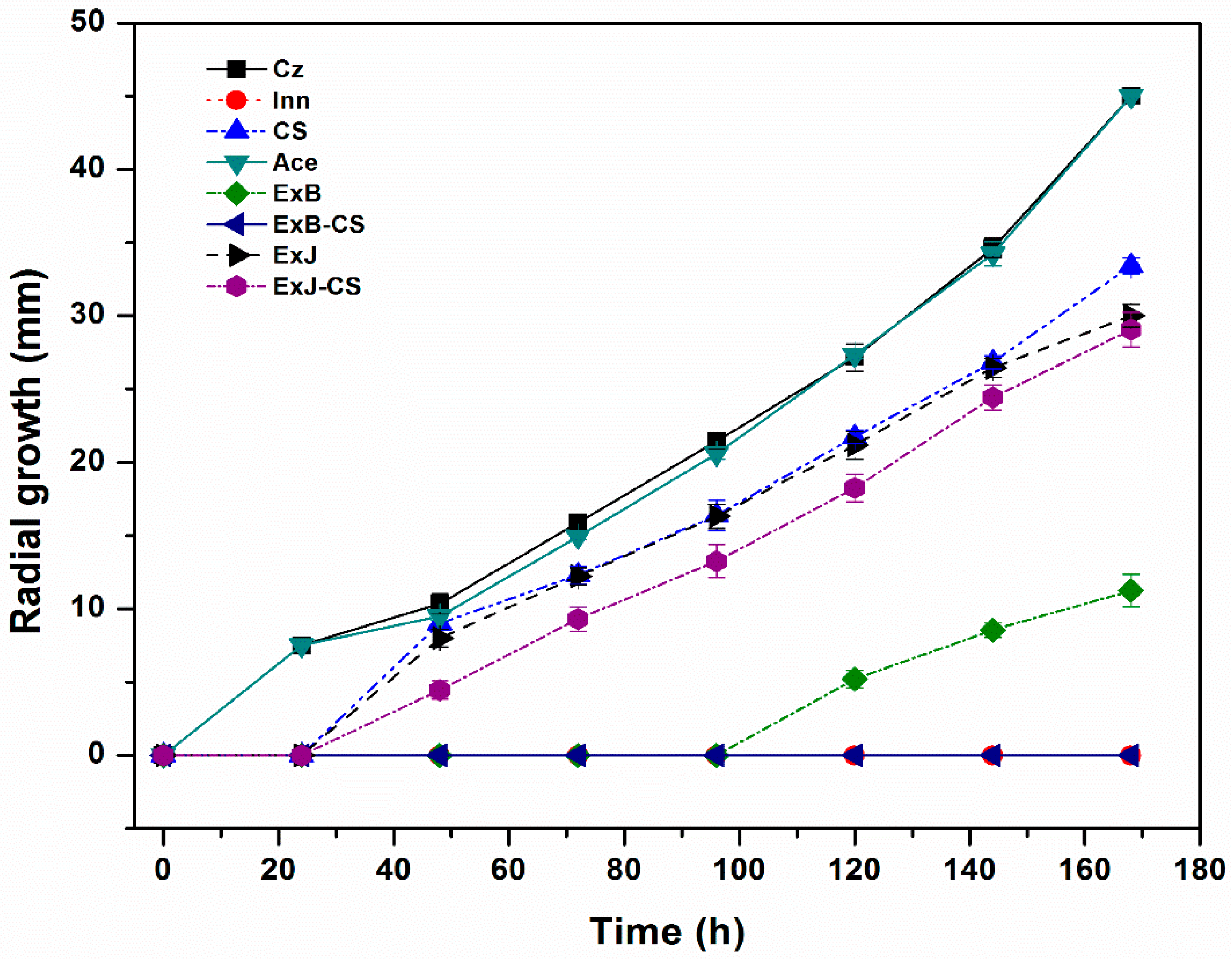
3.1.1. Radial Growth Kinetics

One of the most important parameters in the study of filamentous fungus is their mycelial growth. The impact on the radial growth of C. siamense was tested separately with chitosan (CS) and extracts (ExB and ExJ) to find the concentrations that stopped the growth before comparing the ExB-CS and ExJ-CS combinations. Table 1 shows the percentage of radial growth inhibition of C. siamense on CS, ExB, and ExJ during a period of 168 h. The CS solution and ExB at 4 mg/mL demonstrated a significantly greater inhibitory effect on mycelial development (p < 0.05). Nonetheless, with ExJ, the increased inhibition was noted at 2 mg/mL. After analyzing the results of radial growth with each treatment, it was decided to use the 4 mg/mL concentration to test the ExB-CS and ExJ-CS combinations in a 1:1 proportion.
Figure 1 shows the mycelial growth kinetics of C. siamense with each treatment. The control Inn (Innovator®) demonstrated 100% efficiency, as the fungus did not develop. Similarly, the ExB and the ExB-CS combination retarded growth by 100% during the first 72 h. In contrast, the ExJ-CS and ExJ did not exhibit the same pattern, with around 33 mm of radial development at the end of 168 h. Despite this, ExJ-CS and ExB-CS inhibited mycelial growth compared to the normal control (Cz) (p < 0.05).

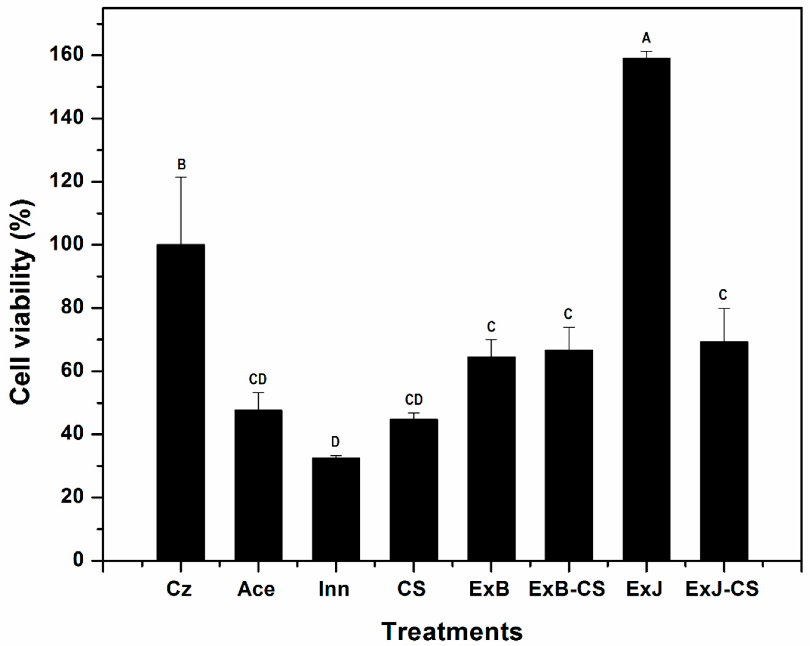
3.1.2. Cell Viability Test

The cell viability assay was performed to evaluate the effect of the ExB-CS and ExJ-CS combinations compared with the individual components at the metabolic level.
The ExJ-CS and ExB-CS combinations reduced the viability of C. siamense cells to 69% and 67%, respectively, which is about a 30% decrease compared to the normal control (Cz) (p < 0.05). The CS solution reduced the viability of C. siamense cells to 50%, while the ExB shows a similar result to the ExJ-CS and ExB-CS combinations. No significant difference was found between ExB-CS, ExJ-CS, ExB (p < 0.05) (Figure 2).

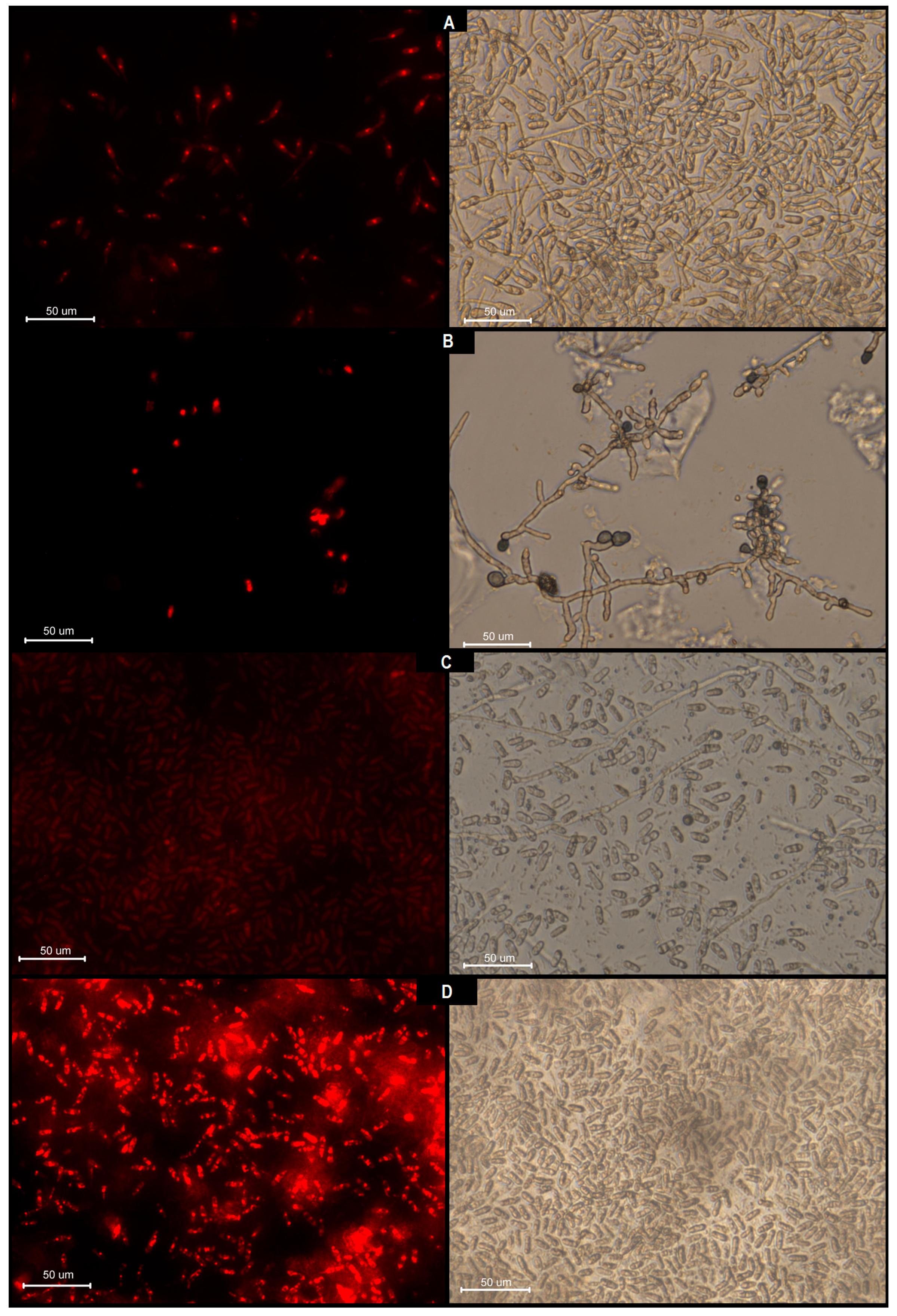
3.1.3. Cell Integrity Damage Analysis

Propidium iodide, a nuclear fluorescence marker that sticks to double-strand DNA in damaged cells [20], was used to confirm if C. siamense exposed to the treatments had damaged the cell membrane. In Figure 3D, C. siamense spores that were exposed to the 1:1 ExJ-CS for 24 h emit an intense red fluorescence, indicating damage; the light field shows non-germinated spores. The C. siamense spores that were treated with a CS solution at 4 mg/mL exhibited fewer damaged cells and more germinated spores (light field) with distorted hyphae (Figure 3B). In contrast to cell viability results, the ExB-CS combinations did not exhibit enough fluorescence to indicate damage.

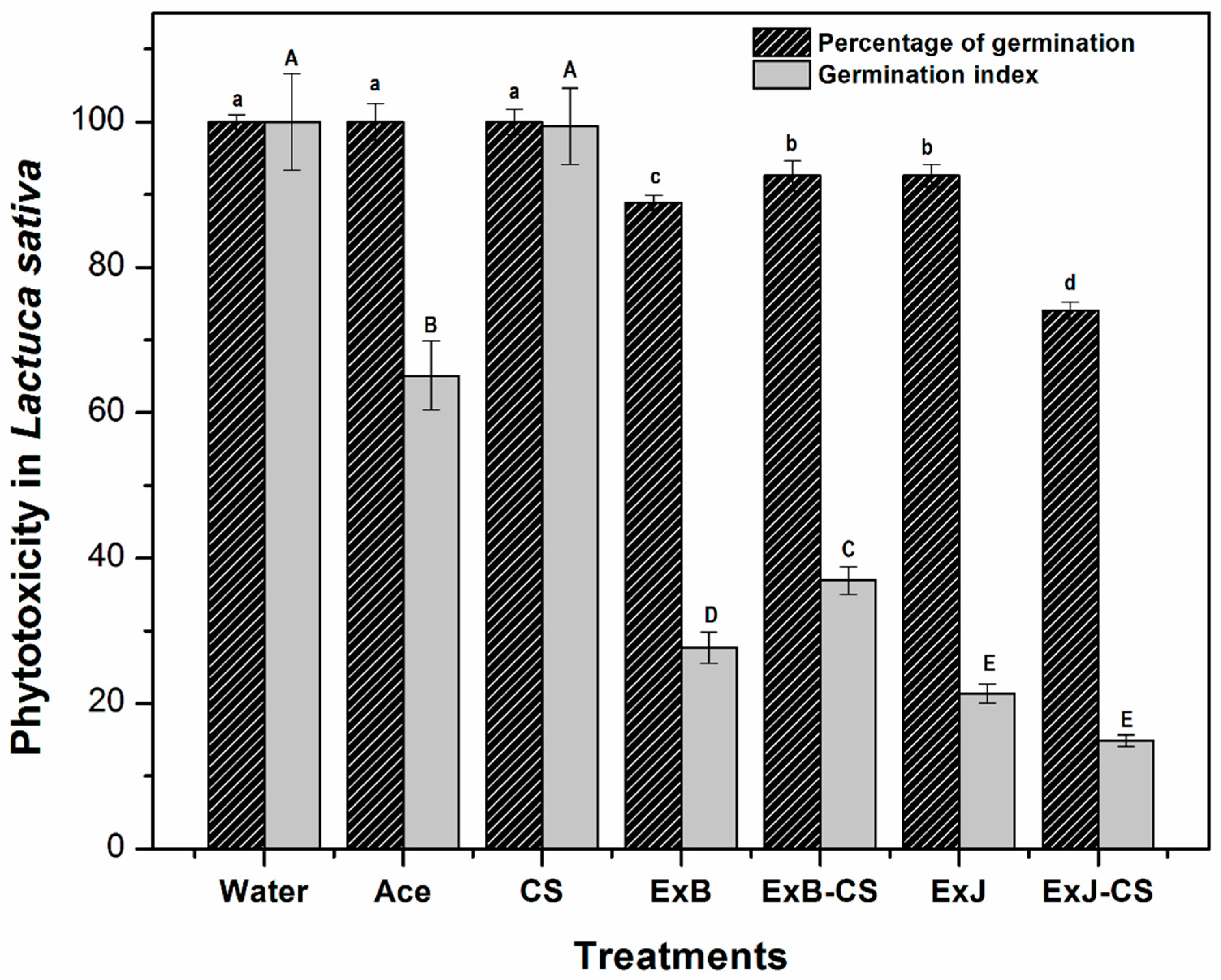
3.2. Phytotoxicity Bioassay

Phytotoxicity is a parameter that indicates possible damage to plant models by determining alteration in the seed and changes in the structure from root elongation [18]. Figure 4 shows that ExB-CS (1:1), ExJ-CS (1:1), ExB, and ExJ reduced the percentage of seed germination, and the germination index of L. sativa compared to the normal control (p < 0.05). The ExJ-CS treatment reduced seed germination by approximately 30%, indicating a higher phytotoxic effect on lettuce seeds (p < 0.05). The CS solution did not exhibit any negative effect on the germination process and index, reaching 100% as the water control.
The lettuce seeds germinated on ExB-CS and ExJ-CS, exhibiting alterations in the elongation of the roots; the RRE percentage was 40% and 20%, respectively. Meanwhile, the lettuce seeds treated with ExB and ExJ both showed similar effects on the root elongation process, with an RRE of 31% and 23%, respectively. Only the CS solution exhibits a root elongation process comparable to the normal control (water) (p > 0.05).

3.3. Acute Toxicity Test in Artemia Salina

The acute toxicity of methanolic plant extracts (ExB and ExJ), CS solution, and their combinations (ExJ-CS and ExB-CS) on Artemia salina nauplii is crucial for understanding their potential ecological impacts and uses. In this study, the methanolic extracts (ExB and ExJ) and their combinations (ExJ-CS and ExB-CS) exhibited toxicity on this model. The survival rates of A. salina were recorded as 39%, 28%, 38%, and 27% for ExJ (4 mg/mL), (1:1) ExJ-CS, ExB (4 mg/mL), and (1:1) ExB-CS, respectively. No significant difference was observed within the groups (p > 0.05); but there was a clear difference concerning the CS solution and the normal control (100%) (p < 0.05) (Figure 5).

4. Discussion

The activity of CS against fungal mycelial growth has been widely demonstrated in previous reports. Chávez-Magdaleno et al. [19] reported that 1% medium molecular weight CS inhibited mycelial growth by 21% in two species of Colletotrichum, close to the results obtained in this study. In contrast, Oliveira et al. [21] reported that low concentrations of CS (0.1, 0.2, and 0.4% w/v) effectively inhibited the growth of C. tamarilloi. This effect depended on the CS molecular weights. For example, Gálvez-Marroquín et al. [22] observed total inhibition of Colletotrichum spp. with 2.5% low molecular weight CS whereas López-Zazueta et al. [23] reported that CS of 13, 25, and 55 kDa (molecular weight) inhibited the development of C. gloeosporioides, which is different from the one in our study. Other authors have mentioned that the antifungal activity of CS varies depending on the type of microorganism, degree of deacetylation, molecular weight, concentration and exposure time, among others [11,23]. With respect to ExJ and ExB, both extracts at the two concentrations tested retarded mycelial growth with respect to the Cz control and there was only significant difference (p < 0.05) between the types of plant (Table 1). There are no other reports about these plant species and the fungi studied. However, Herrera-Parra et al. [24] observed that aqueous and ethanol extracts from Jacquinia flammea (3%, w/v) were highly effective to inhibit mycelial growth of C. magnum up to 94.49%. Buitimea-Cantúa et al. [17] reported antifungal activity in fractions of B. glutinosa and J. macrocarpa and indicated that they act as enzyme inhibitors in fungi, deforming their cell wall. The behavior of the fungus with CS was as expected, since this compound and its derivatives cause leakage of intracellular contents and inhibit microbial growth by affecting endogenous chitinase activity [10,25]. In addition, Nascimento et al. [15], reported that CS caused, among other damages, spore abnormalities, as observed in our study. In the same vein, Rosas-Burgos et al. [16] reported that ExB inhibited by 60% the growth of A. flavus, A. parasiticus and F. verticillioides after 14 days of incubation. Also, in another study was found that an aqueous extract of stem bark of J. flammea inhibited the mycelial growth of three phytopatogenic fungi (Corynespora cassicola, Curvularia lunata and Fusarium equiseti [26]. On the other hand, in the case of J. macrocarpa there are few reports. García-Sosa et al. [27] mentioned that sakurasosaponin is one of the main metabolites responsible for the antifungal activity in J. flammea as well as the jacquinonic acid detected.
In terms of cell viability some authors have shown antifungal activity of B. glutinosa and J. macrocarpa extracts against different phytopathogenic fungi (A. flavus, A. parasiticus, and F. verticillioides) [17,28,29]. The reduction in spore viability observed could be attributed to the loss of cell wall integrity due to the hydrolytic effect of the extracts, as previously reported by some researchers. Both plants have chitinase activity that acts as competitive inhibitors of fungal β-1,3-glucanase. Valenzuela-Cota et al. [29] suggested the defective fungal cell wall is a result of the β-1,3-glucanase inhibition, which affected the β-1,3-glucan synthesis.
With ExJ it was not possible to obtain quantifiable results due to interference from the pigments of this plant, which generated intense green tones. Similar observations were reported by Amiel-Pérez et al. [30], who evaluated plant extracts using the same technique and concluded that methanol solubilizes pigments that are left as residues and affect the reading. There are no other studies reporting these observations, possibly due to the nature of the compounds present in this plant, even though XTT-based cell viability assays have been applied in several fungal species.
Regarding cell integrity damage observed in our study, there are some previous works Xing et al. [31] demonstrated that CS caused irreversible damage to the cell membrane by its ability to increase membrane permeability. This effect may be attributed to the stimulation of enzyme expression by CS, leading to the degradation of key membrane components such as lipids, thereby altering membrane fluidity and structural stability [32].
Additionally, research by Buitimea-Cantúa et al. [17] reported that these extracts adversely affect chitinase and β,1-3 glucanase enzymes involved in the formation of the cell wall and hyphae in filamentous fungi. Damage to the spore membrane can impair organelle-selective functions such as barrier defense and intracellular material transport, among others. These results clearly suggest that CS and ExJ-CS treatments induce cell wall degradation and disrupt cell membrane integrity, which could explain their potential antifungal activity against C. siamense.
The antimicrobial activity of the extracts is associated with their chemical composition, which has been characterized in previous studies. According to Lam-Gutiérrez et al. [33], the most abundant compounds in the methanolic extract of B. glutinosa were 5-(hydroxymethyl)furan-2-carbaldehyde (38.59%), furan-2-carbaldehyde (4.103%), and 5-methylfuran-2-carbaldehyde (2.1%), all of which have also demonstrated antifungal activity [34]. For a fraction of the methanolic extract of J. macrocarpa, Valenzuela-Cota et al. [29] reported that the main compounds identified were γ-sitosterol (22.4%), stephamiersine (14.82%), betulinol (11.28%), and oleic acid (9.88%), some of which have previously been reported to exhibit antifungal activity [35].
On the other hand, in the phytotoxicity test carried out through seed germination, rootlets are the first part of the seedling to emerge from the seed and serve for water and nutrient absorption. The germination index (GR) is inversely proportional to the toxicity level of the tested treatments. In other words, a lower GR indicates higher toxicity, as it partially or completely inhibits seed root growth [36]. In this case, the significantly lower germination index in the treatments compared to the control (p < 0.05), suggests that the extracts of both plants have a phytotoxic effect on seedling development, possibly due to the presence of phenolic compounds. Dias et al. [37] reported that ethanolic extracts at 10% of Baccharis dentata, B. uncinella, and B. anomala inhibit about 90% of the germination rate of L. sativa. Meanwhile, Miranda-Arambula et al. [38] reported that aqueous extracts from dried B. salicifolia at a 1% concentration exhibited significant phytotoxic activity against lettuce seeds. This effect was attributed to the flavonoids present in the plant, such as quercetin, apigenin, and naringenin, whose phytotoxic properties have been confirmed. It was observed that CS presented the highest germination percentage, possibly due to its growth-promoting properties, which is consistent with Nurliana et al. [39], who reported that CS (0.2 g/L) reduced stress and promoted the growth of L. sativa under drought conditions. Chitosan can act as a growth promoter and helps control plant diseases by eliciting defense enzymes and exhibiting fungicidal properties. Its effectiveness, biodegradability, and environmental friendliness make it suitable for both conventional and organic farming [40].
The acute toxicity of methanolic extracts and CS on Artemia salina nauplii indicated negative effects on this model. The plant toxins can manifest through multiple pathways, often involving biological mechanisms that disrupt cellular or physiological functions [41]. For example, extracts with secondary metabolites such as flavonoids, alkaloids, and tannins can elicit differences in toxicity, which has been demonstrated on Artemia nauplii [42]. Nevertheless, the acute toxicity of B. glutinosa and J. macrocarpa on this organism has never been documented, even though there is a report indicating that some Baccharis species possess toxic compounds [43]. In a study by Bhoopathy et al. [44], the toxicity of CS nanoparticles on A. salina was evaluated, reporting survival rates of less than 50%. They suggest that the lethality and growth of the Artemia spp. are influenced by the molar mass of the CS, which is consistent with the difference between percentages obtained in both studies.

5. Conclusions

This study demonstrated that ExB and ExJ, both individually and combined with CS, exhibit antifungal activity against C. siamense in vitro. The treatments reduced fungal radial growth and cell viability, with some combinations showing evidence of damage to cell integrity. These effects suggest potential fungistatic action due to both the bioactive compounds in the extracts and the properties of CS. However, the extracts and their combinations showed phytotoxic effects on L. sativa seeds and caused significant mortality in A. salina nauplii, indicating possible toxicity at the tested concentrations. Overall, while ExB and ExJ combined with CS show promise as natural antifungal agents, further research is needed to optimize their concentrations, evaluate their safety, and test their effectiveness under real agricultural conditions.

Author Contributions

Conceptualization, A.K.L.-M. and M.O.C.-R.; methodology, A.C.G.-I.; R.I.S.-M. and S.E.M.-S.; investigation, S.d.G.-d.l.M.; supervision, M.P.-J. and A.K.L.-M.; writing—original draft preparation, S.d.G.-d.l.M.; writing—review and editing, A.K.L.-M.; M.O.C.-R. and M.P.-J.; project administration, M.P.-J. and A.K.L.-M.; funding acquisition, M.O.C.-R. All authors have read and agreed to the published version of the manuscript.

Funding

This work was supported by the Facultad Interdisciplinaria de Ciencias Biológicas y de Salud of the Universidad de Sonora (project USO313007895). Cortez-Rocha MO has received research support from the Universidad de Sonora.

Institutional Review Board Statement

“Not applicable” for studies not involving humans or animals.

Data Availability Statement

Data can be requested from the corresponding authors.

Acknowledgments

The authors thank to Eber Addí Quintana Obregón of the Centro de Investigación en Alimentación y Desarrollo (CIAD, A.C., Hermosillo) for the donation of the strain of Colletotrichum siamense (H6-1).

Conflicts of Interest

The authors declare that there is no conflict of interest.

References

  1. Khan, R.A.; Najeeb, S.; Hussain, S.; Li, Y. Bioactive secondary metabolites from Trichoderma spp. against phytopathogenic fungi. Microorganisms 2020, 8, 817. [Google Scholar] [CrossRef]
  2. Herrera-González, J.A.; Bautista-Baños, S.; Serrano, M.; Ramos-Bell, S.; Gutiérrez-Martínez, P. Colletotrichum siamense causing anthracnose in postharvest of ‘Hass’ avocado. Rev. Mex. Cienc. Agric. 2024, 15, 5. [Google Scholar]
  3. Tovar-Pedraza, J.M.; Solano-Báez, A.R.; Leyva-Mir, S.G.; Tlapal-Bolaños, B.; Camacho-Tapia, M.; García-León, E.; Ayala-Escobar, V.; Nava-Díaz, C.; Quezada-Salinas, A.; Santiago-Santiago, V.; et al. The need and opportunity to update the inventory of plant pathogenic fungi and oomycetes in Mexico. J. Fungi 2024, 10, 395. [Google Scholar] [CrossRef]
  4. Fuentes-Aragón, D.; Silva-Rojas, H.V.; Guarnaccia, V.; Mora-Aguilera, J.A.; Aranda-Ocampo, S.; Bautista-Martínez, N.; Téliz-Ortíz, D. Colletotrichum species causing anthracnose on avocado fruit in Mexico: Current status. Plant Pathol. 2020, 69, 1513–1528. [Google Scholar] [CrossRef]
  5. García-Estrada, R.S.; Cruz-Lachica, I.; Osuna-García, L.A.; Márquez-Zequera, I. First report of papaya (Carica papaya) anthracnose caused by Colletotrichum plurivorum in Mexico. Plant Dis. 2020, 104, 589. [Google Scholar] [CrossRef]
  6. Pineda-Vaca, D.; Flores-Méndez, G.; Fernández-Pavía, S.P.; Santillán-Mendoza, R.; Montero-Castro, J.C.; Garay-Serrano, E.; Ortega-Arreola, R.; Rodríguez-Alvarado, G. Genetic diversity of Colletotrichum siamense causing mango anthracnose in the state of Jalisco, Mexico, based on ISSR markers. Can. J. Plant Pathol. 2025, 47, 1–17. [Google Scholar] [CrossRef]
  7. Ciofini, A.; Negrini, F.; Baroncelli, R.; Baraldi, E. Management of post-harvest anthracnose: Current approaches and future perspectives. Plants 2022, 11, 1856. [Google Scholar] [CrossRef]
  8. Azeem, S.; Agha, S.; Jamil, N.; Tabassum, B.; Ahmed, S.; Raheem, A.; Khan, A. Characterization and survival of broad-spectrum biocontrol agents against phytopathogenic fungi. Rev. Argent. Microbiol. 2022, 54, 233–242. [Google Scholar] [CrossRef] [PubMed]
  9. El Khetabi, A.; Lahlali, R.; Ezrari, S.; Radouane, N.; Lyousfi, N.; Banani, H.; Askarne, L.; Tahiri, A.; El Ghadraoui, L.; Belmalha, S.; et al. Role of plant extracts and essential oils in fighting against postharvest fruit pathogens and extending fruit shelf life: A review. Trends Food Sci. Technol. 2022, 120, 402–417. [Google Scholar] [CrossRef]
  10. Poznanski, P.; Hameed, A.; Orczyk, W. Chitosan and chitosan nanoparticles: Parameters enhancing antifungal activity. Molecules 2023, 28, 2996. [Google Scholar] [CrossRef]
  11. Aranaz, I.; Alcántara, A.R.; Civera, M.C.; Arias, C.; Elorza, B.; Heras-Caballero, A.; Acosta, N. Chitosan: An overview of its properties and applications. Polymers 2021, 13, 3256. [Google Scholar] [CrossRef] [PubMed]
  12. Pellis, A.; Guebitz, G.M.; Nyanhongo, G.S. Chitosan: Sources, processing and modification techniques. Gels 2022, 8, 393. [Google Scholar] [CrossRef] [PubMed]
  13. Panda, P.K.; Dash, P.; Chang, Y.H.; Yang, J.M. Improvement of chitosan water solubility by fumaric acid modification. Mater. Lett. 2022, 316, 132046. [Google Scholar] [CrossRef]
  14. Li, Z.; Bi, X.; Dai, Y.; Ren, R. Enhancing mango anthracnose control and quality maintenance through chitosan and iturin A coating. LWT 2024, 198, 115955. [Google Scholar] [CrossRef]
  15. Nascimento, J.I.G.; Stamford, T.C.M.; Melo, N.F.C.B.; Nunes, I.D.S.; Lima, M.A.B.; Pintado, M.M.E.; Stamford-Arnaud, T.M.; Stamford, N.P.; Stamford, T.L.M. Chitosan–citric acid edible coating to control Colletotrichum gloeosporioides and maintain quality parameters of fresh-cut guava. Int. J. Biol. Macromol. 2020, 163, 1127–1135. [Google Scholar] [CrossRef]
  16. Rosas-Burgos, E.C.; Cortez-Rocha, M.O.; Cinco-Moroyoqui, F.J.; Robles-Zepeda, R.E.; López-Cervantes, J.; Sánchez-Machado, D.I.; Lares-Villa, F. Antifungal activity in vitro of Baccharis glutinosa and Ambrosia confertiflora extracts on Aspergillus flavus, Aspergillus parasiticus and Fusarium verticillioides. World J. Microbiol. Biotechnol. 2009, 25, 2257–2261. [Google Scholar] [CrossRef]
  17. Buitimea-Cantúa, G.V.; Rosas-Burgos, E.C.; Cinco-Moroyoqui, F.J.; Burgos-Hernández, A.; Plascencia-Jatomea, M.; Cortez-Rocha, M.O.; Gálvez-Ruiz, J.C. In vitro effect of antifungal fractions from the plants Baccharis glutinosa and Jacquinia macrocarpa on chitin and β-1,3-glucan hydrolysis of maize phytopathogenic fungi and on the fungal β-1,3-glucanase and chitinase activities. J. Food Saf. 2013, 33, 526–535. [Google Scholar] [CrossRef]
  18. López-Bermúdez, L.S.; Quintana-Obregón, E.A.; Rosas-Burgos, E.C.; Gálvez-Iriqui, A.C.; Gutiérrez-Martínez, P.; Lizardi-Mendoza, J.; Plascencia-Jatomea, M. Acute phytotoxicity and antifungal effect of nanochitosan particles on Colletotrichum fructicola with low susceptibility to chitosan. Curr. Microbiol. 2024, 81, 445. [Google Scholar] [CrossRef] [PubMed]
  19. Chávez-Magdaleno, M.E.; Luque-Alcaraz, A.G.; Gutiérrez-Martínez, P.; Cortez-Rocha, M.O.; Burgos-Hernández, A.; Lizardi-Mendoza, J.; Plascencia-Jatomea, M. Effect of chitosan-pepper tree (Schinus molle) essential oil biocomposites on the growth kinetics, viability and membrane integrity of Colletotrichum gloeosporioides. Rev. Mex. Ing. Quim. 2018, 17, 29–45. [Google Scholar] [CrossRef]
  20. Zhang, N.; Fan, Y.; Li, C.; Wang, Q.; Leksawasdi, N.; Li, F.; Wang, S. Cell Permeability and nuclear DNA staining by propidium iodide in Basidiomycetous yeasts. Appl. Microbiol. Biotechnol. 2018, 102, 4183–4191. [Google Scholar] [CrossRef]
  21. Oliveira, B.F.; Reis, A.; Costa, C.A.; Moita, A.W.; Pilon, L. Evaluation of chitosan for in vitro control of Colletotrichum tamarilloi and anthracnose on scarlet eggplant fruit. Hortic. Bras. 2023, 41, e2621. [Google Scholar] [CrossRef]
  22. Gálvez-Marroquín, L.A.; Martínez-Bolaños, M.; Cruz-Chávez, M.A.; Ariza-Flores, R.; Cruz-López, J.A.; Magaña-Lira, N.; Cruz-de-la-Cruz, L.L.; Ariza-Hernández, F.J. Inhibition of mycelial growth and conidium germination of Colletotrichum sp. for organic and inorganic products. Agro. Prod. 2022, 15, 25–32. [Google Scholar] [CrossRef]
  23. López-Zazueta, B.A.; Ayón-Reyna, L.E.; Gutiérrez-Dorado, R.; Rodríguez-Gómez, F.A.; López-López, M.E.; López-Velázquez, J.G.; Vega-García, M.O. Effect of chitosan with different molecular weights on the antifungal activity against Colletotrichum gloeosporioides and activation of the non-enzymatic antioxidant system on infected papaya. J. Food Sci. 2023, 88, 1979–1993. [Google Scholar] [CrossRef] [PubMed]
  24. Herrera-Parra, E.; Cristóbal-Alejo, J.; Magaña-Alvarez, A.; Medina-Baizabal, I.L.; Gamboa-Angulo, M. Antifungal effect of Bonellia flammea extracts against Colletotrichum magnum in postharvest fruits of Carica papaya cv. maradol. J. Plant Dis. Prot. 2024, 131, 1685–1694. [Google Scholar] [CrossRef]
  25. El Ghaouth, A.; Arul, J.; Grenier, J.; Asselin, A. Antifungal activity of chitosan on two postharvest pathogens of strawberry fruits. Phytopathology 1992, 82, 398–402. [Google Scholar] [CrossRef]
  26. Moo-Koh, F.A.; Cristóbal-Alejo, J.; Tun-Suárez, J.M.; Medina-Baizabal, I.L.; Arjona-Cruz, A.A.; Gamboa-Angulo, M. Activity of aqueous extracts from native plants of the Yucatan peninsula against fungal pathogens of tomato in vitro and from Croton chichenensis against Corynespora cassiicola on tomato. Plants 2022, 11, 2821. [Google Scholar] [CrossRef] [PubMed]
  27. García-Sosa, K.; Sánchez-Medina, A.; Álvarez, S.L.; Zacchino, S.; Veitch, N.C.; Simá-Polanco, P.; Peña-Rodríguez, L.M. Antifungal activity of sakurasosaponin from the root extract of Jacquinia flammea. Nat. Prod. Res. 2011, 25, 1185–1189. [Google Scholar] [CrossRef]
  28. Muñoz-Ochoa, I.J.; Plascencia-Jatomea, M.; Cinco-Moroyoqui, F.J.; Valenzuela-Cota, D.F.; Buitimea-Cantúa, G.V.; Martínez-Higuera, A.A.; Rosas-Burgos, E.C. Persistence of the antifungal capacity of a fraction of Jacquinia macrocarpa plant against Fusarium verticillioides after continuous exposure. Indian J. Microbiol. 2020, 60, 458–467. [Google Scholar] [CrossRef]
  29. Valenzuela-Cota, D.F.; Buitimea-Cantúa, G.V.; Plascencia-Jatomea, M.; Cinco-Moroyoqui, F.J.; Martínez-Higuera, A.A.; Rosas-Burgos, E.C. Inhibition of the antioxidant activity of catalase and superoxide dismutase from Fusarium verticillioides exposed to a Jacquinia macrocarpa antifungal fraction. J. Environ. Sci. Health B 2019, 54, 647–654. [Google Scholar] [CrossRef]
  30. Amiel-Pérez, J.; Fukusaki, A.; Enciso, N.; Altamirano, C.; Mayanga-Herrera, A.; Marcelo, Á.; Marin-Sánchez, O. Interferencia de pigmentos vegetales al aplicar la técnica XTT a extractos de Buddleja globosa, Senecio tephrosiodes Turcz. y Equisetum giganteum. Científica 2016, 13, 9–29. [Google Scholar]
  31. Xing, K.; Xing, Y.; Liu, Y.; Zhang, Y.; Shen, X.; Li, X.; Qin, S. Fungicidal effect of chitosan via inducing membrane disturbance against Ceratocystis fimbriata. Carbohydr. Polym. 2018, 192, 95–103. [Google Scholar] [CrossRef] [PubMed]
  32. Hernández-López, N.A.; Plascencia-Jatomea, M.; Del-Toro-Sánchez, C.L.; López-Saiz, C.M.; Morales-Rodríguez, S.; Martínez-Téllez, M.Á.; Quintana-Obregón, E.A. Antifungal activity of nanochitosan in Colletotrichum musae and Colletotrichum chrysophillum. Polysaccharides 2025, 6, 4. [Google Scholar] [CrossRef]
  33. Lam-Gutiérrez, A.; Ayora-Talavera, T.R.; Garrido-Ramírez, E.R.; Gutiérrez-Miceli, F.A.; Montes-Molina, J.A.; Lagunas-Rivera, S.; Ruíz-Valdiviezo, V.M. Phytochemical profile of methanolic extracts from chilca (Baccharis glutinosa) roots and its activity against Aspergillus ochraceus and Fusarium moniliforme. J. Environ. Biol. 2019, 40, 302–308. [Google Scholar] [CrossRef]
  34. Eseyin, A.E.; Steele, P.H. An overview of the applications of furfural and its derivatives. Int. J. Adv. Chem. 2015, 3, 42–47. [Google Scholar] [CrossRef]
  35. Yadav, R.P.; Huo, C.; Budhathoki, R.; Budthapa, P.; Bhattarai, B.R.; Rana, M.; Kim, K.H.; Parajuli, N. Antibacterial, antifungal, and cytotoxic effects of endophytic Streptomyces species isolated from the Himalayan regions of Nepal and their metabolite study. Biomedicines 2024, 12, 2192. [Google Scholar] [CrossRef]
  36. Mendes, P.M.; Ribeiro, J.A.; Martins, G.A.; Lucia, T.; Araujo, T.R.; Fuentes-Guevara, M.D.; Corrêa, L.B.; Corrêa, É.K. Phytotoxicity test in check: Proposition of methodology for comparison of different method adaptations usually used worldwide. J. Environ. Manag. 2021, 291, 112698. [Google Scholar] [CrossRef]
  37. Dias, M.P.; Nozari, R.M.; Santarém, E.R. Herbicidal activity of natural compounds from Baccharis spp. on the germination and seedlings growth of Lactuca sativa and Bidens pilosa. Allelopathy J. 2017, 42, 21–36. [Google Scholar] [CrossRef]
  38. Miranda-Arámbula, M.; Reyes-Chilpa, R.; Anaya-Lang, A.L. Phytotoxic activity of aqueous extracts of ruderal plants and its potential application to tomato crop. Bot. Sci. 2021, 99, 487–498. [Google Scholar] [CrossRef]
  39. Nurliana, S.; Fachriza, S.; Hemelda, N.M. Chitosan application for maintaining the growth of lettuce (Lactuca sativa) under drought condition. In Proceedings of the 4th International Conference on Food and Agriculture (ICOFA), Jember, Indonesia, 6–7 November 2021. IOP Conf. Ser. Earth Environ. Sci. 2022, 980, 012013. [Google Scholar] [CrossRef]
  40. Kocięcka, J.; Liberacki, D. The potential of using chitosan on cereal crops in the face of climate change. Plants 2021, 10, 1160. [Google Scholar] [CrossRef]
  41. Regueiras, A.; Pereira, S.; Costa, M.S.; Vasconcelos, V. Differential toxicity of cyanobacteria isolated from marine sponges towards echinoderms and crustaceans. Toxins 2018, 10, 297. [Google Scholar] [CrossRef] [PubMed]
  42. Lopes, V.R.; Antunes, A.; Welker, M.; Martíns, R.; Vasconcelos, V. Morphological, toxicological and molecular characterization of a benthic Nodularia isolated from Atlantic estuarine environments. Res. Microbiol. 2010, 161, 9–17. [Google Scholar] [CrossRef] [PubMed]
  43. Barros, C.S.L.; Jarvis, B.B. Livestock Intoxication by Baccharis. In Baccharis: From Evolutionary and Ecological Aspects to Social Uses and Medicinal Applications; Fernandes, G.W., Oki, Y., Barbosa, M., Eds.; Springer International Publishing: Cham, Switzerland, 2021; ISBN 978-3-030-45373-7. [Google Scholar]
  44. Bhoopathy, S.; Inbakandan, D.; Thirugnanasambandam, R.; Kumar, C.; Sampath, P.; Bethunaickan, R.; Raguraman, V.; Vijayakumar, G.K. A comparative study on chitosan nanoparticle synthesis methodologies for application in aquaculture through toxicity studies. IET Nanobiotechnol. 2021, 15, 418–426. [Google Scholar] [CrossRef] [PubMed]
Figure 1. Kinetics of C. siamense radial growth in Czapek agar added with CS, ExB, ExB-CS, ExJ, and ExJ-CS incubated at 27 ± 2 °C. Controls: Cz = Czapek agar, Inn = Fungicide, Ace = Acetone.
Polysaccharides 06 00052 g001
Figure 2. Cell viability of C. siamense exposed at different treatments incubated at 27 ± 2 °C. The bars represent means (n = 3) ± standard deviation. Different letters represent a significant difference (p < 0.05) between the treatments and control Czapek.
Polysaccharides 06 00052 g002
Figure 3. Propidium iodide staining (fluorescence microscopy) showing the effect of treatments on membrane permeability of C. siamense: (A): Czapek agar, (B): CS (4 mg/mL), (C): 1:1 ExB-CS, (D): 1:1 ExJ-CS. The C. siamense images in light field and fluorescence were captured using the 40× magnification objective.
Polysaccharides 06 00052 g003
Figure 4. Phytotoxicity effect in L. sativa seeds (percentage of germination and germination index). The bars represent means (n = 3) ± standard deviation. Different capital letters indicate a significant difference in germination index and lower-case letters indicate a significant difference in percentage of germination between treatments (p < 0.05).
Polysaccharides 06 00052 g004
Figure 5. Survival percentage of Artemia salina exposed to CS, ExB-CS and ExJ-CS. The bars represent means (n = 3) ± standard deviation. Different letters represent a significant difference (p < 0.05) between the treatments and seawater control.
Polysaccharides 06 00052 g005
Table 1. Radial growth inhibition percentage of C. siamense inoculated in Czapek agar added with CS, ExB and ExJ incubated at 27 ± 2 °C.
Time
(h)
Chitosan (CS)B. glutinosa (ExB)J. macrocarpa (ExJ)
2 mg/mL4 mg/mL2 mg/mL4 mg/mL2 mg/mL4 mg/mL
24100 ± 0.0 a100 ± 0.0 a100 ± 0.0 a100 ± 0.0 a100 ± 0.0 a100 ± 0.0 a
4860.9 ± 2.7 b62.4 ± 2.3 b100 ± 0.0 a100 ± 0.0 a43.7 ± 2.3 c42.1 ± 2.7 c
7229.5 ± 0.8 d47.0 ± 3.7 b79.6 ± 2.5 a81.1 ± 1.5 a45.1 ± 5.9 bc38.3 ± 1.7 c
9626.6 ± 1.4 d46.3 ± 1.8 b71.8 ± 0.7 a72.6 ± 1.8 a42.3 ± 6.0 bc33.8 ± 5.6 cd
12022.4 ± 1.5 d42.5 ± 2.6 bc63.3 ± 1.5 a67.2 ± 2.3 a41.8 ± 3.9 b33.7 ± 2.6 c
14425.7 ± 1.0 d39.8 ± 11.2 c62.5 ± 0.8 a66.8 ± 2.5 a45.4 ± 1.2 b38.2 ± 2.3 c
16827.6 ± 0.0 f38.6 ± 0.9 e63.5 ± 0.6 b67.6 ± 0.4 a47.4 ± 0.6 c43.3 ± 0.4 d
Values are means (n = 3) ± standard deviation. The means followed by different letters in the same row are significantly different according to Tukey’s multiple comparison tests (p < 0.05).
Disclaimer/Publisher’s Note: The statements, opinions and data contained in all publications are solely those of the individual author(s) and contributor(s) and not of MDPI and/or the editor(s). MDPI and/or the editor(s) disclaim responsibility for any injury to people or property resulting from any ideas, methods, instructions or products referred to in the content.

