Article

Exploring the Potential of Carboxymethyl Chitosan and Oxidized Agarose to Form Self-Healing Injectable Hydrogels

by
Eduard A. Córdoba
1,
Natalia A. Agudelo
1,
Luis F. Giraldo
2 and
Claudia E. Echeverri-Cuartas
3,*
1
Grupo de Investigación en Síntesis Orgánica de Polímeros y Biotecnología Aplicada (SINBIOTEC), Escuela de Ingeniería y Ciencias Básicas, Universidad EIA, Envigado 055428, Colombia
2
Laboratorio de Investigación en Polímeros (LIPOL), Instituto de Química, Universidad de Antioquia, Medellín 050010, Colombia
3
Grupo de Investigación en Ingeniería Biomédica (GIBEC), Escuela de Ciencias de la Vida y Medicina, Universidad EIA, Envigado 055428, Colombia
*
Author to whom correspondence should be addressed.
Polysaccharides 2025, 6(2), 49; https://doi.org/10.3390/polysaccharides6020049
Submission received: 7 February 2025 / Revised: 11 March 2025 / Accepted: 30 April 2025 / Published: 11 June 2025

Abstract

:
Localized treatment has emerged as an excellent alternative to minimize the side effects associated with the systemic dispersion of therapeutic agents, which can damage healthy tissues. Injectable hydrogels offer a promising solution because they can encapsulate and release therapeutic agents in a controlled manner. In this context, this study focuses on the development and characterization of an injectable hydrogel based on carboxymethyl chitosan (CMCh) and oxidized agarose (OA), in which chemical crosslinking through imine bond formation avoids the use of external crosslinking agents. Several polymer ratios were evaluated to obtain hydrogels (OA:CMCh), and stable gels were formed at physiological temperatures in all cases. The hydrogels were injectable through a 21 G needle with forces below 30 N, formed porous structures, and exhibited a self-healing capacity after 48 h. Additionally, the hydrogels displayed compressive strengths ranging from 26 to 71 kPa and elastic moduli similar to those of human tissues (6–20 kPa). Swelling percentages of up to 3090% were achieved owing to the high hydrophilicity of CMCh and OA, and strong chemical crosslinking maintained the gel stability for two weeks with low mass loss rates (<21%). Furthermore, polymer ratio variation and storage at 4 °C were observed to affect the hydrogel characteristics, allowing for property modulation according to the application needs. These results indicate that the proposed polymeric combination enables the formation of hydrogels with the potential for localized drug delivery.

Graphical Abstract

1. Introduction

Hydrogels are three-dimensional crosslinked polymeric networks with functional groups that help retain large amounts of water and biological fluids within their structures [1]. These systems do not dissolve because of chemical bonds or physical interactions between the polymer chains [2]. Hydrogels, with this unique characteristic and employing biomaterials, have been used in various biomedical applications, including drug delivery, tissue engineering, and regenerative medicine [3]. Hydrogels have become a platform of great interest for releasing active ingredients because of their previously mentioned characteristics and other properties, such as their ability to modulate pore size and ease of chemical modification [4].
In particular, the use of injectable hydrogels has gained prominence in the study of localized delivery systems. This type of hydrogel can be extruded through a syringe or needle, and some can undergo a sol-to-gel transition [5], which grants them properties such as malleability, allowing them to fill irregular volumes and be implanted through minimally invasive methods [6,7,8]. In recent years, a special type of injectable hydrogel has emerged as a novel alternative for implantable applications, called self-healing hydrogels [9]. Self-healing hydrogels have the ability to fully or partially repair themselves after structural damage without the need for external intervention. This property extends the hydrogel’s lifespan and enhances its reliability by preventing failures caused by material fatigue [5,10]. Due to these advantages, this type of hydrogel has been studied for applications such as localized drug delivery, tissue engineering, and tissue regeneration [9].
Several materials, including natural and synthetic polymers, have been evaluated for the preparation of self-healing hydrogels. However, those derived from natural sources offer superior biocompatibility and biodegradability due to their similarity to components of the extracellular matrix [11,12,13]. Among the natural polymers that have been studied are hyaluronic acid [14], cellulose [15], alginate, gelatin [16], chitosan, and agarose [17].
Chitosan is a polysaccharide derived from the deacetylation of chitin and has been extensively studied for biomedical applications, such as tissue engineering, cell culture, tissue regeneration, and controlled release systems [18,19]. The chemical structure of chitosan includes amino and hydroxyl groups that facilitate the crosslinking of hydrogels based on this polymer. Additionally, the carboxymethylation of these functional groups can improve the solubility of chitosan in water because it generally requires an acidic medium for dissolution [20]. Furthermore, agarose is a polysaccharide derived from brown algae. In addition to its excellent biological properties, it can self-gel at low temperatures and be blended with other polymers, which is of great interest for its use in health research, especially for injectable hydrogels [21,22,23]. Similar to chitosan, agarose can be modified to adapt its properties to specific requirements. For example, oxidation with periodate introduces aldehyde groups into the chemical structure of agarose [24], which can react with the amino groups in chitosan to form imine bonds, thereby facilitating polymer network crosslinking without the use of external crosslinking agents that may be toxic to biological environments. Moreover, this type of crosslinking involves dynamic covalent bond formation, imparting self-healing capabilities to the hydrogels [25].
In 2015, Priya et al. developed an injectable hydrogel composed of amorphous chitin and agarose for soft-tissue regeneration. They reported that, up to that point, chitosan–agarose hydrogels lacked injectable properties and that chitosan required extensive chemical modifications [26]. In 2020, Lima-Sousa et al. proposed an injectable chitosan–agarose hydrogel incorporating graphene for breast cancer treatment [27]. However, the use of unmodified chitosan requires an acidic medium for solubility, necessitating subsequent purification steps to eliminate the acidic medium. Finally, in 2023, Karimi et al. developed a triple hydrogel of carboxymethyl chitosan, sodium carboxymethyl cellulose, and agarose as a wound dressing. In this case, only the effect of the agarose proportion was evaluated, and its characterization as an injectable hydrogel was not evaluated [28].
A literature review revealed no carboxymethyl chitosan (CMCh) blend with oxidized agarose (OA)-based injectable hydrogel synthesized utilizing Schiff base chemistry. According to the above, this research focused on the synthesis and characterization of an injectable hydrogel based on carboxymethyl chitosan (CMCh) and oxidized agarose (OA). The modification of OA enabled crosslinking with carboxymethyl chitosan (CMCh) via imine bonds, forming porous structures with self-healing capacity and avoiding the purification process at the end of the formation of the hydrogels. Additionally, in this investigation, a systematic and rigorous study was conducted on the effect of polymer proportions on characteristics such as rheology and injectability. The polymer combination resulted in hydrogels that could be easily injected through a needle and possessed mechanical properties suitable for soft-tissue applications. Their high water absorption and long-term stability make them promising candidates for local therapies, such as the localized delivery of agents.

2. Materials and Methods

2.1. Materials

Chitosan ( M ¯ w = 176,776 Da, %DD = 81.12%; lot STBH9838) and type II-A agarose (lot SLBV2496) were obtained from Sigma-Aldrich (St. Louis, MO, USA). Monochloroacetic acid (ClCH2COOH) was obtained from Alfa Aesar (Ward Hill, MA, USA). Sodium periodate (NaIO4) and ethylene glycol 99% were obtained from PanReac AppliChem (Barcelona, Spain). Ethanol and sodium hydroxide (NaOH) were obtained from Emsure (Oakville, ON, Canada), and isopropanol was obtained from Químicos JM (Antioquia, Colombia).

2.2. Synthesis of Carboxymethyl Chitosan

Carboxymethyl chitosan was synthesized according to the methodology proposed by Fei et al. [29]. Five grams of chitosan was dispersed in 50 mL of isopropanol under magnetic stirring at 400 rpm; then, 12.5 mL of a 10 M sodium hydroxide solution was added. This solution was added in six equal portions over 20 min, and the mixture was stirred for 45 min. Subsequently, 6 g of monochloroacetic acid was added in five equal portions, with one portion added every 5 min. The mixture was then immersed in a water bath at 30 °C for 3 h. For purification, the mixture was centrifuged at 9000 rpm for 5 min at 4 °C, and the solid obtained was washed three times with ethanol. Subsequently, 150 mL of cold water at 4 °C was added, and the mixture was subjected to dialysis in distilled water for 7 days using 6–8 kDa regenerated cellulose membranes. Finally, the sample was filtered through a coffee paper filter, and the dried polymer was obtained by lyophilization for 72 h at −50 °C and 105 mTorr (VirTis, SP Scientific, Ipswich, UK).

2.3. Synthesis of Oxidized Agarose

The methodology presented in [30] was used for the oxidation of agarose. Type II-A agarose (3 g) was dissolved in 290 mL distilled water. The mixture was stirred in a water bath at 60–70 °C until a homogeneous mixture was obtained. Subsequently, 4.2 g of sodium periodate was dissolved in 10 mL of distilled water. The resulting mixture was stirred at 350 rpm for 6 h. To stop the reaction, 1 mL of 99% ethylene glycol was added, and the mixture was stirred for another hour. For purification, a dialysis process was performed against distilled water for 7 days using 6–8 kDa membranes, and the solid polymer was recovered by lyophilization for 72 h at −50 °C and 105 mTorr (VirTis, SP Scientific, Ipswich, UK).

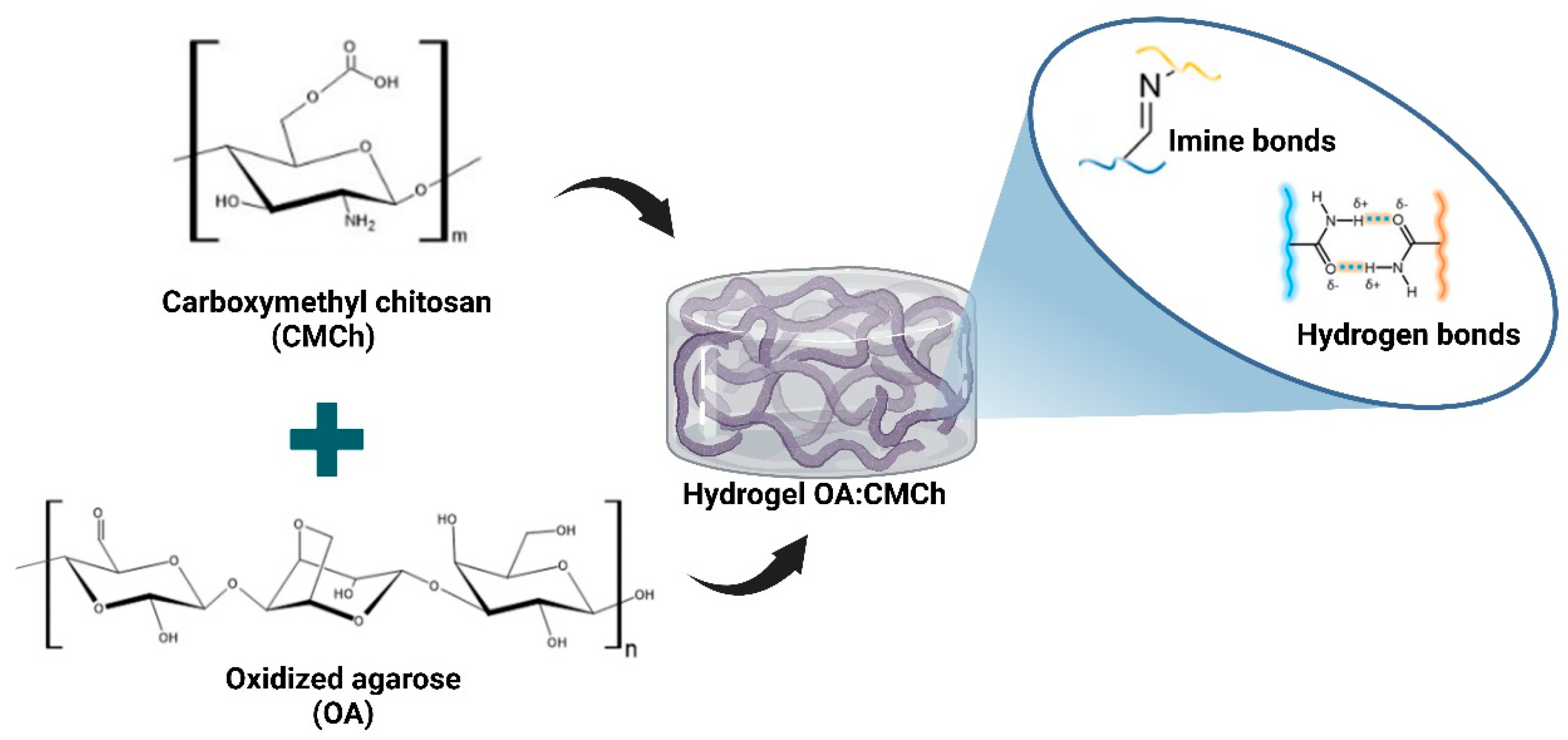
2.4. Preparation of OA:CMCh Hydrogel

After modification, the polymers were mixed to induce hydrogel crosslinking (Figure 1). Hydrogel preparation was performed according to a previously reported method [31]. Hydrogels were prepared at concentrations of 20 and 30 mg/mL, and the following OA:CMCh weight ratios were evaluated: 80:20, 60:40, 50:50, 40:60, and 20:80. To obtain the hydrogel, the required amount of OA (Table 1) was directly weighed into a glass container (approximately 5 mL). Then, 1 mL of distilled water was added, the container was sealed, and it was placed in a water bath at 75 °C, where it was stirred magnetically for 1 h.
Subsequently, the required amount of CMCh (Table 1) was added; the container was resealed, submerged again in the 75 °C water bath, and stirred magnetically for another hour. Once the hydrogels were ready, they were stored until further characterization.

2.5. Gelation Evaluation

Gelation was evaluated by simulating physiological temperature (37 °C) and room temperature (25 °C) using the inverted tube method [32]. The mixture was removed from the water bath (75 °C), and the temperature was measured with a thermocouple; once the mixture reached the evaluation temperature, the container was inverted to observe whether the mixture still flowed along the walls of the container. For evaluation at 37 °C, the container was immersed in a water bath at the same temperature, and the inversion process was repeated. Finally, gel formation occurred when the sample no longer flowed after the container was inverted [32,33]. All tests were performed in triplicate.

2.6. Injectability

The injectability of the hydrogels was evaluated using a universal testing machine (Instron 3345) with a 5 kN load cell. This test measured the maximum force required to extrude the hydrogel using a 1 mL syringe with a 21 G × 1 ½” needle (internal diameter of 0.6 mm). A load was applied to the syringe plunger at a displacement speed of 1.0 mm/s [34]. For this test, hydrogels were prepared at 30 mg/mL in the following OA:CMCh weight ratios: 80:20, 60:40, 50:50, 40:60, and 20:80.
The effect of storage temperature on the injectability of the hydrogels (25 and 4 °C) was also evaluated. The mixtures were prepared and loaded into syringes immediately after removal from the water bath (75 °C) and left to cool to room temperature. The 25 °C test was conducted once the samples had cooled, and for the 4 °C test, the syringes were stored overnight in a refrigerator at 4 °C. The following day, the syringes were removed from the refrigerator and allowed to reach room temperature before the injectability test was conducted. All tests were performed in triplicate.
The shear stress and viscosity of the hydrogels were theoretically calculated using the obtained force values. For the calculation of shear stress, Equation (1) was used, where τ is the shear stress in Pa, F is the maximum load force in N, and A is the perimeter area of the needle in m2. In this case, r is the internal radius of the needle (0.6 mm), and L is the length (40 mm).
τ = F A = F 2 π r L
Furthermore, for the calculation of viscosity, the Hagen–Poiseuille equation was used, which relates the flow rate ( Q ), pressure difference, radius ( r ), length ( L ), and viscosity ( η ). By solving for the viscosity from the Hagen–Poiseuille equation, Equation (2) was obtained, which was used with the values of the parameters r and L mentioned previously. The flow corresponds to the extrusion speed (1 mm/s) multiplied by the cross-sectional area of the syringe ( π r 2), and pressure 1 ( P 1 ) was taken as the calculated shear stress plus atmospheric pressure. By contrast, pressure 2 ( P 2 ) is the atmospheric pressure. Thus, the pressure difference corresponded to the calculated shear stress.
η = π P 1 P 2 r 4 8 Q L = π τ r 4 8 Q L

2.7. Syringeability

Syringeability was determined according to the methodology proposed by Moreira et al. [34] with some modifications. The constant load used in this case was 18.24 N, and the load application time (15 s) was determined using the sample that presented the lowest injectability. A 1 mL syringe with a 21 G × 1 ½” needle (0.6 mm internal diameter) was used. Syringeability was calculated using Equation (3) [34].
S y r i n g e a b i l i t y % = M a s s   e x p e l l e d   f r o m   t h e   s y r i n g e M a s s   b e f o r e   i n j e c t i o n × 100 %
The syringes were weighed using empty needles to determine their initial mass. Subsequently, hydrogels were prepared with OA:CMCh weight ratios of 60:40, 50:50, and 40:60 (these mass ratios were selected for subsequent analyses). The hydrogels were then stored in 1 mL syringes and weighed to determine the mass of the syringes with needles and the sample; thus, the mass of the sample before injection was calculated as the difference between the full and empty syringes. Next, a load of 18.24 N was applied for 15 s. Finally, the syringes were weighed again to determine the mass expelled from the syringe (the difference between the mass of the syringes with needles and the sample before and after injection). Using these data and Equation (3), the syringeability percentage was calculated. The effect of storage temperature on the hydrogels (25 and 4 °C) was also evaluated for this test, following the previously mentioned process. All tests were performed in triplicate.

2.8. Compression Test

The mechanical properties of the hydrogels were measured using a universal testing machine (Instron 3345) according to ASTM D695 [35] with a 10 N load cell and a strain rate of 5 mm/min. Cylindrical hydrogel samples were obtained from molds measuring 20 mm in height and 10 mm in diameter. The effect of storage temperature on the hydrogels (25 and 4 °C) was also evaluated in this test, following the previously mentioned process. All tests were performed in triplicate.
Stress was calculated as the ratio of the measured force to the cross-sectional area of the sample, and strain was calculated as the ratio of the displacement recorded by the equipment to the initial length of the sample to obtain the stress–strain curves. Finally, the modulus of elasticity was determined as the slope of the first linear segment of the stress–strain curve [36].

2.9. Rheological Characterization

An oscillatory rheometer (MCR 92, Anton Paar, Graz, Austria) with a parallel-plate geometry (25 mm in diameter) and a gap of 0.5 mm was used to evaluate the viscoelastic properties of the hydrogels. For this analysis, samples were prepared the day before measurement. Oscillatory mode experiments were conducted to gather information on the viscoelastic response of the material under different conditions, including a frequency sweep (1–100 rad/s) with 1% strain, an amplitude sweep with strain ranging from 0 to 100%, a frequency of 10 Hz, and a flow sweep with a shear rate of 0–100 Hz. Additionally, measurements were taken at 25 and 37 °C to assess the influence of temperature on the rheological properties of the hydrogels. All tests were performed in triplicate.

2.10. Morphological Analysis

Morphological analysis of the dry hydrogels by freeze-drying (xerogels) was performed using a scanning electron microscope (Phenom ProX, Utrecht, The Netherlands) with a scanning voltage of 15 kV. For morphological evaluation, three sample areas were analyzed: the top, bottom, and middle sections; the latter was accessed by making a cross-sectional cut with a blade. The obtained micrographs were processed using ImageJ 1.53e software to measure the pore sizes. Three samples were evaluated at each ratio.

2.11. Cute-Heal Method

For this analysis, once the hydrogels were prepared, half of the sample was placed in cylindrical silicone molds 20 mm in height and 10 mm in diameter. Two drops of methylene blue were added to half, and the sample was agitated until it was evenly stained, after which the remaining portion was added to the molds. Once the samples had cooled, a self-healing test was performed using the cut–heal method [37]. The technique involved removing the hydrogels from the molds and making a transverse cut in the middle of the sample using a blade. The halves were then separated and reassembled using halves of different colors. The new (bicolored) samples were placed back into the mold and covered with plastic wrap to prevent moisture loss. Finally, the samples were left at 37 °C for 48 h, and self-healing was evaluated qualitatively using optical microscopy and manual traction with tweezers from both ends of the samples.

2.12. Swelling Test

The swelling ratio (SR) was determined gravimetrically using dry hydrogel discs (10 mm diameter and 5 mm height). Initially, the dry weight of each sample was obtained, and the samples were immersed in 10 mL of a phosphate-buffered saline (PBS) solution (pH 7.43) and stored in an oven at 37 °C to simulate physiological conditions. Subsequently, the hydrogels were removed at specific time intervals (0.5, 1, 2, 3, 24, 48, 72, 168, and 336 h), and the surface water was removed with filter paper and weighed to record the wet weight. The swelling ratio was calculated using Equation (4):
S R   % = W t W 0 W 0 × 100 %
where W 0 is the dry weight of the hydrogel before immersion in the PBS solution, and W t is the weight of the hydrogel at different swelling evaluation times. Independent samples were used at each time point, and all tests were performed in triplicate.

2.13. Degradation Test

The mass loss of the hydrogels was determined gravimetrically using dry hydrogel discs (10 mm in diameter and 5 mm in height). These discs were weighed to obtain the dry weight of all samples and then immersed in 10 mL of a PBS solution with a pH of 7.43 and stored in an oven at 37 °C. At specific time intervals (0.5, 1, 2, 3, 24, 48, 72, 168, and 336 h), the samples were extracted from the buffer solution, washed twice with distilled water, and stored in a freezer with 5 mL distilled water to remove any salts that could affect the weight of the samples. The samples were then lyophilized for 72 h at −50 °C and 105 mTorr (VirTis, SP Scientific, Ipswich, UK). Once dry, each sample was weighed, and the degradation percentage was calculated using Equation (5):
W e i g h t   l o s s % = W i W f W i × 100 %
where W i is the initial dry mass of the sample, and W f is its dry mass after storage in the buffer solution. Independent samples were used at each time point, and all tests were performed in triplicate.

2.14. Statistical Analysis

The data obtained from the triplicate experiments are presented as the mean ± standard deviation. Statistical analyses were performed using Minitab® software version 21.2, where values were considered significantly different with a p-value of less than 0.05.

3. Results

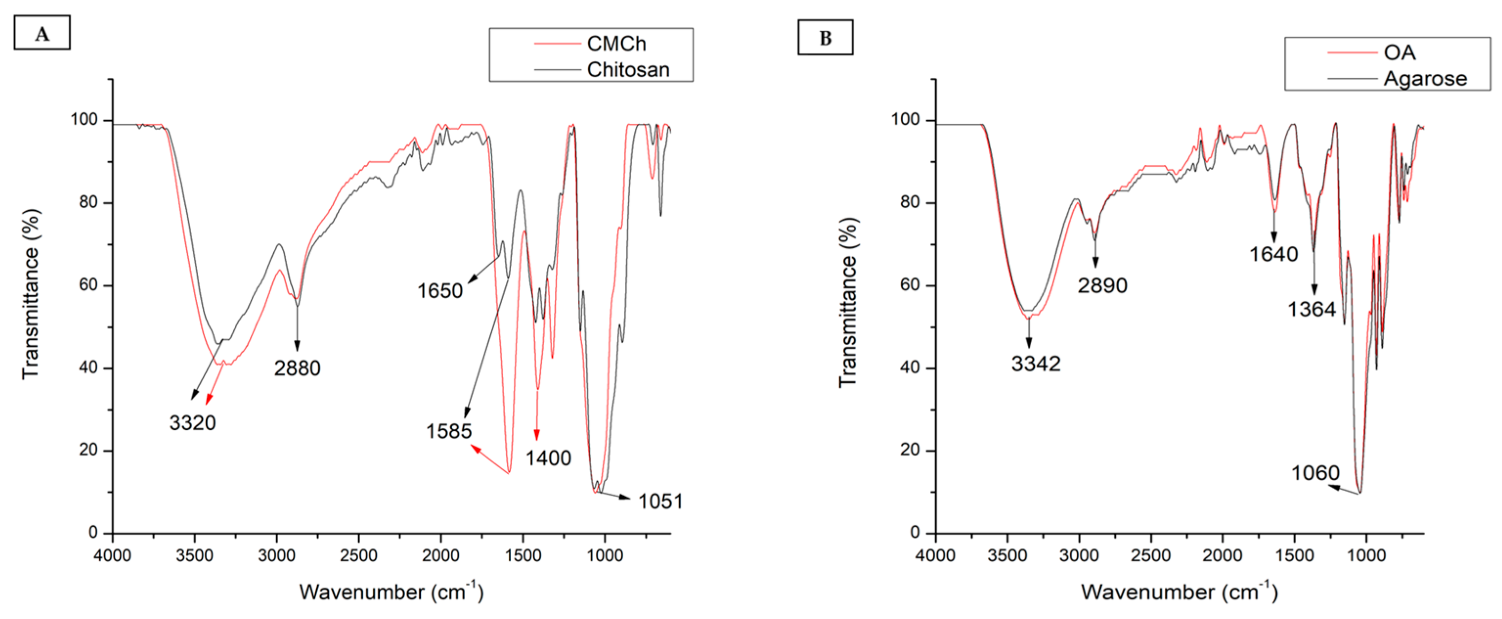
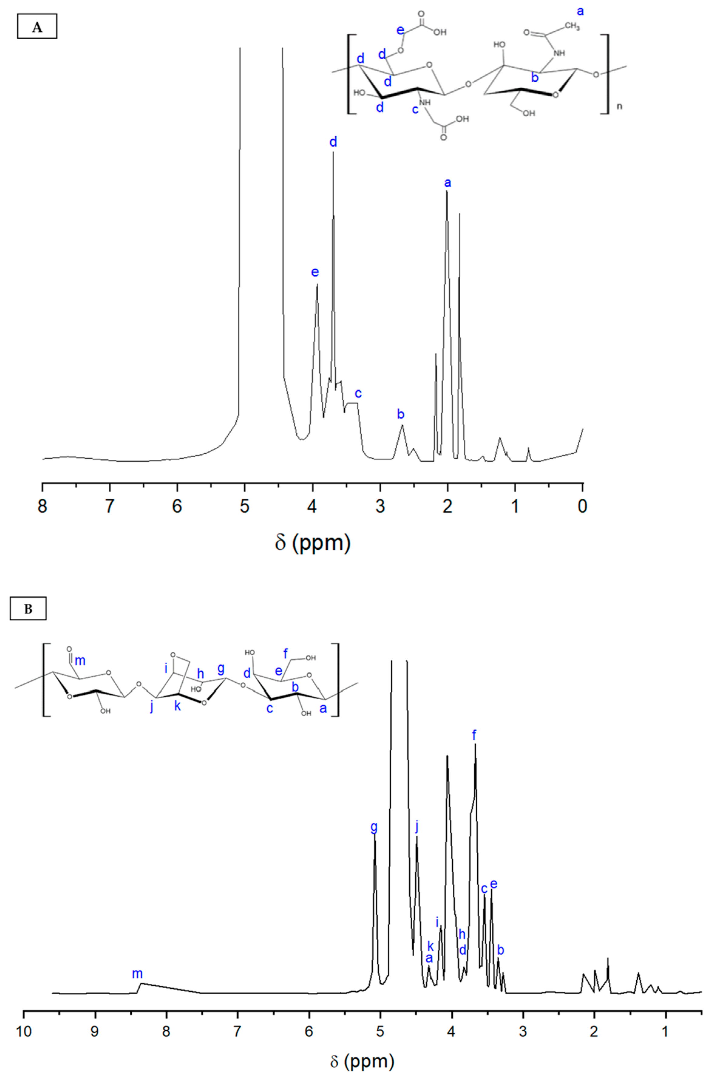
3.1. Characterization of Carboxymethyl Chitosan

The modification was confirmed by Fourier-transform infrared (FTIR) spectroscopy of the modified and unmodified polymers according to their chemical structures (Figure S1). For chitosan, Figure 2A shows the spectrum with characteristic bands at 3320 cm−1, which is due to the stretching of the O-H and N-H groups, and at 2880 cm−1, which represents the stretching of the C-H bonds, and the peak at 1051 cm−1, which is due to the stretching of the C-O-C bond [38]. The same bands were observed for CMCh; additionally, an increase in intensity was observed at 1585 cm−1 owing to the overlapping of the COO and N-H groups. The bands at 1400 cm−1 were attributed to the symmetric and asymmetric deformation of -COO, confirming the carboxymethylation of the hydroxyl and amino groups of chitosan [39]. Additionally, a solubility test of CMCh in distilled water was performed (Figure S2A), which verified that the modification due to the incorporation of -COO groups favored interactions with water, facilitating its solubility.
Figure 3A shows the 1H NMR (proton nuclear magnetic resonance) spectrum of CMCh, where the signal at 2 ppm (a) is attributed to the -CH3 groups of the N-acetyl group residues. Moreover, the signal at 2.68 ppm (b) corresponds to the hydrogen bond to the carbon of the N-acetyl glucosamine unit [40]. The signal at 3.68 ppm (d) corresponds to hydrogen atoms attached to the carbons of the deacetylated glucosamine unit cycle. Finally, in the CMCh spectrum, signals at 3.35 and 3.94 ppm (c, e) are observed, which may be associated with the binding of the carboxyl group to the amino and hydroxyl groups, respectively [41]. This result confirmed the addition of carboxyl groups to the chitosan structure.

3.2. Characterization of Oxidized Agarose

In addition, in Figure 2B, significant bands were observed in the infrared spectra of agarose and OA: the band at 3342 cm−1 was attributed to the stretching of O-H bonds; the band at 2890 cm−1 corresponded to the stretching of C-H single bonds; the signals at 1640 and 1364 cm−1 indicated the bending of O-H and C-C groups, respectively; and the highest intensity peak at 1060 cm−1 corresponded to the stretching vibration of C-O bonds [42,43,44]. As shown in the spectrum, no visually significant changes were observed, which verified the oxidation of agarose, as reported by other authors [30]. Therefore, a qualitative test was performed using an oxide reduction reaction via the Fehling test to identify the aldehyde groups, where a change in the coloration of the sample was observed with OA (Figure S2B). This is owing to the oxidation of the aldehyde groups present in OA, which generates copper oxide, which explains the color change in the solution. This qualitatively confirms the modification of agarose. However, because no noticeable difference was observed in the FTIR spectrum, it is assumed that few aldehyde groups were formed.
Furthermore, in Figure 3B, the 1H NMR spectrum of OA is presented, where the signals between 3.34 and 3.82 ppm (b, c, d, e) correspond to hydrogens attached to carbons in the β-D-galactose subunit. The signals at 3.93 to 4.48 ppm (h, i, k, j) are attributed to the hydrogens present in the 3,6-anhydro-α-galactose subunit [45]. Additionally, the signal at 5.07 ppm (g) corresponds to the hydrogen of the anomeric carbon at the non-reducing end of the 3,6-anhydro-α-galactose [46]. Li et al. [30] reported that the presence of aldehyde groups in the structure of oxidized agarose results in a slight peak at 8.7 ppm. In this case, a slight increase in the signal intensity was observed at 8.36 ppm (m), which corresponded to the presence of aldehyde groups.

3.3. Hydrogel Gelation Time

The inverted tube method [32] was used to evaluate the gelation of the hydrogels, and it was observed that the hydrogel stopped flowing down the walls of the container after inversion. Figure 4 shows the appearance of the samples that were gelled and those that were not.
In the 25 °C test, all samples, regardless of the concentration or polymer ratio, were gelled before reaching room temperature (25 °C), except for the 0:100 sample (OA:CMCh) at a concentration of 20 mg/mL, which did not gel after 20 min. This result indicates that CMCh did not respond to temperature and that the thermosensitive properties of the hydrogels can be attributed to the presence of OA in the mixture.
Furthermore, in the 37 °C test, none of the mixtures with a concentration of 20 mg/mL gelled after being in the water bath at that temperature for 20 min; however, most of the samples at a concentration of 30 mg/mL gelled before reaching 37 °C, except for the 100:0 and 80:20 ratios (OA:CMCh). The fact that the 100:0 ratio did not gel at 37 °C indicates that the gelation temperature of OA ranges from 25 to 37 °C. It was found that OA gels at approximately 30 °C, using the inverted tube method. Furthermore, the 80:20 ratio gelled after 47 ± 6 s, which is an acceptable time for the biomedical applications of injectable hydrogels [47,48]. The other mixtures with a concentration of 30 mg/mL showed gel behavior before reaching 37 °C, which is mainly attributed to the high viscosity of CMCh. Even at high temperatures, the 0:100 (OA:CMCh) sample exhibited high viscosity and gel consistency. Thus, because the 20 mg/mL samples did not gel at 37 °C, the temperature of interest for the application, characterization was continued with the samples at a concentration of 30 mg/mL. In addition, it is essential to note that the gels were injected through a 21 G gauge needle, allowing the formation of the letters E, I, and A (Figure 4C).

3.4. Injectability of Hydrogel

The injectability test enabled the determination of the maximum force required to inject the material through a needle, and the shear stress and viscosity of the hydrogels were determined [34] through theoretical calculations (Table 2).
For the tests at 25 °C, the maximum compression load, shear stress, and viscosity were observed as the amount of CMCh in the mixture increased (Table 2), as noted in another study [49]. Therefore, a higher viscosity required a greater injection force [50]. However, no significant differences were found between the 50:50, 40:60, and 20:80 ratios (p < 0.05), suggesting that a higher proportion of CMCh in the mixture did not substantially affect the injectability of the hydrogels.
Additionally, when evaluating the effect of storage temperature at 4 °C on the samples, it was observed that for the 80:20, 60:40, and 50:50 ratios, there was a significant increase in the injection force required compared to the 25 °C test. By contrast, no significant temperature effects were observed for the 40:60 and 20:80 ratios (p > 0.05). Notably, exposure to low temperatures causes OA to gel; consequently, storage at 4 °C may result in increased viscosity in hydrogels with a higher polymer content, increasing the force needed for injection. However, it should be noted that as the amount of OA in the mixture decreases, the effects caused by the change in storage temperature may not follow the same pattern, which could explain the decrease in injectability at a 20:80 ratio.
As shown in Table 2, all evaluated hydrogels exhibited injection forces below 30–40 N, which is the maximum force allowed for manual injection applications [51,52]. Therefore, for subsequent characterization, 60:40, 50:50, and 40:60 ratios were used, as the analysis of these ratios can provide more practical information on the effect of varying the polymer content used in hydrogel preparation.

3.5. Syringeability

Syringeability is a parameter related to the ease with which the hydrogel is injected through the needle and evaluates the performance of the syringe during injection [34]. As mentioned above, syringeability was assessed by applying a fixed load of 18.24 N to the syringe piston for 15 s. Table 3 shows the results of the syringeability test for the hydrogels.
Statistical analyses indicated that both the polymer ratio and storage temperature had a significant effect on the syringeability of the hydrogels (p < 0.05), demonstrating that an increase in the amount of CMCh led to a lower syringeability percentage, which was associated with a higher viscosity of the hydrogel produced by CMCh and, consequently, greater difficulty in extrusion through the needle. Additionally, the storage temperature of the hydrogel results in a decrease in syringeability because the thermosensitivity of agarose may cause the viscosity to increase further. Furthermore, this is consistent with the injectability results, where it was observed that a greater extrusion force was required in the mixtures with a higher amount of CMCh. Therefore, when a fixed load was applied, the samples with more CMCh presented greater difficulty for injection and, consequently, a lower syringeability percentage. To date, the methods for evaluating syringeability have not been standardized, limiting comparison with other studies [51]. However, syringeability can be affected by the viscosity of the sample, syringe type, and needle diameter used for the test [53].

3.6. Mechanical Properties

Compression tests were conducted using a universal testing machine to determine the Young’s modulus and maximum compressive strength of the OA hydrogels with different ratios. Table 4 shows that for the test at 25 °C, there is an increase in the compressive strength for the 50:50 and 40:60 ratios compared to the 60:40 ratio, indicating that an increase in the amount of CMCh leads to an improvement in the mechanical properties of the material [54]. However, no significant differences were observed between the 50:50 and 40:60 ratios (p > 0.05). A similar behavior was reported in [55], where an increase in the compressive strength with the increase in CMCh was observed; however, with further increments, the strength decreased. Additionally, this is consistent with the findings of the injectability test, where the shear strength increased with the amount of CMCh.
By contrast, Young’s modulus showed that an increase in CMCh in the mixture caused a decrease in the stiffness of the material; however, no significant differences were found between the 60:40 and 50:50 ratios (p-value > 0.05), indicating that excess CMCh leads to reduced stiffness. This behavior was also reported by the authors of [56], who mentioned that an increase in the amount of CMCh may produce greater flexibility among the hydrogel polymer chains. Furthermore, it was observed that after storage at 4 °C, the compressive strength and Young’s modulus increased, indicating that exposure to low temperatures affects the mechanical properties of the hydrogels, which is associated with the thermosensitive characteristics of agarose. In addition, the stiffness moduli obtained were close to those found in soft tissues, such as adipose tissue, cardiac muscle, liver, or breast [57], which may favor biomechanical compatibility between the tissue and hydrogel, preventing unwanted signaling pathways associated with cellular mechanotransduction processes.

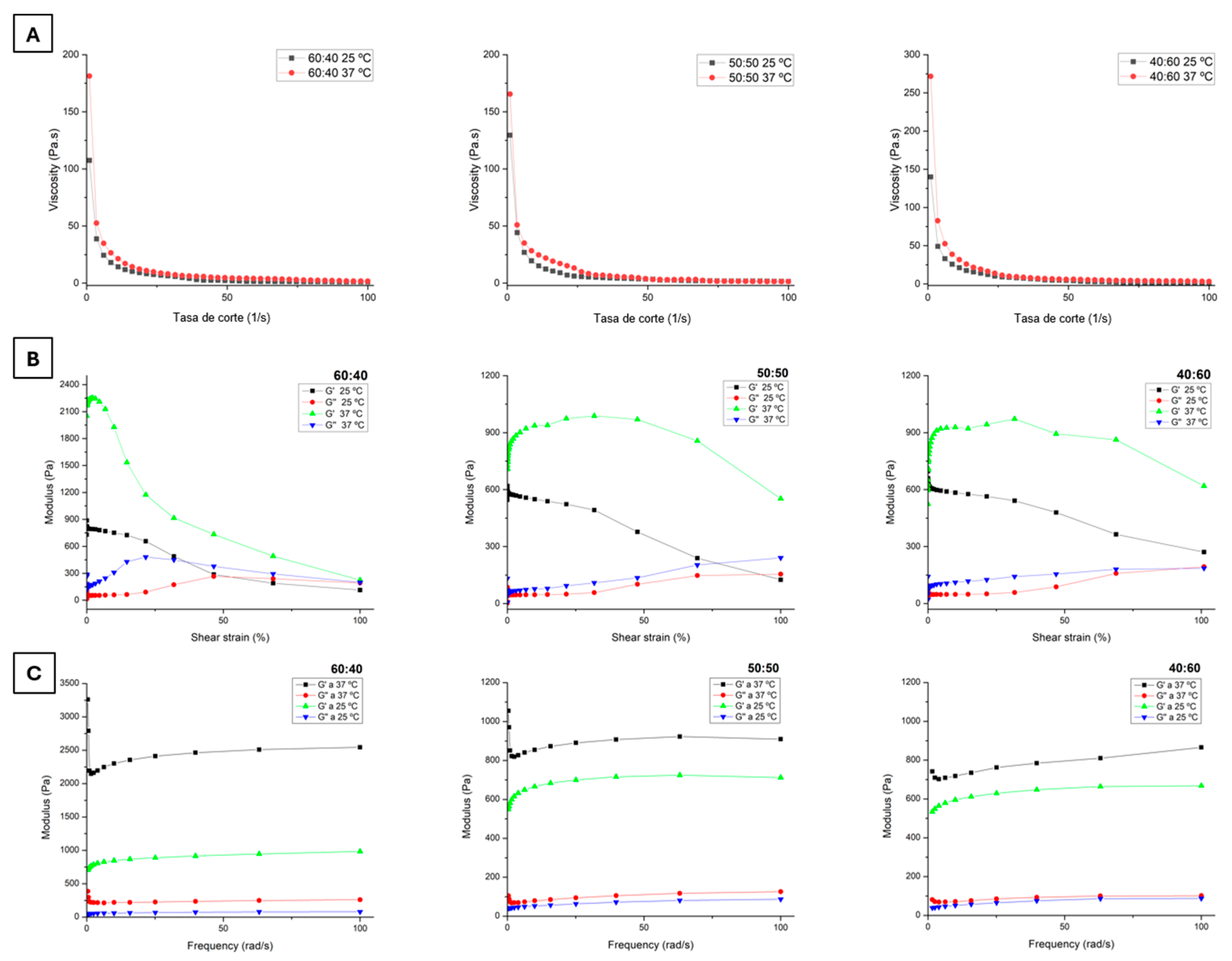
3.7. Rheological Properties

The rheological properties of the hydrogels were evaluated by flow, amplitude, and frequency sweeps. In Figure 5A, it can be observed that for all ratios, the viscosity decreases logarithmically with the increasing shear rate, which is also known as shear thinning and is a characteristic of injectable hydrogels and non-Newtonian fluids [17,58]. With an increase in the shear rate, the polymer chains tend to extend and align in the direction of flow, thereby reducing the flow resistance and, consequently, the viscosity [59]. Additionally, the effect of temperature on the viscosity of the hydrogels can be observed; at 37 °C, all ratios show higher viscosity values at low shear rates, indicating that at physiological temperature, the gels can acquire a firmer consistency.
By contrast, during the amplitude sweep, changes in the storage modulus (G′) and loss modulus (G′) were observed with respect to the shear strain to which the hydrogels were subjected to at different ratios. Figure 5B shows that G′ decreases with increasing strain, meaning that the hydrogels tend to become less elastic at higher strains. Notably, in tests at 25 °C, for the 60:40 and 50:50 ratios (OA:CMCh), a gel–sol phase transition was observed, as there was an intersection point where G″ exceeded G′, indicating that the sample exhibited more viscous than elastic behavior, which can be attributed to the breakdown of the internal structure of the hydrogel [60]. This behavior is not observed in the 40:60 ratio at 25 °C, nor in any ratio in the test at 37 °C, indicating that at this last temperature, the hydrogels can maintain a gel consistency without turning into a fluid, which is suitable for local application interventions where the hydrogel is expected to maintain its gel form.
In Figure 5C, the results of the frequency sweep for each hydrogel ratio can be seen. It was observed that all ratios present a greater G′ than G″, and in both cases, there is no dependence on frequency variations, which is a typical mechanical behavior for gels [61]. Finally, in both the amplitude and frequency sweeps, it can be observed that the value of G′ significantly increases in the test at 37 °C compared to the test at 25 °C, indicating that the samples exhibit more elastic behavior with increasing temperature, which is consistent with the previously mentioned findings.

3.8. Scanning Electron Microscopy Analysis

Scanning electron microscopy (SEM) was employed for the morphological analysis of the OA:CMCh hydrogels, and micrographs of the surface sections, cross-section, and bottom part of the sample were obtained (Figure 6). The micrographs show that the top part of the sample tended to be smooth, although some holes were caused by the rupture of bubbles that may have formed during sample preparation and subsequently burst during vacuum freeze-drying. Through measurements performed using ImageJ, it was found that the holes on the surface of the hydrogels had sizes of 120.46 ± 88.78, 47.18 ± 30.73, and 110.15 ± 49.33 µm for the 60:40, 50:50, and 40:60 ratios, respectively.
Additionally, in the cross-sections, pores of varying sizes and forms can be observed, with average pore openings measuring 176.87 ± 73.59, 215.14 ± 93.01, and 212.07 ± 86.71 µm for the 60:40, 50:50, and 40:60 ratios, respectively. It was also noted that these types of pores appeared to form compartments within the hydrogel. Such spaces may promote water retention within the polymeric structure, as many of these compartments seemed to be closed or partially closed, with most cases showing no apparent interconnection between the pores. In a study conducted by Huang et al. [46], where a benzaldehyde-modified agarose hydrogel crosslinked via a Schiff base was prepared, the matrix had an average pore size of 207.38 ± 44.89 μm and a morphology similar to that obtained in this study, confirming that the results exhibit behavior consistent with previous research on this type of polymer. Furthermore, highly porous surfaces were observed at the bottom for all three ratios, with significant heterogeneity in pore shape and size. The obtained sizes for these pores were 43.68 ± 19.75, 71.44 ± 18.48, and 86.11 ± 40.28 µm for the 60:40, 50:50, and 40:60 ratios, respectively.
As seen in the results of the pore size measurements, the deviations were quite high because when they appeared, the formation of the hydrogel structure was not homogeneous. However, the pores or compartments were randomly formed.

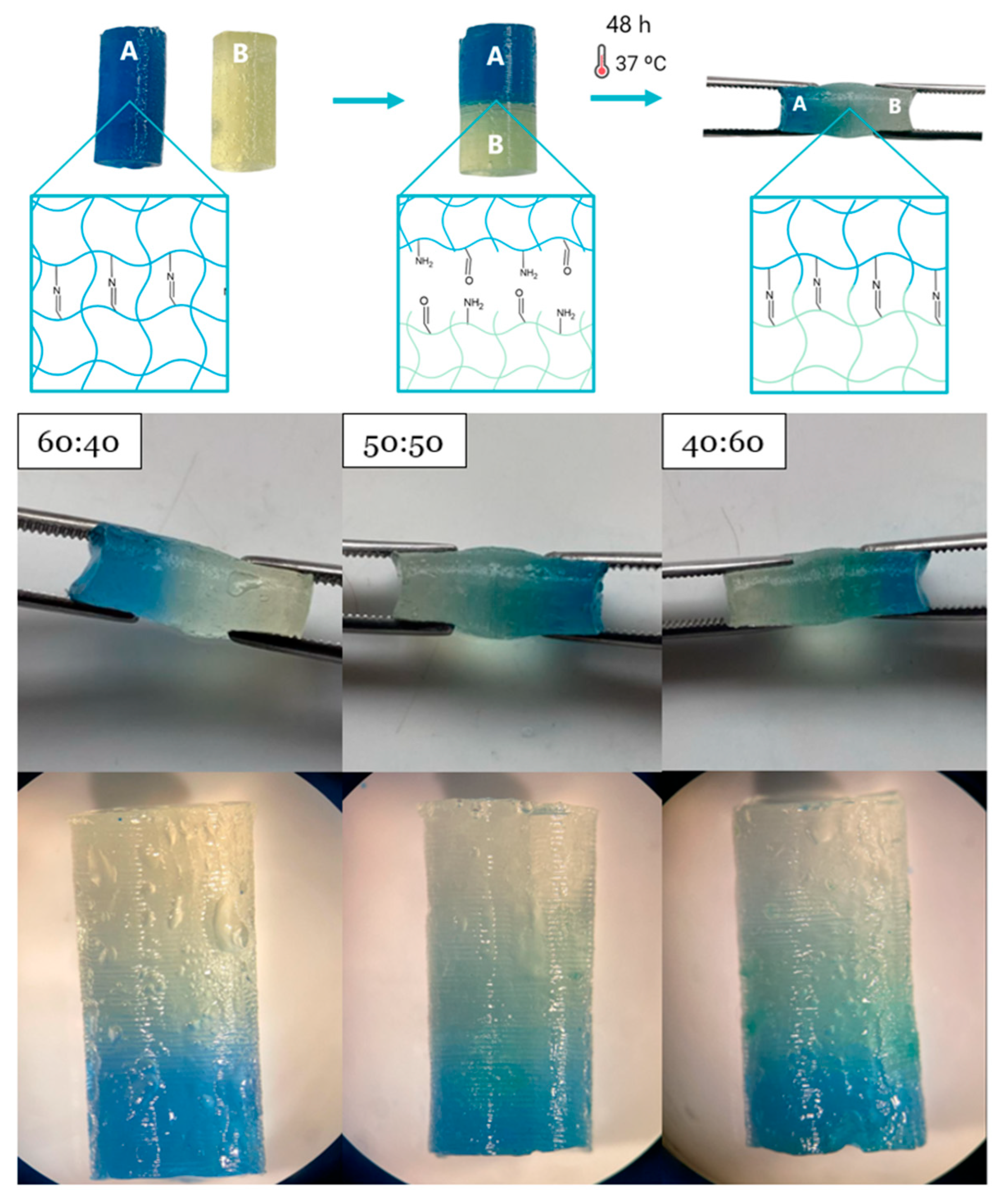
3.9. Self-Healing Evaluation
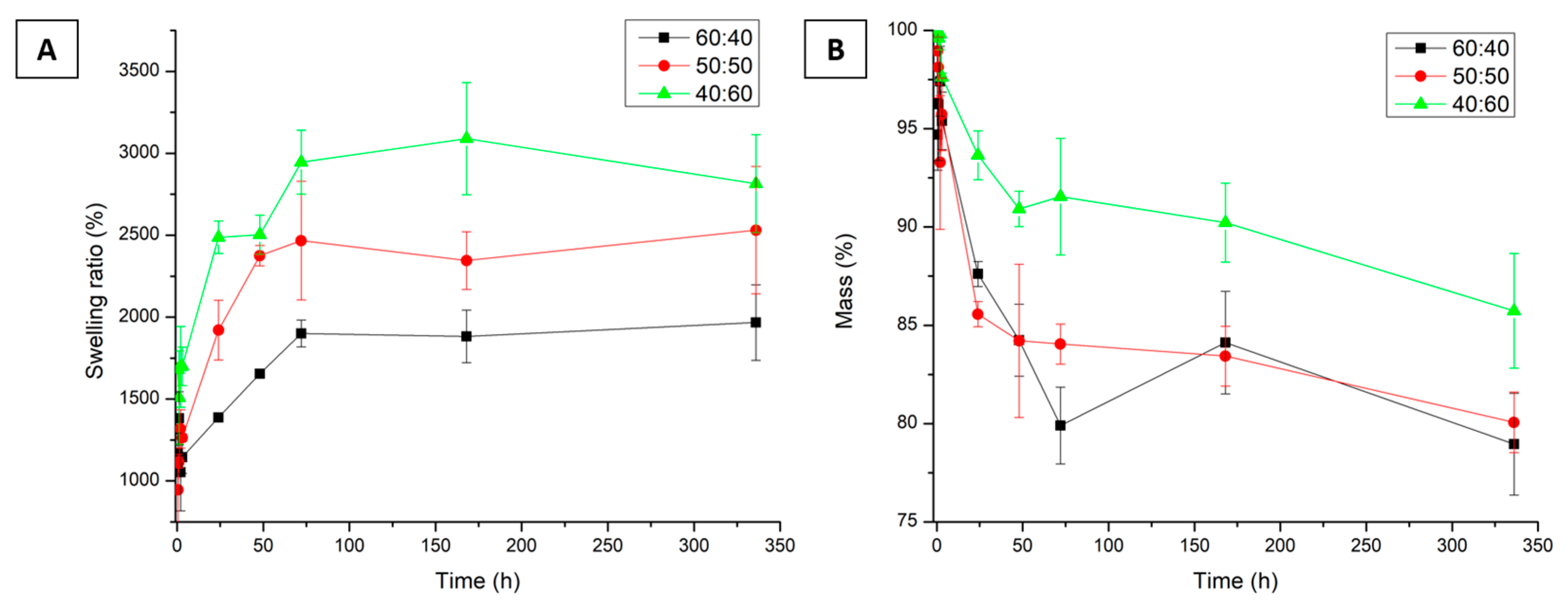
Self-healing materials can repair physical damage to their structures and recover all or part of their mechanical properties, which is beneficial for biomedical applications involving implantable materials [25,62]. In this case, a self-healing test was conducted qualitatively using the cut–heal method, and verification was performed through optical microscopy observations and manual traction applied from both ends. Figure 7 shows the procedure that describes the methodology, in which stained and unstained samples were prepared, cut in half, brought into contact, and left to allow the hydrogel to repair itself.
Initially, self-healing was evaluated after 24 h. However, after collecting the samples, the halves were still separated, indicating that the self-healing process was incomplete. The evaluation was repeated at 48 h, and the hydrogels remained joined after manual traction with tweezers, confirming their self-repairing ability. Microscopy revealed no cracks between the halves (Figure 7). This is because of Schiff base crosslinking, which is based on the formation of a dynamic covalent bond (imine in this case) and is characterized by high selectivity in a chemical reaction. Therefore, in the event of a rupture, the amino groups present in CMCh can react again with the aldehyde groups of OA they come into contact with, reforming the imine bond and generating the self-healing effect [25,63]. This behavior is highly interesting because such materials can recover their original shape after disintegration without external intervention, offering valuable advantages for in situ applications [64]. Additionally, a longer lifespan is favored by maintaining the integrity of the structure and preserving its mechanical stability.

3.10. Swelling Test

A swelling test was conducted to determine the absorption of the hydrogel and the water retention capacity, as this is an essential parameter in the study of the release of active ingredients. A hydrogel with a high swelling capacity is likely to release greater amounts of active ingredients [65].
The swelling evaluation was carried out over two weeks, taking measurements with independent samples for each time point. In Figure 8A, it can be seen that over time, all ratios tend to increase their water retention capacity within their structure, reaching maximum swelling percentages of 1967 ± 231%, 2530 ± 388%, and 3090 ± 343% for the 60:40, 50:50, and 40:60 ratios, respectively. These high percentages are related to the number of hydrophilic functional groups in the OA and CMCh polymer structures. Furthermore, OA has -OH groups in its structure that attract water molecules [43,66], and CMCh has -NH2, -COO, and -OH groups, which are also hydrophilic, favoring interactions between water molecules and the hydrogel [65].
Additionally, a higher amount of CMCh in the mixture significantly increased the swelling capacity of the hydrogels (p < 0.05). The presence of amino and hydroxyl groups in CMCh that can form hydrogen bonds with water and the carboxylate groups (-COO) further increases the interactions with water molecules, leading to more significant attraction and absorption of this molecule within the polymeric network [67]. This behavior was also observed in [67,68], where an increase in the CMCh concentration increased the swelling capacity of the hydrogels.
Furthermore, the high swelling percentages may also be associated with the porous structure of the hydrogels observed in the SEM images, as the formation of such pores may facilitate water permeation into the three-dimensional network and its retention [69]. Importantly, although they can absorb large amounts of water, the hydrogels maintain their shape, can be easily handled without breaking, and do not show significant changes in size.

3.11. Degradation Test

It is crucial to evaluate the degradation of the hydrogels since they are expected to maintain their functionality and integrity during the desired application period for which they were designed [70]. In Figure 8B, the results obtained from the degradation test are presented. It was observed that over the two-week evaluation period, the hydrogels at different ratios did not exceed a mass loss of 21.04%. Therefore, the mass loss can be attributed to the superficial erosion of the hydrogel or the solubilization of noncrosslinked polymer chains, and the integrity of the samples was maintained during handling. These results indicate that the obtained hydrogel has a stable and well-crosslinked structure, which could be beneficial for drug encapsulation, as the slow degradation of the hydrogel ensures a longer protection time for the active ingredients, preventing, for example, the enzymatic degradation of these substances [71].
Moreover, it is essential to note that chitosan and agarose are polysaccharides that do not undergo hydrolytic degradation, which necessitates the use of enzymes such as lysozymes or agarases that can break the glycosidic bonds of these polymers [72,73]. The same result was confirmed by Vivcharenko et al. [74], who found that the degradation of a chitosan/agarose film in PBS medium without enzymes was significantly lower than that with enzymes. By contrast, Lima-Sousa et al. [27] observed that an injectable hydrogel made from chitosan and agarose lost up to 56% of its mass in 14 days in a PBS medium with lysozyme, indicating greater degradation than that observed in this study without the use of lysozyme.
By contrast, as shown in Figure 8B, the 40:60 ratio was the least degraded during the evaluation period, whereas no statistically significant differences were observed between the 60:40 and 50:50 ratios (p > 0.05). This result may be associated with greater stability due to CMCh; however, in most cases, degradation tests report the use of enzymes such as lysozyme, which can break chitosan chains [75]; therefore, the behavior observed in this case may not be generalized. Therefore, this is an important factor to consider in future degradation tests where the evaluation is conducted in a medium that better simulates the final application of the hydrogels [51].

4. Conclusions

In this study, injectable hydrogels were prepared using OA and CMCh. The combination of these two natural polymers exhibits the significant advantage of avoiding the use of crosslinking agents that may be toxic and affects the biocompatibility of the hydrogels. This is because the formation of aldehyde groups in OA facilitates the formation of imine bonds by reacting with the amino groups present in chitosan, which allows for the establishment of a stable crosslinked structure. Additionally, these types of bonds are characterized by dynamic covalent bonds, which endow the hydrogel with a self-healing capability, are favorable for maintaining the structural integrity of the gel, and are essential for applications where a long lifespan is expected. Furthermore, it was possible to inject the hydrogels with forces below 30 N, meeting the standard for manual injection applications. The syringeability test demonstrated that the 60:40 ratio could be injected more easily than the other evaluated ratios.
Furthermore, the mechanical properties of the hydrogels showed stiffness moduli close to those of biological tissues, which may enhance the biomechanical compatibility between the material and tissue, and the rheological properties confirmed the viscoelastic behavior of the injectable hydrogels. Additionally, the hydrogels exhibited a thermosensitive response, as alterations in their properties were observed after storage at 4 °C, with an increase in viscosity and stiffness owing to the response of OA to low temperatures. Finally, high swelling percentages were observed, which were attributed to the high hydrophilicity of the employed polymers. The SEM micrographs showed porous structures, which may facilitate the entry and retention of water in the matrices. Furthermore, the hydrogels exhibited low degradation over 14 days, demonstrating their structural stability. Additionally, it was found that the OA:CMCh polymer ratio had a significant effect on the final properties of the hydrogel, as each polymer component possessed intrinsic properties that could complement the others, demonstrating the versatility of the obtained hydrogel and allowing the modulation of the hydrogel properties from its design to meet the specific needs of the application under study. Based on these results, it can be concluded that OA- and CMCh-based hydrogels can be used for localized drug delivery in applications like the treatment of solid tumors.

Supplementary Materials

The following supporting information can be downloaded at: https://www.mdpi.com/article/10.3390/polysaccharides6020049/s1, Figure S1: Scheme of polymer modification. Figure S2: (A) CMCh solubility test. (B) Fehling OA test.

Author Contributions

Conceptualization, N.A.A. and C.E.E.-C.; methodology, E.A.C., N.A.A., C.E.E.-C. and L.F.G.; validation, E.A.C., N.A.A. and C.E.E.-C.; formal analysis, E.A.C., N.A.A., C.E.E.-C. and L.F.G.; investigation, E.A.C., N.A.A. and C.E.E.-C.; resources, N.A.A., C.E.E.-C. and L.F.G.; writing—original draft preparation, E.A.C.; writing—review and editing, N.A.A., C.E.E.-C. and L.F.G.; supervision, N.A.A. and C.E.E.-C.; project administration, N.A.A. and C.E.E.-C.; funding acquisition, N.A.A., C.E.E.-C. and L.F.G. All authors have read and agreed to the published version of the manuscript.

Funding

This research was funded by Universidad EIA, grant number INVIM0472022.

Institutional Review Board Statement

Not applicable.

Data Availability Statement

Data is contained within the article or Supplementary Materials.

Conflicts of Interest

The authors declare no conflicts of interest. The funders had no role in the design of this study; in the collection, analyses, or interpretation of data; in the writing of this manuscript; or in the decision to publish the results.

Abbreviations

The following abbreviations are used in this manuscript:
CMChCarboxymethyl chitosan
FTIRFourier-transform infrared spectroscopy
OAOxidized agarose
PBSPhosphate-buffered saline
1H NMRProton nuclear magnetic resonance
SEMScanning electron microscopy
SR Swelling ratio

References

  1. Ahmad, U.; Sohail, M.; Ahmad, M.; Minhas, M.U.; Khan, S.; Hussain, Z.; Kousar, M.; Mohsin, S.; Abbasi, M.; Shah, S.A.; et al. Chitosan Based Thermosensitive Injectable Hydrogels for Controlled Delivery of Loxoprofen: Development, Characterization and in-Vivo Evaluation. Int. J. Biol. Macromol. 2019, 129, 233–245. [Google Scholar] [CrossRef] [PubMed]
  2. Singha, I.; Basu, A. Chitosan Based Injectable Hydrogels for Smart Drug Delivery Applications. Sens. Int. 2022, 3, 100168. [Google Scholar] [CrossRef]
  3. Dimatteo, R.; Darling, N.J.; Segura, T. In Situ Forming Injectable Hydrogels for Drug Delivery and Wound Repair. Adv. Drug Deliv. Rev. 2018, 127, 167–184. [Google Scholar] [CrossRef]
  4. Chen, N.; Wang, H.; Ling, C.; Vermerris, W.; Wang, B.; Tong, Z. Cellulose-Based Injectable Hydrogel Composite for PH-Responsive and Controllable Drug Delivery. Carbohydr. Polym. 2019, 225, 115207. [Google Scholar] [CrossRef]
  5. Córdoba, E.A.; Agudelo, N.A.; Echeverri-Cuartas, C.E. Fundamental Concepts of Injectable Hydrogel Based on Natural Polymer for Malignant Solid Tumor: Types and Characterization—Review. J. Drug Deliv. Sci. Technol. 2025, 105, 106587. [Google Scholar] [CrossRef]
  6. Mellati, A.; Hasanzadeh, E.; Gholipourmalekabadi, M.; Enderami, S.E. Injectable Nanocomposite Hydrogels as an Emerging Platform for Biomedical Applications: A Review. Mater. Sci. Eng. C 2021, 131, 112489. [Google Scholar] [CrossRef]
  7. Pourbadiei, B.; Adlsadabad, S.Y.; Rahbariasr, N.; Pourjavadi, A. Synthesis and Characterization of Dual Light/Temperature-Responsive Supramolecular Injectable Hydrogel Based on Host-Guest Interaction between Azobenzene and Starch-Grafted β-Cyclodextrin: Melanoma Therapy with Paclitaxel. Carbohydr. Polym. 2023, 313, 120667. [Google Scholar] [CrossRef] [PubMed]
  8. Wang, H.; Zhang, H.; Xie, Z.; Chen, K.; Ma, M.; Huang, Y.; Li, M.; Cai, Z.; Wang, P.; Shen, H. Injectable Hydrogels for Spinal Cord Injury Repair. Eng. Regen. 2022, 3, 407–419. [Google Scholar] [CrossRef]
  9. Tu, Y.; Chen, N.; Li, C.; Liu, H.; Zhu, R.; Chen, S.; Xiao, Q.; Liu, J.; Ramakrishna, S.; He, L. Advances in Injectable Self-Healing Biomedical Hydrogels. Acta Biomater. 2019, 90, 1–20. [Google Scholar] [CrossRef]
  10. Anupama Devi, V.K.; Shyam, R.; Palaniappan, A.; Jaiswal, A.K.; Oh, T.H.; Nathanael, A.J. Self-Healing Hydrogels: Preparation, Mechanism and Advancement in Biomedical Applications. Polymers 2021, 13, 3782. [Google Scholar] [CrossRef]
  11. Samadian, H.; Maleki, H.; Allahyari, Z.; Jaymand, M. Natural Polymers-Based Light-Induced Hydrogels: Promising Biomaterials for Biomedical Applications. Coord. Chem. Rev. 2020, 420, 213432. [Google Scholar] [CrossRef]
  12. Tong, X.; Pan, W.; Su, T.; Zhang, M.; Dong, W.; Qi, X. Recent Advances in Natural Polymer-Based Drug Delivery Systems. React. Funct. Polym. 2020, 148, 104501. [Google Scholar] [CrossRef]
  13. Cirillo, G.; Spizzirri, U.G.; Curcio, M.; Nicoletta, F.P.; Iemma, F. Injectable Hydrogels for Cancer Therapy over the Last Decade. Pharmaceutics 2019, 11, 486. [Google Scholar] [CrossRef] [PubMed]
  14. Yang, R.; Liu, X.; Ren, Y.; Xue, W.; Liu, S.; Wang, P.; Zhao, M.; Xu, H.; Chi, B. Injectable Adaptive Self-Healing Hyaluronic Acid/Poly (γ-Glutamic Acid) Hydrogel for Cutaneous Wound Healing. Acta Biomater. 2021, 127, 102–115. [Google Scholar] [CrossRef]
  15. Li, L.; Wang, L.; Luan, X.; Pang, Y.; Zhang, K.; Cheng, Y.; Ji, Z.; Pang, J. Adhesive Injectable Cellulose-Based Hydrogels with Rapid Self-Healing and Sustained Drug Release Capability for Promoting Wound Healing. Carbohydr. Polym. 2023, 320, 121235. [Google Scholar] [CrossRef]
  16. Gao, L.T.; Chen, Y.M.; Aziz, Y.; Wei, W.; Zhao, X.Y.; He, Y.; Li, J.; Li, H.; Miyatake, H.; Ito, Y. Tough, Self-Healing and Injectable Dynamic Nanocomposite Hydrogel Based on Gelatin and Sodium Alginate. Carbohydr. Polym. 2024, 330, 121812. [Google Scholar] [CrossRef]
  17. Cao, J.; Wu, P.; Cheng, Q.; He, C.; Chen, Y.; Zhou, J. Ultrafast Fabrication of Self-Healing and Injectable Carboxymethyl Chitosan Hydrogel Dressing for Wound Healing. ACS Appl. Mater. Interfaces 2021, 13, 24095–24105. [Google Scholar] [CrossRef]
  18. Shariatinia, Z. Carboxymethyl Chitosan: Properties and Biomedical Applications. Int. J. Biol. Macromol. 2018, 120, 1406–1419. [Google Scholar] [CrossRef]
  19. Farhaj, S.; Agbotui, T.L.; Nirwan, J.S.; Mahmood, Q.; Yousaf, A.M.; Hussain, T.; Shahzad, Y.; Khan, N.; Conway, B.R.; Ghori, M.U. Carbohydrate Polymer-Based Targeted Pharmaceutical Formulations for Colorectal Cancer: Systematic Review of the Literature. Polysaccharides 2022, 3, 692–714. [Google Scholar] [CrossRef]
  20. Li, H.; Cheng, F.; Wei, X.; Yi, X.; Tang, S.; Wang, Z.; Zhang, Y.S.; He, J.; Huang, Y. Injectable, Self-Healing, Antibacterial, and Hemostatic N,O-Carboxymethyl Chitosan/Oxidized Chondroitin Sulfate Composite Hydrogel for Wound Dressing. Mater. Sci. Eng. C 2021, 118, 111324. [Google Scholar] [CrossRef]
  21. Bagheri, B.; Zarrintaj, P.; Surwase, S.S.; Baheiraei, N.; Saeb, M.R.; Mozafari, M.; Kim, Y.C.; Park, O.O. Self-Gelling Electroactive Hydrogels Based on Chitosan–Aniline Oligomers/Agarose for Neural Tissue Engineering with on-Demand Drug Release. Colloids Surf. B Biointerfaces 2019, 184, 110549. [Google Scholar] [CrossRef] [PubMed]
  22. Beaumont, M.; Tran, R.; Vera, G.; Niedrist, D.; Rousset, A.; Pierre, R.; Shastri, V.P.; Forget, A. Hydrogel-Forming Algae Polysaccharides: From Seaweed to Biomedical Applications. Biomacromolecules 2021, 22, 1027–1052. [Google Scholar] [CrossRef] [PubMed]
  23. Gericke, M.; Witzler, M.; Enkelmann, A.; Schneider, G.; Schulze, M.; Heinze, T. Functional Agarose Hydrogels Obtained by Employing Homogeneous Synthesis Strategies. Polysaccharides 2024, 5, 184–197. [Google Scholar] [CrossRef]
  24. Ghasemzadeh, H.; Afraz, S.; Moradi, M.; Hassanpour, S. Antimicrobial Chitosan-Agarose Full Polysaccharide Silver Nanocomposite Films. Int. J. Biol. Macromol. 2021, 179, 532–541. [Google Scholar] [CrossRef]
  25. Mo, C.; Xiang, L.; Chen, Y. Advances in Injectable and Self-Healing Polysaccharide Hydrogel Based on the Schiff Base Reaction. Macromol. Rapid Commun. 2021, 42, 2100025. [Google Scholar] [CrossRef]
  26. Priya, M.V.; Kumar, R.A.; Sivashanmugam, A.; Nair, S.V.; Jayakumar, R. Injectable Amorphous Chitin-Agarose Composite Hydrogels for Biomedical Applications. J. Funct. Biomater. 2015, 6, 849–862. [Google Scholar] [CrossRef] [PubMed]
  27. Lima-Sousa, R.; de Melo-Diogo, D.; Alves, C.G.; Cabral, C.S.D.; Miguel, S.P.; Mendonça, A.G.; Correia, I.J. Injectable in Situ Forming Thermo-Responsive Graphene Based Hydrogels for Cancer Chemo-Photothermal Therapy and NIR Light-Enhanced Antibacterial Applications. Mater. Sci. Eng. C Mater. Biol. Appl. 2020, 117, 111294. [Google Scholar] [CrossRef]
  28. Karimi, T.; Mottaghitalab, F.; Keshvari, H.; Farokhi, M. Carboxymethyl Chitosan/Sodium Carboxymethyl Cellulose/Agarose Hydrogel Dressings Containing Silk Fibroin/Polydopamine Nanoparticles for Antibiotic Delivery. J. Drug Deliv. Sci. Technol. 2023, 80, 104134. [Google Scholar] [CrossRef]
  29. Fei Liu, X.; Lin Guan, Y.; Zhi Yang, D.; Yao, K.D. Antibacterial Action of Chitosan and Carboxymethylated Chitosan. J. Appl. Polym. Sci. 2001, 79, 1324–1335. [Google Scholar] [CrossRef]
  30. Li, C.; Li, X.; Gu, Q.; Xie, L.; Cai, Y.; Liao, L. Synthesis, Characterization and Potential Applications for Oxidized Agarose. Int. J. Biol. Macromol. 2023, 242, 124643. [Google Scholar] [CrossRef]
  31. Giraldo, J.C. Hidrogel Inyectable con Posible Aplicación en El Cáncer de Mama. Trabajo de Grado, Universidad EIA, Envigado, 2022. Available online: https://repository.eia.edu.co/bitstream/handle/11190/5359/GiraldoJuan_2022_HidrogelInyectablePosible.pdf?sequence=10&isAllowed=y (accessed on 27 September 2022).
  32. Taymouri, S.; Amirkhani, S.; Mirian, M. Fabrication and Characterization of Injectable Thermosensitive Hydrogel Containing Dipyridamole Loaded Polycaprolactone Nanoparticles for Bone Tissue Engineering. J. Drug Deliv. Sci. Technol. 2021, 64, 102659. [Google Scholar] [CrossRef]
  33. Li, X.; Fan, D.; Ma, X.; Zhu, C.; Luo, Y.; Liu, B.; Chen, L. A Novel Injectable PH/Temperature Sensitive CS-HLC/β-GP Hydrogel: The Gelation Mechanism and Its Properties. Soft Mater. 2014, 12, 1–11. [Google Scholar] [CrossRef]
  34. Moreira, C.D.F.; Carvalho, S.M.; Sousa, R.G.; Mansur, H.S.; Pereira, M.M. Nanostructured Chitosan/Gelatin/Bioactive Glass in Situ Forming Hydrogel Composites as a Potential Injectable Matrix for Bone Tissue Engineering. Mater. Chem. Phys. 2018, 218, 304–316. [Google Scholar] [CrossRef]
  35. ASTM D695-23; Standard Test Method for Compressive Properties of Rigid Plastics. American Society for Testing and Materials: West Conshohocken, PA, USA, 2023. Available online: https://www.astm.org/d0695-23.html (accessed on 4 September 2024).
  36. Aranzana, S.P. Modulación Mecánica En Hidrogeles de Polietilenglicol. Master’s Thesis, Universidad de Zaragoza, Zaragoza, Spain, 2016. [Google Scholar]
  37. Sharma, S.; Kumar, R.; Kumar Rana, N.; Koch, B. The Consequence of Imine Bond Origination: Fabrication of Rapid Self-Healing Chitosan Hydrogel as a Drug Delivery Candidate for Water-Soluble Drug. Eur. Polym. J. 2022, 180, 111605. [Google Scholar] [CrossRef]
  38. Mourya, V.K.; Inamdar, N.N.; Tiwari, A. Carboxymethyl Chitosan and Its Applications. Adv. Mater. Lett. 2010, 1, 11–33. [Google Scholar] [CrossRef]
  39. Nadira, P.P.; Mujeeb, V.M.A.; Rahman, P.M.; Muraleedharan, K. Effects of Cashew Leaf Extract on Physicochemical, Antioxidant, and Antimicrobial Properties of N, O–Carboxymethyl Chitosan Films. Carbohydr. Polym. Technol. Appl. 2022, 3, 100191. [Google Scholar] [CrossRef]
  40. Bukzem, A.L.; Signini, R.; dos Santos, D.M.; Lião, L.M.; Ascheri, D.P.R. Optimization of Carboxymethyl Chitosan Synthesis Using Response Surface Methodology and Desirability Function. Int. J. Biol. Macromol. 2016, 85, 615–624. [Google Scholar] [CrossRef]
  41. Yan, Y.; Guan, S.; Wang, S.; Xu, J.; Sun, C. Synthesis and Characterization of Protocatechuic Acid Grafted Carboxymethyl Chitosan with Oxidized Sodium Alginate Hydrogel through the Schiff’s Base Reaction. Int. J. Biol. Macromol. 2022, 222, 2581–2593. [Google Scholar] [CrossRef]
  42. Hu, Z.; Hong, P.; Liao, M.; Kong, S.; Huang, N.; Ou, C.; Li, S. Preparation and Characterization of Chitosan—Agarose Composite Films. Materials 2016, 9, 816. [Google Scholar] [CrossRef]
  43. Hu, Y.; Kim, Y.; Hong, I.; Kim, M.; Jung, S. Fabrication of Flexible Ph-Responsive Agarose/Succinoglycan Hydrogels for Controlled Drug Release. Polymers 2021, 13, 2049. [Google Scholar] [CrossRef]
  44. Sivashankari, P.R.; Prabaharan, M. Three-Dimensional Porous Scaffolds Based on Agarose/Chitosan/Graphene Oxide Composite for Tissue Engineering. Int. J. Biol. Macromol. 2020, 146, 222–231. [Google Scholar] [CrossRef]
  45. Gericke, M.; Heinze, T. Homogeneous Tosylation of Agarose as an Approach toward Novel Functional Polysaccharide Materials. Carbohydr. Polym. 2015, 127, 236–245. [Google Scholar] [CrossRef] [PubMed]
  46. Huang, F.; Chen, J.; Mao, X.; Tang, S. Preparation and Biological Properties of Schiff-Base Hydrogels Crosslinked by Benzaldehyde Substituted Agarose Oligosaccharides. React. Funct. Polym. 2023, 193, 105745. [Google Scholar] [CrossRef]
  47. Fathi, A.; Mithieux, S.M.; Wei, H.; Chrzanowski, W.; Valtchev, P.; Weiss, A.S.; Dehghani, F. Elastin Based Cell-Laden Injectable Hydrogels with Tunable Gelation, Mechanical and Biodegradation Properties. Biomaterials 2014, 35, 5425–5435. [Google Scholar] [CrossRef]
  48. Zhang, F.; Zhang, S.; Lin, R.; Cui, S.; Jing, X.; Coseri, S. Injectable Multifunctional Carboxymethyl Chitosan/Hyaluronic Acid Hydrogel for Drug Delivery Systems. Int. J. Biol. Macromol. 2023, 249, 125801. [Google Scholar] [CrossRef] [PubMed]
  49. Naghizadeh, Z.; Karkhaneh, A.; Khojasteh, A. Simultaneous Release of Melatonin and Methylprednisolone from an Injectable in Situ Self-Crosslinked Hydrogel/Microparticle System for Cartilage Tissue Engineering. J. Biomed. Mater. Res. A 2018, 106, 1932–1940. [Google Scholar] [CrossRef] [PubMed]
  50. Chen, M.H.; Wang, L.L.; Chung, J.J.; Kim, Y.H.; Atluri, P.; Burdick, J.A. Methods to Assess Shear-Thinning Hydrogels for Application as Injectable Biomaterials. ACS Biomater. Sci. Eng. 2017, 3, 3146–3160. [Google Scholar] [CrossRef]
  51. Alonso, J.M.; Andrade del Olmo, J.; Pérez Gonzáles, R.; Sáez-Martínez, V. Injectable Hydrogels: From Laboratory to Industrialization. Polymers 2021, 13, 650. [Google Scholar] [CrossRef]
  52. Tanga, S.; Aucamp, M.; Ramburrun, P. Injectable Thermoresponsive Hydrogels for Cancer Therapy: Challenges and Prospects. Gels 2023, 9, 418. [Google Scholar] [CrossRef]
  53. Hikmawati, D.; Maulida, H.N.; Putra, A.P.; Budiatin, A.S.; Syahrom, A. Synthesis and Characterization of Nanohydroxyapatite-Gelatin Composite with Streptomycin as Antituberculosis Injectable Bone Substitute. Int. J. Biomater. 2019, 2019, 7179243. [Google Scholar] [CrossRef]
  54. Kłosiński, K.K.; Wach, R.A.; Girek-Bąk, M.K.; Rokita, B.; Kołat, D.; Kałuzińska-Kołat, Ż.; Kłosińska, B.; Duda, Ł.; Pasieka, Z.W. Biocompatibility and Mechanical Properties of Carboxymethyl Chitosan Hydrogels. Polymers 2023, 15, 144. [Google Scholar] [CrossRef] [PubMed]
  55. Gong, C.; Fang, S.; Xia, K.; Chen, J.; Guo, L.; Guo, W. Enhancing the Mechanical Properties and Cytocompatibility of Magnesium Potassium Phosphate Cement by Incorporating Oxygen-Carboxymethyl Chitosan. Regen. Biomater. 2021, 8, rbaa048. [Google Scholar] [CrossRef]
  56. Suriyatem, R.; Auras, R.A.; Rachtanapun, P. Improvement of Mechanical Properties and Thermal Stability of Biodegradable Rice Starch–Based Films Blended with Carboxymethyl Chitosan. Ind. Crops Prod. 2018, 122, 37–48. [Google Scholar] [CrossRef]
  57. Guimarães, C.F.; Gasperini, L.; Marques, A.P.; Reis, R.L. The Stiffness of Living Tissues and Its Implications for Tissue Engineering. Nat. Rev. Mater. 2020, 5, 351–370. [Google Scholar] [CrossRef]
  58. Stojkov, G.; Niyazov, Z.; Picchioni, F.; Bose, R.K. Relationship between Structure and Rheology of Hydrogels for Various Applications. Gels 2021, 7, 255. [Google Scholar] [CrossRef]
  59. Zheng, T.; Tang, P.; Shen, L.; Bu, H.; Li, G. Rheological Behavior of Collagen/Chitosan Blended Solutions. J. Appl. Polym. Sci. 2021, 138, 50840. [Google Scholar] [CrossRef]
  60. Bonhome-Espinosa, A.B.; Campos, F.; Durand-Herrera, D.; Sánchez-López, J.D.; Schaub, S.; Durán, J.D.G.; Lopez-Lopez, M.T.; Carriel, V. In Vitro Characterization of a Novel Magnetic Fibrin-Agarose Hydrogel for Cartilage Tissue Engineering. J. Mech. Behav. Biomed. Mater. 2020, 104, 103619. [Google Scholar] [CrossRef]
  61. Dave, P.N.; Macwan, P.M.; Kamaliya, B. Synthesis and Rheological Investigations of Gum-Ghatti-Cl-Poly(NIPA-Co-AA)-Graphene Oxide Based Hydrogels. Mater. Adv. 2023, 4, 2971–2980. [Google Scholar] [CrossRef]
  62. Bertsch, P.; Diba, M.; Mooney, D.J.; Leeuwenburgh, S.C.G. Self-Healing Injectable Hydrogels for Tissue Regeneration. Chem. Rev. 2023, 123, 834–873. [Google Scholar] [CrossRef]
  63. Malik, U.S.; Niazi, M.B.K.; Jahan, Z.; Zafar, M.I.; Vo, D.V.N.; Sher, F. Nano-Structured Dynamic Schiff Base Cues as Robust Self-Healing Polymers for Biomedical and Tissue Engineering Applications: A Review. Environ. Chem. Lett. 2021, 20, 495–517. [Google Scholar] [CrossRef]
  64. Maiz-Fernández, S.; Pérez-álvarez, L.; Ruiz-Rubio, L.; Vilas-Vilela, J.L.; Lanceros-Mendez, S. Polysaccharide-Based in Situ Self-Healing Hydrogels for Tissue Engineering Applications. Polymers 2020, 12, 2261. [Google Scholar] [CrossRef]
  65. Sampath, T.M.; Ching, Y.C.; Chuah, C.H. Enhancement of Curcumin Bioavailability Using Nanocellulose Reinforced Chitosan Hydrogel. Polymers 2017, 9, 64. [Google Scholar] [CrossRef] [PubMed]
  66. Aslam, M.; Barkat, K.; Malik, N.S.; Alqahtani, M.S.; Anjum, I.; Khalid, I.; Tulain, U.R.; Gohar, N.; Zafar, H.; Paiva-Santos, A.C.; et al. PH Sensitive Pluronic Acid/Agarose-Hydrogels as Controlled Drug Delivery Carriers: Design, Characterization and Toxicity Evaluation. Pharmaceutics 2022, 14, 1218. [Google Scholar] [CrossRef]
  67. Azizullah; Al-Rashida, M.; Haider, A.; Kortz, U.; Joshi, S.A.; Iqbal, J. Development and in Vitro Anticancer Evaluation of Self-Assembled Supramolecular PH Responsive Hydrogels of Carboxymethyl Chitosan and Polyoxometalate. ChemistrySelect 2018, 3, 1472–1479. [Google Scholar] [CrossRef]
  68. Ghosh, T.; Mohammed, Y.; Murahari, M.; Samual, S.E.; Deveswaran, R.; Basavaraj, B.V. Vanillin Based Crosslinked Films of CMCh-PVA for Wound Healing Application. J. Drug Deliv. Sci. Technol. 2023, 83, 104400. [Google Scholar] [CrossRef]
  69. Yin, H.; Song, P.; Chen, X.; Huang, Q.; Huang, H. A Self-Healing Hydrogel Based on Oxidized Microcrystalline Cellulose and Carboxymethyl Chitosan as Wound Dressing Material. Int. J. Biol. Macromol. 2022, 221, 1606–1617. [Google Scholar] [CrossRef]
  70. Hosseinzadeh, B.; Ahmadi, M. Degradable Hydrogels: Design Mechanisms and Versatile Applications. Mater. Today Sustain. 2023, 23, 100468. [Google Scholar] [CrossRef]
  71. Li, J.; Mooney, D.J. Designing Hydrogels for Controlled Drug Delivery. Nat. Rev. Mater. 2016, 1, 16071. [Google Scholar] [CrossRef]
  72. Gámiz-González, M.A.; Guldris, P.; Antolinos Turpín, C.M.; Ródenas Rochina, J.; Vidaurre, A.; Gómez Ribelles, J.L. Fast Degrading Polymer Networks Based on Carboxymethyl Chitosan. Mater. Today Commun. 2017, 10, 54–66. [Google Scholar] [CrossRef]
  73. Jiang, C.; Liu, Z.; Cheng, D.; Mao, X. Agarose Degradation for Utilization: Enzymes, Pathways, Metabolic Engineering Methods and Products. Biotechnol. Adv. 2020, 45, 107641. [Google Scholar] [CrossRef]
  74. Vivcharenko, V.; Benko, A.; Palka, K.; Wojcik, M.; Przekora, A. Elastic and Biodegradable Chitosan/Agarose Film Revealing Slightly Acidic PH for Potential Applications in Regenerative Medicine as Artificial Skin Graft. Int. J. Biol. Macromol. 2020, 164, 172–183. [Google Scholar] [CrossRef] [PubMed]
  75. Wang, G.; Lu, G.; Ao, Q.; Gong, Y.; Zhang, X. Preparation of Cross-Linked Carboxymethyl Chitosan for Repairing Sciatic Nerve Injury in Rats. Biotechnol. Lett. 2010, 32, 59–66. [Google Scholar] [CrossRef] [PubMed]
Figure 1. Preparation of carboxymethyl chitosan (CMCh) and oxidized agarose (OA) hydrogel. Created in BioRender. https://BioRender.com/a26f379 (accessed on 30 September 2024).
Polysaccharides 06 00049 g001
Figure 2. Infrared spectra of modified and unmodified (A) chitosan and (B) agarose.
Polysaccharides 06 00049 g002
Figure 3. 1H RMN spectra of (A) CMCh and (B) OA. The letters marked in the structure correspond to the protons, which are correlated in the spectrum.
Polysaccharides 06 00049 g003
Figure 4. Physical appearance of samples that did not gel (A) and those that did gel (B). Qualitative injectability test (C).
Polysaccharides 06 00049 g004
Figure 5. Rheological test results of OA:CMCh hydrogels. Flow (A), amplitude (B), and frequency (C) sweep.
Polysaccharides 06 00049 g005
Figure 6. SEM images of the OA:CMCh hydrogel proportions, where the micrograph on the left corresponds to the top, the middle to the middle (cross-section), and the right to the bottom.
Polysaccharides 06 00049 g006
Figure 7. Cut–heal method procedure and results of the self-healing evaluation of OA:CMCh hydrogels.
Polysaccharides 06 00049 g007
Figure 8. Swelling (A) and degradation (B) test results of OA:CMCh hydrogels.
Polysaccharides 06 00049 g008
Table 1. Amount of OA and CMCh polymers for each weight ratio and concentration.
Concentration 20 mg/mLConcentration 30 mg/mL
Ratio
(OA:CMCh)
Amount OA (mg)Amount CMCh (mg)Amount OA (mg)Amount CMCh (mg)
80:20164246
60:401281812
50:5010101515
40:608121218
20:80416624
Table 2. OA:CMCh hydrogel injectability test results.
Maximum Compression Load (N)Shear Stress (kPa)Viscosity (Pa.s)
OA:CMCh25 °C4 °C → 25 °C25 °C4 °C → 25 °C25 °C4 °C → 25 °C
80:205.95 ± 0.848.86 ± 0.8681.64 ± 11.53121.52 ± 11.7721.45 ± 3.0331.94 ± 3.09
60:4012.85 ± 0.3620.66 ± 0.55176.26 ± 5.00283.41 ± 7.5446.32 ± 1.3274.48 ± 1.98
50:5018.51 ± 1.2924.50 ± 0.77253.92 ± 17.74336.19 ± 1.5666.73 ± 4.6688.36 ± 2.78
40:6020.94 ± 0.5619.49 ± 0.59287.30 ± 7.75267.36 ± 8.1075.51 ± 2.0470.27 ± 2.13
20:8022.12 ± 1.0817.96 ± 0.25303.49 ± 14.75246.42 ± 3.4579.76 ± 3.8864.76 ± 0.91
Table 3. Syringeability results of the OA:CMCh hydrogels.
OA:CMCh25 °C4 °C → 25 °C
60:4089.74 ± 2.8347.30 ± 3.51
50:5054.08 ± 3.7328.90 ± 4.25
40:6037.36 ± 2.3714.55 ± 3.50
Table 4. Compressive strength and Young’s modulus results from mechanical testing.
Compressive Strength (kPa)Young’s Modulus (kPa)
OA:CMCh25 °C4 °C → 25 °C25 °C4 °C → 25 °C
60:4026.92 ± 6.0449.15 ± 3.729.63 ± 1.3920.98 ± 1.88
50:5046.28 ± 7.4471.06 ± 11.628.67 ± 0.7316.87 ± 3.74
40:6046.36 ± 10.5470.14 ± 3.236.60 ± 1.129.57 ± 0.24
Disclaimer/Publisher’s Note: The statements, opinions and data contained in all publications are solely those of the individual author(s) and contributor(s) and not of MDPI and/or the editor(s). MDPI and/or the editor(s) disclaim responsibility for any injury to people or property resulting from any ideas, methods, instructions or products referred to in the content.

